Note: Descriptions are shown in the official language in which they were submitted.
CA 02578597 2007-02-28
WO 2006/024102 PCT/AU2005/001330
- 1 -
A NEURAL EVENT PROCESS
FIELD
The present invention relates to a neural event process and a system for
performing the
process. The process may advantageously be used to extract data representing a
response
produced by a patient's auditory or vestibular system.
BACKGROUND
Systems have been developed to obtain an auditory evoked response (AER) or
brainstem
auditory evoked response (BAER) for a patient representing activity of the
patient's
auditory system. The AER is an electrical brain wave or neural response
obtained from
electrodes placed on the patient in response to a stimulus, normally a sound.
Depending of
the latency of the response and the placement of the electrodes, different
classes or types of
AERs can be obtained. Those with the shortest latency are generated by the
inner ear and
the auditory nerve, and are referred to as electrocochleography responses. The
next
response reflects activity within the auditory brainstem and is referred to as
an auditory
brainstem response (ABR). Further detail is provided in Hall, James W, III;
Handbook of
Auditory Evoked Responses; Allyn and Bacon; Needham Heights, Massachusetts,
1992.
Electrocochleography ("ECOG" or "ECochG") systems are currently used to
perform
diagnoses of the cochlea and vestibular apparatus. In the case of the
vestibular system,
recently analysis for this specific part of the ear has been referred to as
electrovestibulography (EVestG), being a specific sub-class of ECOG. The
systems are
used to produce a patient neural response which involves placing a recording
electrode as
close as practical to a patient's cochlea. An acoustic transducer, eg an
earphone, is used to
provide an auditory stimulus to evoke the response. For EVestG the patient is
however
tilted, in different directions, to evoke a specific response from the
vestibular apparatus. It
is not necessary to also use an auditory stimulus for EVestG. An ECOG signal
representing the neural response is used to determine an Sp/Ap ratio that can
be used for
CA 02578597 2007-02-28
WO 2006/024102 PCT/AU2005/001330
- 2 -
the diagnosis of a number of conditions, particularly Meniere's disease. The
first wave,
normally labelled Ni, of the response signal is examined to determine the
summating
potential (Sp), the action potential (Ap) and the second summating potential
(Sp2), as
shown in Figure 1. The response is only of the order of a few V and is
received with
considerable unwanted noise making it difficult to determine and isolate.
For example, the ECOG signal is normally assessed by obtaining multiple
samples from a
patient in response to acoustic stimuli, and then obtaining an average Sp/Ap
ratio for
diagnosis. This process, however, is neither very sensitive nor specific, as a
patient can
have Meniere's disease and a normal ECOG, and alternatively the patient could
also have
an abnormal ECOG, but not have Meniere's disease. Accordingly, an alternative
process
("the Franz process") has been developed by Professor Burkhard Franz, as
described in
International Patent Publication WO 02/47547, which seeks to analyse directly
the
vestibular response, rather than the cochlea response, as Meniere's disease is
a pathology of
the vestibular system. The Franz process uses an ECOG system to record the
response
obtained from a patient asked to tilt their head either forward, backward,
contralaterally or
ipsilaterally. The process seeks to identify a periodic signal in the response
which is
believed to come from either the semi-circular canals (SCCs) or the otolith
organs at
predominantly 23 Hz, but also at 11.5 Hz and 46 Hz. This analysis is done by
averaging
the ECOG response over a number of intervals at the frequency of interest, eg
1/23 Hz at
repeated intervals.
There are, however, a number of difficulties with the Franz process. Firstly,
the process is
not considered to be reliable for all patients, and particularly for
inhibitory head tilts and
especially for involuntary head tilts. The process also cannot be easily
adopted by an
audiologist without significant training. Also, more significantly, it has
been found that
the frequencies of interest, 11.5, 23 and 46 Hz, do not have characteristic
signals that can
be reliably located once the background signal for ambient noise has been
removed. This
indicates that these frequency components of the ECOG response are primarily
induced by
background noise and/or muscle (premotor and/or motor) activity, and any
response from
the SCCs and otolith organs is extremely difficult to detect or isolate at
these frequencies.
CA 02578597 2013-10-11
66718-88
- 3 -
Similar problems exist with determining and analyzing other AERs, such as the
ABR.
Accordingly, it is desired to address the above, or provide at least a useful
alternative.
SUMMARY
In accordance with some aspects of the present invention there is provided a
neural event
process, including:
receiving a neural response signal;
decomposing said signal using at least one wavelet;
differentiating phase data of said wavelets and said response signal to
determine maxima and minima of said phase data and said signal; and
processing said maxima and minima to determine peaks representing neural
events.
Some aspects of the present invention also provides a neural event process,
including:
receiving a neural response signal produced by an ECOG system;
decomposing said signal into at least one wavelet representing a centre
frequency having a low frequency in the spectrum of said signal, said wavelet
having a small
bandwidth factor;
differentiating phase data of said wavelet and said response signal to
determine
maxima and minima of said phase data and said signal; and
processing said maxima and minima to determine an Sp/Ap ratio.
Some aspects of the present invention also provides an auditory brain stem
response (ABR)
process, including;
CA 02578597 2013-10-11
66718-88
- 4 -
receiving an ABR signal produced by an ABR system;
decomposing said signal into at least one wavelet representing a centre
frequency having a low frequency in the spectrum of said signal, said wavelet
having a small
bandwidth factor;
differentiating phase data of said wavelet and said response signal to
determine
maxima and minima of said phase data and said signal; and
processing said maxima and minima to determine Sp and Ap data.
Some aspects of the present invention also provides a system for performing
the process.
Some aspects of the present invention also provides a computer readable medium
having
computer program code for use in performing the process.
Some aspects of the present invention also provides a neural response system,
including:
electrodes for connecting to a person to obtain a neural response signal;
an amplifier for receiving and producing a sampled form of said signal for
processing; and
an analysis module for decomposing said signal using at least one wavelet,
differentiating phase data of said wavelets and said response signal to
determine maxima and
minima of said phase data and said signal, and processing said maxima and
minima to
determine peaks representing neural events.
Some aspects of the present invention also provides a neural response system,
including:
electrodes for connecting to a person to obtain a neural response signal;
an amplifier for receiving and producing a sampled form of said signal for
processing; and
CA 02578597 2016-05-25
66718-88
- 5 -
an analysis module for processing said signal to generate a TAP marker to
indicate whether a person has a disorder.
Some aspects of the present invention also provides a neural response system,
including:
electrodes for connecting to a person to obtain a neural response signal;
an amplifier for receiving and producing a sampled form of said signal for
processing; and
an analysis module for processing said signal to generate plot of time and
frequency data for peaks in the 70 to 300 Hz range to display activity of
components of a
person's auditory system and mark any disorder.
Some aspects of the present invention also provides a neural response process,
including
processing a response signal obtained from a person to generate a TAP marker
to indicate
whether said person has a disorder.
Some aspects of the present invention also provides a neural response process,
including
processing a response signal obtained from a person signal to generate plot of
time and
frequency data for peaks in the 70 to 300 Hz range to display activity of
components of a
person's auditory system and mark any disorder.
According to one aspect of the invention, there is provided a neural event
process executed by
a computer system, the process comprising: receiving a neural response signal
obtained from
a person; decomposing said signal using wavelets; obtaining derivatives of
phase data of said
wavelets; obtaining derivatives of said response signal; determining maxima
and minima of
said phase data using the derivatives of said phase data; determining maxima
and minima of
said response signal using the derivatives of said response signal; and
processing said maxima
and minima of said phase data and said maxima and minima of said response
signal with a
computing device to determine peaks representing neural events for diagnosing
at least one of
a condition, a disease and a disorder.
CA 02578597 2016-05-25
66718-88
- 5a -
According to another aspect of the invention, there is provided a neural event
process,
executed by a computer system, the process comprising: receiving a neural
response signal
obtained from a person and produced by an electrocochleography system;
decomposing said
signal using at least one wavelet representing a centre frequency having a low
frequency in
the spectrum of said signal, said at least one wavelet having a bandwidth
factor greater than or
equal to 0.05 and less than 1; obtaining derivatives of phase data of said at
least one wavelet;
obtaining derivatives of said response signal; to determine determining maxima
and minima
of said phase data using the derivatives of said phase data; determining
maxima and minima
of said response signal using the derivatives of said response signal; and
processing said
maxima and minima of said phase data and said maxima and minima of said
response signal
to determine summating potential (Sp) and action potential (Ap) data for
diagnosing at least
one of a condition, a disease and a disorder.
According to another aspect of the invention, there is provided a neural event
process,
executed by a computer system, the process comprising: receiving an auditory
brain stem
response (ABR) signal obtained from a person and produced by an ABR system;
decomposing said signal at least one wavelet representing a centre frequency
having a low
frequency in the spectrum of said signal, said at least one wavelet having a
small bandwidth
factor; obtaining derivatives of phase data of said at least one wavelet;
obtaining derivatives
of said response signal; to determine maxima and minima of said phase data
using the
derivatives of said phase data; determining maxima and minima of said response
signal using
the derivatives of said response signal; and processing said maxima and minima
of said phase
data and said maxima and minima of said response signal to determine peaks
representing
neural events for diagnosing at least one of a condition, a disease and a
disorder.
According to another aspect of the invention, there is provided a computer
readable medium
having computer program code stored thereon that, when executed by at least
one computer,
causes the at least one computer to perform the process as described above or
below.
According to another aspect of the invention, there is provided a neural
response system,
including: electrodes for connecting to a person to obtain a neural response
signal; an
amplifier for receiving and producing a sampled form of said signal for
processing; and an
CA 02578597 2016-05-25
66718-88
- 5b -
analysis module for: decomposing said signal using wavelets; obtaining
derivatives of phase
data of said wavelets; obtaining derivatives of said response signal;
determining maxima and
minima of said phase data using the derivatives of said phase data;
determining maxima and
minima of said response signal using the derivatives of said response signal;
and processing
said maxima and minima of said phase data and said maxima and minima of said
response
signal to determine peaks representing neural events.
BRIEF DESCRIPTION OF THE DRAWINGS
Preferred embodiments of the present invention are hereinafter described, by
way of example only, with reference to the accompanying drawings, wherein:
Figure 1 is a representation of Sp, Ap and Sp2 points related to the first
wave
of a generalized ECOG response signal from an ECOG system and defines the
summating
potentials Sp and Sp2 and the action potential AP;
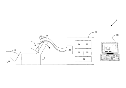
Figure 2 is a schematic diagram of a preferred embodiment of an ECOG
system connected to a patient;
Figure 3 is a response signal recorded by the system;
Figure 4 is a flow diagram of a neural event process performed by the ECOG
system;
Figures 5 to 10 are Sp/Ap plots produced by the neural event process;
Figure 11 is a display of Sp/Ap plots produced using a high pass filter by the
ECOG system;
Figure 12 is a display of Sp/Ap plots produced by the ECOG system by
including DC offsets of the stimulus response;
Figure 13 is a flow diagram of a neural event process performed by a preferred
embodiment of an ABR system connected to a patient;
CA 02578597 2014-08-12
66718-88
- 5c -
Figure 14 is a display produced by the ABR system of detected ABR neural
events;
Figure 15 is a diagram of different ABR components;
CA 02578597 2007-02-28
WO 2006/024102 PCT/AU2005/001330
- 6 -
Figure 16 is a display of Sp/Ap plots for a Parkinson's patient produced by
the
ECOG system;
Figure 17 is a display of Sp/Ap plots produced for a patient suffering
depression by
the system;
Figures 18 and 19 are displays of Sp/Ap plots produced for a Meniere's patient
by
the system;
Figures 20 to 24 are displays of TAP measurement markers produced by the
system
for a number of patients;
Figure 25 is a diagram of averaged wavelet coefficients against frequency
generated
by the system; and
Figure 26 is a display of HF/LF ratio data markers produced for a number of
different patients by the system.
DETAILED DESCRIPTION OF PREFERRED EMBODIMENTS
An ECOG system 2, as shown in Figure 2, is used to obtain Sp/Ap plots, as
shown in
Figures 5 to 10, from a patient who is subjected to a single stimulus, eg an
involuntary
head tilt. The Sp/Ap plots are generated from the ECOG signal produced in
response to
the stimulus. The ECOG signal is obtained from electrodes 10, 12 and 14
electrically
connected to an amplifier circuit 22 of a computer system 20 of the ECOG
system 2. A
first electrode 10 (eg a ECochG Electrode produced by Bio-Logic Systems Corp
(http://wwvv.blsc.com/pdfs/HearCatalog.pdf) is placed on the tympanic membrane
of an
ear of a patient 4. A second electrode 12 is placed on the patient's earlobe,
as a reference
point, and a third electrode 14 is connected to the patient's forehead and to
the common
point of the amplifier. A shield connection 16 is also made to an electrical
isolation shield
18 normally placed around the testing room. The shield 18 is connected to the
shield of
the amplifier 22. To obtain an auditory ECOG signal a continuous auditory
signal is
applied to the ear, comprising alternating polarity acoustic clicks. However,
for a
vestibular ECOG signal (ie a EVestG signal) the patient 4, as shown in Figure
2, is placed
on a chair 6, such as a recliner lounge chair, that allows the patient's head
to be tilted either
voluntarily or involuntarily. Tilt chairs have been specifically produced by
Neuro Kinetics
CA 02578597 2007-02-28
WO 2006/024102 PCT/AU2005/001330
- 7
Inc. (http://www.neuro-kinetics.com) that enable a patient to be tilted and
produce a
response to this stimulus which is less corrupted by muscle artefact. An
involuntary head
tilt is obtained by an assistant manipulating the chair 6 so as to induce the
head tilt without
any patient neck muscle activity. A typical sequence is 20 seconds in a
neutral position, 20
seconds tilted and 20 seconds neutral when tilted back up. The head tilt is
done for
approximately the same angle as a maximum voluntary head tilt that could be
achieved by
the patient themself. Tilts are back, forward, ipsilateral and contralateral.
Though less
effective and less location specific, it is, however, also possible for the
ECOG system 2 to
produce Sp/Ap plots derived from a response from the combined auditory and
vestibular
system that is produced without any specific stimulus. This is based on
recorded
spontaneous background activity of the auditory and vestibular system. For a
voluntary
head tilt,to obtain a stimulated response, the patient is asked to sit in the
chair upright with
their head in the neutral position for 20 seconds, and then their head tilted
forward for 20
seconds, back to neutral for 20 seconds, backwards again for 20 seconds,
neutral for 20
seconds, ipsilateral to the electrode 10 for 20 seconds, neutral for 20
seconds, contralateral
to the electrode 10 for 20 seconds and then neutral for 20 seconds.
The neural response produced on electrodes 10 to 14 is continuously recorded
by the
ECOG system 2. The neural response signal for each tilt is a time domain
voltage signal
having multiple frequency components. The main components of interest are up
to 22,500
Hz, and accordingly the sampling rate used by the system 2 is chosen to be
44.1kHz. With
this rate the Sp peak (depending on the signal to noise ratio (S/N)) is only a
few samples
wide. The signal is characterised by distinct regions in time: (i) a
background region
comprising primarily background ambient noise; (ii) an onset region for the
start of tilt
(approximately 0-5 seconds after tilt onset) which includes the response of
the semi
circular canals and otolith organs; (iii) a transient region for the remainder
of the tilt
(approximately 5-10 seconds after tilt onset) which includes the response of
the semi
circular canals (decaying) and otolith organ; and (iv) a steady state region
(approximately
10-20 seconds after tilt onset) which includes essentially the response of the
otolith organs.
An example of a recorded response signal for an involuntary tilt is shown in
Figure 3, and
the elements of the signal are described in the table below
CA 02578597 2007-02-28
WO 2006/024102 PCT/AU2005/001330
- 8 -
Tilt Segment Time (sec)
Background 5-20
Onset 20-25
Transient 20-30
Steady state _ 30-40
Onset (tilting back up) 40-45
Transient (tilting back up) 40-50
Steady state (tilting back up) 50-60
The computer system 20 of the ECOG system 2 includes the amplifier 22 and a
communications module 24 for handling the output of the amplifier 22 and then
storing the
response as a voltage signal over time as a wave file using a computer program
such as
Adobe Audition (hftp://www.pacific.adobe.com/products/audition/main.html)
provided by
a capture module 26. The amplifier 22 includes a CED 1902 isolated pre-
amplifier and a
CED Power 1401 analogue to a digital converter (ADC). Both the CED 1902 and
CED
1401 ADC are produced by Cambridge Electronic Design Limited
(http://www.ced.co.uk).
The CED 1401 ADC has an excellent low frequency (less than a few Hz) response.
The
computer system 20 has further software modules, including an analysis module
28 and a
display module 30. The analysis module 28 includes computer program code (eg.
MATLABC code, http://www.mathworks.com) responsible for performing neural
event
extraction processes, as shown in Figures 4 and 13, in conjunction with the
other software
modules. The analysis module 28 also executes a number of different filters
used to filter
the response signal samples, as discussed below. The graphics display module
30
generates a user interface 32 for an operator of the ECOG system 2 to provide
input
controls so that the operator can control the neural event extraction process,
and to
generate displays of neural event data, such as the Sp/Ap plots shown in
Figures 5 to 10.
The computer program code of the software modules 24 to 30 of the computer
system 20
are run on an operating system 34, such as Microsoft Windows or Linux, and the
hardware
used may include the amplifier 22 and a standard personal computer 20, such as
that
produced by IBM Corporation (http://www.ibm.com). ECOG recording systems are
produced by Bio-Logic Systems Corp (hftp://vvww.blsc.com/hearing/). Whilst the
neural
event extraction process may be performed under the control of the software of
the
modules 24 to 34, it will be understood by a skilled addressee that steps of
the process can
CA 02578597 2007-02-28
WO 2006/024102 PCT/AU2005/001330
- 9 -
be performed by dedicated hardware circuits, such as ASICs and FPGAs, and also
performed by components or modules distributed across a computer
communications
network, such as the Internet.
The neural event extraction process uses known temporal and frequency
characteristics of
an Sp/Ap plot to try to accurately locate an evoked response from the patient.
Basically
only a rough shape of the plot and the expected latency between the points of
interest is
known. Latency between the points corresponds to a frequency range of
interest.
Accordingly, the Sp/Ap plot is known to exhibit a large phase change across a
frequency
range of interest at points on the Sp/Ap plot, in particular, the Sp, Ap,
onset of Sp, offset of
Ap and beginning of Sp2 points. The neural event extraction process operates
to produce a
representative data stream that can be used to determine neural events
occulting in the
right time frame and with appropriate latency that can be considered to
constitute
characteristic parts of an evoked response. The same principle can also be
applied to other
AERs, as discussed below.
The neural event extraction process, as shown in Figure 4, involves recording
the voltage
response signal output by the amplifier 22 in response to a head tilt (step
302). Where
necessary a 50 or 60 Hz mains power notch filter is applied to the recording
in the
amplifier stage to remove power frequency harmonics. The response signal from
the
amplifier 22 may also be high pass filtered (for example by a 120 Hz 1 pole
Butterworth
filter) to enable the extraction process to generate improved Sp/Ap peak plots
(eg as shown
in Figure 11) at step 350. If the very low frequency data is retained, ie < 10
Hz, then this
can be used to plot (at step 350) discriminate "dc" magnitude threshold shifts
prior to a
neural event. These threshold shifts are shown in Figure 12 and relate to the
onset region
(largest shift and therefore at the bottom of Figure 12), the transient region
(next largest
shift) and the steady state region (lowest shift and at top). Examination of
this very low
frequency data, and in particular the magnitude shifts, can be used to aid the
diagnosis of
central nervous system disorders, as described below, and in particular
illustrate more
cortical influences on the vestibular system. Absence or enhancement of the
shifts tend to
indicate a disorder.
CA 02578597 2007-02-28
WO 2006/024102 PCT/AU2005/001330
- 10 -
The recorded response signal is decomposed in both magnitude and phase using a
complex
Monet wavelet (step 304) according to the definition of the wavelet provided
in equation
(1) below, where t represents time, Fb represents the bandwidth factor and Fe
represents the
centre frequency of each scale. Other wavelets can be used, but the Monet is
used for its
excellent time frequency localisation properties. The neural response signal
x(t) is
convolved with each wavelet.
t2
j27rFc t
1
Monet (t)=. ____________________________________ 2Fb
(1)
v27-cFb
To directly measure the vestibular system, seven scales are selected to
represent wavelets
with centre frequencies of 12000 Hz, 6000 Hz, 3000 Hz, 1500 Hz, 1200 Hz, 900
Hz and
600 Hz. Different frequencies can be used provided they span the frequency
range of
interest and are matched to appropriate bandwidth factors, as discussed below.
The
wavelets extend across the spectrum of interest of a normal vestibular Sp/Ap
response
signal, and also include sufficient higher frequency components so that the
peaks in the
waveform can be well localised in time. Importantly, the bandwidth factor is
set to less
than 1, being 0.1 for the scales representing 1500 to 600 Hz and 0.4 for all
remaining
frequencies. Using a bandwidth factor that is so low allows for better time
localisation at
lower frequencies, at the cost of a frequency bandwidth spread, which is
particularly
advantageous for locating and determining neural events represented by the
response
signal. Magnitude and phase data is produced for each scale representing
coefficients of
the wavelets.
The phase data for each scale is unwrapped and differentiated (306) using the
"unwrap"
and "cliff" functions of MATLAB. Any DC offset is removed, and the result is
normalised
for each scale to place it in a range from -1 to +1. This produces therefore
normalised,
zero average data providing a rate of phase change measurement for the
response signal.
CA 02578597 2007-02-28
WO 2006/024102 PCT/AU2005/001330
- 11 -
A first derivative of the phase change data (actually a derivative of a
derivative) is obtained
for each scale (308), and normalised in order to determine local maxima/minima
rates of
phase change (320). To eliminate any false peaks, very small maxima/minima are
removed at a threshold of 1% of the mean absolute value of the first
derivative (322). All
positive slopes from the first derivative (308) are set to 1, negative slopes
to -1 and then a
second derivative of the phase change data is obtained (310) to produce -2 and
+2 step
values. Each scale is then processed to look for resulting values of -2 and +2
which
represent points of inflexion for the determined maxima and minima (320). For
these
particular loci, a value of 1 is stored for all scales. For the low frequency
scale, ie 600 Hz,
the actual times for both the positive and negative peaks are also stored for
analysis to
further isolate the driven responses as discussed below.
The original response signal in the time domain (312) is also processed to
detect points
which may be points of maximum phase change for comparative analysis with the
extracted phase peaks from the wavelet analysis. Firstly the mean and maximum
of the
original signal is determined. The signal is then adjusted to have a zero
mean. Using this
signal, the process locates and stores all points where the signal is greater
than the mean
minus 0.1 of the maximum in order to identify regions where an Ap point is
least likely
(positive deviations above axis) and to exclude in later derivatives maxima as
a
consequence of noise. The slope of the original response is obtained by taking
the
derivative of the original response, and then also determining the absolute
mean of the
slope. For the result obtained, all data representing a slope of less than 10%
of the absolute
mean slope is set to 0. A derivative is then obtained of this slope threshold
data (314)
which is used to define the local maxima/minima of the slope (316). Similarly,
the
absolute mean of this result is also obtained and a threshold of 10% of the
mean used to
exclude minor maxima/minima (step 318). All positive slopes of the original
response are
set to 1 and the negative slopes are set to -1, and then a second derivative
obtained (314).
From this derivative each scale is examined to find values of -2 and +2,
representing points
of inflexion. The position of these loci are stored for the positive and
negative peaks.
For each scale, if there is a positive peak, ie a maximum, determined from the
first slope
CA 02578597 2007-02-28
WO 2006/024102 PCT/AU2005/001330
- 12 -
derivative, then for any peaks corresponding to these times (+1 or -1) these
are set to 0 in
any scale in which they appear in order to initially selectively look for the
Ap point which
will be a minima. The same is also done for points that were previously deemed
unlikely
regions for an Ap point found during the original processing of the time
domain response
signal (312). The times of the peaks determined during processing of the phase
data, and
that determined during processing of the time domain signal, are compared
(step 324).
Because of scale dependant phase shifts inherent in detecting each wavelet
scales phase
maxima, the wavelet scale maxima are compared with those detected in the time
domain
and shifted to correspond to a magnitude minima in the time domain. Thus
potential Ap
loci (326) are determined.
The loci times for the low frequency scale, scale 7 representing 600 Hz, are
searched to
attempt to locate the Sp point, as it is most likely that the preceding steps
have determined
the Ap point, due to the size of the signal and the difficulty of normally
locating the Sp
point. This search is undertaken over a range of normally 0.1 to 0.9 ms
(depending on the
noise level; for example the lower limit of 0.1 may be increased, say to 0.5)
before the
potential Ap point looking for +2 values (i.e. negative peaks) in this range.
If the value of
the original response signal at the potential Sp point is greater than 0.9 of
the potential Ap
point (a negative value), then both the Ap loci and the potential Sp loci are
stored. If an Sp
point is located 0.1 to 0.9 ms before the Ap point, then the 600 Hz scale loci
time for the
Ap point and the time domain minima, proximal to that Ap point, are checked to
determine
whether they are at the same point in time. If this is not the case, then the
scale loci is reset
to match the time domain loci to take into account any limitations in time
localisation
properties associated with the wavelet decompositions. For verification,
similar location
procedures for the Sp point can be performed on the other scales, but this is
not needed in
all cases.
All of the scales are then processed (step 330) to look for maxima across the
scales and
link them to form a chain across as small a time band as possible. This allows
false Aps
associated with all of the scales to be eliminated. The analysis module 28 is
able to use a
"Chain maximum-eliminate "false" maxima" routine of MATLAB to perform this
step.
CA 02578597 2007-02-28
WO 2006/024102 PCT/AU2005/001330
- 13 -
As described below, a Sp/Ap plot is formed by processing the time domain
signal (or
averaging the time domain signals obtained) centred on the local maxima
determined
previously. Following the Sp/Ap plot formation process, maxima/minima values
are
further determined to establish the baseline (ie the average level before the
evoked
response, as shown in Figure 1) necessary to calculate the Sp/Ap.
Using firstly the +2 values, and then the -2 values if no +2 values are found,
for the points
of inflexion determined from the phase data, the loci is searched in the range
allocated to
the Sp previously determined (typically 0.5 to 0.9 ms before Ap). For each Ap,
remaining
after the elimination process (330) the Sp times are found and averaged to
record an Sp.
The baseline is found (340) by starting at the Sp point -0.2 to -0.6 ms (based
on average
Sp/Ap shape), and again beginning with the +2 point inflexion values, and then
-2 point
inflexion values (if necessary) of the phase data in a time range initially
allocated to the
baseline. For each Sp plus offset, the potential baseline times are found and
averaged to
record an initial baseline time. If the baseline time does not meet a baseline
check, then
the process is repeated starting with the new baseline time estimate. This
process is
repeated until a baseline check is met, which may be whether a baseline is
within a
predetermined time range from the Ap and Sp. The average magnitude at the
determined
time is used. Alternatively, the baseline can be determined as being the mean
of the first
300 samples of the Sp/Ap plot.
Sp2 is found (330) by also using firstly the +2 values for the points of
inflexion of the
phase data, and then if there a no +2 values using the -2 values, and
searching for loci in
the range allocated (initially 1.3 ms after the Ap). For each Ap plus offset,
the Sp2 times
are determined and then averaged to record an Sp2 time. The average magnitude
at the
determined time is used.
An artefact, being a spike of about 3 samples wide, is produced at the tip of
Ap due to the
selection of local minima in the time domain based on scale determined loci
proximal
CA 02578597 2007-02-28
WO 2006/024102 PCT/AU2005/001330
- 14 -
thereto. The samples corresponding to the spike (which may be up to 5 samples)
should be
removed, and this is done (342) by using the values of the points on either
side of the spike
to interpolate values into the removed sample positions. A filter, such as a
15 point
moving average filter, can then be applied after removal to smooth the
response.
Based on the Sp, Sp2 and Ap neural events determined, the ratios Sp/Ap and
Sp2/Ap are
calculated and displayed with the plot of the vestibular response (350). The
plot is
generated by the display module 30 using the times/loci of the maxima and
minima
determined by the neural event extraction process.
In summary, the neural event extraction process uses a complex time frequency
approach
with a variable bandwidth factor to determine the points where maximum/minimum
phase
changes occur across a range of frequencies characteristic of neural events
associated with
an Sp/Ap plot. The maximum/minimum phase change is used to establish the Ap,
Sp, Sp2
and baseline points. Being able to determine these points enables elimination
of other
phase change events that are not related to an Sp/Ap plot, such as those
produced by
background noise. Also, maximum/minimum phase change points are correlated
with
events in the time domain to reduce time localisation error inherent in the
use of the
frequency domain representation provided by the wavelet analysis.
Figure 5 shows an example of a display produced by the ECOG system 2 following
analysis of a 1 second region of a steady state response (14.4 seconds after
head tilt onset)
to a voluntary backwards head tilt (patient's eyes open). The Sp/Ap ratio is
determined to
be 22.6% by the analysis module. The horizontal scale is 1 ms, equivalent to
44.1 samples
of the evoked response signal. Figure 9 shows a similar display produced
following
analysis of a 10 second region of a steady state response (10 seconds after
head tilt onset)
where the Sp/Ap ratio is determined to be 28%.
Figures 6, 7 and 8 also show Sp/Ap plots produced using the ECOG system 2. The
plots
are for a non voluntary movement on a tilt chair. Figure 6 is a plot for the
onset region,
Figure 7 is a plot for the transient region and Figure 8 is a plot for the
steady state region.
CA 02578597 2007-02-28
WO 2006/024102 PCT/AU2005/001330
- 15 -
All the plots have a baseline, Ap and Sp (and Sp2 seen normally only with tone
stimulus
responses and also for the onset period or component of excitatory tilt
responses) point
marks that can be determined by the neural event extraction process of the
analysis module
28. Figure 10 also shows Sp/Ap plots produced for the onset region (dark),
steady state
region (light) and for the transient region (medium). In this Figure, the Sp
and Ap is
shown as only that determined for the steady state response.
The system 2 as described is able to perform an accurate analysis of a
response from the
vestibule that not only can be used for the detection of Meniere's disease,
but can also be
used for diagnosis of Parkinson's disease and depression as discussed below.
Also other
neural events can be sought and determined, such as those produced by other
auditory
nuclei. The system 2 can be configured to obtain other AERs and the analysis
module 28
used to accurately process the AER obtained, such as an ABR.
Latency considerations relevant to the Auditory Brainstem Response (ABR) allow
for the
separation then generation of Sp/Ap like waveforms from each main nuclei.
Responses
from subnuclei like the Medial Nucleus of the Trapeziod Body, Lateral Superior
olive and
Medial superior olive of the superior olivary complex are also separable.
Responses could
also be obtained from the visual pathway and its nuclei, indeed most evoked
response
pathways.
For the ABR, the system 2 is adjusted so the analysis module 28 executes an
ABR process,
as shown in Figure 13, and the electrodes 10 and 12 are rearranged to obtain
an ABR
response, instead of an ECOG response. In particular, the patient 4, remains
at rest, and
the electrodes 10 and 12 are used as surface electrodes, with one being placed
on each
mastoid, and the additional electrode 14 used on the forehead. The patient's
leg is again
connected to the shield 18. The stimulus produced by the computer system 20 is
an
audible click (100us) or tone pip (5 ms), eg 80dB SPL (sound pressure level),
repeated
about 300-1000 times. Each stimulus is 200ms apart. The first 10ms post
stimulus is
recorded. The ABR process, as shown in Figure 13, is primarily the same as the
neural
event process described above with reference to Figure 4, except for the
following:
CA 02578597 2007-02-28
WO 2006/024102 PCT/AU2005/001330
- 16 -
The first stage of the process (302) performs segmentation by recording the 10
ms
of interest from the 200 ms of each response signal received. The recorded 10
ms
time domain signal is then filtered using a 500Hz - 4kHz bandpass 6 pole
Butterworth filter.
(ii) The wavelet scales used in the step 304 have the same bandwidth
factors, except a
very small bandwidth factor of 0.05 is used for the lowest frequency scale,
600 Hz.
(iii) Additional processing (802) is performed after step 330 to determine the
Ap point
marks corresponding to each of the subnuclei of interest. This is done on the
basis
of the latencies of the Aps in comparison with the time domain data in order
to
construct an Sp/Ap plot for the nuclei and subnuclei of interest, eg 3.2 ms to
4.4 ms
for peak III of an ABR.
Figure 14 is a display produced of a plot showing the neural events detected
by the system
2 from 25 ABR stimulus recordings, with the neural events corresponding to
peaks II to V
of an ABR delineated. The data for each of the detected neural events, at
times associated
with the events, can be averaged to produce Sp/Ap plots for the nucleus of
interest. Figure
15 illustrates the timing, ie the latency, of the different ABR components for
the different
nuclei and subnuclei, which include the auditory nerve (AN), the dorsal
cochlea nucleus
(DCN), ventral cochlea nucleus (VCN), medial nucleus of trapezoid body (MNTB),
lateral
superior olive (LSO), medial superior olive (MSO), lateral lemniscus (LL),
central nucleus
of inferior colliculus (IC C), pericentral nucleus of inferior colliculus
(ICP), external
nucleus of inferior colliculus (ICX) and medial geniculate body (MGB). The
responses
from the nuclei and subnuclei are separable into different events as shown in
Figure 14.
Using a tone instead of a click enables a response to be evoked from the
lamina in the
nucleus of interest. The neural event detected using a click is a response
from the entire
tonotopic region of the nucleus. However by using a tone only, one lamina or
layer of the
nucleus is excited allowing for the localisation within the nucleus of any
departures from a
normal response.
A further application of the ECOG system 2 is detecting the degeneration of
cells in the
Basal Ganglia (eg Substantia Nigra in Parkinson's Disease) by accurately
detecting the 70-
CA 02578597 2007-02-28
WO 2006/024102 PCT/AU2005/001330
- 17 -
300Hz inter-event intervals (time-frequency representations) and changes in
the neural
Sp/Ap response characteristics (including Ap width, Sp peak height, etc)
consequent to
changes in the Basal Ganglia and other connected structures observed in the
vestibular
response and believed to be modulated by Basal Ganglia outputs via the
reticular formation
to the vestibular nuclei. This is particularly useful for quantitatively
measuring the efficacy
of therapies and drugs to treat, as well as for the early detection of,
Parkinson's disease.
Figure 16 shows two Sp/Ap plots produced by the system 2 for a Parkinsons
patient, one
where the patient is without medication (upper), and another where the patient
is with
levodopa medication (lower with a deliberate offset for clarity). The effect
of the
medication is indicated by the Ap width, ie the TAP measurement, the Sp
magnitude
change and the general change in the Sp/Ap plots. The TAP is a time measure
from the
minima peak ("notch") before the Sp peak horizontally to the upward arm of the
Ap, as
shown in Figure 16. Alternatively, a different TAP measure could be the
internal width of
the Ap horizontally at the Sp notch vertical level used in the preceding
definition.
Another application is detecting the decrease or increase in activity of cells
in the Basal
Ganglia (eg Thalmus in depression) co-incident with changes in depressive
state by again
accurately detecting changes in the 70-300 Hz inter-event intervals (time-
frequency
representations) and changes in the neural Sp/Ap response characteristics
(including Ap
width, Sp peak height, etc) consequent to changes in the Basal Ganglia and
other
connected structures observed in the vestibular response and believed to be
modulated by
Basal Ganglia outputs via the reticular formation to the vestibular nuclei.
This is
particularly useful for quantitatively measuring the efficacy of therapies and
drugs to treat
depression, as well as the detection of depression (particularly in
intellectually disabled
and those with limited communication skills). Figure 17 shows two Sp/Ap plots
produced
by the system 2 for a patient suffering depression. One plot is before the
patient is
medicated (lower and light), and the second plot has been taken three hours
after the
patient has been medicated with SSRIs (Selective Serotonum Uptake Inhibitors)
(upper
and dark). Again, the effect of the medication is indicated especially by the
Ap width, ie
the TAP measurement marker.
CA 02578597 2007-02-28
WO 2006/024102 PCT/AU2005/001330
- 18 -
Figure 18 shows an Sp/Ap plot produced by the system for a Meniere's patient
with
(lower) and without medication (upper), this again shows the stark differences
between the
Sp/Ap plots, and the Ap width measure, TAP. The medication used was AVILTM
(43.5
mg). Figure 19 shows Sp/Ap plots comparing a Meniere's patient with symptoms
on the
left side (upper) but not on the right side (lower).
The analysis module 28 of the system 2 is able to produce a series of markers
to
discriminate between patients that have, or to determine whether they have, a
disorder,
such as a central nervous system (CNS) disorder, and in particular whether
they are
depressed, suffering Meniere's disease, or suffering Parkinson's disease. The
markers
include (i) the Sp/Ap point marks, (ii) the TAP measurement, being the time
and duration
of Ap (plus the Sp peak depending on the TAP period definition used), and
(iii) a HF/LF
ratio being the ratio of the high frequency energy to the low frequency energy
of the
average wavelet coefficients of the scales, as shown in Figure 25. The HF/LF
ratio is a
ratio for the response signal of the high frequency and low frequency areas
beneath the plot
of the averaged wavelet coefficients against frequency for the respective
ranges 50 to 500
Hz and 2 to 28 Hz, as shown in Figure 25. Figures 20 to 24 show a variety of
TAP
measurements obtained for different patients, and illustrate how they can be
discriminated.
Figure 26 shows how the HF/LF ratio can be used as a discriminating marker.
Other
markers are provided by analysis of scales for the 70 to 300 Hz range to
determine
alterations to the response signal and Sp/Ap plots due to modulation by the
Basal Ganglia
components. The alterations may be the presence or absence of peaks or
distribution
changes for time against frequency representations for this range. Peaks
within this range,
particularly proximal the ranges 70-90 Hz, 110-150 Hz and 200-300 Hz, may
indicate
activity of the Basal Ganglia components. If those markers are used,
additional scales are
used by the neural event process for the 70 to 300 Hz range.
To assist diagnosis, the magnitude, phase, frequency and time data extracted
by the neural
event process can be used to generate three dimensional or four dimensional
(with color)
plots for responses obtained from patients.