Note: Descriptions are shown in the official language in which they were submitted.
CA 02578759 2007-03-01
WO 2006/027678 PCT/IB2005/002679
Button Anchor System for Moving Tissue
Field of the Invention
This invention relates generally to a system and method for moving or for
moving
and stretching human or animal plastic tissue that exerts a relatively
constant tension over
a given distance and that is readily adjustable, and more specifically to an
anchor for use
with such systems.
Back2round
In general, surgery and surgical treatment involve one or both of tissue
separation
and tissue joining. In surgery, medical treatment, and medical research, it is
desirable to
retract tissue, stabilize tissue, and present tissue in a variety of specific
orientations to
provide access to the area under investigation or repair, ideally in a method
that creates
minimal trauma beyond what is necessary for exposure and visualization of the
operative
area. Ultimately, the procedures should allow for immediate, or primary,
closure of the
wound. Unfortunately, the latter option is not always available in surgical or
trauma
wound scenarios.
Moving tissue presents unique challenges, as tissues often resist joining, or
closure, depending on the nature of the tissue structure, the circumstances of
the tissue
separation, and general patient health. Complications related to wound closure
and
healing generally result from major forces, minor forces and/or compromised
healing
responses. Major forces are retractive forces created beyond the viscoelastic
properties of
the tissue, and may be created by: (1) increased internal volume, such as in
the case of
obesity, which elevates containment forces on the skin system; (2) changes in
aspect
1
CA 02578759 2007-03-01
WO 2006/027678 PCT/IB2005/002679
ratio, such as increased abdominal circumference created in prone, non-
ambulatory
patients due to muscular atrophy; (3) respiratory muscular activity; (4)
muscular
response; (5) loss of fascia structure; (6) muscular-skeletal deformation; (7)
fleshy
appendages; (8) tumors; and (9) severe burns.
Minor forces are internal forces created by the viscoelastic properties of the
tissue,
which can cause the skin to retract. Elastic tissues, such as skin, comprised
mostly of
extracellular matrix (ECM) components along with cells, return to a minimum
elastic, or
relaxed, state when released from tension. In this relaxed state, tissue
tensions are
minimized and balanced. Skin tissue in this minimum elastic state will remain
relaxed,
demonstrating behavior similar to a non-elastic material. The force required
to elongate
the tissue in this state often approaches a force that will rupture or sheer
structural
connective elements, causing localized failures or tears. Soft tissue in this
minimum
elastic state provides minimum surface coverage and has the highest reluctance
to stretch.
It is known that a gentle but constant force below the sheer force threshold
applied to
tissue in combination with adequate hydration will, over time, restore certain
tissues to
near-original or original elastic state. Additionally, this force can be
applied to stretch
tissue past the point of equilibrium (normal elastic range) to the maximum
elastic range
and create the thinnest possible configuration, covering the maximum surface
area. If
tensions in the tissue do not exceed the point at which the connective
structural elements
are compromised, the tissue remains at the maximum elastic state as healthy
tissue, and
normal biological processes will allow cell regeneration and associated ECM
production
to restore normal skin thickness and tension, which is described below as
biological
creep.
2
CA 02578759 2007-03-01
WO 2006/027678 PCT/IB2005/002679
Plastic tissues, such as skin and muscle, possess certain viscous and elastic
rheological properties, and are therefore viscoelastic. Certain plastic
tissues are able to
increase surface area over time, which can be termed "creep." "Mechanical
creep" is the
elongation of skin with a constant load over time beyond intrinsic
extensibility, while
"biological creep" refers to the generation of new tissue due to a chronic
stretching force.
A constant and unrelenting force applied to a body tissue, such as skin or
muscle, may
result in both mechanical and biological creep. Mechanical creep restores the
tension
originally present but lost in the skin across the incision or wound by
retensioning skin,
thereby increasing skin coverage. Biological creep occurs more slowly and
involves the
creation of new tissue. Tissue expansion has long been part of the art of
plastic surgery,
traditionally accomplished with balloon-type tissue expanders embedded under
the skin
and externally inflated and increased over time to create expanded pockets of
skin for
procedures such as breast reconstruction after radical mastectomies, and
stretching
healthy tissue prior to plastic surgery for the creation of flaps for soft
tissue closure.
Finally, compromised healing responses may complicate wound closure or
healing. A surgical or other incision becomes a complicated wound as soon as
it falls
behind normal healing progression. Wound management, including treatment and
care of
large skin defects and severely retracted incisions, is an area of increasing
importance to
the health care community. An aging population and an increase in diseases
related to
obesity and inactivity have increased the occurrence of complicated wounds and
placed
an increased burden on health care resources. Factors contributing to
compromised
wound healing include patient age, weight, nutritional status, dehydration,
blood supply
to the wound site, immune response, allergies to closure materials, chronic
disease,
debilitating injuries, localized or systemic infection, diabetes, and the use
of
3
CA 02578759 2007-03-01
WO 2006/027678 PCT/IB2005/002679
immunosuppressive, corticosteroid or antineoplastic drugs, hormones, or
radiation
therapy. Complicate(i wounds inch;de, but are not limited to: surgical wounds,
diabetic
ulcers and other chronic ulcers; venous stastis ulcers; decubitis or pressure
sores or ulcers;
bums; post traumatic lesions, cutaneous gangrene, crush wounds with ischemic
necrosis;
wounds having exposed plates or bones; keloids; skin lesions; blunt abdominal
trauma
with perforations; and other acute, subacute or chronic wounds. Treatment and
care of
these tissue defects is challenging due to difficulties in closure of open
wounds.
Two common methods of closure of wounds and skin defects include split
thickness skin grafting and gradual closure. A split thickness skin graft
involves
removing a partial layer of skin from a donor site, usually an upper leg or
thigh, and
leaving a portion of the dermis at the donor site to regenerate and re-
epithelialize. In this
manner, a viable skin repair patch can be transferred or grafted to cover a
wound area.
The graft is often meshed, (which involves cutting the skin in a series of
rows of offset
longitudinal interdigitating cuts) allowing the graft to stretch to cover two
or three times
greater an area as well as provide wound drainage while healing. Normal
biological
function of the skin heals the holes after the graft has been accepted. A
meshed graft of
this type requires a smaller donor area than a conventional non-meshed or full
thickness
skin graft. However, these methods do not provide optimal cosmesis or quality
of skin
cover. Other disadvantages of this method include pain at the donor site,
creation of an
additional disfiguring wound, and complications associated with incomplete
"take" of the
graft. In addition, skin grafting often requires immobilization of the limb,
which
increases the likelihood of contractures. The additional operation and
prolongation of
hospital stay is an additional economic burden.
4
CA 02578759 2007-03-01
WO 2006/027678 PCT/IB2005/002679
Gradual, or progressive, closure is a second method of closure. This technique
may involve suturing vessel loops to the wound edge and drawing them together
with
large sutures in a fashion similar to lacing a shoe. In addition, the wound
edges may be
progressively approximated with suture or sterile paper tape. The advantages
of this
gradual, or progressive, technique are numerous: no donor site is required for
harvest of a
graft, limb mobility is maintained, and superior cosmetic result, more durable
skin
coverage, better protection from full skin thickness and the maintenance of
normal skin
sensation may all be achieved.
Existing devices for effecting a gradual closure have many disadvantages.
Current
methods and devices draw wound edges together using mechanical devices such as
screw-actuated systems that require repeated periodic adjustment because a
relatively
small skin movement substantially eliminates much of the closure force. Widely
used
existing closure techniques involve use of relatively inelastic materials,
such as sutures or
surgical tape. Excessive tension may cut the skin or cause necrosis due to
point loading
of the tissue. Current solutions include suture bolsters, suture bridges, use
of staples as
anchors at the wound edge, and the use of ligature wire to distribute the load
along the
wound margins. These approaches all rely on static ribbon or suture material,
which must
repeatedly be readjusted in order to function effectively, and even with this
constant
readjustment, maintenance of near constant tension over time is difficult, if
not
impossible, to achieve. Widely used traditional gradual closure methods rely
on static
force through fixed distance reduction, and do not provide continuous or
dynamic tension.
Many current methods of open wound reduction employ static or non-yielding
devices such as sutures or hard approximators, which reduce the distance
between the
wound margins and rely on the skin's natural elasticity to compensate for
movement. One
5
CA 02578759 2007-03-01
WO 2006/027678 PCT/IB2005/002679
problem with these devices has been that when they are at the point of being
most
effective, when the skin is at the point of maximum stretch, additional skin
tension
created through motion, such as breathing or walking, creates stress points
where the
mechanical fasteners meet the wound margins, causing tearing and wound edge
necrosis.
This has generally required patients to remain immobile during the course of
treatment.
Existing systems for treating animals need not consider cosmetic result to
such a degree
as the healthy patient typically masks the wound site with fur, but cosmesis
is a critical
criteria in the measurement of a successful result from the system in the
human
application.
One existing method for effecting closure of a wound utilizes a constant
tension,
low-grade force to draw wound edges together. One device for practicing this
method
includes a pair of hooks carried by a pair of sliders that move along a path
pulled by a
pair of springs. This spring device is enclosed in a plastic housing and is
available having
various curvatures. The sharp hooks used in this system may damage the skin.
The
constant force used is a dictated force that is not variable. Other closure
devices use
elastomeric material, including rubber bands and other types of compressive
and non-
compressive materials, to approximate wound margins. One kit requires bonding
to the
skin with an adhesive and also requires periodic adjustment to tighten the
straps. Other
known closure devices use hooks and elastic loops, which must be replaced with
smaller
elastic loops to maintain tension, or a motor power source to provide a
tightening means.
Finally, another current device consists of two surgical needles, two U-shaped
lexan
polycarbonate arms with hooks on the bottom surface, a threaded tension bar
and a
polycarbonate ruler. The needles are threaded along the wound margin and each
arm is
6
CA 02578759 2007-03-01
WO 2006/027678 PCT/IB2005/002679
positioned above a needle, with the hooks piercing the skin and engaging the
needles.
The tension bar is then locked, and tension can be adjusted using the screw.
Existing methods of gradual wound closure fail to provide an effective gradual
closure that restores original skin tensions lost across the wound. For
example, one
system has a single tension of 460 grams. In many instances, such as with the
elderly or
with compromised skin, this force is too great, resulting in localized
failures, tears and
necrosis. Many current devices are cumbersome, restrict patient mobility, must
be
completely removed for wound dressing and cleaning, and are usable in a
relatively
limited number of situations because of size constraints. Many also require a
surgeon for
reinstallation after removal for wound dressing. Finally, many current devices
cannot
readily be used for radial closure of wounds due to their limited ability to
pull in a single
direction along an overhead beam, thereby restricting their application to
parallel pulls
along the same axis.
Summary of the Invention
This invention provides manipulation and control of tissue positions and
tensions
on a living person or animal, utilizing both tissue stretch and creep to
restore and move
tissues. This invention provides methods and devices for moving or for moving
and
stretching tissue that are simple, easy to use, cost=effective, extremely
versatile, self-
adjusting and capable of exerting relatively constant force or tension over a
variety of
distances and at various intersecting angles in wounds having simple or
complex
geometry.
Components of this invention exert a dynamic force on the tissue, providing
and
maintaining a maximum safe counter-traction pressure or force across a wound
margin or
7
CA 02578759 2007-03-01
WO 2006/027678 PCT/IB2005/002679
other area. Systems of this invention create controlled constant and
unrelenting tension,
which can be applied to counteract major or minor retraction forces or to
achieve
maximum mechanical and biological yields to move or to move and stretch
plastic tissue,
including closure of large retracted skin defects.
Terms used herein are generally defmed and, in some cases, abbreviated, as
they
are introduced. For convenience, selected terms are also defined here. A force
applying
component ("fac") generally stores energy in a manner that exerts force and
transmits the
force. An elastic force applying component ("efac") combines these two
functions in a
single elastic component. The tissue manipulation system of this invention
utilizes facs
coupled to force coupling components ("anchors") that couple to tissue the
force exerted
by the force applying component. The term "elastomer" refers to relatively
elastic
material, such as silicone, or latex rubber. The term "non-reactive" is used
to describe
components that are either immunologically inert or hypoallergenic.
Coupling a fac to tissue can occur simply by passing a fac or a portion of a
fac
such as a suture through a hole created to penetrate tissue. However, such
rudimentary
coupling works poorly for several reasons, importantly including the extremely
poor force
distribution across the tissue and the absence of any practical means for
adjusting the
force exerted by the suture over a period of time.
Anchors of this invention include structures for coupling to force applying
components that permit quick, easy attachment and reattachment of various
facs,
particularly including facs made of silicone, which is extremely difficult to
secure.
Anchors of this invention provide distribution of force applied and bolster
tissue
proximate holes through which an fac passes.
8
CA 02578759 2007-03-01
WO 2006/027678 PCT/IB2005/002679
This invention provides advances over current methods for moving or moving and
stretching plastic tissue through the introduction of gradual but unrelenting
tension that is
readily adjustable. When tension adjustment is required, it can be
accomplished quickly,
and the force applying components can include an easily read quantitative
visual
indicator. Utilizing dynamic force to move and stretch tissue offers the
advantage of a
relentless countertraction force, while allowing for expansion and contraction
of the
wound site, which greatly enhances patient mobility and is compliant with
respiratory
movements.
This invention can be used to apply dynamic force for closure or remodeling of
tissue to close dermal wounds, incisions, or defects that may be associated
with a variety
of conditions, as well as in the stretching of healthy skin in preparation for
a skin graft,
flap or other remodeling procedure. In one example, this invention includes a
system of
button anchor assemblies for moving or for moving and stretching plastic
tissue,
particularly including deep fascia and muscle layers of the abdominal or
thoracic cavity
wall, in surgical, post surgical, and post traumatic reconstruction where the
wound
margins are beyond a distance that permits normal re-approximation.
Prior patent applications assigned to Canica Design Inc. describe in detail
the use
of elastomers and anchors to move and stretch tissue. While the structures
disclosed are
highly effective, this invention extends the principles disclosed in the
earlier patent
applications to additionally provide different anchors for the re-
approximation of severely
retracted abdominal wall and full thickness thoracic wounds where a closure
force is
required to be applied to the sub-dermal layers. Systems of this invention
allow for such
a force to be applied and externally controlled during treatment.
9
CA 02578759 2007-03-01
WO 2006/027678 PCT/IB2005/002679
Systems of this invention include a system for moving tissue comprising: (a)
at
least one non-reactive force applying component; and (b) at least one anchor
for
attachment to the tissue, the anchor comprising (i) a first slot sized to
allow the force
applying component to freely pass through the anchor, and (ii) a second slot
sized to
capture the force applying component without knotting or tearing the force
applying
component and formed to provide adjustable attachment of the force applying
component; and (c) at least one anchor pad coupled to the at least one anchor
and adapted
to distribute force across an area of the tissue, the pad comprising a pad
slot
corresponding to the first slot of the anchor.
Systems of this invention also include a system for moving tissue comprising:
(a)
at least one non-reactive force applying component; and (b) at least one
anchor for
attachment to the tissue, the anchor comprising an opening sized to allow the
force
applying component to freely pass through the anchor, wherein the anchor
distributes
force applied to the tissue and bolsters a perimeter of a transcutaneous
opening through
with the force applying component passes.
In another example, this invention provides an anchor assembly for attachment
to
tissue to transmit force for moving the tissue, the anchor assembly
comprising: (a) at least
one anchor for attachment to the tissue, the anchor comprising (i) a first
slot sized to
allow a force applying component to freely pass through the anchor, (ii) a
second slot
sized to capture the force applying component without knotting or tearing the
force
applying component and formed to provide adjustable attachment of the force
applying
component, and (b) an anchor tail attachable to the anchor and comprising
adhesive for
attachment to the surface of the skin.
CA 02578759 2007-03-01
WO 2006/027678 PCT/IB2005/002679
In another example, this invention provides an anchor assembly for attachment
to
tissue to transmit force for moving the tissue, the anchor assembly
comprising: (a) at least
one anchor for attachment to the tissue, the anchor comprising (i) a first
slot sized to
allow a force applying component to freely pass through the anchor, (ii) a
second slot
sized to capture the force applying component without knotting or tearing the
force
applying component and formed to provide adjustable attachment of the force
applying
component, and (iii) a hook for coupling the anchor tail to the anchor; and
(b) an anchor
pad adapted to distribute force across an area of the tissue comprising a pad
slot
corresponding to the first slot of the anchor; and (c) an anchor tail
comprising adhesive
for attachment to the surface of the skin and a loop for engaging the anchor.
Methods of this invention include a method for moving and stretching plastic
tissue comprising: threading a force applying component through the skin and
through
muscle or fascia, the force applying component exiting the skin on the
opposite side of a
wound or incision, securing a first end of the force applying component to a
first anchor
and securing a second end of the force applying component to a second anchor
without
knotting or tearing the force applying component; adjusting tension by
removing and re-
securing the at least one force applying component to the at least one anchor.
Brief Description of the Drawings
Figure 1 is a perspective view of a system of this invention for moving
tissue.
Figure 2 is a perspective view of a button anchor and anchor tail of the
system of Figure
1.
Figure 3 is a top view of the button anchor of Figure 2.
Figure 4 is a front view of the button anchor of Figure 2.
11
CA 02578759 2007-03-01
WO 2006/027678 PCT/IB2005/002679
Figure 5 is a top view of the anchoring portion of the button anchor of Figure
2.
Figure 6 is a top view of the anchor pad of the button anchor of Figure 2.
Figure 7 is a perspective view of the anchor pad of the button anchor of
Figure 2.
Figure 8 is a top view of the anchor tail of the button anchor of Figure 2.
Figure 9 is an enlarged detail perspective view of a portion of the anchoring
portion of the
button anchor of Figure 2 showing the anchor tail locking interface.
Figure 10 is a perspective view of installation of part of the system of
Figure 1.
Detailed Description
Anchors of this invention are used to transmit and distribute force to the
tissue to
be moved or stretched. A force applying component according to this invention
may be
formed in rods, cords, bands, loops, sheets, nets, wires, strands, cables,
tubes or other
suitable structure. In one embodiment, the fac is an elastic strand that
flattens out at the
point of maximum load and becomes load dissipating. In one embodiment, a rod-
shaped
fac is driven through the tissue using a cannula-like device and is attached
at each end to
an anchor.
Force applying components ("facs") of this invention may have elastic
properties
"efacs") and may be made from any suitable elastomeric material, including,
without
limitation, latex rubber, silicone, natural rubber and materials of similar
elasticity, GR-S,
neoprene, nitrile-butyl-polysulfide, ethylene-polyurethane, polyurethane, or
any other
suitable material that exhibits the property of exerting a return force when
held in an
elongated state at tensions and distances that are useful in the context of
this invention.
Efacs may provide a dynamic opposing force equal to or greater than the
naturally
occurring elastomeric traction forces of the tissue. The efacs of this
invention generally
12
CA 02578759 2007-03-01
WO 2006/027678 PCT/IB2005/002679
are not endless loops but rather are lengths of a single strand, sometimes
called a
"monostrand," and may be either solid or hollow. In some instances, multiple
strands or
endless loops or bands may be used. Significantly, the efacs used in
practicing this
invention may be secured to a tissue attachment structure at virtually any
point along the
efac, providing variable tension within the elastic limits of the elastomer
used. Use of a
non-reactive fac is generally desirable. Non-reactive facs include components
that are
either immunologically inert or hypoallergenic, such a elastomers formed from
silicone or
a hypoallergenic form of latex rubber.
Elastomers having various durometers may be used for the force applying
components of this invention. Although other elastomeric materials and sizes
of material
may be used, polyurethane, thermoplastic (TPE) or rubber elastomer in
monofilaments
1mm - 8mm in diameter have been found to be useful in practicing this
invention.
In one embodiment, an efac has a 0.125 inch diameter with a nominal durometer
of 40. Other efacs, such as efacs having a smaller diameter, may also be
provided and
differentiated one from another based on color. Alternative shapes, sizes and
strengths
may be appropriate in some situations. An extruded silicone efac may have a
durometer
of 40 (which allows a 5:1 stretch ratio). A molded silicone efac may have a
durometer of
5 (which allows a 12:1 stretch ratio). In one exanlple, a secure mechanical
lock may be
achieved by restraining the efac within a constricting aperture of a size
greater than the
tensioned diameter but less than the untensioned diameter, such that the
untensioned end
of the elastomer acts as a restraint upon the aperture.
Force applying components can include marks indicating tension or stretch. The
indicia may be formed from colorant, including any means for providing visual
contrast,
such as ink, dye, paint, or the like. Force applying components may also be
disposable.
13
CA 02578759 2007-03-01
WO 2006/027678 PCT/IB2005/002679
As noted above, it is generally desirable to use a non-reactive elastomeric
force
applying component such as a silicone, but silicone is normally difficult to
secure. The
viscoplastic properties of low durometer material, such as silicone, fall
below the
threshold where the material will hold a knot. Adequate constricting force may
not be
applied upon the material by the material itself to retain it under load
because the
application of the load reduces the material diameter beyond the minimum
compression
diameter of the constricting loop. This precludes the use of conventional
surgical knot
tying techniques because such knots will not hold. An additional complication
is the
tendency of the material to creep, or slip, when alternative capture methods
are used.
Thus, it is difficult to secure a silicone efac when a force is applied to the
efac without the
efac being cut or otherwise caused to fail by the securing structure.
Successful structures for securing a silicone elastomer (or other low
durometer
material) must clamp the silicone elastomer structure with enough force to
hold it in place
(avoiding creep) but with sufficiently distributed force that the elastomer is
not severed.
This invention provides structures that result in sufficient contact between
an efac
(including a silicone efac) and anchor structure that the two do not slide
relative to each
other while avoiding cutting or tearing the efac. Such structure can be
provided by
squeezing the efac between, or forcing it against, planar or relatively large
radius arcuate
surfaces while avoiding contact between the efac and arrises (intersections of
planar
surfaces) that might cut the elastomer.
Such a structure can be achieved with opposed planar or arcuate surfaces
forming
a Vee-shape and oriented so that tension on the efac forced into the gap
between the
surfaces will cause any reduction in outer diameter of the efac, such as
occurs with added
load, to result in the efac securing purchase lower in the Vee. In this
manner, the efac-to-
14
CA 02578759 2007-03-01
WO 2006/027678 PCT/IB2005/002679
anchor structure contact is maintained, thereby improving the lock between the
elastomer
and anchor structure. Similarly, parallel surfaces may be engineered to
provide an
entrapment force and prescribed release tension for the efac in order to
provide a
maximum applicable tension and integral safety release.
The opposed surfaces can be provided by a variety of structures, such as
arcuate
surfaces provided by suitably rigid round wire or rod or by rounded opposed
edges of
plates of metal, plastic or other suitable material. Such structure can also
be provided in
other forms. For instance, the opposed surfaces between which the efac is
trapped can
also be provided by opposed flanges, typically positioned on a post or column
and shaped
so that the opposed flange surfaces get progressively closer together at
points nearer the
column. In such a structure, a first one of the opposed surfaces can be planar
and can be,
for instance, a flat base, provided that the other flange or other efac
contact structure
provides a surface that gets progressively closer to the first surface as the
efac moves in
the direction force applied to it during use will cause it to tend to move.
For instance, the
other flange can present a truncated conical surface.
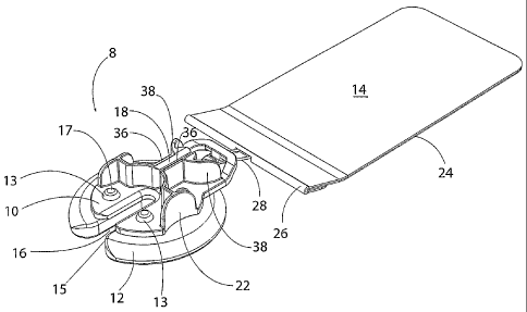
As shown in Figures 1-3, a button anchor 8 of this invention comprises an
anchoring portion 10, which rests on an anchor pad 12 and which can optionally
engage a
load distributing anchor tail 14. This button anchor 8 remains external to the
human or
animal tissues, and comprises specific features for anchoring a fac traveling
across a
wound or through tissues that, by its presence and ability to apply a reducing
force,
provides the specific benefit of moving or moving and stretching tissue to
bring reduction
or closure of a full thickness wound where the wound margins lie beyond a
distance
where they can be primarily closed without undue force. In one example, a fac
is passed
through the skin, engaging or encircling the sub-dermal structures requiring
closure, and
CA 02578759 2007-03-01
WO 2006/027678 PCT/IB2005/002679
returned through the skin on the other side of a wound or incision. The button
anchors 8
are applied to the ends of the fac, allowing the fac to be tensioned and
anchored, thereby
applying a sub-dermal reduction force, as illustrated in Figure 1. In an
alternative
embodiment, button anchors 8 positioned on opposite sides of a wound secure a
fac that
passes over the wound and that does not penetrate the tissue.
As shown in Figures 2-5, the anchoring portion 10 has a large slot 16 and a
smaller slot 18 for engagement of an efac, such as an elastomer. Slot 18
includes walls
36 and is a metered tension, elastomer-locking slot, with a shape, length and
size such
that the slot 18 captures and anchors the elastomer but allows the elastomer
to migrate if
tension exceeds a pre-determined level, thereby creating a limit to the amount
of force
that can be applied by the system. This limit is determined at the time of
manufacture of
the anchoring portion 10 by controlling the relationship between the size of
the slot 18
and the diameter or cross-sectional area of the elastomer. The cross-sectional
area of the
untensioned portion of the elastomer decreases as the elastomer elongates
under increased
tension. If a force applied to the elastomer exceeds the therapeutic force
range,
elongation and resulting reduction in diameter cause the elastomer to release
within the
slot, returning the quantity of tension to one within the therapeutic limit of
the elastomer.
Convex upstanding regions 38 (visible in Figures 1 and 4) of the anchoring
portion
10 prevent other objects from catching the edges of the button anchor 8.
The anchoring portion 10 may be molded of polycarbonate plastic or any other
appropriately rigid and strong polymeric material suitable for use in the
surgical
applications for which the present invention is intended. Alternatively it may
be molded,
machined or otherwise formed or fabricated of any other suitably strong,
surgically
acceptable material such as stainless steel.
16
CA 02578759 2007-03-01
WO 2006/027678 PCT/IB2005/002679
While the size of the button anchor 8 of this invention may be varied
depending on
the situation in which it is used, anchoring portion 10 may be approximately
32mm in
diameter. An anchoring portion 10 for use with an elastomeric three mm
diameter, 40
durometer silicone cord may have a slot 18 one mm in width (i.e., the distance
between
walls 36), 7.3 mm in height and 11mm in length. Many other dimensions are also
usable
provided that the desired coupling with elastomer is achieved (generally as
described
above).
Various arcuate or curved surface shapes for anchor efacs attachment
structures
are described above. It should be understood that functionally equivalent
shapes can also
be used, such as, for instance, a rod having a cross-section that is not
curved but rather is
a polygon.
As shown in Figures 6 and 7, anchor pad 12 includes a slot 15 that corresponds
to
slot 16 of the anchoring portion 10. Anchor pad 12 dissipates the compression
load
exerted by one or more facs connected to the anchoring portion 10 over the
surface of the
patient's skin and works to prevent maceration or undue restriction of the
underlying
blood circulation. The anchor pad 12 is generally the same size and shape as
the
anchoring portion 10, but it may be smaller or larger in alternative
embodiments. For
example, larger pads may be used in patients with compromised skin tissues,
including
the elderly or those with associated co-morbidities, such as diabetes.
. The anchor pad 12 may be made of a compressible material such as silicone,
or
any other suitable material. The skin contact surface (i.e., the underside) of
anchor pad
12 may be smooth or it may be textured in order to accommodate fluid
dissipation. The
skin contact surface may be flat, convex, concave or multi-planar to
accommodate
anatomical contour. The skin-contacting surface of pad 12 may also be coated
or treated
17
CA 02578759 2007-03-01
WO 2006/027678 PCT/IB2005/002679
to provide antimicrobial properties. In one embodiment, the skin-contacting
surface of
the anchor pad includes an adhesive.
As shown in Figure 5, the anchoring portion 10 is penetrated by apertures 20
that
secure the anchoring portion 10 to the anchor pad 12. Tabs 13 (shown in Figure
7)
project from anchor pad 12 and are received in apertures 20 of anchoring
portion 10.
Enlarged diameter end 17 of tabs 13 retain anchoring portion 10 on pad 12. In
an
alternative embodiment, the anchor pad 12 is adhered, adhesively bonded, or
molded to
anchoring portion 10. In one example, the anchor pad 12 and anchoring portion
10 are an
integral unit.
As shown in Figures 2 and 5, finger grips 22 facilitate gripping and
manipulating
the button anchor 8 by opposed digits. Finger grips 22 are concave in the
embodiment
illustrated in the drawings, but the gripping portion may also be convex,
multi-planar or
textured.
Optional anchor tail 14, shown in Figures 2, 3 and 8, may be utilized to
further
dissipate and distribute the shear-load placed on the skin by performing wound
closure
over the maximum possible surface area. In one embodiment, the anchor tail 14
is
formed from polyurethane foam having an adhesive for attachment to the skin
and
includes a wire that forms a loop 28 at end 26. In alternative embodiments,
the anchor
tail 14 may be formed from any suitable fabric, foam or film. Such material
may be
elastic or inelastic. Preferably the anchor tail 14 material conforms to the
skin surface
and mimics the elasticity of the skin. In addition, the loop 28 may be formed
or molded
as a separate or integral component.
Anchoring portion 10 of button anchor 8 includes structure for engaging anchor
tail 14. Such structure may include a hole, tab, cleat or other suitable
structure. In one
18
CA 02578759 2007-03-01
WO 2006/027678 PCT/IB2005/002679
embodiment, shown in the Figures, and particularly in Figure 9, the anchoring
portion 10
includes a hook 30 having a ramp 32 for guiding the wire loop 28 of tail 14 up
and into
depression 34 of anchoring portion 10. In use, the anchor tail 14 is attached
to the
anchoring portion 10 via the engagement hook 30 and is adhered to the skin. In
this
manner, anchor tail 14 bolsters the button anchor 8 and dissipates the forward
force load
(a force vector that travels toward the wound edge and parallel to the skin
surface) over a
large area of healthy skin located behind the button anchor 8. While the hook
30 and loop
28 provide one example of structure to couple the anchor tail and anchor, any
suitable
structure may be used.
The system of this invention may be used to provide deep fascia repair and
deep
fascia dynamic wound reduction. In one embodiment, illustrated in Figures 10,
a silicone
elastomer 13 is coupled to a cannula-like device 42 and is passed through the
dermis 44,
fat layer 46, and fascia 48 at an optional anchor placement mark 50 placed on
the skin
prior to installation of the system. After passing through the area of the
wound 7, the
elastomer 13 is presented through slot 16 of anchoring portion 10 and slot 15
of anchor
pad 12 of button anchor 8, where it is then captured and secured in smaller
slot 18 of
anchoring portion 10. In this manner, closure force is applied to a wound or
incision 7.
Multiple sets of anchors and elastomers may be used, as shown in Figure 1.
The elastomer 13 may either be presented through the skin and through the slot
16
of an anchor previously placed, or the elastomer 13 may exit the skin, at
which time the
slot 16 and the pad slot 15 of the anchor 8 may be moved into place around the
elastomer
13. The efac may be used to apply tension to sub-dermal structures (deep
fascia) but the
efac tension may be adjusted from above the skin by increasing or decreasing
the tension
at the smaller slot 18. The anchor 8 acts as a grommet, removes the point load
from the
19
CA 02578759 2007-03-01
WO 2006/027678 PCT/IB2005/002679
exit hole to reduce the occurrence of localized failures, and also allows
adjustment of the
tension across the wound. In this manner, the anchor bolsters the perimeter of
the
transcutaneous opening through which the elastomer passes, reducing localized
failures
and also reducing scarring.
A system according to this invention may provide wound stabilization of
abdominal procedures. For example, this system may be used to restore radial
abdominal
integrity during prolonged interventions for complications such as abdominal
infections
management or which require large abdominal access. This system increases
patient
comfort and mobility by providing abdominal containment and support, and
maintains
normal skin tensions during intervention to minimize retraction.
Another system of this invention may provide stability to sternal or chest non
unions as can arise after open heart surgical procedures. In addition, systems
of this
invention may be used with conventional primary wound closure methods to
distribute
skin system tensions to healthy skin beyond the wound, thereby minimizing
stress at the
wound site and reducing dehiscence. A system of this invention may be applied
pre-
operatively to tension skin and create surplus tissue, allowing excisions to
be covered and
closed in a conventional manner. Embodiments of this invention may also be
used as a
dressing retention system by providing efac lacing across the wound site,
which passes
over the wound dressing and secures it in position. Adhesives may be used on
the skin
contacting surface of the anchor pad but such adhesives normally would not be
required,
thereby further facilitating the periodic inspection and cleaning of tissues
under the
anchor pads.
All of the tissue attachment structure and anchor designs described herein may
be
produced in a variety of sizes.
CA 02578759 2007-03-01
WO 2006/027678 PCT/IB2005/002679
The systems and methods of moving or moving and stretching plastic tissue
according to this invention are not confined to the embodiments described
herein but
include variations and modifications within the scope and spirit of the
foregoing
description and the accompanying drawings. For instance, the scale of the
components of
the invention can vary quite substantially depending on the nature and
location of the
tissue with which the invention is used. The configuration of the tissue
attachment
structures can also be varied for the same reasons and for aesthetic reasons.
While most
of the elements of the illustrative embodiments of the anchors of this
invention depicted
in the drawings are functional, aspects of the shape and appearance of the
illustrative
embodiments are nonfunctional and ornamental.
The materials from which the components used in practicing this invention are
made can be those described above as well as others, including materials not
yet
developed that have appropriate properties of strength, elasticity and the
like that will be
apparent to those skilled in the art in light of the foregoing. For instance,
useful materials
generally must be sterile or sterilizable and non-reactive. The illustrated
components are
typically intended to be disposable, but the invention can also be practiced
using reusable
components.
21