Note: Descriptions are shown in the official language in which they were submitted.
CA 02578817 2007-03-01
WO 2006/031500 PCT/US2005/031658
RECONSTITUTING INFUSION DEVICE
Field of the Invention
The present invention relates generally to medical devices and methods for the
preparation and adininistration of therapeutic or other coinpounds to a
patient, and
more particularly to drug reconstitution and administration systems and
inethods
which facilitate optimal reconstitution, mixing and dilution of a drug and/or
other
coinpound with a liquid diluent, and subsequent adininistration of the
resultant
mixture from an infusion type device.
Background of the Invention
Infusion therapy is a widely known therapy for patients who require
medicaments to be delivered over some'time period. Diabetic infusion pump
therapy,
which entails the purchase of an expensive puinp that lasts for about three
years, has
possibly the largest population of outpatient infusion therapy. The initial
cost of the
pump is a high barrier to this type of therapy. From a user perspective,
however, the
overwhelming majority of patients who have used pumps prefer to remain with
pumps
for the rest of their lives. This is because infusion pumps, although more
complex
than syringes and pens, offer the advantages of continuous infusion of
insulin,
precision dosing and programmable delivery schedules. This results in closer
glucose
control and an improved feeling of wellness.
As patients on oral agents eventually move to insulin and their interest in
intensive therapy increases, users typically look to insulin puinps. However,
in
addition to their high cost (roughly 8 to 10 times the daily cost of syringe
therapy) and
limited lifetime, insulin pumps represent relatively old technology and are
cumbersome to use. Also, fiom a lifestyle standpoint, the tubiilg (known as
the
"infusion set") that links the pump witlz the delivery site on the user's
abdomen is very
inconvenient and the puinps are relatively heavy, making carrying the puinp a
burden.
Therefore interest in better therapy is on the rise, accounting for the
observed
growth in pump therapy and increased number of daily injections. In this and
similar
infusion examples, what is needed to fully meet this increased interest is a
forin of
insulin delivery or infusion that combines the best features of daily
injection therapy
1
CA 02578817 2007-03-01
WO 2006/031500 PCT/US2005/031658
(low cost and ease of use) with those of the insulin pump (continuous infusion
and
precision dosing) and that also avoids the disadvantages of each.
Several attempts have been made to provide ambulatory or "wearable" drug
infusion devices that are low in cost and convenient to use. Some of these
devices are
intended to be partially or entirely disposable. In theory, devices of this
type can
provide many of the advantages of an infusion puinp without the attendant cost
and
inconvenience. Unfortunately, however, many of these devices suffer from
disadvantages including user discomfort (due to the gauge and/or length of
injection
needle used), compatibility and interaction between the substance being
delivered and
the materials used in the' construction of the infusion device, and possible
malfunctioning if not properly activated by the user (e.g., "wet" injections
resulting
from premature activation of the device). Long-term drug stability has also
been an
issue for these types of devices, and therefore a majority of drugs, when in
liquid form
must be refrigerated.
In order to combat the drug stability problem, storage-stability can be
imparted
to medicaments by placing them in a dry powder form. Techniques for doing this
include freeze-drying, spray freeze-drying, lyophilization and the like.
However,
reconstitution of such medicaments has been difficult and involves many steps.
Additionally, reconstituted liquids typically do not have the same properties
as a
liquid drug formulation, at least because bubbles may be formed during
reconstitution.
Various methods to disrupt bubbles in reconstituted forinulations have been
atteinpted in the past. Most of these inethods use application of ultrasonic
energy. The
ultrasonic effect is based on what is lcnown as cavitation, i.e., cavities
containing gas
forined by sound waves. These cavities collide with each other forming larger
cavities
that then rise to the surface and dissipate. These methods require specialized
and
bulky equipment and power sources. Yet another drawbaclc of cavitation is the
momentary, yet intense, burst of heat generated as each bubble collapses. The
heat
generated can certainly destroy some active coinponents or unstable drugs in
the
product. It would be desirable to have a method for reinoving bubbles from a
reconstituted solution, which did not have such issues as high-energy input
and heat
generation. Other inethods to reduce bubble fonnation that have been attempted
are
2
CA 02578817 2007-03-01
WO 2006/031500 PCT/US2005/031658
application of a high pressure. It is theorized that high pressures reduces
bubble
fomiation because the rate of bubble collapse is proportional to G, the
gradient
between external tension and bubble internal pressure. The higher external
tension can
shrinlc bubbles. With a decrease in diaineter of the bubbles, the iricreased
internal gas
pressure forces the gas inside the bubble to dissolve, resulting in bubble
collapse as
the gas is forced into solution. Nevertheless, this approach also requires
additional
equipment safeguards and is not feasible for many applications due to either
safety or
cost concerns. Additionally, a bubble will form again after high pressure is
removed,
such as when the reconstituted product is drawn out of a pressured vial.
To date, however, there remains a need for a systein for the administration of
medicaments where the medicament is in a storage stable dry form, which can be
readily reconstituted and directly administered via an infusion type device.
Additionally, the reconstituted drug should have properties, which nzimic the
pre-
inixed liquid formulation. Accordingly, a need exists for an alternative to
current
infusion devices, such as infusion pumps for insulin that further provides
siniplicity in
manufacture and ease-of-use for both insulin and non-insulin applications.
Suimnary of the Invention
The present drug reconstitution and administration system is a inulti-
component arrangement normally enclosed witllin a housing which permits a
concentrated drug or other composition to be mixed with a liquid diluent from
a pre-
filled cartridge assembly, with the systein further permitting the infuser
reservoir to be
filled with the resultant mixture for patient administration. The system
permits drugs
to be efficiently stored and handled in concentrated forin, and further
facilitates
dilution or reconstitution of the drugs to the desired concentration just
prior to
adininistration through the use of the integrated coinponents of the systein.
In accordance with the illustrated embodiments, the present system includes a
container, or cartridge for containing a drug or other medicament, witll the
container
having a pierceable stopper for closing the container. The system f-urther
includes an
infuser asseinbly including a reservoir having a filling end, an.d a patient
needle end
defining a flow passage therebetween. The reservoir defines an internal
chainber in
fluid coimnunication with the flow passage so that liquid can be moved into
and out
of the internal chainber via the flow passage. The reservoir may be
constructed in
3
CA 02578817 2007-03-01
WO 2006/031500 PCT/US2005/031658
accordance with the reservoir construction of US Patent application of
Cindrich et al.,
Serial Nos. 10/916,649 and 10/916,648, filed on August 12, 2004, the entire
content
of which is incorporated herein by reference.
The present system furtlier includes a mixing adapter assenibly for inixing a
liquid in the reservoir asseinbly with a inedicament in the cartridge. The
adapter
assembly includes a generally cylindrical receiving inlet having an access
needle for
fluid connection with the pierceable stopper of the associated cartridge. By
this
arrangement, the end of the cartridge, and the stopper positioned therein, can
be
positioned in one end of the receiving inlet sleeve of the adapter assembly.
The
receiving inlet has an inside diameter larger than the outside diameter of the
associated cartridge, tllus permitting the cartridge asseinbly to be
positioned generally
telescopically within the receiving inlet during use of the systein.
The access needle of the receiving inlet and the cartridge assembly connect to
the flow passage of the reservoir assembly and the patient needle in selective
fluid
conununication with each other. Optionally, a valve is placed in the fluid
path
between the patient needle and the reservoir. By this aiTangement, the present
system
pennits reconstitution of a concentrated drug by positioning the drug filled
cartridge
asseinbly generally within the open end of the receiving inlet with the
components
slidably engaged to each other. In this configuration, a liquid, such as a
diluent, pre-
filled in the internal chainber of the reservoir assembly, can be caused to
flow through
the flow passage of access needle into the cartridge assembly by action of a
low-
pressure condition in the cartridge assembly. The reconstitution is effected
by the
diluent becoming in contact with the drug. The cartridge asseinbly is then
slid
longitudinally further into the receiving inlet, however, the stopper is
prevented from
further translation due to the interaction with the access needle hub. Thus
the stopper
is fixed in relation to the housing and is now translated with respect to the
cartridge
asseinbly such that the stopper forces the now drug/diluent mixture from the
internal
chamber of the cartridge and through the access needle into the reservoir
assembly.
The liquid from the reservoir assembly is tlius mixed with the medicament from
the
cartridge within the chainber of the cartridge, and forced under pressure back
into the
reservoir assembly. The desired diluted drug mixture is thus provided witllin
the now
filled reservoir assembly, with the now-empty chamber fitted within the
receiving
inlet.
4
CA 02578817 2007-03-01
WO 2006/031500 PCT/US2005/031658
When inixing is complete, the present system facilitates administration of the
mixture by methods and devices according to US Patent application of Cindrich
et al.,
Serial Nos. 10/916,649 and 10/916,645, filed on August 12, 2004, the entire
conteilt
of which is incorporated herein by reference. A general description of the
action of
the infuser is as follows: The device is self-contained and is attached to the
skin
surface of the user by adhesive disposed on a bottom surface. Once properly
positioned and activated by the user, a pressurizing system acts on a
reservoir surface
within the device can be used to empty the contents of the partially flexible
reservoir
through one or more patient needles via a needle manifold. The substance
within the
reservoir is then delivered through the skin of the user by the needles, which
are
driven into the skin. It will be understood that other embodiments are
possible in
which the pressurizing systein is replaced with a different type of energy
device,
which may be mechanical, electrical and/or cheinical in nature inter alia gas
generation pressurizing means, mechanical actuators, or shape inemory alloys.
In the preferred form, the reservoir asseinbly is provided in a closed form to
maintain its sterility, such as by the preferred provision of a blister
paclcage with pair
of peel-away seals or like closing elements positioned at respective opposite
ends of
the adapter assembly. The arrangement is preferably configured for single-use,
and to
this end, a locking arrangeinent is provided which prevents removal of the
cartridge
from the adapter assembly after it has been connected with the receiving
inlet.
Additionally, it has been found that pressure treatments of the medicament
within the cartridge have an unexpected benefit to the quality of the
reconstituted
drug/diluent solution. For exainple, low pressure conditions in the drug
reservoir not
only serve the purpose of filling the cartridge with the diluent upon fluid
connection
to the reservoir, the resultant mixture has a lower observable amount of
bubbles.
Additionally in certain applications, it may be desirable to replace the
atinospheric
gasses normally present with the drug in the cartridge with inert gasses,
intef alia
argon, heliuin to further iinprove the reconstitution characteristics.
These and other aspects of the invention are substantially achieved by
providing systems and methods for a patch-like, wearable, self-contained
reconstituted substance infusion device which provides one or more
substantially
hidden patient needles which can be placed in fluid coinmunication with a
content
5
CA 02578817 2007-03-01
WO 2006/031500 PCT/US2005/031658
reservoir assetnbly that includes a rigid bladder portion used in conjunction
with a
non-distensible bladder film, such as a metallized film. A connection is
provided for a
reconstitution fluid and/or dry powdered drug. A push type activation assembly
is
provided which can then be used to remove a retaining pin and allow a Disk
spring
asseinbly to apply an essentially even and constant pressure to the contents
of a
reservoir assembly. The push type activation asseinbly then releases and seats
one or
more spring-loaded patient needles into the patient's skin and establishes a
fluid
cominunication patll between the patient needles and the pressurized reservoir
contents, thereby delivering an infusion of contents into the skin of the
user. Upon
completion aiid removal of the infusion device, a number of safety mechanisms
can
be engaged to cover the needles for disposal.
Brief Description of the Drawings
The various objects, advantages and novel features of the preferred
embodiinents of the present invention will be more readily appreciated from
the
following detailed description when read in conjunction with the appended
drawings,
in which:
Fig. lA is a top perspective view a patch-like injector or infusor system
using
a two component mixing systein prior to activation.
Fig. 1B is a bottom perspective view of the patch-like injector of Fig lA.
Figs 2A is a first exploded view of the patch-like injector of Fig lA showing
the reservoir asseinbly and upper housing.
Figs 2B is a second exploded view of the patch-like injector of Fig lA
showing the button assembly, needle manifold and lower housing.
Figs 2C is a third exploded view of the patch-like injector of Fig lA showing
the cartridge assembly and access needle.
Fig. 3 is a plan view of the patch-like injector of Fig 1A, showing axes A-A,
C-C and B-B.
Fig.3A is a cross-sectional side view along axis A-A of the patch like
injector
of Fig. 3, shown prior to insertion of the cartridge.
6
CA 02578817 2007-03-01
WO 2006/031500 PCT/US2005/031658
Fig.3B is a cross-sectional side view along axis B-B of the patch like
injector
of Fig. 3, shown prior to insertion of the cartridge.
Fig.3C is a cross-sectional side view along axis C-C of the patch like
injector
of Fig. 3, shown prior to insertion of the cartridge.
Fig.3D is a cross-sectional top perspective view along axis B-B of the patch
like injector of Fig. 3, shown prior to insertion of the cartridge.
Fig.4A is a cross-sectional side view along axis A-A of the patch like
injector
of Fig. 3, shown after insertion of the cartridge.
Fig.4B is a cross-sectional side view along axis B-B of the patch like
injector
of Fig. 3, shown after insertion of the cartridge.
Fig.4C is a cross-sectional side view along axis C-C of the patch like
injector
of Fig. 3, shown after insertion of the cartridge.
Fig.4D is a cross-sectional top perspective view along axis B-B of the patcll
like injector of Fig. 3, shown after insertion of the cartridge.
Fig.5A is a cross-sectional side view along axis A-A of the patch like
injector
of Fig. 3, shown after access to the reservoir.
Fig.5B is a cross-sectional side view along axis B-B of the patch like
injector
of Fig. 3, shown after access to the reservoir.
Fig.5C is a cross-sectional side view along axis C-C of the patch like
injector
of Fig. 3, shown after access to the reservoir.
Fig.5D is a cross-sectional top perspective view along axis B-B of the patch
like injector of Fig. 3, shown after access to the reservoir.
Fig.6A is a cross-sectional side view along axis A-A of the patch like
injector
of Fig. 3, shown after transfer of the fluid.
Fig.6B is a cross-sectional side view along axis B-B of the patch like
injector
of Fig. 3, shown after transfer of the fluid.
7
CA 02578817 2007-03-01
WO 2006/031500 PCT/US2005/031658
Fig.6C is a cross-sectional side view along axis C-C of the patch like
injector
of Fig. 3, shown after transfer of the fluid.
Fig.6D is a cross-sectional top perspective view along axis B-B of the patcll
like injector of Fig. 3, shown after transfer of the fluid.
Fig.7A is a cross-sectional side view along axis A-A of the patch like
injector
of Fig. 3, shown after pressurization of the reservoir.
Fig.7B is a cross-sectional side view along axis B-B of the patch like
injector
of Fig. 3, shown after pressurization of the reseivoir.
Fig.7C is a cross-sectional side view along axis C-C of the patch like
injector
of Fig. 3, shown after pressurization of the reservoir.
Fig.7D is a cross-sectional top perspective view along axis B-B of the patch
like injector of Fig. 3, shown after pressurization of the reservoir.
Fig.8A is a cross-sectional side view along axis A-A of the patch like
injector
of Fig. 3, shown after deployment of the needle.
Fig.BB is a cross-sectional side view along axis B-B of the patch like
injector
of Fig. 3, shown after deployrnent of the needle.
Fig.8C is a cross-sectional side view along axis C-C of the patch like
injector
of Fig. 3, shown after deployinent of the needle.
Fig.8D is a cross-sectional top perspective view along axis B-B of the patch
like injector of Fig. 3, shown after deploylnent of the needle.
Throughout the drawings, like reference numerals will be understood to refer
to like parts, coinponents or structures.
8
CA 02578817 2007-03-01
WO 2006/031500 PCT/US2005/031658
Detailed Description of the Exemplary Embodiments
The aspects of the present device described below can be used as a convenient,
patch-like device to deliver a pre-measured dose of a substance, such as a
drug or
medication, which has been separated into at least two coinponents (typically
a liquid
diluent, and a dry powder), to a user through an adhesive attached infusion
device.
The device is self-contained and is attached to the skin surface of the user
by adhesive
disposed on a bottom surface. Typically, the two components are mixed by the
device
and then transferred to the reservoir. Once properly positioned and activated
by the
user, a pressurization system on a reservoir surface within the device can be
used to
enlpty the contents of the partially flexible resetvoir through one or more
patient
needles via a needle manifold. The mixed substance within the reservoir is
then
delivered through the skin of the user by the needles, which are driven into
the skin.
It will be understood that other embodiments are possible in wllich the
pressurization system is a variation of a disk spring, disk spring, or
different type of
stored energy device, which may be mechanical, electrical and/or cheinical in
nature.
It will also be understood that the terins mixing and reconstitution are used
interchangeably herein w11en referring to the mixture of drug coinponents in
the
cartridge and the reservoir. Medicament components to be mixed may be gasses,
solids, liquids, dry powders, suspensions, or a mixture of any of these.
Although many
of the examples herein are of binary systeins e.g. dry powder and diluent, it
will be
understood that multiple mixing operations may be performed so that more than
two
components may be mixed in a sequential fashion. As will be appreciated by one
skilled in the art, there are nuinerous ways of carrying out the patch-like
injection or
infusor systein disclosed herein. Although reference will be made to the
einbodiments depicted in the drawings and the following descriptions, the
embodiments disclosed herein are not meant to be exhaustive of the various
altemative designs and embodiinents that are encoinpassed by the disclosed
invention.
In each disclosed embodiment, the device is referred to as an infusor;
however, the
device may also inject substances at a inuch faster bolus rate than is
coininonly
accoinplished by infusor devices. For exainple, the contents can be delivered
in a
period as short as several seconds, or as long as several days.
9
CA 02578817 2007-03-01
WO 2006/031500 PCT/US2005/031658
As shown in Figs. 1A through 8D, the einbodiment of certain aspects of
present invention can be constructed to provide a patch-like, wearable, self-
contained
substance infusion device that can be used to deliver a variety of multiple-
component
medications to a patient e.g. dry powder and diluent. The device also provides
for a
separated drug container called a cartridge, which is filled with at least one
coinponent of the drug to be delivered to the patient. The device provides a
hidden
patient needle or needles prior to and during use, and can be secured to a
patient via
an adhesive surface. The pressurization of the contents of the reservoir can
be
achieved by reinoving or displacing the spring retention disk, as described in
greater
detail below, to pressurize the device contents and the device can then be
further
activated via a reasonable force applied to the top push surface to seat the
patient
needles. Alternatively, the patient may push on the side of the device to
allow a
mechanism to seat the needles. In doing so, the device facilitates self-
injection and
reduces or eliminates variations in injection techniques between users.
In an exemplary embodiment of aspects of the present invention shown in
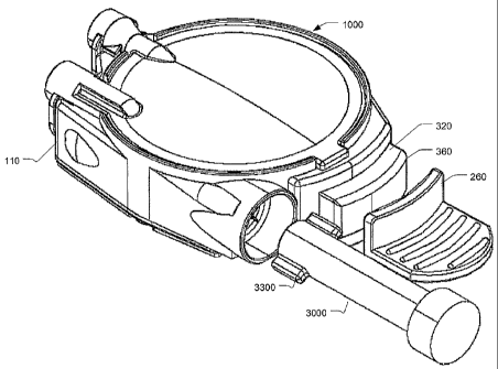
FIGS. 1A through 8D, an infusion device 1000 includes a reservoir subassembly
100,
including an upper housing 110, a reservoir base surface 120, at least one
Disk spring
130, a retaining pin 140, fill plug 150, septuin 160 and reservoir film 170.
The
infusion device 1000 furtller includes a housing subassembly 200, including a
lower
housing 210, aild patient needle maizifold 220 having at least one patient
needle 222
and a manifold fihn 224. The housing subassembly 200 f-urther includes a
needle
shield 230, needle shield drive spring 232 and an adjustable needle cap 240.
An
adhesive layer 250 is disposed upon the lower surface of the lower housing
210, and
can be covered by a reinovable film (not shown), and a pull handle 260. A
clip, such
as an "E" clip can be used to secure the retaining pin 140 to the pull handle
260.
Alternatively, pin 140 may be integrally fonned into pull handle 260. The
infusion
device 1000 further includes a push button subassembly 300, including at least
one
patient needle inanifold drive spring 310, a push button slide 320, at least
one septuin
needle 330, and a fluid communication tube 350. A button face 360 can be
provided
to coinplete the push button subassembly 300. The infusion device further
includes
cartridge asseinbly 3000, which contains a portion of the inedicainent to be
delivered
to the patient. Cartridge 3000 is inserted into housing 110. Subsequently,
througli a
series of steps, medicainent coinponents in the reservoir 100 are mixed with
CA 02578817 2007-03-01
WO 2006/031500 PCT/US2005/031658
medicament components in the cartridge 3000, and are finally deposited into
the
reservoir 100 for infusion into the patient via microneedles 222. In the
description
below, the term reservoir is often used to describe the asseinbled and
separate
reservoir base surface 120, fill plugs 150, 151, septums 160, 161 and
reservoir film
170 of the reservoir subassembly 100.
FIG. lA is a top perspective view of a first einbodiment of the infusion
device
1000. In FIG. lA, the assembled upper and lower housing 110 and 210
respectively is
shown, between which the push button subassembly 300 is contained. The pull
handle
260, described in greater detail below, is shown in a pre-energized, pre-
activated
position and serves to secure the retaining pin 140 within the device and
shield the
push button 360 fronl any applied forces. As more clearly illustrated in FIG.
1B,
which is a bottom perspective view of the first embodiment, the pull handle
260 is
further interlocked with the needle cap 240 and the retaining pin 140 via clip
270.
Also, the pull handle 260 is optionally further interlocked with the push
button slide
320.
As shown in FIGS. 2A through 2C, the embodiinent of the present invention
1000 can be constructed of these subassemblies to provide a patch-like,
wearable,
self-contained substance infusion device that can be used to deliver a variety
of
medications to a patient. The device 1000, shown in a pre-reconstitution, pre-
energized, pre-activated position in FIG. lA, provides a hidden patient needle
or
needles prior to and during use, and can be secured to a patient via an
adhesive
surface. The reconstitution of the finished drug to be delivered can be
aclzieved by the
insertion of cartridge 3000 into housing 110. The pressurization of the
contents of the
reservoir can be achieved by removing the pull handle 260 to "energize" the
device
and device contents, and the device can then be "activated" via a reasonable
force
applied to the push-button 360 to seat the patient needles and establish a
flow path
between the reservoir and needles. In doing so, the device 1000 facilitates
self-
injection and reduces or eliminates variations in injection techniques between
users.
In FIG. 2A, the reservoir subasseznbly 100 of the infusion device 1000 is
shown, and can be comprised of a rigid portion 120 used in conjunction with
one or
more non-distensible but flexible films 170, such as metallized films. The
reservoir
subassembly 100 can contain any number of substances between either a first
and
11
CA 02578817 2007-03-01
WO 2006/031500 PCT/US2005/031658
second film, where either the first or second film is also positioned against
the rigid
portion, or between a first film and the rigid portion. The reservoir is
preferably filled
with a liquid diluent. The rigid portion 120, or reservoir base, caii be
comprised of and
serve as a hard portion of the reservoir against which the flexible film 170
can be
pressed as described in greater detail below. As noted above, the reservoir of
the
embodiment shown in FIG. 2A can be constructed to preferably have a hard shell
or
inner surface, and at least one flexible film attached about the perimeter of
the hard
shell or inner surface. The flexible film 170 can be heat sealed to the rigid
portion 120
to create a chainber, or bladder, for storage of device contents. As at least
one wall of
the chaniber comprises a flexible film 170, and at least one wall of the
cllamber
coinprises a rigid surface, one or more Disk springs 130 can be placed
adjacent to the
flexible fihn 170 and used to apply a substantially constant pressure to the
flexible
film 170, and pressurize the reseivoir chainber and contents. Although a disk
spring is
primarily disclosed, any type of pressurization systein may be used with
aspects of the
invention. Septum 160 is inserted into rigid portion 120 at recess 1600 to
seal the
access path to inicroneedles 222, and septum 161 is inserted into rigid
portion at
recess 1610 to seal the access path to cartridge 3000. Additionally, fill plug
150 is
inserted into recess 1500 and fill plug 151 is inserted into recess 1510 to
provide
closure of chamber 127 in reservoir 100.
The reservoir of the reservoir subassembly 100 is further preferably able to
be
stored for the prescribed shelf life of the reseivoir contents in applicable
controlled
environments without adverse effect to the contents and is capable of
applications in a
variety of envirorunental conditions. Additionally, the barrier provided by
the
coniponents of the reservoir do not pennit the transport of gas, liquid and
solid
materials into or out of the contents at a rate greater than that allowable to
meet the
desired shelf life. In the embodiment shown in FIG. 2A, the resetvoir
subasseinbly
materials are capable of being stored and operated in a temperature range of
approximately 0 to 120 degrees F, and can have a shelf life of two or more
years.
Other variations of materials may be selected which allow for therinal cycles
to room
teinperature and back to cold storage, as well as other teinperature operating
ranges,
beyond 0 to 120 degrees F.
Now referring to Fig. 2C, which shows the cartridge assembly 3000. The
cartridge assembly provides cartridge barre13100, which is prismatic in nature
witli a
12
CA 02578817 2007-03-01
WO 2006/031500 PCT/US2005/031658
cylindrical cross-section, although any shape may be used. Cartridge
barre13100 has a
button end 3175 and an access end 3125, and an internal portion 3150. At the
access
end 3125 of cartridge body 3100 is fitted a slidable, pierceable stopper 3160.
Stopper
3160 is slidably engaged to the internal diameter 3150 of cartridge barrel
3100.
Stopper 3160 is slidable from access end 3125 to button end 3175. At the
button end
3175 of cartridge barrel 3100 is plug 3200, which seals button end 3175 of
cartridge
baiTe13100. Cartridge barre13100 also has at least one tang 3300, and as shown
in the
drawings two tangs 3300A and 3300B. Tangs 3300 are provided for guiding
cartridge
3000 into housing 110 and optionally for locking cartridge 3000 into housing
110 at
the end of the mixing step. In an alternate einbodiment, cartridge barrel 3100
is a test-
tube like structure, with a single opening which forms the access end, thus
eliminating
the need for plug 3200. Optionally, plug cover 3150 covers plug 3200 for a
more
pleasing aesthetic appearance. In another alternate embodiment, cartridge
barre13100
is an evacuated blood collection tube-like structure, with a single opening
which
forms the access end, and barrier properties which mimic evacuated blood
collection
tubes, thus eliininating the need for plug 3200. When assembled, the
components of
cartridge assembly 3000 fonn internal chamber 3500 which is contains a portion
of
the medicament to be mixed and delivered inter alia a dry powder.
The materials of the cartridge subassembly 3000 are further preferably able to
be stored for the prescribed shelf life of the cartridge contents in
applicable controlled
envirorunents without adverse effect to the contents. Additionally, the
barrier
provided by the coinponents of the reservoir do not permit the transport of
gas, liquid
and solid materials into or out of the contents at a rate greater than that
allowable to
meet the desired shelf life. In the embodiment shown in FIG. 2C, the cartridge
subassembly materials are capable of being stored and operated in a
temperature
range of approximately 0 to 120 degrees F., and can have a shelf life of two
or more
years. Preferably the cartridge subasseinbly is adapted to contain a vacuum
for the
entire shelf life of the system. Other variations of materials may be selected
which
allow for thermal cycles to room teniperature and baclc to cold storage, as
well as
other temperature operating ranges, beyond 0 to 120 degrees F.
Fig. 2C also shows an exploded view of access needle assembly 2000 which is
a hub having double pointed needle assembly. In this embodiment the double
pointed
needle asseinbly has separated stopper needle 2400 and septum needle 2330
affixed to
13
CA 02578817 2007-03-01
WO 2006/031500 PCT/US2005/031658
hub 2500. In an alternate embodiment, access needle 2000 is formed from a
single
double pointed needle having a stopper end 2450 and a septum end 2350. Access
needle 2000 is contained within and slidably engaged to housing 110. Stopper
end
2450 is adapted to penetrate stopper 3160 and Septum end 2350 is adapted to
penetrate septum 161. Access Needle 2000 provides for a selectable fluid
conduit
from the chamber 127 of reservoir 1000 to chamber 3500 of cartridge 3000.
The reservoir of the reservoir subassembly 100 is preferably evacuated prior
to
filling, as described in greater detail below. In addition, the shape of the
reservoir may
be configured to adapt to the type of energizing mechanisin used, e.g., a disk
or
Belleville spring 130 having any number of diameters and height dimensions.
Additionally, using an evacuated flexible reservoir during filling mininiizes
any air or
bubbles within the filled reservoir. The use of a flexible reservoir is also
very
beneficial when the device is subjected to external pressure or tenlperature
variations,
which can lead to increased internal reservoir pressures. In such case, the
flexible
reservoir expands and contracts with the contents, thereby preventing possible
leaks
due to expansion and contraction forces exerted on the fill plugs 150, 151 and
septum
160, 161. This also helps to eliminate dose variation due to teinperature and
pressure
fluctuations in the enviromnent. Additionally, a flexible reservoir ensures
the ability
of a vacuuin in the cartridge to enable temporary filling of cartridge 3000
for
reconstitution, and subsequent re-filling of reservoir 100.
Yet anotlier feature of the reservoir subasseinbly 100 includes the ability to
permit automated particulate inspection at the tiine of fill, or by a user at
the time of
use. One or more reservoir barriers, such as the rigid portion 120, can be
molded of a
transparent, clear plastic material, which allows inspection of the substance
contained
within the reservoir. The transparent, clear plastic material is preferably a
cyclic olefin
copolymer that is characterized by high transparency and clarity, low
extractables and
biocompatibility with the substance contained in the reservoir.
The rigid portion 120 of the reservoir subassembly 100 of FIG. 2A further
coinprises at least one fluid path 128 as shown in FIG. 2A, which accesses the
main
chainber 127 of the reservoir. In the embodiment shown in FIG. 2A, the fluid
patll
128 exits the main chamber 127 of the reservoir, passing under or through the
heat
seal area provided about the perizneter of the rigid portion 120 for securing
the
14
CA 02578817 2007-03-01
WO 2006/031500 PCT/US2005/031658
flexible film 170, and into a chamber 129 between a fill-head stopper 150 and
a
septum~ 160, allowing fluid of the reservoir to travel from the reservoir to
the septum
160. In the einbodiYnent shown in FIG. 2A, the fluid path 128 is preferably
constructed to reduce dead volume and incorporates a fill-head receiving
geomet-ry.
The septum 160 of FIG. 2A, is positioned between the first fluid path 128 and
a second fluid path coinprised of the septum needle 330, septum needle
manifold 322,
and tube 350, and can be an elastomeric plug that when penetrated by a septum
spike
or septuin needle 330, creates a sterile flow path between the reservoir and
the patient
needles 222. The septuin needle 330, which is used to penetrate the septum 160
and
create a flow path between the first and second fluid paths, can include a
septum
needle boot that maintains the sterility of the septuin needle prior to, and
after the boot
is collapsed and the fluid path is created.
As described in greater detail below, the septum needle 330 can be
significantly larger than the patient needles 222, such as 25-29 gauges, to
allow easier
handling and preventing flow restriction. As more clearly shown in FIG. 8D,
the
septum needle is sized to engage the septum 160 and reinain buried in the
septum 160.
This engageinent between the septuin 160 and septum needle 330 creates a
sterile
environment through which the septum needle 330 travels when piercing the
septum
160, such that at no time is the septum needle exposed to a non-sterile
enviromnent.
Returning to FIG. 2B, a bottom, or lower housing 210 is provided that can
mate with the upper housing 110 and the reservoir subassembly 100 described
above.
The lower housing 210 can be used to trap and contain all remaining
components, and
can provide snap features to receive and attach components and housing
members.
The lower housing 210 can also include one or more guiding features for
securing,
releasing, and directing the button slide 320 and patient needle manifold 220
as
described in greater detail below. A break line between units, sucli as
between the
upper and lower housing units, can be positioned toward vertical center of the
device,
which creates a more stable asseinbly since the push button subasseinbly
described
below can be top down loaded into a substantial housing instead of onto a
plate. The
upper and lower housings 110 and 210 respectively, can then be snap fit or
bonded
ultrasonically to one another.
CA 02578817 2007-03-01
WO 2006/031500 PCT/US2005/031658
The disclosed device also contains at least one patient needle 222, or
microneedle, but may contain several, such as the three microneedles shown in
the
push button subassembly 300 of FIG. 2B. Each microneedle 222 is preferably at
least
31 gauge or smaller, such as 34 gauge, and is anchored within a patient needle
manifold 220 which can be placed in fluid coininunication with the reservoir.
Each
microneedle is secured to prevent disassembly from the manifold 220 at any
force less
than 1 pound. The microneedles 222, when more than one is included in the
device,
may also be of differing lengths, or gauges, or a combination of both
differing lengths
and gauges, and can contain one or more ports along a body lengtll, preferably
located
near the tip of the needle or near the tip bevel if the needle has one.
In the embodiment described above, the use of multiple 34 gauge needles to
deliver the reservoir contents is practical as the infusion occurs over a
longer period
than typically associated with an iminediate syringe injection requiring a
much larger
cannula, or needle. In the disclosed einbodiments, any needle can be used
which
targets preferably either the intradennal or subcutaneous space; however, the
einbodiment shown in FIG. 2B includes microneedles of between 1 and 4 mm in
exposed length (i.e., 2 mm), and the arrangement of these patient needles can
be in a
linear or nonlinear array, and can include any number of needles as required
by the
specific application. Other ranges of needle lengths may be uses such as 0.5
to 1 mm.
Furthermore, injections made by the device may be in any tissue space, as it
is not
required that injection be limited to tissue spaces discussed in the context
of the
specification. The mixing and reconstitution aspects of the invention could be
useful
in parenteral administration in general (e.g., subcutaneous, intravenous,
intramuscular
and intradennal delivery) or direct administration of inedicaments to orifices
in the
body (e.g. intranasal adininistration). Thus, although the specific
embodiments
disclosed herein relate to an intradennal infusion apparatus and method, it
should be
noted that the invention is not to be limited to only an intradermal infusion
device, as
devices having aspects of the invention may be useful in devices perforining
parenteral adininistration in general.
In FIG. 2B, a push button subassembly 300 is shown and integrates a septuin
needle 330, septuin needle inanifold 322, and push button slide 320 into one
piece;
however, fabrication of the push button subasseinbly 300 may be siinplified
somewhat by providing a snap-on push button face plate 360 to allow for two or
more
16
CA 02578817 2007-03-01
WO 2006/031500 PCT/US2005/031658
simpler molded button parts. The push button slide 320 also provides a
mechanism to
secure the patient needle manifold in a retracted position, and release ttie
manifold
when the device is properly activated. Tubing 350 which is used to establish a
fluid
path as described in greater detail below exits the septuxn needle manifold
322 on the
same side as a tubing entry to the patient needle manifold 220 allowing easier
assembly and creating a flexible fluid path between the septuin needle
inanifold and
the patient needle manifold. The patient needle manifold 220 containing the
patient
needles 222 is assembled into tracks 324 provided by the button slide 320 and
creates
a stable securing and release mechanism, as described in greater detail below.
Thus,
septum needle 330, septum needle manifold 332, tubing 350, needle manifold 220
and
needle 222 provide a selective fluid conduit between cliamber 127 of reservoir
100
and the patient.
A top view of the first embodiment shown in FIG. 3 that illustrates the
aligninent and travel between the push button slide 320 and the device, which
is
required for activation. FIG. 3A is a side elevational view of the first
einbodiment and
illustrates the low profile of the device and the centered positioning of the
patient
needle opening, which is more clearly illustrated in the bottom view of the
first
embodiment shown in FIG. 1B. FIGS. 3A through 8D, illustrate a nuinber of
cross-
sectional views (A-A, B-B, and C-C in FIG. 3) of the present embodiment and
illustrate the construction, positioning and operation of each subassembly in
a pre-
mixed (FIGS. 3A-3D), mixing (FIGS. 4A-4D), mixing (FIGS. 5A-5D), filled (FIGS.
6A-6D), energized (FIGS. 7A-7D), and post activated position (FIGS. 8A-8D),
each
described in greater detail in separate sections below.
As shown in FIG 3A-3D, the infuser device is in a pre mixed state. The
cartridge is outside of the housing and the chambers of the both the resei-
voir and the
cartridges are sealed. Cartridge 3000 is aligned for insertion into housing
110 to begin
the mixing of the medicament constituents.
As shown in FIGS. 4A to 4D, whicll is the first portion of the mixing step.
Cartridge 3000 is at least partially inserted into housing 110 such that llub
2500 of
access needle 2000 is abutting stopper 3160 and stopper end 2450 of access
needle
has entered chamber 3500 of cartridge 3000 such that coinmunication between
chainber 3500 and the interior of access needle 2000 is enabled. As hub 2500
is
17
CA 02578817 2007-03-01
WO 2006/031500 PCT/US2005/031658
abutting stopper 3160, any further insertion of cartridge 3000 into housing
110 will
cause septum end 2350 of access needle 2000 to penetrate septum 151, since
access
needle 2000 is slidably engaged to housing 110.
As shown in FIGS. 5A to SD, which is the second portion of the mixing step
which shows Cartridge 3000 inserted into housing 110 slightly further than in
FIGS
4A-4D. Consequently, hub 2500 has been pushed by stopper 3160 such that septum
end 2350 of access needle 2000 has breached septum 151. A fluid path between
chamber 3500, access needle 2000, and cllamber 127 of reservoir 100 is
established;
thereby fluid communication between chamber 3500 and the chamber 127 of
reservoir 100 is now enabled, allowing mixing of medicament constituents.
Preferably, to draw inedicament constituents from chamber 127, into chamber
3500,
chamber 3500 has a pre-selected lower pressure with respect to chainber 127.
Alternatively, the pressures in chainber 127 and chamber 3500 may be
substantially
equal and a fluid flow is established by manipulation of cartridge 3000 to
draw
constituents into chamber 3500. Alternatively, the pressures in chamber 127
and
chamber 3500 may be substantially equal and substantially no fluid flow occurs
and
mixing occurs substantially as shown in FIG. 6A-6D.
As shown in FIGS. 6A to 6D, the contents of chainber 3500 have been
substantially injected into chamber 127 of reservoir 100 by the further
insertion of
cartridge 3000 into housing 110. The further insertion of cartridge 3000 into
housing
110 has caused translation of stopper 3160 within cartridge 3000 to reduce the
volume
of chamber 3500. As the stopper translates within carriage 3000, the voluine
of
chamber 3500 is reduced until it reaches a pre-deterinined dead voluine.
Preferably,
the dead volume of chamber 3500 is minimized. As shown, the medicainent
mixture
is substantially contained within chamber 127 of reservoir 100. At this point
the
medicament mixture may be viewed by the patient through he clear portions of
the
reseivoir for proper mixture characteristics. Further aspects of the invention
described
herein provide for optimization of these cllaracteristics.
As shown in FIG. 7A-7C, whicll deinonstrates the pressurized state, in which
an exemplary pressurization systern in the form of a Disk spring 130 is
included in the
device 1000 for applying an essentially even, constant force to the reservoir
to force
the contents from the reservoir, and is hereinafter sometimes referred to as a
"constant
18
CA 02578817 2007-03-01
WO 2006/031500 PCT/US2005/031658
force spring". The constant force spring 130 is used to store energy that,
when
released by device activation, pressurizes the reservoir at the time of use.
The spring
130 is held in a flexed state by a pin 140 positioned at the center of a
plurality of
spring fingers. In doing so, the spring is prevented from putting stress on
the fihn 170
of the reseivoir subassembly 100 or any remaining device components during
storage
and reconstitution.
The pin 140, or retaining pin, can be any suitable pin, tube or ring, that is
sufficiently rigid to resist spring tension and deformation, and secure the
pin to a
removal mechanism, such as a pull handle 260 described in greater detail
below. The
pin 140 should not fail under normal tensile load or, if part of an asseinbly,
should not
disassemble at forces that can be induced by shipping and handling, and
resulting in
inadvertent activation. Pull handle 260 is provided to aid in the removal of
the
retaining pin 140 described above. The pull handle 260 is positioned adjacent
to the
bottom surface of the device, and includes one or more members, which extend
to one
side of the device creating a mechanical advantage for the removal of the
retaining pin
140. In the embodiineiit shown, the pull handle 260 includes a member 262 that
extends and shields the button head 360 of the push button subassembly 300. In
doing
so, the pull handle 260 prevents the application of a force to the push button
360 until
the pull handle is reinoved. This prevents accidental activation of the device
via the
push button prior to proper placement. Optionally, the pull handle 260
includes a
member, which prevents the application of a force to the push button. In other
versions of this embodiment, the pull handle can include a member that extends
between the pus11 button and the device housing to prevent inoveinent of the
push
button when a force is applied to the push button.
When the retaining pin 140 is pulled fiee of the Disk spring 130, the fingers
of
the spring are released and free to bias towards the film, and in doing so,
exert a force
on the film lid 170 of the reservoir subassembly 100. The edge of the spring
130 is
trapped between the reservoir and the upper housing, and can be configured to
preferably create a pressure within the reservoir of from about 1 to 50 psi,
and more
preferably from about 2 to about 25 psi, and most preferably from about 15 to
about
20 psi for intradermal delivery of the reservoir contents. For sub-cutaneous
injection
or infusion, a range of about 2 to 5 psi may be sufficient. The Disk spring
can be sized
between about 1.15 to 1.50 inches in diaineter, preferably 1.26 inches, to
allow for a
19
CA 02578817 2007-03-01
WO 2006/031500 PCT/US2005/031658
full 750 microliter delivery. A Belleville washer, or disk spring, exhibits a
load
characteristic, shown as a percentage of flat position load deflection, as the
spring
travels from a flat or flexed state to a relaxed state. One skilled in the art
may select a
spring size and rate to deliver a range of volumes.
As shown in FIGS. 7A to 7D, a disk spring 130 is provided to apply a
substantially even and constant pressure to the flexible fihn 170 of the
reservoir
subassembly 100, compressing the contents of the reservoir between the
flexible film
170 and the rigid portion 120, and forcing the contents from the reservoir
through one
or more flow paths as shown in greater detail in FIG. 8D, which illustrates a
partial
cross-sectional view of the fluid path and reservoir. As noted above, the
reservoir of
FIG. 1A can also be made up of two or more flexible, non-distensible films,
wherein
the contents can be contained between the films where at least one film is
attached to
the rigid portion 120 to provide a rigid base for compressing and pressurizing
the
contents of the reseivoir. h1 yet anotller embodiment of the reservoir
subassembly
100, the flow rate is automatically adjusted from an initial high rate to one
or more
stepped-down lower flow rates. Additional details of an adjusting flow rate
are further
discussed in U.S. patent application Ser. No. 10/396,719, entitled "Multi-
Stage Fluid
Delivery Device and Method", filed on Mar. 26, 2003, the entire content of
which is
incorporated herein by reference.
As shown in Fig. 8A-8D an activated position is provided, or in-use position.
As the patient needle manifold 220 reinains stationary relative to the
slidable
movement of the button slide 320, the activated position is provided as the
button
slide is slidably engaged and detented in this position. In the activated
position, the
septuin 160 is penetrated, and the manifold are released and forced downward
towards the user's skin surface, driven by the spring 310. In the embodiment
shown,
the force required to penetrate the septuin 160, move the needle within the
septuin and
release the patient needle manifold 220, in moving to this activated position
is
typically between 2 and 4 pounds.
The patient needle and septum needle manifold asseinblies 220 and 322
respectively, enable access and discharge of fluid contained within the
reservoir and
delivery of the fluid to the patient needles 222. Each manifold housing
therefore
contains a number of fluid flow paths for routing reservoir contents received
from the
CA 02578817 2007-03-01
WO 2006/031500 PCT/US2005/031658
septum needle 330, or other protuberance, and any associated tubing or
conduits 350,
and delivering the contents to the patient needles 222 and into the skin of
the user.
The patient needle manifold 220 in which the patient iieedles 222 are anchored
is in
fluid communication with the septuin needle manifold 322, in which the septum
needle 330 is anchored, by way of a flexible tubing 350. Alternatively, tubing
350
could be a conduit fonned by the fluid-tight assembly of two or more
components.
The patient needle manifold 220 is held in a pre-release, or "up" state, under
load, provided by one or more springs 310, by the push button subassembly 300
and
lower housing 210. In the first version of securing the patient needle
manifold 220 in
an up state described above, the patient needle manifold 220 slidably engages
a set of
tracks 324 disposed on the button slide 320. As the patient needle manifold
220
remains stationary within a chute 212 provided by the lower housing 210, the
button
slide 320 slidably travels until a track opening 325 aligns with the patient
needle
manifold 220, releasing the patient needle manifold 220 from the traclcs 324
within
the chute.
In each version described above, one or more drive springs 310 exert a force
on the top of the patient needle manifold 220 to drive the manifold when
activated, or
released from the up state, allowing for patient needle 222 seating when the
manifold
is released, and creating a fluid path between the septum needle, septum
needle
manifold, flexible tubing, patient needle manifold and the array of patient
needles.
The drive springs 310 serve to "plant" the needles into the slcin via the
spring-loaded
patient needle manifold 220 which can travel at a speed ranging between 15 and
26
miles per hour (between 6 and 12 meters per second)
The slidable motion of the button slide 320 also pushes the septum needle 330
through the septum 160, creating a flow path between the reservoir and the
septum
needle. A septuin needle containing maiiifold 322 can be attached or
constructed as a
component of the button slide 320, and moves with the button slide during
activation
steps until the septum needle 330 penetrates the septum boot 340, and
subsequently
the septuin 160. Depending upon the sequence desired, prior to, concurrent
with, or
slightly after the septum needle 330 penetrates the septum 160, the patient
needle
inanifold 220 is released and bottoms out against the skin surface, seating
the patient
needles 222 and thereby initiating flow of energized fluid from the reservoir,
through
21
CA 02578817 2007-03-01
WO 2006/031500 PCT/US2005/031658
the septum needle and septum needle manifold, througll the flexible tubing
attached to
the septum needle manifold, and to the patient needles of the patient needle
manifold.
One or more septum needles 330 can be provided, separate fiom the patient
microneedles 222, allowing greater flow within the complete fluid path between
reservoir and patient needles. In the embodiment described above, the
coinplete fluid
path includes in part, two or more needles, specifically, at least one septum
needle
330, and at least one patient microneedle 222. This allows the device to
incorporate
needles of different constructions depending upon the fluid path
cliaracteristics
desired. For example, the patient microneedles 222 can include one or more 34
gauge
needles, where the septum needle 330 can include one or more equal or larger
needles
as required. Additionally, the separation of the patient and septum needles
allows
further freedoin of moveinent of the patient needles during operation of the
device.
Furtl7ermore, one or more reservoirs may be employed in the device, allowing
greater
or altered flow characteristics within the complete fluid path between
reservoir and
patient needles.
A flexible tube 350 can be used to connect the septum needle 330 and/or
septum needle lnanifold 322 to the patient needle manifold 220. The flexible
nature of
the tube coupling allows the patient needle manifold 220 to move with greater
independence from the remaining components of the device, allowing more
effective
needle seating. As such, the term "tubing" 350 encompasses any conduit which
may
be formed by the fluid-tight assembly of two or more coinponents and allows
flow
between the desired manifolds. Once properly seated, the patient needle
manifold 220
coinpletes the fluid path between the flexible tubing 350 and the array of
patient
microneedles 222, and the user's skin. As noted above, the patient needle
inanifold
220 is guided into position by features in the lower housing 210, and the
drive springs
310 described above exert a force on top of the patient needle manifold 220
allowing
for needle seating when the manifold is released. A variety of drive spring
options
exist, including the use of as few as one or as many as four coil springs, or
one or
more leaf springs.
The subasseinbly embodiments presented above are not restrictive, and can be
reconfigured as required in a given application. The embodiment of aspects of
the
present invention described above is a push-button design wherein the device
is first
22
CA 02578817 2007-03-01
WO 2006/031500 PCT/US2005/031658
energized, then positioned and affixed to a skin surface, and activated by
gently
pressing a slide button as shown in FIGS. 7A through 8D. Specifically, the
user first
removes the device from a sterile packaging and energizes the system prior to
adhering the device to the skin by removing the pull handle 260 from the
bottom
surface of the device as shown in FIG. 7A-&C, in a motion similar to opening a
soda
can or peeling open an orange. The pull handle 260 is positioned and extends
to one
side of the device thereby creating a mechanical advantage for the removal of
the pull
handle and attaclied retaining pin 140, which can be removed with no more than
a
reasonable ainount of force that can be exerted by a wide range of users (i.e.
typically
less than 3 pounds). As shown in FIG. 7A, the removal of the pull handle 260
removes the retaining pin 140, and can also simultaneously remove an adhesive
cover
(not shown) and/or a needle cap 240, as described in greater detail below. In
yet
another version of this embodiment, the pull handle 260 can be incorporated
with the
product packaging, sucll that when the package is opened and the device is
removed,
the retaining pin 140, adhesive cover and/or the needle cap 240 is also
removed.
Upon removal of the device fiom the paclcage and prior to use, the features
described above allows the user to then inspect both the device and the
contents
therein, including inspection for missing or damaged components, expiration
dates(s),
hazy or color-shifted drugs, and so forth. After use, the user can once again
inspect
the device to ensure the entire dose was delivered. In this regard, the device
can
include an administered dose indicator for exainple, a readable gauge area
that is at
least 20% of the surface area of the device housing and accurate to within +/-
10% of
the labeled dose. Both cartridge 3000 and reservoir 100 may be inspected in
this
manner.
After inspection, cartridge 3000 is inserted into housing 110 which allows the
low pressure of chamber 3500 in cartridge 3000 to draw the medicament from
chainber 127 of reservoir 100 into cartridge 3000. Upon inixture of
inedicament
constituents in cartridge 3000 with medicament constituents fonnally in
reservoir 100,
cartridge 3000 is inserted further into housing 110, which moves stopper 3160
within
cartridge 3000 and expels the inixture from cartage 3000 back into reseivoir
100. As
the mixture is ready for injection it may be further inspected via observation
tllrough
clear portions of reservoir 100. Once the inspection is complete the user may
pull
retaining pin 140, thereby pressurizing reservoir 100. Once pin 140 has been
pulled a
23
CA 02578817 2007-03-01
WO 2006/031500 PCT/US2005/031658
sufficieint distance fiom the device to disengage from the spring, the fingers
of the
Disk spring 130 are released and are free to drop against the reservoir film
170 within
the device. The activation button 360 and button slide 320 of the button
subassembly
300 can be either interlocked with, and/or shielded by the pull handle 260,
such that
the activation button 360 cannot be pushed until the pull handle 260 has been
reinoved, thus preventing inadvertent activation or incorrect order of
operation by the
user. Once removal of the pull handle 260, retaining pin 140, adhesive cover
and
needle cap 240 is accomplished as showii in FIG. 7A, the device is energized
and
ready for positioning and activation. This energizing step releases the Disk
spring 130
allowing it to press against the flexible film 170 of the reservoir
subassembly 100,
pressurizing the reservoir and the substance conununication path up to the
septum
160, and prepares the device for activation.
After pressurization, the device is positioned and applied to the user's skin
surface. Like a patch, the user firmly presses the device onto the slcin and
the lower
housing 210 includes a bottom surface that allows for the adhesive layer 250
to secure
the device to the skin of the user. This bottom surface of the lower housing
210 can be
flat, contoured, or shaped in any suitable fasliion, and includes an adhesive
layer 250
thereon, which would most likely be covered prior to shipping. Prior to use,
the user
peels back the adhesive covering, such as a film covering the adhesive,
thereby
exposing the adhesive for placement against the skin. The adhesive should
preferably
adhere to the bottom surface of the device with a peel force of not less than
2 pounds,
and include a covering that should preferably release from the adhesive with a
peel
force of less than 1/2 pound. Once removed, the user is then able to place the
device
against the skin and press to ensure proper adhesion (i.e. application of a
vertical load
of 3 pounds). In versions of the embodiment in which a reinovable needle cover
240
is provided, the needle cover should preferably remove from the device with a
force
not to exceed 2 pounds.
Once properly positioned, the device is activated by sliding the button 360
and
attached button slide 320 of the push button subasseinbly 300 towards the
center of
the device as shown in FIG. 8A. With no more than a reasonable amount of force
applied by the user (i.e. between 2 and 4 pounds), the activation button can
be
depressed completely to allow activation. The button and button slide extends
within
the device and includes at least one slot wllich, in a non-release position,
holds the
24
CA 02578817 2007-03-01
WO 2006/031500 PCT/US2005/031658
patient needle manifold 220 up against the coinpressive force of one or more
driving
springs 310.
As the user pushes the button, the first event to occur is the button pushing
the
septum needle 330 tlu=ough the septuni 160, creating a flow path between the
reservoir
and the patient needles. As noted above, the "shipping" position has already
brought
the septum needle and septum into contact. Further motion of the button then
releases
the patient needle manifold 220 as described above, allowing the patient
needles 222
to seat into the skin of the patient driven by the force of one or more
driving springs
310. At this point, the button 360 and button slide 320 locks into place
giving a
positive audible and tactile feedback to the user indicating that infusion has
begun.
The button subasseinbly 300 sequence of operation described above can be
varied in other einbodiments of the same or similar device. In one such
embodiment
for example, as the button is pushed by the user, the first event to occur is
the patient
needle manifold 220 releasing and allowing the patient needles 222 to seat
into the
skin of the patient driven by the force of the driving springs 310. Further
motion of
the button then pushes the septum needle 330 through the septum needle boot
340 and
septum 160 to create a fluid path. Either method can be implemented, but
failure
modes of each can be different. For example, in an operation sequence in which
flow
is initiated before the patient needle manifold is released, if the patient
needles fail to
seat properly a wet injection will typically occur.
The flexible tubing 350 in each embodiment connects the septum needle 330
or septum needle manifold 322 now in fluid communication with the reservoir,
to the
patient needle manifold 220 now in fluid conununication with the user, and is
sufficiently flexible to allow the patient needle manifold to move
independently of
any other device coinponent. In addition, as with the tortuous path
established by the
patient needle manifold channels described above, the tubing 350 can also
serve as a
flow restriction where required.
Once activated, the user typically leaves the device in position, or "wears"
the
device, for some period of time, such as five minutes to seventy-two hours for
coinplete delivery of the device contents, and then removes and discards the
device
with no damage to the underlying tissue. However, upon intentional or
accidental
removal, one or more safety features can deploy as described in greater detail
below
CA 02578817 2007-03-01
WO 2006/031500 PCT/US2005/031658
to shield the exposed needles resulting from activation. The safety features
however
can be configured to not deploy if the button and button slide has not been
pushed and
the patient needles extended.
In addition to the perfonnance advantages described above, another advantage
of the embodiment of FIG. 1 described above is the ability to make two or more
distinct, self-contained subassemblies that allow for assembly flexibility.
Each
subassembly is self-contained and stable, and provides the ability to separate
the
reservoir assembly fiom remaining colnponents, allowing separate filling and
inspection of the reservoir, while preventing the unnecessary handling of the
remaining components. Additionally, should any of the additional coinponents
be
discarded, the costly reseivoir contents can be retained in used in another
assembly.
Also, the reservoir contains no unnecessary parts and as a result, brings a
low particle
load into filling operations. Also, all stored energy coinponents are in the
body
subasseinbly so they cannot be inadvertently deployed during filling of the
reservoir.
Specifically, no springs are included in the reservoir, which prevents the
cliance of
unwanted spring release during filling. As noted, minimal extraneous
components in
the reservoir reduce particle load, and only contains necessary components,
such as
the reservoir, lid, septum and stopper. No dangling parts are present, and
reinaining
parts for remaining subassemblies typically require only drop-in assembly
steps.
A further advantage of the embodiment of FIG. 1 described above includes the
location of patient needles near the center of the device footprint. Such
placement
reduces the effects of needle displacement due to device movement, such as
"rocking". The patient needle manifold is constructed having a low mass, due
in part
to providing a separate manifold for the septum, thus providing a higher
patient
needle inanifold velocity during activation. The patient needle manifold is
provided
with independent direct drive of patieiit needles, as the drive springs are
located
directly over the patient manifold, and serve to drive the patient needle
manifold
exclusively. The septuin penetration force and boot collapse force have no
influence
on patient needle manifold movement. Additionally, there is room to include
larger
needle spacing and a lower activation force is sufficient, however,
inadvertent
activation due to such lower forces is prevented by nuinerous activation
lockouts.
26
CA 02578817 2007-03-01
WO 2006/031500 PCT/US2005/031658
Sufficient room is also provided for a traditional septum, as well as
sufficient
room allowing the use of flexible tubing, or any number of flow restrictors,
such as
capillary tubes, for flow restriction. This can be provided while still
maintaining a
smaller device footprint. Additionally, the reservoir can be located on top of
the
device, whiclz can allow full and un-obscured view of the drug reservoir
through a
transparent coinponent, allowing view of the reservoir contents to the user or
manufacturer.
In each embodiment described above, the reservoir subassembly of the
infusion device can be coinprised of a rigid portion used in conjunction with
one or
more non-distensible but flexible films, such as metallized films, and can
contain any
nuinber of substances between either a first and second fihn, where either the
first or
second film is also positioned against the rigid portion, or between a first
film and the
rigid poi-tion. The rigid portion, or reservoir base, can be comprised of and
serve as a
hard portion of the reservoir against which the flexible film can be pressed
as
described in greater detail below. The rigid portion can contain a dished out
central
section and a flange, provided about the perimeter of the rigid portion to
allow for
heat-sealing the flexible film, or fihn lid to the rigid portion and forining
a content
reservoir, or chamber, therebetween. As at least one wall of the chamber
comprises a
flexible film and at least one wall of the chamber comprises a rigid surface,
one or
more pressurization systems can be placed adjacent to the flexible film and
used to
apply a substantially constant pressure to the flexible film, and pressurize
the
reservoir chamber and contents. As noted above, the reservoir can also be made
up of
two or more flexible, non-distensible films, wherein the contents can be
contained
between the films and at least one film is attaclied to the rigid portion to
provide a
rigid base for compressing and pressurizing the contents of the reservoir. In
yet
another embodiment of the reservoir subasseinbly, the flow rate is
automatically
adjusted from an initial high rate to one or more stepped-down lower flow
rates.
Additional details of an adjusting flow rate are further discussed in a U.S.
patent
application of Jim Fentress et al., Serial No. 10/396,719, filed March 26,
2003,
entitled "Multi-Stage Fluid Delivery Device And Method", the entire content of
whicli is incorporated herein by reference.
The flexible film of the reservoir subasseinbly can be inade of non-
distensible
materials or laininates, such as metal-coated films or other similar
substances. For
27
CA 02578817 2007-03-01
WO 2006/031500 PCT/US2005/031658
example, one possible flexible laminate film which can be used in the
reservoir
subasseinbly of the first embodiment can be coinprised of a first polyethylene
layer, a
second chemical layer as known to those skilled in the art to provide an
attachment
mechanism for a third metal layer which is chosen based upon barrier
characteristics,
and followed by a fourth layer comprised of eitller polyester or nylon. By
utilizing a
metal-coated or metallized film in conjunction with a rigid portion, the
barrier
properties of the reservoir are improved, thereby increasing or iinproving the
shelf life
of the contents contained within. For example, where reservoir content
includes
insulin, the primary materials of contact in the reservoir subassembly of the
embodiment described above include linear, low-density polyethylene (LLDPE),
low-
density polyethylene (LDPE), cyclic olefin copolymer (COC) and Teflon. As
described in greater detail below, the primary materials of contact in the
remaining
flow path of the reservoir contents include polyethylene (PE), medical grade
acrylic,
and stainless steel. Such materials wllich are in extended contact with the
contents of
the reservoir subassembly preferably pass ISO 10-993 and other applicable
biocompatibility testing.
The reseivoir of the reservoir subassembly is further preferably able to be
stored for the prescribed shelf life of the reseivoir contents in applicable
controlled
enviroiunents witliout adverse effect to the contents and is capable of
applications in a
variety of enviroiunental conditions. Additionally, the barrier provided by
the
components of the reservoir do not permit the transport of gas, liquid and
solid
materials into or out of the contents at a rate greater than that allowable to
meet the
desired shelf life. In the embodiments shown above, the reservoir subassembly
materials are capable of being stored and operated in a temperature range of
approximately 0 to 120 degrees F, and can have a shelf life of two or more
years.
Other variations of materials may be selected which allow for thennal cycles
to room
teinperature and baclc to cold storage, as well as otller teinperature
operating ranges,
beyond 0 to 120 degrees F.
In addition to satisfying stability requirements, the reseivoir can further
ensure
operation by successfully passing any nuinber of lealc tests, such as holding
a 30 psi
sample for 20 minutes without leaking. Additional filling, storage and
delivery
benefits resulting from the configuration of the reservoir subasseinbly
include
minimized headspace and adaptability as described in greater detail below.
28
CA 02578817 2007-03-01
WO 2006/031500 PCT/US2005/031658
The reservoir of the reservoir subassembly is preferably evacuated prior to
filling, as described in greater detail below. By evacuating the reservoir
prior to
filling, and having only a slight depression in the hard floor of the rigid
portion,
headspace and excess waste within the reservoir can be minimized. In addition,
the
shape of the reseivoir may be configured to adapt to the type of energizing
nieclianism used, e.g., a disk or Disk spring having any number of diameter
and
height dimensions. Additionally, using an evacuated flexible reservoir during
filling
minimizes any air or bubbles within the filled reservoir. The use of a
flexible
reservoir is also very beneficial when the device is subjected to external
pressure or
temperature variations, which can lead to increased internal reseivoir
pressures. In
such case, the flexible reservoir expands and contracts with the contents,
thereby
preventing possible lealcs due to expansion and contraction forces. Alternate
filling
methods may also be employed, such as described in U.S. Patent Application
Serial
No. 10/679,271, filed on October 7, 2003 the entire contents of which is
incorporated
herein by reference in its entirety.
Yet another feature of the reservoir subasseinbly includes the ability to
permit
automated particulate inspection at the time of fill, or by a user at the time
of use.
One or more reseivoir barriers, such as the rigid portion, can be molded of a
transparent, clear plastic material, wllich allows inspection of the substance
contained
within the reservoir. The transparent, clear plastic material is preferably a
cyclic
olefin copolymer that is characterized by high transparency and clarity, low
extractables and biocompatibility with the substance contained in the
reservoir. In
such applications, the reservoir includes minimal features, which could
possibly
obstruct inspection (i.e. rotation during inspection is pennitted).
A fluid patli between the reservoir and the patient microneedles in the
embodiments described above is constructed of materials similar or identical
to those
described above for the reservoir subassembly, and that satisfy nuinerous
biocoinpatibility and storage tests. For exarnple, as shown in Table 1 below,
where a
device content includes insulin, the primary materials of contact in the
reseivoir
subassembly of the einbodiinents include linear, low-density polyethylene,
cyclic
olefin copolymer and Teflon, and can also include a transparent, clear
plastic. The
primary materials of contact in the remaining flow path between the reservoir
29
CA 02578817 2007-03-01
WO 2006/031500 PCT/US2005/031658
subasseinbly and the microneedles of the patient needle manifold include
polyethylene, medical grade acrylic, and/or stainless steel.
Table 1
Path Component Material
Reservoir Polyethylene, cyclic olefin
copolymer and/or Teflon
Reservoir Film metal-coated film, suc11 as
polyethylene, aluininum,
polyester and/or nylon witli a
chemical tie layer, such as the
product such as the product
A83, manufactured by Beacon
Converters of Saddle Brook
N.J.
Cartridge Glass or Plastic (saine as
reservoir) or combination
thereof.
Cartridge Stopper Elastomer
Patient Needle Polyethylene and/or medical
Manifold grade acrylic
Patient Needle Stainless steel
Access Needle Stainless Steel
Specifically, the patient needles can be constructed of stainless steel, and
patient needle inanifold can be constructed of polyethylene and/or medical
grade
acrylic. Such materials when in extended contact with the contents of the
reseivoir
subasseinbly preferably pass ISO 10-993 biocompatibility testing.
As shown in each einbodiinent above, a disk or Disk spring is included in the
device for applying an essentially even, constant force to the reservoir to
force the
contents from the reservoir, and is hereinafter sometimes referred to as a
constant
force spring. The constant force spring is used to store energy that, when
released by
device energizing, pressurizes the reservoir at the time of use. The spring is
held in a
flexed state by a retention disk, or handle, that is positioned at the center
of a plurality
CA 02578817 2007-03-01
WO 2006/031500 PCT/US2005/031658
of spring fingers. In doing so, the spring is prevented from putting stress on
the film
of the reservoir subasseinbly or any reinaining device components during
storage.
The retaining disk is sufficiently rigid to resist spring tension and
deformation, and
should not fail under normal tensile load.
Each embodiment described above also contains at least one patient needle, or
microneedle, but may contain several, such as the tluee microneedles. Each
microneedle is preferably at least 31 gauge or smaller, such as 34 gauge, and
is
anchored within a patient needle manifold which can be placed in fluid
communication with the reservoir. The microneedles, when more than one is
included
in the device, may also be of differing lengths, or gauges, or a coinbination
of both
differing lengths and gauges, and can contain one or more ports along a body
length,
preferably located near the tip of the needle or near the tip bevel if the
needle has one.
In the embodiments described above, the use of inultiple 34 gauge needles to
deliver the reservoir contents is practical as the infusion occurs over a
longer period
than typically associated witlz an iminediate syringe injection requiring a
much larger
cannula, or needle. In the disclosed embodiments, any microneedles can be used
wliich target either an intradermal or subcutaneous space, however, the
embodiments
shown above include intradermal. microneedles of between 0.5 and 4 mm in
length
(i.e., 2 mm), and the arrangement of these patient needles can be in a linear
or
nonlinear array, and can include any number of needles as required by the
specific
application. Alternatively, other lengths and gages may be used for parenteral
delivery
to other tissue spaces.
The patient needles are positioned in a patient needle manifold. In the
patient
needle manifold of each embodiment described above, at least one fluid
communication path, or feed channel, is provided to each patient needle. The
manifold may siinply have a single path to one or more patient needles, or may
provide multiple fluid paths or chatinels routing contents to each needle
separately.
These paths or channels may further coinprise a tortuous path for the contents
to
travel, thereby affecting fluid pressures and rates of delivery, and acting as
a flow
restrictor. The channels or paths within the patient needle manifold can range
in
width, depth and configuration depending upon application, where channel
widths are
31
CA 02578817 2007-03-01
WO 2006/031500 PCT/US2005/031658
typically between about 0.015 and 0.04 inch, preferably 0.02 inch, and are
constructed
to minimize dead space within the manifold.
The devices and methods described herein are suitable for use in
adininistering
various substances, including medications and pharmaceutical agents, to a
patient, and
particularly to a human patient. As used herein, a pharmaceutical agent
includes a
substance having biological activity that can be delivered through the body
meinbranes and surfaces, and particularly the skin. Examples, listed in
greater detail
below, include antibiotics, antiviral agents, analgesics, anesthetics,
anorexics,
antiarthritics, antidepressants, antihistamines, anti-inflammatory agents,
antineoplastic
agents, vaccines, including DNA vaccines, and the like. Other substances that
can be
delivered intradennally or subcutaneously to a patient include human growth
hormone, insulin, proteins, peptides and fraglnents thereof. The proteins and
peptides
can be naturally occurring, synthesized or recoinbinantly produced.
Additionally, the
device can be used in cell therapy, as during intradermal infusion of
dendritic cells.
Still other substances which can be delivered in accordance with the method of
the
present invention can be selected from the group consisting of drugs, vaccines
and the
like used in the prevention, diagnosis, alleviation, treatment, or cure of
disease, with
the drugs including Alpha-1 anti-trypsin, Anti-Angiogenesis agents, Antisense,
butorphanol, Calcitonin and analogs, Ceredase, COX-II inllibitors,
dennatological
agents, dihydroergotamine, Dopamine agonists and antagonists, Enkephalins and
other opioid peptides, Epidennal growth factors, Erythropoietin and analogs,
Follicle
stiinulating honnone, G-CSF, Glucagon, GM-CSF, granisetron, Growth hormone and
analogs (including growth horinone releasing hoi7none), Growth hormone
antagonists, Hirudin and Hirudin analogs such as hirulog, IgE suppressors,
Insulin,
insulinotropin and analogs, Insulin-like growth factors, Interferons,
Interleulcins,
Leutenizing honnone, Leutenizing honnone releasing horinone and analogs, Low
inolecular weight heparin, M-CSF, metocloprainide, Midazolam, Monoclonal
antibodies, Narcotic analgesics, nicotine, Non-steroid anti-inflainmatory
agents,
Oligosaccharides, ondansetron, Parathyroid honnone and analogs, Parathyroid
hormone antagonists, Prostaglandin antagonists, Prostaglandins, Recombinant
soluble
receptors, scopolamine, Serotonin agonists and antagonists, Sildenafil,
Terbutaline,
Throinbolytics, Tissue plasminogen activators, TNF--, and TNF--antagonist, the
vaccines, with or without carriers/adjuvants, including prophylactics and
therapeutic
32
CA 02578817 2007-03-01
WO 2006/031500 PCT/US2005/031658
antigens (including but not limited to subunit protein, peptide and
polysaccharide,
polysaccharide conjugates, toxoids, genetic based vaccines, live attenuated,
reassortant, inactivated, whole cells, viral and bacterial vectors) in
connection with,
addiction, arthritis, cholera, cocaine addiction, diphtheria, tetanus, HIB,
Lyine
disease, meningococcus, measles, inumps, rubella, varicella, yellow fever,
Respiratory syncytial virus, tick borne japanese encephalitis, pneunlococcus,
streptococcus, typhoid, influenza, hepatitis, including hepatitis A, B, C and
E, otitis
media, rabies, polio, HIV, parainfluenza, rotavirus, Epstein Barr Viius, CMV,
chlamydia, non-typeable haemophilus, moraxella catarrhalis, human papilloma
virus,
tuberculosis including BCG, gonorrhoea, asthma, atheroschlerosis malaria, E-
coli,
Alzheimer's, H. Pylori, salmonella, diabetes, cancer, herpes simplex, human
papilloma and the like other substances including all of the major
therapeutics such as
agents for the common cold, Anti-addiction, anti-allergy, anti-emetics, anti-
obesity,
antiosteoporeteic, anti-infectives, analgesics, anesthetics, anorexics,
antiarthritics,
antiasthinatic agents, anticonvulsants, anti-depressants, antidiabetic agents,
antihistamines, anti-inflammatory agents, antiinigraine preparations,
antimotion
sicla-iess preparations, antinauseants, antineoplastics, antiparkinsonism
drugs,
antipruritics, antipsychotics, antipyretics, anticholinergics, benzodiazepine
antagonists, vasodilators, including general, coronary, peripheral and
cerebral, bone
stimulating agents, central nervous system stimulants, hormones, hypnotics,
iinmunosuppressives, inuscle relaxants, parasynlpatholytics,
parasympathomimetrics,
prostaglandins, proteins, peptides, polypeptides and other macromolecules,
psychostimulants, sedatives, sexual hypofunction and tranquilizers and major
diagnostics such as tuberculin and other hypersensitivity agents as described
in U.S.
Patent No. 6,569,143, entitled "Method of Intraderinally Injecting
Substances", the
entire content of which is expressly incorporated herein by reference.
Vaccine fonnulations which can be delivered in accordance with the systems
and methods of aspects of the present invention can be selected from the group
consisting of an antigen or antigenic coinposition capable of eliciting an
immune
response against a huinan pathogen, which antigen or antigenic coinposition is
derived from HIV-1, (such as tat, nef, gp120 or gp160), human herpes viruses
(HSV),
such as gD or derivatives thereof or Iminediate Early protein such as ICP27
from
HSVI or HSV2, cytomegalovirus (CMV (esp. Huinan) (such as gB or derivatives
33
CA 02578817 2007-03-01
WO 2006/031500 PCT/US2005/031658
thereof), Rotavirus (including live-attenuated viruses), Epstein Barr virus
(such as
gp350 or derivatives thereof), Varicella Zoster Virus (VZV, such as gpl, II
and IE63)
or from a hepatitis virus such as hepatitis B virus (for example Hepatitis B
Surface
antigen or a derivative thereof), hepatitis A virus (HAV), hepatitis C virus
and
hepatitis E virus, or from other viral pathogens, such as paramyxoviruses:
Respiratory
Syncytial virus (RSV, such as F and G proteins or derivatives thereof),
parainfluenza
virus, measles virus, mumps virus, huinan papilloma viruses (HPV for example
HPV6, 11, 16, 18), flaviviruses (e. g. Yellow Fever Virus, Dengue Virus, Ticlc-
borne
encephalitis virus, Japanese Encephalitis Virus) or Influenza virus (whole
live or
inactivated virus, split influenza virus, grown in eggs or MDCK cells, or
whole flu
virosonles or purified or recombinant proteins thereof, such as HA, NP, NA, or
M
proteins, or combinations thereof), or derived from bacterial pathogens such
as
Neisseria spp, including N. gonorrhea and N. meningitidis (for example
capsular
polysaccharides and conjugates thereof, transferrin-binding proteins,
lactoferrin
binding proteins, Pi1C, adhesins) ; S. pyogenes (for example M proteins or
fragments
thereof, C5A protease, lipoteichoic acids), S. agalactiae, S. mutans; H.
ducreyi;
Moraxella spp, including M catarrhalis, also known as Branhamella catarrhalis
(for
example high and low inolecular weight adhesins and invasins); Bordetella spp,
including B. pertussis (for example pertactin, pertussis toxin or derivatives
thereof,
filainenteous hemagglutinin, adenylate cyclase, fimbriae), B. parapertussis
and B.
bronchiseptica; Mycobacterium spp., including M. tuberculosis (for example
ESAT6,
Antigen 85A, -B or-C), M. bovis, M. leprae, M. avium, M. paratuberculosis M.
sineginatis ; Legionella spp, including L. pneumophila ; Escherichia spp,
including
enterotoxic E. coli (for exainple colonization factors, heat- labile toxin or
derivatives
tlZereof, heat-stable toxin or derivatives thereof), enterohemorragic E. coli,
enteropathogenic E. coli (for example shiga toxin-like toxin or derivatives
thereof)
Vibrio spp, including V. cholera (for example cholera toxin or derivatives
thereof)
Shigella spp, including S. sormei, S. dysenteriae, S. flexnerii; Yersinia spp,
including
Y. enterocolitica (for example a Yop protein), Y. pestis, Y.
pseudotuberculosis;
Cainpylobacter spp, including C. jejuni (for exainple toxins, adhesins and
invasins)
and C. coli; Salmonella spp, including S. typhi, S. paratyphi, S.
choleraesuis, S.
enteritidis; Listeria spp., including L. monocytogenes; Helicobacter spp,
including H.
pylori (for example urease, catalase, vacuolating toxin); Pseudomonas spp,
including
P. aeruginosa; Staphylococcus spp., including S. aureus, S. Epideiinidis
34
CA 02578817 2007-03-01
WO 2006/031500 PCT/US2005/031658
Enterococcus spp., including E. faecalis, E. faecium; Clostridium spp.,
including C.
tetani (for example tetanus toxin and derivative thereof), C. botulinum (for
example
Botulinum toxin and derivative tllereof), C. difficile (for example
clostridium toxins
A or B and derivatives thereof) ; Bacillus spp., including B. antllracis (for
example
botulinum toxin and derivatives thereof) ; Corynebacterium spp., including C.
diphtheriae (for example diphtheria toxin and derivatives thereof) ; Borrelia
spp.,
including B. Burgdorferi (for example OspA, OspC, DbpA, DbpB), B. garinii (for
exa2nple OspA, OspC, DbpA, DbpB), B. afzelii (for example OspA, OspC, DbpA,
DbpB), B. andersonii (for example OspA, OspC, DbpA, DbpB), B. Herinsii ;
Ehrlichia spp., including E. equi and the agent of the Human Granulocytic
Ehrlichiosis ;.Rickettsia spp, including R. rickettsii ; Chlamydia spp.,
including C.
Trachomatis (for example MOMP, heparin-binding proteins), C. pneumoniae (for
exainple MOMP, heparin-binding proteins), C. psittaci; Leptospira spp.,
including L.
interrogans; Treponema spp., including T. palliduin (for exainple the rare
outer
meinbrane proteins), T. denticola, T. hyodysenteriae; or derived from
parasites such
as Plasmodium spp., including P. Falciparuin Toxoplasma spp., including T.
gondii
(for example SAG2, SAG3, Tg34); Entamoeba spp., including E. histolytica;
Babesia
spp., including B. microti; Trypanosoma spp., including T. cruzi; Giardia
spp.,
including G. lamblia; Leshmania spp., including L. major; Pneuinocystis spp.,
including P. Carinii ; Trichomonas spp., including T. vaginalis; Schisostoma
spp.,
including S. mansoni, or derived from yeast such as Candida spp., including C.
albicans; Cryptococcus spp., including C. neoformans, as described in PCT
Patent
Publication No. WO 02/083214, entitled "Vaccine Delivery System", the entire
content of which is expressly incorporated herein by reference.
These also include other preferred specific antigens for M. tuberculosis, for
example Tb Ra12, Tb H9, Tb Ra35, Tb38-1, Erd 14, DPV, MTI, MSL, mTTC2 and
hTCC1. Proteins for M. tuberculosis also include fusion proteins and variants
thereof
where at least two, preferably t1u-ee polypeptides of M. tuberculosis are
fused into a
larger protein. Preferred fusions include Ra12-TbH9-Ra35, Erdl4-DPV-MTI, DPV-
MTI-MSL, Erdl4-DPV-MTI-MSL-inTCC2, Erdl4-DPV-MTI-MSL, DPV-MTI-
MSL-mTCC2, TbH9-DPV-MTI. Most preferred antigens for Chlainydia include for
exainple the High Molecular Weight Protein (HWMP), ORF3, and putative
ineinbrane proteins (Pmps). Preferred bacterial vaccines coinprise antigens
derived
CA 02578817 2007-03-01
WO 2006/031500 PCT/US2005/031658
from Streptococcus spp, including S. pneumoniae (for example capsular
polysaccharides and conjugates thereof, PsaA, PspA, streptolysin, choline-
binding
proteins) and the protein antigen Pneumolysin (Biochem Biophys Acta,
1989,67,1007
; Rubins et al., Microbial Patliogenesis, 25,337-342), and inutant detoxified
derivatives thereof. Other preferred bacterial vaccines comprise antigens
derived from
Haeinophilus spp., including H. influenzae type B("Hib", for exainple PRP and
conjugates thereof), non typeable H. influenzae, for example OMP26, high
molecular
weight adhesins, P5, P6, protein D and lipoprotein D, and fimbrin and fimbrin
derived
peptides or inultiple copy variants or fusion proteins thereof. Derivatives of
Hepatitis
B Surface antigen are well laiown in the art and include, inter alia, PreS1,
PreS2 S
antigens. In one preferred aspect the vaccine foi7nulation of the invention
comprises
the HIV-1 antigen, gp120, especially when expressed in CHO cells. In a further
embodiment, the vaccine formulation of the invention comprises gD2t as
hereinabove
defined.
The embodiments of the present invention described herein include a push-
surface (i.e. push button) design wherein the device can be positioned and
affixed to a
skin surface, and energized and/or activated by gently pressing a push button
or push
surface. Specifically, the user first removes the device from a sterile
packaging and
may also remove an adhesive cover (not shown) and/or a needle cap. Upon
removal
of the device from the package and prior to use, the features described above
allows
the user to inspect both the device and the contents therein, including
inspection for
missing or damaged components, expiration dates(s), hazy or color-shifted
drugs, and
so forth. After use, the user can once again inspect the device to ensure the
entire
dose was delivered. In this regard, the device can include an administered
dose
indicator for exainple, a readable gauge area that is at least 20% of the
surface area of
the device housing and accurate to within +/- 10% of the labeled dose. The
next step
is the reconstitution step. The device can include a receiving port for the
receipt of a
drug container, which interacts with the contents of the reservoir within the
housing to
form a reconstituted medicament solution.
The next step is the positioning and application of the device to the user's
skin
surface. Like a patch, the user firinly presses the device onto the skin. The
device
includes a bottom surface having an adhesive layer to secure the device to the
skin of
the user. This bottom surface can be flat, contoured, or shaped in any
suitable
36
CA 02578817 2007-03-01
WO 2006/031500 PCT/US2005/031658
fashion, and includes an adhesive layer thereon, which would most likely be
covered
prior to shipping. Pi-ior to use, the user peels back the adhesive covering,
such as a
film covering the adhesive, thereby exposing the adhesive for placeinent
against the
skin, if it has not already been removed in conjunction with the devices de-
shielding
or sterile package removal.
Once removed, the user is then able to place the device against the skin and
press to ensure proper adhesion. As noted above, once properly positioned, the
device
may be activated by sliding the button or pressing a push surface. This
activation step
releases the Disk spring allowing it to press against the flexible film of the
reservoir
subassembly, pressurizing the reservoir. This activation step also may also
serve to
release the patient needle manifold and seat the patient needles. Finally, the
activation step may also serve to open one or more of the valve assemblies
described
above, establishing a fluid communication path between the reservoir and the
patient
needles. A significant benefit to each embodiment described above includes the
ability to achieve each step in a single push button action. Additionally,
another
significant benefit includes the use of a continuous fluid cominunication path
coinprised of the reservoir subassembly.
Once activated, the user typically leaves the device in position, or wears the
device, for some period of time, such as ten minutes to seventy-two hours for
complete delivery of the device contents, and then removes and discards the
device
with no dainage to the underlying tissue. However, upon intentional or
accidental
removal, one or more safety features can deploy as described in greater detail
in US
Patent application of Cindrich et al., Serial Nos. 10/916,649 and 10/916,648,
filed on
August 12, 2004.
In addition to the perforinance advantages described above, another advantage
of the einbodiments described above is the ability to inalce two or more
distinct, self-
contained subassemblies that allow for asseinbly flexibility. Each
subasseinbly is
self-contained and stable, and provides the ability to separate the reservoir
assembly
or the cartridge assembly from reinaining coinponents, allowing separate
filling and
inspection of the reservoir and/or cartridge assembly, while preventing the
unnecessary handling of the remaining coinponents. Additionally, should any of
the
additional coinponents be discarded, the costly reservoir contents can be
spared and
37
CA 02578817 2007-03-01
WO 2006/031500 PCT/US2005/031658
used in anotlier asseinbly. Also, the reservoir contains no unnecessary parts
and as a
result, brings a low particle load into filling operations. Also, all stored
energy
components are in the body subassembly so they cannot be inadvertently
deployed
during filling of the reservoir. Specifically, no springs are included in the
reservoir,
which prevents the chance of unwanted spring release during filling.
Another aspect of the invention provides methods to reduce bubble fonnation
upon product reconstitution by using at least a partial vacuum while the
product is
packaged as a dry form. It is theorized that dried products whicll contain
some bubble
forming components, e.g. surfactant, will upon product reconstitution,
generate
bubbles, especially when agitation is needed for reconstitution. For most
applications,
parenteral solutions inust be free of all visible particulate material.
Particles
measuring 50 microns or larger can be detected by visual inspection.
Specialized
equipment is needed to detect particles less than 50 inicrons in size. The USP
27/NF
22 Section <788> sets limits on the number and size of particulates that are
pennissible in parenteral formulations. For small volume parenterals, the
limit is 3000
particles/container that are equal to or larger than 10 microns, and not more
than
300/container that are equal to or larger than 25 microns. Therefore, the
healthcare
professional or the patient need to malce sure the solute is completely
dissolved and
the solution looks clear before adininistration. The presence of bubbles in
the
reconstituted solution can make the solution appear turbid, which in turn
interferes
with the observation and determination of complete dissolution of the product.
The
turbidity of the solution makes it very difficult to deterinine whether
bubbles or
insoluble particles are present. As previously mentioned, the latter would not
be
desirable in an injected product.
Additionally, injection of bubbles can cause serious problems as well.
Therefore, healtllcare professional and patient have to wait until bubbles
dissipate and
the solution looks transparent. It may take quite a long time for bubbles to
dissipate
especially in a viscous solution. Aspects of methods of present invention
dictate that if
the dried product is paclcaged and sealed under vacuum or partial vacuum, and
if the
vacuuin is not coinpletely released upon reconstitution (e.g. using a syringe
to
introduce diluent by punching into the stopper of a sealed vial), the vacuum
helps to
enliance the dissipation of bubbles. Details can be found in the following
exainple.
38
CA 02578817 2007-03-01
WO 2006/031500 PCT/US2005/031658
EXAMPLE I
This example used a freeze dried formulation which contained 216 mg of an
anti-HIV peptide, 200 ing PEG1500 and trace amount of sodium hydroxide and
acetic
acid in each vial for a desired pH. PEG 1500 helps enhance the solubility of
the anti-
HIV peptide. Upon reconstitution of this formulation, the freeze-dried cake
wetted
instantly and dissolved rapidly. Nevertheless dissolution of PEG 1500
generated large
amounts of bubbles and these bubbles took approximately 20 min to dissipate.
To
prepare sainples for the experiment, the formulation was reconstituted and
reprocessed by freeze-drying and spray freeze-drying using 3m1 or 5m1 Iyo
vials. The
vials were sealed under 2000mT (partial vacuum) or atmospheric pressure. Then
a
dissolution test was performed and results can be found in Table 2. It was
observed
that a higher partial vacuum reinaining in the vial after reconstitution
reduces bubble
forination. For instance, the product dissolution and bubble dissipation with
the
samples in the 5m1 vial was more rapid than those in the 3ml vial and sealed
under
2000mT, despite that the sainple amount in both vials were comparable. The
volume
effect of vials on bubble dissipation was unexpected, and it is theorized that
the bigger
vial provides more capacity to maintain higher partial vacuum than smaller
vials after
diluent was added. Wit11 the same vial size of 3 ml, the vials sealed under
2000mT
had less bubble forrnation than those sealed at 1 ATM. It is theorized that
the vacuum
helps the bubbles escape from the solution and therefore, the solution clears
faster.
Table 2. Result of reconstitution test of freeze-dried and spray freeze-dried
anti-HIV
peptide and PEG 1500 forinulations.
39
CA 02578817 2007-03-01
WO 2006/031500 PCT/US2005/031658
Recons. Visual Observation of
Water Dissolution
Dry Powder For Pro erties, in minutes Vacuum Qualitative
Powder Weight Injection Purity Fully Completely Bubbl in vial Vial type Bubble
Processing (g) es formation
volume Wet Dissolve clear
(ul) (min) (inin)
(min)
186 384 100% <1 -10 20 ATM 3m1 ++
Wheaton
214 443 ND <1 -10 18 2000 mT 3m1 ++
Freeze Wheaton
Dried 3ml
211 436 ND < l -10 18 2000 inT ++
Wheaton
207 428 ND <1 -10 18 2000 mT 3m1 ++
Wheaton
184 381 100% 3 12 15 ATM 3m1 .+
Wheaton
Spray 174 359 ND <1 5 8 2000 mT 5m1 +
Freeze Kiinble
Diied Sml
168 347 ND <1 8 11 2000 inT +
ICiinble
133 276 ND <1 10 20 ATM 3m1 ++.
Wheaton
Unlike prior art methods; the methods and devices of bubble reduction
according to aspects of the present invention can be conveniently applied
during small
and large-scale product manufacturing and packaging. The vacuuin may be
applied
during the initial bottle/vial sealing, as in the manufacturing of the drug
container or
cartridge. In another embodiment, the vacuum may be also induced by special
device
upon reconstitution.
CA 02578817 2007-03-01
WO 2006/031500 PCT/US2005/031658
EXAMPLE II
It may be desirable to maintain the pressure in the drug container below
atmospheric pressure throughout the process of reconstitution of the
medicament. For
this purpose, initial evacuation of the container to a pressure less than
about 50 Torr is
expected to be adequate as the vapor pressure introduced by the diluent is
ininimal.
The vapor pressure of water at 22 C is 20 Torr. Once mixed the vapor pressure
of the
reconstituted aqueous medicament would generally be lower.
For example, in a drug reservoir of 1 ml volume, and an initial pressure of 2
Torr, the ideal gas law allows an estimate of the expected final pressure:
P1V1=P2VZ
Filling the container with 0.99 ml diluent, the final pressure due to the
initial
gas in the reservoir (P2) is:
2 T x 1m1=P2x(1-0.99)ml
The total pressure can be estimated as the sum of the partial pressures from
the
initial gas in the reservoir and the diluent.
Ptotal= Pvap + P2 = 20 T+ 2 x 1/0.01= 220 T
As a second example, with 50 Torr initial pressure, adding 0.9 ml diluent:
P2 = 20 + 50 x 1/0.1 =520 T
In botli examples, the final pressure reinains sub atmospheric.
Although only a few exemplary einbodiments of the present invention have
been described in detail above, those skilled in the art will readily
appreciate that
many modifications are possible in the exemplary embodiments without
materially
departing from the novel teaeliings and advantages of this invention.
Accordingly, all
such modifications are intended to be included witllin the scope of this
invention. We
claim:
41