Note: Descriptions are shown in the official language in which they were submitted.
CA 02578847 2011-02-03
CONSTANT CURRENT ELECTROPORATION DEVICE AND METHODS OF
USE
BACKGROUND
[0002] The present invention relates to an electroporation device and
its use for
facilitating the introduction of a macromolecule into cells of a selected
tissue in a body or
plant. The electroporation device comprises an electro-kinetic device ("EKD")
which
provides a series of programmable constant-current pulse patterns between
electrodes in an
array, user control and input of the pulse parameters, and storage and
acquisition of data. The
electroporation device also comprises a replaceable, or exchangeable,
electrode disk having
an array of needle electrodes, a central injection channel for an injection
needle, and a
removable guide disk.
[0003] Plasmid transfer technology has traditionally been limited in
scope because
in vivo expression levels resulting from the naked DNA transfer have been low,
only fractions
of that achieved by viral gene transfer. Numerous investigators have outlined
the safety and
toxicological concerns with injecting viruses as DNA vectors into animals and
humans (Pilaro
and Serabian, 1999). Consequently, direct injection of plasmid DNA has become
more
attractive as a viable alternative. Persistent plasmid DNA transfer is
accomplished with the
application of a series of electric pulses to drive the DNA into a stable, non-
dividing,
population of cells. Skeletal muscle cells have provided an ideal target for
direct plasmid
transfer for DNA vaccines and other applications (Mor and Eliza, 2001; Stoll
and Cabs,
2002). Enhancement of plasmid delivery using electroporation allows the
injected muscle to
be used as a bioreactor for the persistent production and secretion of
proteins into the blood
stream. The expression levels are increased by as much as two to three orders
of magnitude
over plasmid injection alone, to levels comparable to those of adenoviral-
mediated gene
delivery and may in some cases reach physiological ranges.
[0004] The method of plasmid delivery in vivo, termed electroporation,
electro-
permeabilization, or electrokinetic enhancement, is simple, efficient and
reproducible. It has
1
CA 02578847 2007-03-01
WO 2005/025669
PCT/US2004/028998
become valuable for basic research, with great potential for gene transfer and
DNA
vaccination. Electroporation has been used very successfully to transfect
tumor cells after
injection of plasmid or to deliver the anti-tumor drug bleomycin to cutaneous
and
subcutaneous tumors in humans. Electroporation has been extensively used in
mice, rats,
dogs and pigs to deliver therapeutic genes that encode for a variety of
hormones, cytokines,
enzymes or antigens. The numerous tissues and organs that have been targeted
include liver,
skin, eye, testis, cardiac muscle, smooth muscle, tumors at different
locations, and skeletal
muscle.
[0005] Broadly, electroporation is the use of a transmembrane electric
field pulse
to induce microscopic pathways (pores) in a bio-membrane. These pores are
commonly
called "electropores." Their presence allows macromolecules, ions, and water
to pass from
one side of the membrane to the other. Thus, electroporation has been used to
introduce
drugs, DNA or other molecules into multi-cellular tissues, and may prove to be
effective for
the treatment of certain diseases. However, the use of electroporation in
living organisms has
several problems, including cell death that results from generated heat and
the inability of
electropores to reseal. The beneficial effects of the drug or macromolecule
are extremely
limited with prior art electroporation methods where excessive cell heating
and cell death
Occurs.
[0006] To better understand the process of electroporation, it is
important to look
at some simple equations. When a potential difference (voltage) is applied
across the
electrodes implanted in a tissue, it generates an electric field ("E"), which
is the applied
voltage ("V") divided by the distance ("d") between the electrodes.
[0007] E = V / d
[0008] The electric field intensity E has been a very important value
in prior art
when formulating electroporation protocols for the delivery of a drug or
macromolecule into
the cell of the subject. The field intensity is inversely proportional to the
distance between the
electrodes in that given a voltage, the field strength increases as the
distance between the
electrodes is decreased. However, a caveat is that an electric field can be
generated in a tissue
with insulated electrodes (i.e. flow of ions is not necessary to create an
electric field).
Although not wanting to be bound by theory, it is the flow of ions that opens
the electropores
2
CA 02578847 2007-03-01
WO 2005/025669
PCT/US2004/028998
and allows movement of molecules into the cells of a subject during
electroporation. The
flow of electric charge in a conductor or medium between two points having a
difference in
potential is called the current. The current between electrodes is achieved by
the ions or
charged particles in the tissues, which can vary among tissues and patients.
Furthermore, the
flow of conducting ions in the tissue can change between electrodes from the
beginning of the
electric pulse to the end of the electric pulse.
[0009] When tissues have a small proportion of conducting ions,
resistance is
increased, heat is generated and cells are killed. Ohm's law expresses the
relationship
between current ("I"), voltage ("V"), and resistance ("R"):
R = V / I
[0010] The resistance in the tissue between two electrodes varies
depending on the
charged particles present therein. Thus, the resistance in the tissue changes
from the
beginning of the electric pulse to the end of the electric pulse.
[0011] Heating is the product of the inter-electrode impedance (i.e.
combination of
resistance and reactance and is measured in ohms), and is proportional to the
product of the
current, voltage and pulse duration. Heating can also be expressed as the
square of the
current, and pulse duration ("t", time). For example, during electroporation
the heating or
power ("W", watts) generated in the supporting tissue can be represented by
the following
equation:
W = /2Rt
[0012] Broadly, prior art teaches that metallic electrodes are placed
in contact with
tissues and short pulses of predetermined voltages are imposed on the
electrodes initiating the
cells to transiently open membrane pores. The protocols currently described
for
electroporation are defined in terms of the resulting field intensities E,
which are dependent
on short pulses of voltage proportional to the distance between the
electrodes, and regardless
of current. Accordingly, the resistance or heating cannot be determined for
the electroporated
tissue, which leads to varied success with different pulsed voltage
electroporation protocols.
Certainly, the difference in upper limit amplitudes of a voltage pulse between
electroporation
3
CA 02578847 2007-03-01
WO 2005/025669
PCT/US2004/028998
protocols that facilitate effective electroporation and electroporation
protocols that cause the
cells to die are very small. Additionally, a definite correlation has been
observed between
death of cells and the heating of cells caused by the upper limit amplitudes
of the short
voltage pulses. Thus, the over heating of cells between across electrodes
serves as a principal
cause for the ineffectiveness of any given electroporation voltage pulsing
protocol.
Furthermore, the current between electrodes serves as a primary determinant of
the
effectiveness of any given pulsing protocol, not the voltage across the
electrodes.
[0013] When electricity is delivered to the cells of a subject, the
dose of electricity
can be accurately described in terms of charge ("Q"), which is the current
("/") and the time
("t"), according to the formula:
Q = /t
[0014] If the current is not constant, as is the case in prior art
electroporators, Q
represents the time integral of I. In this respect, charged particles, be they
ions or molecules,
behave in a similar fashion. For example, when silver ions are deposited on an
electrode to
define the standard unit of electrical charge (the coulomb), only the charge,
as defined above,
is of importance. A certain minimum voltage must be present to generate a
current, but the
quantity of ions deposited can not be determined from a pre-determined
voltage.
Correspondingly, the quantity of charged particles delivered to cells in an
electroporator can
not be derived from the voltage imposed on the electrodes.
[0015] Although electroporation is widely used for laboratory gene
transfection
and gaining increased importance for non-viral gene therapy, it is generally
employed using
trial-and-error optimization schemes for lack of methods to predict
electroporation's effects
on cells (Canatella and Prausnitz, 2001). For example, it has been shown that
the efficiency
of plasmid gene transfer to skeletal muscle can be significantly improved by
the application of
an electrical field to the muscle following injection of plasmid DNA. However,
this
electrotransfer is associated with significant muscle damage that may result
in substantial loss
of transfected muscle fibers (McMahon et al., 2001). The reduction of the
voltage used in the
technique can result in a decrease in muscle damage, with a concomitant
reduction in
expression, but without a significant decrease in the number of transfected
fibers.
4
CA 02578847 2007-03-01
WO 2005/025669
PCT/US2004/028998
[0016] The effectiveness of electroporation is limited by the fact
that there is a
threshold value for the pulse intensity below which electroporation does not
occur, and an
upper limit above which the cells are destroyed. Experimental evidence shows
that the
difference between the upper and lower limits is so small that it is very
difficult to design
effective pulsing protocols without undue experimentation. This makes use of
the technique
difficult.
[0017] References in the art directed toward an electroporation
apparatus illustrate
the usefulness of both an electrode apparatus and an in vivo method of
electroporation.
Correspondingly there are many U.S. Patents that claim either specific
electrodes, or methods
for electroporation. For example, U.S. Patent 6,302,874 is a method and
apparatus for
electrically assisted topical delivery of agents for cosmetic applications;
U.S. Patent
5,676,646; is a flow through electroporation apparatus for implanting
molecules into living
blood cells of a patient; U.S. Patents 6,241,701 & 6,233,482 describe a method
and apparatus
for electroporation mediated delivery of drugs and genes. More specifically,
they describe a
method and apparatus for electroporation therapy ("EPT") for treating tumors
with a
combination of electroporation using the apparatus of the invention and a
chemotherapeutic
agent to produce regression of tumors in vivo; U.S. Patent 6,216,034 describes
a method of
programming an array of needle electrodes for electroporation therapy of
tissue; U.S. Patent
6,208,893 describes an electroporation apparatus with a connective electrode
template; U.S.
Patent 6,192,270 describes an electrode assembly for an apparatus and a method
of trans-
surface molecular delivery; U.S. Patent 6,181,964 describes a minimally
invasive apparatus
and method to electroporate drugs and genes into tissue. Using EPT as
described in the
invention, tumors treated by a combination of electroporation using the
apparatus of the
invention and a chemotherapeutic agent caused regression of tumors in vivo.
U.S. Patent
6,150,148 describes an electroporation apparatus for control of temperature
during the
process, by generating and applying an electric field according to a user-
specified pulsing and
temperature profile scheme; U.S. Patent 6,120,493 describes a method for the
introduction of
therapeutic agents utilizing an electric field electroporation apparatus; U.S.
Patent 6,096,020
describes an electroporation method and apparatus for generating and applying
an electric
field according to a user-specified pulsing scheme; U.S. Patent 6,068,650
describes a method
of selectively applying needle array configurations for in vivo
electroporation therapy; and
U.S. Patent 5,702,359 describes an electrode apparatus for the application of
electroporation
CA 02578847 2011-02-03
to a portion of the body of a patient with a sensing element for sensing a
distance between the
electrodes and generating a distance signal proportionate to the distance
between said
electrodes, and means responsive to said distance signal for applying pulses
of high amplitude
electric signal to the electrodes proportionate to the distance between said
electrodes.
[0018]
Significant progress in the enhancement of plasmid expression in vivo and
the achievement of physiological levels of a secreted protein has been
recently obtained using
electroporation (Draghia-Akli et al., 2002). Studies show that injection of a
plasmid that
expresses growth hormone releasing hormone ("GHRH"), followed by
electroporation, is
scalable and represents a promising approach for stably producing secreted
proteins for
treating large mammals (Draghia-Akli et al., 2003a; Draghia-Akli et al.,
2003b). Despite the
recent advances in naked plasmid delivery (Dean et al., 2003; Fattori et al.,
2002), additional
improvements in electroporation techniques are needed.
[0019] Previous
investigators have utilized electroporation devices for plasmid
DNA transfer, all of which are conceptually based on constant voltage systems,
utilizing a
predetermined voltage between the electrodes. Because the impedance between
electrodes
that are embedded in a tissue can vary from case-to-case, or tissue-to-tissue,
a predetermined
voltage does not produce a predetermined current. A predetermined voltage
pulse causes an
unregulated increase in the current flowing through a muscle tissue during the
duration of the
pulse in addition to the loss of the perfect square wave fivaction. By
contrast, a constant-
current source actually maintains a square wave function constant current
through muscle
tissue. However, the existing commercial electroporation devices do not have
the firmware
design to enable measurement of the exact amount of current to which the cells
are exposed.
The unregulated current generated with conventional electroporation devices
may generate
amounts of heat in tissues that can easily kill cells (Martin et al., 2002;
Pliquett et al., 2002).
For example, a typical electronic 50 milliseconds (ms) pulse with an average
current of 5
Amperes (A, or Amp) across a typical load impedance of 25 ohms (0) can
theoretically raise
the temperature in tissue 7.5 C, which is enough to kill cells. The physics of
tissue injury
caused by electrical shock is reviewed by Lee et al. (Lee et al., 2000). By
contrast, the power
dissipation is less in a constant-current system and controls heating of a
tissue, which may
reduce tissue damage and contribute to the overall success of the procedure.
Thus, there is a
6
CA 02578847 2007-03-01
WO 2005/025669
PCT/US2004/028998
need to avoid the technological problems associated with constant voltage
electroporation by
providing a means to control effectively the amount of electricity delivered
to the cells and
thereby achieve proficient electroporation.
[0020] The difficulties present in prior-art electrodes stem from the
fact that the
pulse energy is concentrated in the center of the array, the point where the
material to be
transfected is deposited. As a result, the spatial distribution of energy
delivery assumes a very
non-uniform character. Therefore, only a fraction of the cells in the volume
encompassed by
the electrode assembly is electroporated. Thus, there is also a need to
provide a means to
effectively control the dosage of electricity delivered to the cells in the
inter-electrode space
by precisely controlling the ionic flux that impinges on the conduits in the
cell membranes.
[0021] Furthermore, commercially available electroporation devices and
needle
arrays typically do not permit injection and electroporation in one combined
operation. With
these instruments, the procedure requires that the injection needle be
inserted into the target
muscle for plasmid delivery, and then removed. Subsequently, the electrodes
are inserted into
the muscle in the proximity of the injected area, usually identified by a skin
tattoo. However,
the underlying muscle may more or contract so the injection site may not be
completely
circumscribed by the needle electrodes. Thus, there is a need for an
electroporation device
that permits injection and electroporation in one combined operation so that
the needle
electrodes delineate the injection area during the entire electroporation
procedure.
[0022] In addition, electroporation devices which use skin and muscle
invasive
replaceable needle arrays as electrodes to deliver the electric current
require maintenance of
sterile conditions when the needle array replacement occurs. This is necessary
from both a
medical practice and regulatory compliance viewpoint. Typically, if there is
an orifice in the
electroporator handle and electrode disk through which the injection needle
must pass to
deliver solutions to the tissue, the orifice is not sterile. Depending on the
skill of the operator,
the injection needle may or may not touch the non-sterile surfaces of the
orifice. Furthermore,
replacement of the electrode disk is typically done manually, risking
contamination of the
needle array. Thus, there is also a need to provide an electrode disk that
allows delivery of the
medicinal solution and replacement of the needle array under sterile
conditions.
7
CA 02578847 2007-03-01
WO 2005/025669
PCT/US2004/028998
SUMMARY
[0023] The present invention pertains to an electroporation device and
its use for
facilitating the introduction of a macromolecule into cells of a selected
tissue in a body or
plant. The electroporation device comprises an electroporation enducer, or
electro-kinetic
device ("EKD"), which provides constant current to produce an electric field
and has
hardware which enables the user, through a controller, such as a single-chip
microcontroller
and a software or firmware application, to control the electric pulse
parameters and to
document the electrical features of each pulse. The present invention also
pertains to a
replaceable, or exchangeable, electrode disk comprising an array of needle
electrodes
mounted on a support structure having a central injection channel, or central
port, for an
injection needle. The central channel allows an injection needle to be
inserted simultaneously
with the insertion of the needle electrodes to allow both sterile delivery of
the medicinal
solution and delineation of the injection area by the electrodes. A guide disk
of variable
thickness is also provided for the electrode disk, allowing the operator to
control the depth of
penetration of the needle electrodes and replace the electrode disk without
touching the sterile
needles.
[0024] The electroporation device and the EKD can be used to
electroporate cells
and deliver plasmid DNA without causing permanent damage to adjacent cells.
Furthermore,
use of the electroporation device causes an increase in electroporation
efficiency, meaning
that a smaller amount of plasmid is needed to generate adequate levels of
target proteins.
[0025] The electro-kinetic device ("EKD") provides a constant-current
electric
field through the electrode needle array in various user-programmable pulse
patterns and
facilitates the introduction of a macromolecule into cells of a selected
tissue in a body or
plant. The EKD comprises an electrode assembly having a plurality of needle
electrodes; a
current waveform generator for applying a pattern of constant-current pulses
or a current
pulse train waveform that runs through the electrode array; a power source; a
controller for
controlling the operation of the current waveform generator and other
peripheral devices; and
a waveform logger for recording the electroporation voltage and current
waveforms that are
generated. The controller of the EKD operates through a software or firmware
application
which enables users to input desired parameters and control the operation of
the EKD. The
EKD may also include an impedance tester for optional monitoring of the amount
of
8
CA 02578847 2007-03-01
WO 2005/025669
PCT/US2004/028998
resistance in the target tissue. Other components of the EKD may include: an
input device for
inputting operating parameters; a status-reporting device for reporting status
information; a
communications port; memory; and a power switch. Another aspect of the EKD is
the
electrode assembly, which preferably includes a non-conductive handle. The
electrode handle
assembly contains the needle electrode array and may contain a status-
reporting device and an
activation switch. The handle assembly may also be adapted to receive the
replaceable
electrode disk.
[0026] The central channel, or port, of the electrode disk allows the
user to inject
the medicinal solution and electroporate the tissue area in one operation,
which ensures that
the injection area will be delineated by the needle electrodes. The electrode
disk also
eliminates cross-contamination between subjects in a group receiving
injectable solutions
accompanied by electroporation with the same equipment. Each disk can be
sterilized,
inserted into the handle by grasping the guide disk, and used to inject the
medicinal solution
through the central channel without risk of contamination of either the
injection needle or the
electrodes.
[0027] The EKD produces a constant current pulse train waveform that
sequences
between at least any two electrodes of the needle electrode assembly. The EKD
can deliver a
decentralized constant-current pulse to an area of a tissue such that
electroporation overlap
points do not develop. Furthermore, the utilization of a constant-current
pulse has several
advantages over prior art, one advantage being reduced heating and subsequent
death of the
electroporated tissue. The present invention also allows the entire modular
electrode system
to be portable and operated via a battery pack.
[0028] The present invention also pertains to a method for
facilitating the transport
of a macromolecule into cells of a selected tissue in the body or plant.
Briefly, an operator
can firmly insert the plurality of needle electrodes into the selected tissue
in a body or plant.
In preferred embodiments, the needle electrodes are part of the replaceable
electrode disk.
The removable guide disk may be used to control the depth of penetration of
the needle
electrodes. An injection needle may then be passed through the sterile central
channel of the
electrode disk. The macromolecules are delivered via the injection needle into
the selected
tissue. The EKD is activated, an electroporation sequence is entered, and the
electrode-firing
sequence is applied to the plurality of needle electrodes. The applied
constant-current
9
CA 02578847 2007-03-01
WO 2005/025669
PCT/US2004/028998
electrical pulse facilitates the introduction of the macromolecule into the
cell between the
plurality of electrodes. Cell death due to overheating of cells is prevented
by keeping the
constant-current below a certain critical value. When electrode disks are
replaced, they can be
grasped by the guide disk to ensure continued sterility of the needle
electrodes.
CA 02578847 2007-03-01
WO 2005/025669
PCT/US2004/028998
BRIEF DESCRIPTION OF THE DRAWINGS
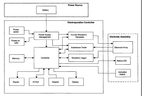
[0029] Figure 1 shows a system diagram, or flow chart, of a preferred
embodiment
of the EKD.
[0030] Figure 2 shows an example of a controller which may be used in
the EKD.
[0031] Figure 3 shows a side view of the replaceable electrode disk.
[0032] Figure 4 shows a top view of the replaceable electrode disk.
[0033] Figure 5 shows a side view of the guide disk.
[0034] Figure 6 shows a top view of the guide disk.
[0035] Figure 7 shows a side view of the guide disk mounted on the
replaceable
electrode disk.
[0036] Figure 8 shows an example of a programmed electrode pulse
pattern,
labeled Program 0000, for the EKD.
[0037] Figure 9A shows an artistic representation of current pulses and
Figure 9B
shows an artistic representation of current waveform, both of which are
produced by the pulse
pattern of Figure 8.
[0038] Figure 10 shows an example of a programmed electrode pulse
pattern,
labeled Program 0001, for the EKD.
[0039] Figure 11 shows an example of a programmed electrode pulse
pattern,
labeled Program 0002, for the EKD.
[0040] Figure 12 shows an example of a programmed electrode pulse
pattern,
labeled Program 0003, for the EKD.
[0041] Figure 13 shows an example of a programmed electrode pulse
pattern,
labeled Program 0004, for the EKD.
11
CA 02578847 2007-03-01
WO 2005/025669
PCT/US2004/028998
[0042] Figure 14 shows an example of a programmed electrode pulse
pattern,
labeled Program 0005, for the EKD.
[0043] Figure 15 shows an example of a programmed electrode pulse
pattern,
labeled Program 0006, for the EKD.
[0044] Figure 16 shows an example of a programmed electrode pulse
pattern,
labeled Program 0007, for the EKD.
[0045] Figure 17 shows an example of a programmed electrode pulse
pattern,
labeled Program 0008, for the EKD.
[0046] Figure 18 shows an example of a programmed electrode pulse
pattern,
labeled Program 0009, for the EKD.
[0047] Figure 19 shows a diagram of the first half of a sample
delivery operation
sequence for the EKD.
[0048] Figure 20 shows a diagram of the second half of a sample
delivery
operation sequence for the EKD.
[0049] Figure 21 shows an example of a first set of data that can be
acquired and
stored by the EKD during a sample electroporation procedure.
[0050] Figure 22 shows an example of a second set of data that can be
acquired
and stored by the EKD during a sample electroporation procedure.
[0051] Figure 23 shows a formatted example of a third set of data that
can be
acquired and stored by the EKD during a sample electroporation procedure.
[0052] Figure 24 shows the expression levels of SEAP in animals which
were
injected with different amounts of the plasmid pSP-SEAP and electroporated
with the EKD in
pulse pattern Program 0000.
[0053] Figure 25 shows the expression levels of SEAP in animals which
were
injected with the same amount of plasmid pSP-SEAP in different volumes and
electroporated
with the EKD in pulse pattern Program 0000.
12
CA 02578847 2007-03-01
WO 2005/025669
PCT/US2004/028998
[0054] Figure 26 shows the expression levels of SEAP in animals which
were
injected with the same amount of plasmid pSP-SEAP and electroporated with the
EKD in
pulse pattern Program 0000 at different electric field intensities.
[0055] Figure 27 shows the expression levels of SEAP in animals which
were
injected with the same amount of plasmid pSP-SEAP and electroporated with the
EKD in
different pulse patterns and at different electric field intensities.
[0056] Figure 28 shows the expression levels of SEAP in animals which
were
injected with the same amount of plasmid pSP-SEAP and electroporated with the
EKD after
different lag times between plasmid injection and the first pulse of pulse
pattern Program
0000.
[0057] Figure 29 shows the expression levels of SEAP in animals which
were
injected with the same amount of plasmid pSP-SEAP and electroporated either
contemporaneously or after electrode removal and repositioning in the muscle.
[0058] Figure 30 shows the expression levels of SEAP in animals which
were
injected with different amounts of plasmid pSP-SEAP and electroporated with an
alternative
electroporation device.
13
CA 02578847 2007-03-01
WO 2005/025669
PCT/US2004/028998
DETAILED DESCRIPTION OF PREFERRED EMBODIMENTS
[0059] The term "current" as used herein refers to the flow or rate of
flow of
electric charge in a conductor or medium between two points having a
difference in potential,
generally expressed in amperes.
[0060] The term "ampere" as used herein refers to the standard unit
for measuring
the strength of an electric current. It is the rate of flow of charge in a
conductor or conducting
medium of one coulomb per second.
[0061] The term "coulomb" as used herein refers to the meter-kilogram-
second
unit of electric charge equal in magnitude to the charge of 6.28 x 10¹⁸
electrons or the charge
transported through a conductor by a current of one ampere flowing for one
second.
[0062] The term "voltage" as used herein refers to the electromotive
force, or
difference in electrical potential, expressed in volts, which are the
practical units of
electromotive force or difference in potential between two points in an
electric field that
requires one joule of work to move a positive charge of one coulomb from the
point of lower
potential to the point of higher potential.
[0063] The term "power" as used herein refers to a source of physical
or
mechanical force or energy that is at, or can be put to, work, e.g. "electric
power, water
power."
[0064] The term "impedance" as used herein refers to the total
opposition offered
by an electric circuit to the flow of an alternating current of a single
frequency. It is a
combination of resistance and reactance and is measured in ohms.
[0065] The term "field" as used herein refers to physical quantity
specified at
points throughout a region of space.
[0066] The term "quick-release mechanism" as used herein refers to any
connector
mechanism that allows the plurality of needle electrodes to be fastened
securely and released
quickly from the constant-current pulse generator subsystem. When the needle
electrodes are
fastened securely, the quick release mechanism also maintains electrical
communication with
14
CA 02578847 2007-03-01
WO 2005/025669
PCT/US2004/028998
the constant-current pulse generator subsystem. Many different types of quick-
release
mechanisms are well known in the art of engineering.
[0067] The term "amplitude" as used herein refers to the extreme range
of a
fluctuating quantity, as an alternating current or the swing of a pendulum,
generally measured
from the average or mean to the extreme. It is the amount or degree to which a
thing extends.
[0068] The term "frequency" as used herein refers to the number of
periodic
oscillations, vibrations, or waves per unit of time. It is usually expressed
in hertz (Hz).
[0069] The term "macromolecule" as used herein refers to nucleic acid
sequences,
proteins, lipids, microbubbles (e.g. drug-loaded vesicles), and
pharmaceuticals.
[0070] The present invention pertains to an electro-kinetic device
("EKD") for
providing a constant-current electric field through an electrode needle array
and facilitating
the introduction of a macromolecule into cells of a selected tissue in a body
or plant. The
EKD produces a current pulse train waveform that passes through the electrodes
of the
electrode needle array in accordance with a programmed sequence and can be
monitored and
recorded during the procedure.
[0071] The present invention also pertains to a replaceable, or
exchangeable,
electrode disk having a needle array which may be used in association with an
electroporation
device, such as an EKD. The electrode disk has a central channel or port,
through which an
injection needle may be inserted to allow sterile delivery of the medicinal
solution, and a
removable guide disk, for controlling the depth of penetration of the needle
electrodes and
facilitating replacement of the disk.
[0072] Figure 1 shows a system diagram of one preferred embodiment of
the
EKD. Major functional elements of the EKD are shown in the diagram. Each
element is
described in terms of the hardware functionality of each element. The
sequences of events
that are enabled by the hardware are controlled by software or firmware, as
described below.
[0073] The central element of the EKD is the controller, which is
responsible for
controlling various peripheral devices connected to it. The controller is
responsible for
managing the electroporation procedure, which includes operations such as: (1)
Generating
CA 02578847 2007-03-01
WO 2005/025669
PCT/US2004/028998
the electroporation firing sequence or constant-current pulse pattern for the
electrode
assembly by controlling the current waveform generator; (2) Performing
impedance testing to
determine if electroporation should be performed; (3) Sensing and processing
user commands;
(4) Providing the user with status information; (5) Transmitting
electroporation data to an
external electronic device via the communications port; and (6) Saving and
retrieving
electroporation data (e.g. voltage and current curves) to and from memory.
[0074] The controller is preferably a single-chip microcontroller
(such as, Texas
Instruments msp430F149, or Motorola 68HC908AZ60A), such as the microcontroller
shown
in Figure 2. The boxes labeled "Peripheral" in Figure 2 represent any of the
peripheral
devices of the EKD which are shown in Figure 1 and discussed below. The
software directing
the steps of the electroporation procedure is preferably firmware, because it
resides
permanently within and runs from the single-chip microcontroller.
[0075] Another component of the EKD is the current waveform generator.
The
current waveform generator generates a current pulse train waveform that
passes through the
electrodes of the electrode array in accordance with a programmed sequence.
The pulse train
is square in shape and the number of pulses is limited by the software or
firmware. One pulse
is applied to each electrode set. Typically, each pulse is 52 ins in duration
and occurs at a rate
of 1 Hz. The amplitude of the pulse train is programmable by the operator and
ranges from
0.1 A to 1.5 A in increments of 0.1 A. The current waveform generator may be
composed of
general power-transistor analog circuits which function as directed by the
controller.
[0076] An additional component of the EKD is the impedance tester. The
impedance tester determines if the resistance of the load (e.g. muscle tissue)
is sufficiently
low. If the resistance is too high, the resulting voltage across the
electrodes might be too high
and cause heating and cell damage. Electroporation treatment may therefore be
preceded by
an impedance test. If any of the impedance measurements exceeds 1000 5 Q,
the
impedance test fails and the electroporation sequence is not initiated. The
impedance test is
an operator programmable feature controlled by software or firmware that may
be disabled
during the operation. The impedance tester maybe composed of general
operational amplifier
("op-amp") analog circuits which function as directed by the controller.
16
CA 02578847 2007-03-01
WO 2005/025669
PCT/US2004/028998
[0077] The impedance tester also functions as a safety feature in the
EKD in order
to make it a safe device to operate. It indicates whether all of the
electrodes have penetrated
the same tissue and a circuit can be established. Electrodes in contact with
air, especially dry
air, have an extremely high resistance. If electroporation starts and one or
more electrodes
have not penetrated the tissue, the resulting electrode voltages can be
thousands of volts,
which might have lethal consequences and also damage the EKD. For this reason,
overload
voltage protection may be implemented to prevent excessive voltages on the
electrodes. For
example, regardless of the electrical load (e.g. air or muscle tissue), the
over-voltage
protection may be engaged if Vu exceeds 200V for a period of no more than 1
ms. V is the
voltage across electrode i and j where i, j = 1 to 5. If the over-voltage
protection engages,
I/if goes to approximately 0 V until the next electroporation pulse is fired.
While the EKD is
in the off state, the voltage across any electrode pair preferably does not
exceed 10V.
[0078] A further component of the EKD is a waveform logger. The
waveform
logger records electroporation voltage and current waveforms, which are to be
continuously
sampled during electroporation treatment. By sampling and monitoring the
parameters of the
electroporation procedure, an operator can more easily analyze possible
problems and adjust
the settings in the event that an electroporation procedure fails or doesn't
achieve desirable
results. An exemplary sample rate is 2000 samples per second, about 104
samples for each of
the 5 current pulses. An exemplary total sample period is 4152 ins with
sampling starting
approximately 50 ins before the first pulse is fired and stopping about 50 ins
following the last
pulse. The voltage and current waveforms may be quantified into a 12-bit
digital
representation with 1 least significant bit ("LSB") linearity. The current
waveform
resolution should preferably be at least 5 mA and the voltage waveform
resolution should
preferably be at least 0.25 V. The waveform logger may be composed of general
op-amp
analog circuits and an analog to digital ("A/D") converter suitable for use
with the controller.
[0079] Another component of the EKD is an input device for inputting
user
commands. For example, the EKD operating parameters may be entered by an
operator via a
numeric keypad (such as, Grayhill 88AB2). In a preferred embodiment, the
numbers input
into the keypad are displayed on a liquid crystal display ("LCD"). Typical
parameters that
can be programmed are the electroporation pulse current, impedance test
enable/disable, and
17
CA 02578847 2007-03-01
WO 2005/025669
PCT/US2004/028998
electroporation firing delay. The features related to the keypad are also
directed by the
controller.
[0080] Other possible components of the EKD include status-
reporting devices for
displaying or otherwise notifying the operator as to the status of the system.
These status-
reporting devices may include an information display panel, such as a liquid
crystal display
("LCD") (such as, Lumex LCM-S02004DSF, or Optrex DMC-20434N). The LCD is
preferably of the character display type and is preferably capable of
displaying 4 lines of 20
characters each. The LCD is also preferably equipped with a back-light that
can be switched
on and off by means of a toggle switch. Status information may also be
provided by audible
notification, such as a buzzer (such as, CUT CEP-2202AS) sounding at various
pitches. Each
pitch preferably has a different meaning, as controlled by the software or
firmware. For
example, the volume of the buzzer may have 3 programmable settings and range
roughly from
60 to 80 dB at a distance of 1 meter from the buzzer. The sound pressure level
range is only
given as reference. The sound level is deemed acceptable if it is audible in a
noisy
environment (e.g. a farm) if set to its highest level and it is not too loud
in a quite environment
(e.g. an office) if set to its lowest level. In addition, the EKD may be
equipped with a light
emitting diode ("LED") (such as, Lumex SSI-LXR1612ID, or any panel-mount red
LED) to
designate whether the unit is turned on or off.
[0081] A further component of the EKD is a communications port that
can be used
to upload electroporation waveform data to an external electronic device, such
as a personal
digital assistant ("PDA") or personal computer ("PC"), for viewing purposes.
Preferably, the
communications port is an optical serial communications port, such as an
infrared ("IR") port
(such as, Transceiver: Vishay TFDU4100, or Zilog ZHX1201; Encoder: Microchip
MCP2120, or TI TIR1000).
[0082] The EKD may also possess a memory component. The memory
component stores electroporation waveform data and operating parameters.
Preferably, the
= memory (such as, Atmel AT45DB321B) is nonvolatile, meaning it retains its
data even if the
EKD is off. To conserve memory, electroporation waveform data may only be
saved to
memory during the active periods of the electroporation pulse train. During
the inactive
periods, sampled data may only be stored to memory if either one of the
waveforms exceeds a
specified threshold. For example, these thresholds may be a voltage threshold
of 2 V and a
18
CA 02578847 2007-03-01
WO 2005/025669
PCT/US2004/028998
current threshold of 10 mA. Data stored to memory during the inactive periods
of the current
pulse train may be time stamped so that the time index of the data is known
once the
waveforms are reconstructed. Provision may be made for the storage of up to 40
samples (20
ins) of data that occur during the inactive periods of the pulse train.
Storage can be limited to
20 ms because the software can specify that the remainder of the
electroporation sequence will
be aborted if anyone of the thresholds is exceeded for a cumulative period of
more than 20 ins.
An electroporation waveform data set requires about 2 kB of memory when the
above
compression technique is implemented. The EKD preferably contains sufficient
memory to
save at least 600 electroporation waveform data sets.
[0083] Further components of the EKD are a power source and a power
switch.
The power source is preferably a battery (such as, 2 X Powersonic PS-640 Fl,
or Panasonic
LC-R064R2P) and is responsible for providing power to each of the EKD's
circuits. These
circuits include a low voltage/low power capacity power supply for the
controller and its
peripherals, a low voltage and low power capacity power supply for the
impedance tester, and
a high power capacity power supply for the current waveform generator. The
power switch
(such as, E-Switch R5CBLKBLKEFO, or any DPDT 10A panel-mount rocker switch)
controls power to the EKD and can be either on or off. In the off position,
all electrical
connections to the electrode assembly are electrically neutral within 5
seconds after power is
turned off.
[0084] The EKD also includes an electrode handle assembly. Preferably,
the
electrode handle assembly includes three elements: a needle electrode array, a
status-reporting
device for reporting the status of the EKD, and an activator switch. In a
preferred
embodiment, the needle electrode array is circular and comprises five needle
electrodes. The
status of the EKD is preferably indicated on the handle assembly through the
use of one or
more LED's, which can be in varying colors and programmed to flash
intermittently to signify
various steps of the electrode firing sequence. The handle assembly activator
switch is
preferably used to initiate various steps of the electrode firing sequence.
[0085] Another embodiment of the present invention is a replaceable
electrode
disk which may be removably mounted in the handle of an electroporation
device. In a
preferred embodiment, the replaceable electrode disk is mounted in the
electrode handle
assembly of the EKD. Figure 3 shows a side view of the electrode disk and
Figure 4 shows a
19
CA 02578847 2007-03-01
WO 2005/025669 PCT/US2004/028998
top view of the electrode disk. In Figures 3 and 4, the electrode disk 10 has
a plurality of
needle electrodes 101 mounted on a support structure 102 in a spatial
arrangement for
penetrating the selected tissue. In a preferred embodiment, the spatial
arrangement is a
circular array. Individual electrodes in the needle array on the handle side
of the electrode
disk are blunt-ended and deburred for insertion into the complementary
electrical contact
fittings in the handle. The handle preferably houses an electrical connector
from the needle
electrodes to the pulse generator or EKD. The electrode disk support structure
102 also has a
sterile central injection channel 103 (shown in dotted lines), through which
an injection needle
may be passed for injection of the macromolecules. The channel 103 preferably
extends
outward on the top side of the electrode disk 10, through the support
structure 102 and handle,
to a sufficient length to create a sterile tube that passes through both the
handle and disk.
Thus, the handle provides a user an easy means for implanting the needle
electrodes into a
selected tissue and contemporaneously injecting the macromolecules.
[00861 A guide disk which can be mounted on the replaceable electrode
disk is
also provided. Figure 5 shows a side view of the guide disk and Figure 6 shows
a top view of
the guide disk. As shown in Figures 5 and 6, the guide disk 20 has a plurality
of guide holes
201 corresponding to the physical spacing of the needle electrodes 101
(Figures 3 and 4) and a
central passage 203 corresponding to the central channel 103 (Figures 3 and 4)
of the
electrode disk for the insertion of the injection needle. The guide disk may
be of variable
thickness, allowing the operator to control the depth of penetration of the
needle electrodes.
The guide disk also allows the operator to replace the electrode disk without
touching the
sterile needles. Figure 7 shows the guide disk 20 mounted on the electrode
disk 10.
[0087] In a preferred embodiment, the needle electrodes in the EKD
electrode
assembly as well as in the replaceable electrode disk are in a circular array.
In a further
preferred embodiment, the plurality of needle electrodes consists of five
needle electrodes. In
an additional preferred embodiment, the centers of the five needle electrodes
fall in a circular
array in the shape of a pentagon inscribed by roughly a 1.0 cm diameter
circle.
[0088] Because the waveforms required for electroporation are
specified by
software or firmware, the EKD differs from other electroporation devices,
which rely on
hardware specifications. For example, as shown in Figure 8, in a programmed
sequence
designated Program 0000, the number of pulses is 5. For pulse 1, current flow
is from
CA 02578847 2007-03-01
WO 2005/025669
PCT/US2004/028998
electrode 1 to electrodes 3 and 4. Electrode 1 is therefore positive and
electrodes 3 and 4 are
negative. Electrodes 2 and 5 are electrically isolated from electrodes 1, 3
and 4. Isolation
voltage is at least 200V. The entire sequence is depicted in which electrodes
1 through 5
become the positive electrode successively, with two negatively charge
electrodes at opposite
vertices of the pentagonal array. The code for the electrode configuration is
P =positive, 0 =
off and N = negative. The composite diagram is the sum of all pulses, and the
direction of
current flow, using the conventional physics notation.
[0089] The typical current pulses produced in Program 0000 are shown
in Figure
9A. Figure 9B shows the waveform of each current pulse. The waveform
parameters are:
Period ( t ): 1000 ins 250 ms.
Rise time ( tr ): 20 ,us maximum.
Settling time ( ): 20 us maximum.
Pulse width ( tiy ): 52 ms 100 ,us.
Fall time (t1): 20 ,us maximum.
Nominal current ( /7, ): /õ E {0.1A, 0.2A, 0.3A ... 1.5A} 10% of In during
th and
with R1 100 C2 . R1is the load resistance between anyone of the 5 electrode
sets shown in
Figure 8.
[0090] Only the current waveform is specified in Figure 9B. The shape
of the
voltage waveform depends on the impedance seen by the electrodes while the
current pulse is
firing (during th ). The voltage waveform is not specified during th since the
impedance is
unknown during this period. The voltage across any electrode set during t1 is
0 V.
[0091] The EKD is programmable to utilize a variety of electrode pulse
patterns.
Examples of these pulse patterns are illustrated in Figures 10 ¨ 18. Each
pattern may test
hypotheses related to providing optimum transgene expression by varying the
volume of
tissue electroporated, the potential damage associated with current flow in
opposite directions
through the same tissue volume, and the total current per tissue volume.
[0092] The underlying phenomenon of electroporation is believed to be
the same
in all cases, but the exact mechanism responsible for the observed effects has
not been
21
CA 02578847 2007-03-01
WO 2005/025669
PCT/US2004/028998
elucidated. Although not wanting to be bound by theory, the overt
manifestation of the
electroporative effect is that cell membranes become transiently permeable to
large molecules,
after the cells have been exposed to electric pulses. There are conduits
through cell walls
which, under normal circumstances, maintain a resting transmembrane potential
of 90 mV by
allowing bi-directional ionic migration.
[0093] Although not wanting to be bound by theory, electroporation
makes use of
the same structures, by forcing a high ionic flux through these structures and
opening or
enlarging the conduits. In prior art, metallic electrodes are placed in
contact with tissues and
predetermined voltages, proportional to the distance between the electrodes
are imposed on
them. The protocols used for electroporation are defined in terms of the
resulting field
intensities, according to the formula E=V/d, where ("E") is the field, (" V')
is the imposed
voltage and ("d") is the distance between the electrodes.
[0094] The electric field intensity E has been a very important value
in prior art
when formulating electroporation protocols for the delivery of a drug or
macromolecule into
the cell of the subject. Accordingly, it is possible to calculate any electric
field intensity for a
variety of protocols by applying a pulse of predetermined voltage that is
proportional to the
distance between electrodes. However, a caveat is that an electric field can
be generated in a
tissue with insulated electrodes (i.e. flow of ions is not necessary to create
an electric field).
Although not wanting to be bound by theory, it is the current that is
necessary for successful
electroporation, not electric field per se. The activation of the EKD's
current waveform
generator will distribute a constant-current electrical pulse to the plurality
of needle electrodes
such that a decentralized electroporation event occurs in an area where no
congruent
electroporation overlap points develop. The permeability of the cells in the
area of
decentralized electroporation increases and the macromolecule are delivered
into the cell of
the subject without overheating and damaging the cell or tissue.
[0095] The present invention pertains to an electroporation device for
introducing
macromolecules into one or more cells of an animal or plant. The
electroporation device
comprises the EKD and an electrode assembly. The electroporation device may
also comprise
a replaceable, or exchangeable, electrode disk having a plurality of needle
electrodes, a central
channel or port, and an optional removable guide disk. Together the
replaceable, or
exchangeable, electrode disk and guide disk form a needle electrode assembly
that can be
22
CA 02578847 2007-03-01
WO 2005/025669
PCT/US2004/028998
mounted on a handle of an electroporation device. The handle contains an
electrical
connector from the needle electrode assembly to a constant-current pulse
generator subsystem
or the EKD. The handle is non-conductive and designed to provide a user an
easy means for
implanting the needle electrode assembly into a selected tissue. The
utilization of disposable
needle assembly and snap-on mounts on the handle allows a user to quickly
attach and detach
the needle electrode assembly. The guide disk provides a means for grasping
the electrode
assembly without contaminating the sterile needles. The power source of the
electroporation
device, in particular the EKD, can utilize battery packs for use in the field
where access and
use of a plug in power source is dangerous or inconvenient.
[0096] It should also be understood that numerous changes and
modifications of
the EKD and electrode assembly itself may be made therein without departing
from the spirit
and the scope of the invention as defined in the claims. For example, in
another embodiment,
the invention provides a method for delivery of a macromolecule to a cells
that make up the
blood vessel walls or simply cells in culture. With modifications, the needle
electrode array
could be converted into a catheter electrode array that is connected to the
same EKD
described herein. The catheter could be placed inside a blood vessel and
macromolecules
could then be delivered directly into the vessel wall utilizing a constant-
current protocols
described herein, which would not overheat or destroy the wall of the blood
vessel. The
constant-current pulse would be generated by the EKD. This method will not
cause cell death
due to heating. Such an apparatus and method would be an excellent mechanism
for direct
and more regulated delivery of macromolecules into the blood stream.
[0097] The concept can be extended to any number of electrodes, such
as a three-
electrode array. The distance L is chosen so that the energy intensity at
point B is one third of
that at point A. After three pulses, (1 to 2, 2 to 3 and 3 to 1), point B has
received a
cumulative dose equal to that of point A. As the number of electrodes in the
array is
increased, the distance L necessary to yield a uniform energy distribution
becomes
proportionately longer. L =kxn where 12 is the number of electrodes, and k is
a
proportionality constant. Thus, by selecting a greater number of electrodes a
greater volume
of tissue can be encompassed. The optimal number of electrodes chosen may
depend on the
volume of the material to be transfected and how far it is dispersed between
injection and
electroporation.
23
CA 02578847 2007-03-01
WO 2005/025669
PCT/US2004/028998
[0098] A syringe with a specially designed macromolecule injection
cartridge can
also be used to deliver a single dose concentration of pre-sterilized
macromolecules into a
body or plant. This macromolecule injection cartridge may be a plastic
container portion that
contains the single dose concentration of pre-sterilized macromolecules and a
pre-sterilized
hollow sharp needle extending from the plastic container portion that will
convey fluids from
within the container out through the tip of the hollow needle when the needle
is inserted into
the body or plant. The central injection channel of the electrode disk ensures
that the pre-
sterilized macromolecules and needle are not contaminated during the injection
process.
EXAMPLE 1. OPERATION OF THE ELECTRO-KINETIC DEVICE ("EKD")
[0099] The operation of the Controller is shown in Figures 19 and 20,
which
illustrate a preferred operation sequence of the EKD. In a preferred
embodiment, an
information display panel LCD displays each step of the sequence to promote
user-friendly
operation. Prior to operating the EKD, the electrode assembly is firmly
inserted into the
target tissue.
[00100] First, the power is turned on and the EKD is booted up. The firmware
remains in the idle state until input is received from the user. To start an
electroporation
sequence, a password is entered to obtain an introductory prompt on the LCD.
At this point,
the handle assembly activator switch is pressed. The user then enters a
number, preferably an
animal ID number, which is logged with the data of every pulse stored for
later download.
The number is preferably entered using a numeric keypad. The user is then
prompted, via a
"beep" from the buzzer, to press the activation switch to continue the
electroporation
sequence. After the activation switch is pressed, the firmware establishes
whether or not the
impedance tester is enabled. If the impedance tester is enabled, the software
immediately
performs a series of impedance measurements. The firmware tests the impedance
between
electrodes with a low DC voltage. These measurements are performed as quickly
as possible
to get sufficiently accurate readings. During the impedance testing, a red LED
on the handle
assembly is lit. If any of the 5 impedance measurements fail, a long error
"beep" will sound,
the handle LED will stay red, the LCD will display the error, and the firmware
will return to
the idle state.
24
CA 02578847 2007-03-01
WO 2005/025669
PCT/US2004/028998
[00101] If all 5 measurements pass, a short "beep" is emitted, a green LED on
the
handle assembly is lit, and the display prompts the user to press the
activation switch to
continue. The firmware waits for the handle activation switch to be pushed
again to continue
the sequence. If any key on the keypad is pressed at this time, a long error
"beep" will be
sounded and the unit will return to the idle state.
[00102] Typically, the plasmid is injected into the muscle at this point in
the
sequence. When plasmid injection is complete, the user pushes the activation
switch to
continue the electroporation sequence. A short "beep" emits, and the firmware
counts down
using a programmed firing delay to the actual electrode-firing sequence.
During the firing
delay, the green LED on the handle assembly flashes once per second. If any
key on the
keypad is pressed at this time, a long error "beep" emits and the unit returns
to the idle state.
For the last 5 seconds before the electroporation, the buzzer makes an
intermediate-length
"beep" once per second.
[00103] At the end of the firing delay, the firmware implements the firing
sequence
as proscribed by the pulsing program selected. The red LED on the handle
assembly lights up
every second for roughly 500 ms during the 5-second period of electroporation.
When the
electroporation sequence is completed successfully, the EKD returns to the
idle state. If the
total current delivered was less than that specified by the firmware, an error
message is
displayed. The fraction of current delivered, compared to that specified, is
given as the
percentage complete.
EXAMPLE 2. DATA ACQUISITION AND STORAGE
[00104] The EKD software or firmware enables real time data acquisition and
storage in non-volatile memory. Figure 21 illustrates a first portion of data
that may be
collected during the electroporation process. The first section of the file
header contains the
file name and the animal number. The columnar data describes the pulse in
sequence, the
wait time before pulsing, the pulse width, and the pulse current for each of
the five electrodes.
Figure 22 illustrates a second portion of data, which identifies the
configuration of each
electrode during a given pulse sequence. Reading vertically for the first
pulse, electrode 1 is
positive, 2 is off, 3 is negative, 4 is negative, and 5 is off. The electrode
configurations for
pulses 2, 3, 4, and 5 constitute the remainder of the data columns. Figure 23
illustrates a
CA 02578847 2007-03-01
WO 2005/025669
PCT/US2004/028998
formatted version of a third portion of raw data for the same electroporation,
which consists of
time points, about 20 ms apart, for the five electrodes. Reading vertically
for the first
pulse, column 1 records the voltage and column 2 the current. The voltage-
current data for
pulses 2, 3, 4, and 5 are found in columns 3 and 4, 5 and 6, 7 and 8, and 9
and 10,
respectively.
EXAMPLE 3. PLASMID DESIGN, DELIVERY METHODS, AND
EXPERIMENTAL ANIMALS
[00105] Plasmid construction. The plasmid pSP-SEAP (5019 bp) is a muscle
specific expression plasmid for secreted embryonic alkaline phosphatase
("SEAP"). The
promoter is SPc5-12, a strong, muscle specific, synthetic promoter (Li et al.,
1999), and the 3'
ends of SEAP transcripts are defined by the SV40 late poly(A) signal. The
plasmid was
constructed by inserting a 394 bp Acc65I-HindIII fragment, containing the 334
bp SPc5-12
promoter sequence, between the Acc65I and HindIII sites ofpSEAP-2 Basic Vector
(Clontech
Laboratories, Inc., Palo Alto, CA).
[00106] Electroporation conditions. Square wave pulses were used in all
experiments. Electroporation conditions are stated individually for each
experiment. In all
cases, constant current was used at 0.4 to 1.0 Amps, with 3 or 5 pulses, for
52
milliseconds/pulse, and with one second between pulses. The EKD
electroporation device
contained a circular array (1 cm diameter) of five equally spaced 21-gauge
solid stainless steel
needle electrodes, mounted on a non-conductive material. The electrode disk
had a central
channel through which the injection needle could be inserted into the muscle,
such that the
plasmid was delivered within the area that was delineated by the surrounding
five electrodes.
All electrodes were 2 cm in length and were completely inserted through the
skin into the
muscle during all treatments. In all but one experiment, the EKD (ADViSYS) was
used. In
the last experiment, a different model of electroporation device (ADViSYS
Enducer Model
BB) was used for comparison purposes.
[00107] Intramuscular injection of plasmid DNA in pigs. Young hybrid pigs of
mixed gender, three to six weeks of age, with weights between 15 ¨40 kg, were
used in the
SEAP studies (n =6 to 7/group/experiment). Animals were housed in groups in
pens with ad
libitum access to a 24% protein diet (Producers Cooperative Association,
Bryan, TX) and
26
CA 02578847 2007-03-01
WO 2005/025669
PCT/US2004/028998
water. Endotoxin-free plasmid preparations were diluted in sterile water and
formulated at
1% weight/weight with poly-L-glutamate. On Day 0 of the experiment, the
animals were
manually restrained and the SEAP plasmid solution was directly injected
through the intact
skin into the semimembranosus muscle using a 21-gauge needle. All major
surface blood
vessels were avoided when finding an appropriate injection site. At a pre-
determined time
interval after plasmid injection, electroporation was applied through the 5-
electrode array.
[00108] Blood collection. On days 0, 3, 7, 10, and 14 of each experiment, the
animals were weighed and blood was collected by jugular vein puncture into
MICROTAINER serum separator tubes. Blood was allowed to clot for 10 to 15 min.
at room
temperature and was subsequently centrifuged at 3000 x g for 10 min. The serum
was stored
at -80 C until further analysis.
[00109] Secreted embryonic alkaline phosphatase assay. Serum samples were
thawed and 50 ptI., was assayed for SEAP activity using the Phospha-LightTM
Chemiluminescent Reporter Assay Kit (Applied Bio systems, Bedford, MA), per
manufacturer
instructions. The lower limit of the detection of the assay was 3 pg/mL. More
concentrated
serum samples were diluted 1:10 in mouse serum before assaying for SEAP
activity. All
samples were read using a LLTMIstar GalaxyTM luminometer (BMG Labtechnologies,
Offenburg, Germany).
[00110] Statistics. Data were analyzed using Microsoft Excel m Statistics
package.
Values shown in the figures are the mean SEM. Specific values were obtained
by
comparison using t-test or one-way ANOVA. A value of p <0.05 was set as the
level of
statistical significance.
EXAMPLE 4. EFFECTS OF PLASMID DOSE
[00111] Animals were injected with either 0.5 mg or 1 mg SEAP expressing
plasmid in a total volume of 2 mL. The intensity of the electric field was 0.5
A using
Program 0000 (Figure 8). The lag time between plasmid injection and
electroporation was 80
seconds. As illustrated in Figure 24, the expression of SEAP was dependent on
the amount of
plasmid administered. At Day 7, post-injection SEAP expression in animals
treated with 1
mg plasmid was 2.1 fold that of animal treated with 0.5 mg plasmid (71.6 22
versus 33.7
27
CA 02578847 2007-03-01
WO 2005/025669
PCT/US2004/028998
pg/mL/kg, * P <0.09, due to high inter-animal variability at higher plasmid
doses). In all
subsequent experiments, a total 0.5 mg plasmid dose was used.
EXAMPLE 5. EFFECTS OF PLASMID VOLUME
[00112] Animals were injected with 0.5 mg SEAP expressing plasmid in a total
volume of 2, 3 or 4 mL water. The intensity of the electric field was 0.5 A
using Program
0000 (Figure 8). The lag time in between plasmid injection and electroporation
was 80
seconds. As shown in Figure 25, SEAP expression was dependent on the plasmid
volume. At
Day 3, SEAP expression was significantly higher in animals administered the
plasmid in a 2
mL injection volume: 54 17 pg/mL/kg, * P <0.02 versus 11 3 pg/mL/kg in the
3 mL-
treated group, and 0.13 0.3 pg/mL/kg in the 4 mL-treated group, t P < 0.03;
at Day 7, 38.5
18 pg/mL/kg, t P <0.05 versus 1 0.3 pg/mL/kg in the 4 mL-treated group; and
at Day 10
* P < 0.04 versus 3 mL-treated group.
EXAMPLE 6. EFFECTS OF ELECTRIC FIELD INTENSITY
[00113] Electric field intensity correlates with pulse pattern for optimum
plasmid
uptake and transgene expression. In a first experiment, all animals were
injected with 0.5 mg
SEAP expressing plasmid in a total volume of 2 mL. The lag time between the
injection and
electroporation was 80 seconds. Using the EKD Program 0000 (Figure 8) pulse
pattern, the
intensity of the electric field was decreased from the 1 Amp positive controls
to 0.6 Amp,
while expression is increased, as shown in Figure 26. The 1 Amp condition was
chosen
because of the substantial body of literature with data from constant voltage
electroporation
devices which suggest that this electric field intensity would yield the best
expression level
(Mir et al., 1998). As stated, in this case the best results were obtained
using an electric field
intensity of 0.6 Amps. At Day 3 ¨ * P <0.04 for 0.6 Amp and t P <0.03 for 0.5
Amp; at Day
7 - * P < 0.03 for 0.6 Amp; and at Day 10 - * P <0.01 for 0.6 Amp.
[00114] Further comparison was performed between electric field intensities,
using
a different pulse pattern model. Electric field intensities of 0.4, 0.5 and
0.6 Amp, using the
Program 0005 pulse pattern (Figure 14 ¨ three pulses, no reverse of electric
field), were
compared to 1 Amp using the Program 0000 pulse pattern (Figure 8 ¨ five
pulses, complete
reverse of electric field), as a positive control reference. All animals were
injected with 0.5
28
CA 02578847 2007-03-01
WO 2005/025669
PCT/US2004/028998
mg SEAP expressing plasmid in a total volume of 2 mL. The lag time between
plasmid
injection and electroporation was 80 seconds. Using this pulse pattern, as
shown in Figure 27,
the best expression levels were obtained using an electric field of only 0.4
Amp: at Day 3¨ *
P <0.02 for 0.4 Amp; Day 7¨ * P <0.03 for 0.4 Amp; and Day 10¨ * P <0.03 for
0.4 Amp.
At all time points tested, the group treated at 0.5 Amp had a trend towards
increased SEAP
values (P = 0.06 ¨ 0.07).
EXAMPLE 7. EFFECTS OF LAG TIME
[00115] Previous experiments utilized a 2 minute lag time between plasmid
injection and electroporation (Draghia-Akli et al., 1999; Mir et al., 1999).
To facilitate large-
scale applications of the technology, it is important to consider shortening
the lag time as
much as possible. However, as shown in Figure 28, the lag time between plasmid
injection
and electroporation should be no less than 80 seconds. In this experiment, all
animals were
injected with 0.5 mg SEAP expressing plasmid in a total volume of 2 mL. The
intensity of the
electric field was 0.5 A. SEAP expression levels decreased when the lag time
was reduced
from 80 to 70, 60 or 50 seconds. There was a clear reduction in SEAP
expression levels at lag
times less than 80 seconds: Day 10 ¨ * P < 0.05 between the group
electroporated at 50
seconds and the group that received the electroporation at 80 seconds post-
injection.
Although not wanting to be bound by theory, this lag time may be necessary to
allow to the
injected plasmid to distribute in the muscle and bind to the membrane surface
before it is
electrically and reversibly restructured.
EXAMPLE 8. DELINEATION OF INJECTION AREA
[00116] Commercially available electroporation devices and needle arrays do
not
permit injection and electroporation in one combined operation. With these
instruments, the
procedure required that the injection needle be inserted into the target
muscle for plasmid
delivery, then removed from the muscle. The electrodes were then inserted into
the muscle in
the proximity of the injected area, usually based on a skin tattoo. However,
the underlying
muscle may move or contract, so the injection site may not be completely
circumscribed by
the needle electrodes.
29
CA 02578847 2011-02-03
[00117] In this experiment, all animals were injected with 0.5 mg SEAP
expressing
plasmid in a total volume of 2 mL. The intensity of the electric field was 0.5
A. The lag time
between the injection and electroporation was 80 seconds. One group of animals
had the
needles inserted into the target muscle, and held in place for the entire
procedure. In a second
group the needles were removed immediately after the plasmid injection,
reinserted after 15
seconds into the same injection site visualize on the skin, and the 80 seconds
count down
started. SEAP levels were 43.5 16.8 pg/mL/kg in Group A versus 9 1.7 pg/mL/kg
at Day
7. * P <0.02; and 45.6 16 pg/mL/kg versus 12 2.5 pg/mL/kg at Day 10- * P
<0.02. As
shown in Figure 28, the non-specificity of the electroporation site reduced
plasmid expression
by up to 75%. This finding emphasizes the importance of precisely and
accurately
electroporating the injection site for greater gene expression.
EXAMPLE 9. COMPARATIVE STUDY
1001181 Escalating pSP-SEAP
doses from 0 to 10 mg were administered in
accordance with the previous examples. Plasmid injection was followed by
electroporation
using a different electroporation device (ADViSYS Enducer Model BB, U.S.
Patent
No. 7,245,963). As shown in Figure
30, expression level was dose dependent.
Nevertheless, at Day 7 post-injection, SEAP levels averaged 7 2.2 pg/mL/kg,
while
experiments using the EKD showed SEAP expression at the corresponding time
point
averaging 33.7 10 pg/mL/kg, a 4 fold increase. Although not wanting to be
bound by
theory, this increase in expression may result from the enabling step, which
allows the
operator to be sure that all needles are in the same muscle; the display,
which allows for real
time control of the procedure; the software, which allows for visualization of
each pulse;
and subsequent adjustment of parameters if the first electroporation procedure
fails.