Note: Descriptions are shown in the official language in which they were submitted.
CA 02578963 2007-03-01
WO 2006/028855 PCT/US2005/030991
METHOD AND SYSTEM FOR TREATMENT OF ATRIAL FIBRILLATION AND
OTHER CARDIAC ARRHYTHMIAS
FIELD OF THE INVENTION
This invention relates generally to methods and systems for treatment of
atrial
fibrillation and other cardiac arrhythmias and, in particular, to methods and
systems for
delivering biological material to a chamber inside the heart.
BACKGROUND OF THE INVENTION
Atrial fibrillation is an arrhythmia of the heart in which the atria or upper
chambers
of the heart stop contracting as they fibrillate. Premature atrial contraction
(extra beats)
originating in the pulmonary veins can act as triggers and initiate paroxysms
of atrial
fibrillation. The inability to reproducibly induce premature beats and
precisely identify
the ostium or junction of the pulmonary veins with the left atrium due to the
complex
three-dimensional geometry of the left atrium makes prohibitive the use of
ablation
therapy in many patients. There is also a risk of complications such as
stroke, bleeding
around the heart and narrowing of the pulmonary veins during radio-frequency
catheter
ablation procedures.
Studies have found activity that is suggestive of the presence of conduction
tissue
at the left atrial-pulmonary vein junction. Thus, a new approach directed at
blocking
conduction at a cellular or molecular level by delivering biological material
that would
block conduction across cells could provide significant advantages in the
treatment of this
complex arrhythmia. Such delivery systems could include the transplantation of
cells or
the injection of antibodies.
This approach could also be beneficial to treating other arrhythmias and other
conditions if precise localization and delivery of cells, antibodies and
similar biological
substances including genes were possible.
SUMMARY OF THE INVENTION
One aspect of this invention provides a method for treatment of a heart
arrhythmia
having the steps of (1) obtaining cardiac image data using a digital imaging
system,
CA 02578963 2007-03-01
WO 2006/028855 PCT/US2005/030991
2
preferably a computer tomography (CT) system, (2) generating a 3D model of a
cardiac
chamber and surrounding structures from this cardiac image data, (3)
registering the 3D
model with an interventional system, (4) visualizing this registered 3D model
on the
interventional system, (5) positioning a catheter apparatus within the cardiac
chamber, (6)
visualizing the catheter apparatus over the registered 3D model of the cardiac
chamber
upon the interventional system, (7) navigating the catheter apparatus within
the cardiac
chamber utilizing this registered 3D model, and (8) delivering biological
material through
the catheter apparatus to heart tissue at select locations within the cardiac
chamber.
In certain preferred embodiments, the biological material being delivered by
the
catheter apparatus are transplanted cells that can alter electrical impulses
at these select
locations within the heart. Highly preferred is where the transplanted cells
are myoblasts.
Another desirable embodiment is where the biological material delivered to
heart tissue
within the cardiac chamber are antibodies such that electrical impulses at the
selected
locations are altered by these antibodies.
It is most desirable that the interventional system be a fluoroscopic system.
More
desirable is where the heart arrhythmia is atrial fibrillation and the 3D
model is of the left
atrium and pulmonary veins. Highly desirable embodiments find the catheter
apparatus
having a main body with a central lumen that is adapted to deliver biological
material and
a control mechanism coupled to the main body such that the delivery of the
biological
material from the main body is controlled.
In another aspect of this invention, a system is provided for treatment of a
heart
arrhythmia that has a digital imaging system to obtain cardiac image data, an
image
generation system to generate a 3D model of a cardiac chamber and its
surrounding
structures from this cardiac image data, a workstation to register the 3D
model onto an
interventional system so that the registered 3D model can be visualized upon
the
interventional system, and a catheter apparatus to deliver biological material
to heart tissue
within this cardiac chamber at certain select locations, the catheter
apparatus being
visualized upon the interventional system over the registered 3D model.
Desirable cases of this system find the biological material delivered to be
transplanted cells, most preferably myoblasts. Also highly desirable is where
the
biological material are antibodies.
CA 02578963 2007-03-01
WO 2006/028855 PCT/US2005/030991
3
Preferred embodiments of this system are where the interventional system is a
fluoroscopic system. Most preferred embodiments find the digital imaging
system to be a
computer tomography (CT) system. In certain preferred cases, the heart
arrhythmia is
atrial fibrillation and the 3D model is of the left atrium and pulmonary
veins. Highly
preferred is where the catheter apparatus includes a main body having a
central lumen
adapted to the delivery of the biological material and a control mechanism
coupled to the
main body to control such delivery from the apparatus.
BRIEF DESCRIPTION OF THE DRAWINGS
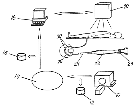
FIG. I is a schematic overview of a system for treatment of a heart arrhythmia
in
accordance with this invention with an enlarged longitudinal cross-section of
a portion of
the catheter.
FIG. 2A depicts 3D cardiac images of the left atrium.
FIG. 2B illustrates localization of a standard mapping and ablation catheter
over an
endocardial view of the left atrium registered upon an interventional system.
FIG. 3 is a flow diagram of a method for treatment of atrial fibrillation and
other
cardiac arrhythmias in accordance with this invention.
DETAILED DESCRIPTION OF PREFERRED EMBODIMENTS
FIG. 1 illustrates a schematic overview of an exemplary system for the
treatment
of a heart arrhythmia such as atrial fibrillation in accordance with this
invention. A digital
imaging system such as a CT scanning system 10 is used to acquire image data
of the
heart. Although the embodiments discussed hereinafter are described in the
context of a
CT scanning system, it will be appreciated that other imaging systems known in
the art,
such as MRI and ultrasound, are also contemplated.
Cardiac image data 12 is a volume of consecutive images of the heart collected
by
CT scanning system 10 in a continuous sequence over a short acquisition time.
The
shorter scanning time through use of a faster CT scanning system and
synchronization of
the CT scanner with the QRS on the patient's ECG signal reduces the motion
artifacts in
images of a beating organ like the heart. The resulting cardiac image data 12
allows for
reconstruction of images of the heart that are true geometric depictions of
its structures.
CA 02578963 2007-03-01
WO 2006/028855 PCT/US2005/030991
4
Cardiac image data 12 is then segmented using protocols optimized for the left
atrium and pulmonary arteries by image generation system 14. It will be
appreciated that
other chambers of the heart and their surrounding structures can be acquired
in a similar
manner. Image generation system 14 further processes the segmented data to
create a 3D
model 16 of the left atrium and pulmonary arteries using 3D surface and/or
volume
rendering. Additional post-processing can be performed to create navigator
(view from
inside) views of these structures.
3D model 16 is then exported to workstation 18 for registration with an
interventional system such as a fluoroscopic system 20. The transfer of 3D
model 16,
including navigator views, can occur in several formats such as the DICOM
format and
geometric wire mesh model. Information from CT scanning system 10 will thus be
integrated with fluoroscopic system 20. Once 3D model 16 is registered with
fluoroscopic
system 20, 3D model 16 and any navigator views can be seen on the fluoroscopic
system
20.
A detailed 3D model of the left atrium and the pulmonary veins, including
endocardial or inside views, is seen in FIG. 2A. The distance and orientation
of the
pulmonary veins and other strategic areas can be calculated in advance from
this 3D image
to create a roadmap for use during the ablation procedure.
Using a transeptal catheterization, which is a standard technique for gaining
access to the
left atrium, a catheter apparatus 22, having a flexible catheter 24 with a
central lumen 26,
is introduced into the left atrium. Catheter 24 is visualized on the
fluoroscopic system 20
over the registered 3D model 16. Catheter 24 can then be navigated in real-
time over 3D
model 16 to the appropriate site within the left atrium. FIG. 2B illustrates
localization of a
standard mapping and ablation catheter over an endocardial view of the left
atrium
registered upon an interventional system.
Catheter apparatus 22 is provided with a control mechanism 28 for opening and
closing the distal end of lumen 26. Upon filling lumen 26 with biological
material 30,
catheter apparatus 22 can be used as a delivery device for the release of
biological material
30 at specifically selected locations within the heart. After catheter 24 has
been guided to
a site identified as a strategic area whose electrical conductivity needs to
be altered or
blocked, control mechanism 28 is actuated to deliver biological material 30
such as
CA 02578963 2007-03-01
WO 2006/028855 PCT/US2005/030991
transplanted cells at that site. Such transplanted cells could be myoblastic
or smooth
muscle cells. Antibodies can also be injected in this manner to alter or block
abnormal
electrical activity at the cellular level, especially in responding to
antigens that may be
responsible for the triggering of impulses that initiate atrial fibrillation.
There is shown in FIG. 3 an overview of a method for ablation of atrial
fibrillation
and other cardiac arrhythmias in accordance with this invention. As seen in
step 110, a 3D
image of the heart is acquired. 3D images of the heart can be created using CT
scan or
MRI. At step 120, a 3D model of the chamber of interest such as the left
atrium is created
through segmentation of the image data using protocols optimized for the
appropriate
structures. Once this 3D model has been obtained, it can be stored as an
electronic data
file using various means of storage. The stored model can then later be
transferred to a
computer workstation linked to an interventional system.
As illustrated in step 130, after it has been transferred to the workstation,
the 3D
model is registered with the interventional system. The registration process
allows
medical personnel to correlate this 3D model of the cardiac chamber with the
interventional system that is being used with a particular patient so that it
can be visualized
during the interventional procedure.
The following step 140 involves visualization of a catheter that has been
positioned
within the left atrium over the registered 3D model. This permits the catheter
to be
navigated inside the chamber in real-time over this registered image to the
locations
selected for the treatment to be performed.
CA 02578963 2007-03-01
WO 2006/028855 PCT/US2005/030991
6
In step 150, transplanted cells such as myoblasts are released from a central
lumen
of the catheter at the selected site to alter or block electrical activity
across that location.
Alternatively, at step 160, antibodies or genes can be inserted at the site in
treatment of the
arrhythmia after being transported to the left atrium within the catheter's
lumen.
It will be appreciated to one skilled in the art that other arrhythmias such
as ventricular
tachycardia can be targeted for treatment in this manner. Furthermore,
automatic
techniques may be used to perform any of the above steps.
Various alternatives and embodiments are contemplated as being within the
scope
of the following claims particularly pointing out and distinctly claiming the
subject matter
regarded as the invention.