Note: Descriptions are shown in the official language in which they were submitted.
CA 02579236 2007-02-19
Gilmore, Andre
P315245
FIRST METATARSAL HEAD LIFT ORTHOTIC
RELATED APPLICATIONS
This application claims priority of U.S. Provisional Serial No.
60/774,767, filed February 17, 2006.
BACKGROUND OF THE INVENTION
Orthotics and foot orthoses are available in a variety of forms
addressing support or accommodate that which exists. The focus of those
past designs was to support the arch only, always with forethought that the
arch is the major issue of misalignment to the foot. Various forms of arch
supports have been found in the prior art, along with other support devices in
an attempt to bio mechanically align the foot and subtalar joint.
As described further here in, the disclosure recites a method for
dynamically providing mechanical alignment of the foot/subtalar joint. All
other
prior art addressing only static alignment to the foot, i.e., the foot
standing still.
As described herein, a Postural Foundational-Alignment System is
provided to engage the first metatarsal head in a manner such that the greater
toe along with the metatarsal head of the greater toe is lifted a certain
height
affecting the subtalar joint and other proximal joints above the base of the
feefi/foundation.
CA 02579236 2007-02-19
2
SUMMARY OF THE DISCLOSURE
An postural foundational alignment device attached to support the
middle foot and anterior foot portions, the interior foot portion comprising a
first metatarsal and first phalanges and second-fifth metatarsals and second-
fifth phalanges. The postural foundational alignment device comprises a front
region having a forefoot engagement surface comprising a first metatarsal
engagement surface to support a first metatarsal and first phalange of the
anterior foot portion, and further comprising a second-fifth metatarsal
engagement surface adapted to engage the second-fifth metatarsals and
second-fifth phalanges.
The surface of the first metatarsal engagement surface is positioned
higher relative to the second-fifth metatarsal engagement surface and
adapted to vertically raise the first metatarsal and first phalanges with
respect
to the second-fifth metatarsals and phalanges where the first metatarsal
engagement surface is substantially level. The postural foundational
alignment device further includes a metatarsal arch portion adapted to engage
the middle foot region where the metatarsal arch portion is provided with an
arch support that extends forwardly to the first metatarsal engagement
surface.
In another form, the postural foundational alignment device is made
where the arch support extends rearwardly to form a heel cup that is adapted
to surround a posterior portion of a foot. In a different embodiment of the
postural foundational alignment device, a first metatarsal base surface is
positioned beneath the first metatarsal engagement surface at a distance
which is greater than the distance between the second-fifth metatarsal
engagement surface and a second-fifth metatarsal base surface.
In another form of the device mentioned above, the first metatarsal
base surface and the second-fifth metatarsal base surface is not contiguous
where an abrupt portion provides a more drastic change in elevation between
these two surfaces. In alternative embodiment, the first metatarsal
CA 02579236 2007-02-19
3
engagement surface and the second-fifth metatarsal engagement surface
collectively comprise a forefoot engagement surface which is in a
substantially
continuous plane when pressure is not applied thereto.
In other forms of the postural foundational alignment device, the arch
support may have a lower support surface that is contiguous with the first
metatarsal base surface. Additionally, the first metatarsal base surface and
the second-fifth metatarsal base surface may not be not contiguous surfaces.
In this form, the lower support surface of the arch support may extend
in the posterior portion to form a heel cup, and the heel cup may have a
lateral region that extends in the anterior direction just short of the cuboid
and
the base region of the fifth metatarsal. The posterior portion of the foot is
adapted to rest within the heel cup of the postural foundational alignment
device.
In various other forms, a heel engagement surface is positioned above
the heel cup and is substantially contiguous when pressure is not applied
thereto, or the heel engagement surface may be rested between a medial and
lateral raised area to support the posterior portion of the foot. The postural
foundational alignment device may be made from a material having a
durometer rating between 30 and 50.
CA 02579236 2007-02-19
BRIEF DESCRIPTION OF THE DRAWINGS
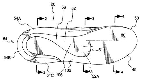
Fig. 1 shows a bottom view of an postural foundational alignment
device;
Fig. 2 shows a cross-sectional view of the postural foundational
alignment device and a heel cup region taken at line 2 -- 2 of Fig. 1;
Fig. 3 shows a cross-sectional view of the postural foundational
alignment device at a metatarsal arch portion taken at line 3-- 3 of Fig. 1;
Fig. 4 shows a cross-sectional view at the forefoot lift region showing
the area which lifts the first metatarsal head of the foot taken at line 4 --
4 of
Fig. 1;
Fig. 4A shows a cross-sectional view at the forefoot lift region showing
the area which lifts the first metatarsal head of the foot taken at line 4 --
4 of
Fig. I where the metatarsals are positioned in a preferred form;
Fig. 5 shows a bottom view of the orthotic with the bone structure
superimposed thereabove as a hatched line.
Fig. 6 shows a side view of the foot positioned on the postural
foundational alignment device.
Fig. 7 shows a view of another embodiment where the orthotic can be
adjusted with the shim inserts;
Figs. 8A -- 8F show various views taken along line 8A -- 8A of Fig. 7
where it can be seen that various lateral and vertical shims are positioned to
adjust the nature of the first metatarsal head lift orthotic.
CA 02579236 2007-02-19
~
DETAILED DESCRIPTION
OF THE PREFERRED EMBODIMENTS
As described herein, there is shown an embodiment where the first
metatarsal head is raised with respect to the surrounding areas. In a
preferred
form, a surface is substantially orthogonal to the flux field of gravity (of
course
with localized surface variations as an option or angled about a lateral
axis).
It is believed by the applicant that as the foot steps off, the greater toe
especially, that toe pronates or rolls more to the inside in a twisting
torquing
shearing moment past 4 degrees of this a normal acceptable inside roll. The
human body in its innate wisdom also recognizes in some people that instead
of allowing the toe to potentially pronate excessively it will protect or
guard
against this roll to the inside and brace or restrict itself and hold the foot
at the
toe off phase of motion thus to hold the foot in a "supinated" position.
Having
a higher arch or walking too long along the outside or lateral border of the
foot.
As a result of this excessive (in one form, more than four degrees of
internal roll) pronation motion, a "shortening" of the inside portion of the
foot
occurs. in the typical gait phase of walking the human body strikes at the
heel
contact phase of forward motion affecting a loss of alignment as the center of
gravity or the weight bearing line passes over the Sustentaculum tali area of
the calcaneus, then on to the navicular bone, the first cuneiform bone, the
greater metatarsal head including the sesamoid bones of the great toe and
along the MIP, PIP, DIP and out to the very distal edge of the great toe an,
excessive pronation motion can occur along this area that describes the
"medial column" border of the medial (inside) of the foot.
It is believed that such an unbalanced, unstable non symmetrical
biomechanical human foundation can contribute to further negative affects of
health through out the human body.
Areas of negative affectations can result in the following:
= Musculoskeletal imbalances
CA 02579236 2007-02-19
~
-'~
o Over worker muscles,
o Under worker muscles
o Contracted tight muscles
o Stretched (past their normal effective length)
muscles
o Painful point of origin and insertion attachments
= Forward leaning posture
= Rounded shoulders
= Sway backs,
= Kyphotic upper backs
= Incongruent joint alignment
= Excessive torque and joint wearing points of abnormal
contact
= Spinal dysfunctions such as scoliosis, pinched nerves,
slipped discs, spondylolesis, boney spurs,
= Lower, middle, upper back, shoulder, neck, head strains
as sprains felt as "pain"
= Bio-implosion of the thoracic cavity collapsing on itself
affecting abnormal compressive forces on the lungs,
heart, and other organs of the thoracic cavity.
It is believed that such negative issues can occur when the foundation
of the foot is not in dynamic symmetrical alignment, i.e., right foot to left
foot is
not symmetrically aligned to each other in the full dynamics of forward
motion.
Present analysis indicates that raising the first metatarsal head and
throughout the greater toe with its special contouring can be at the PIP joint
area designated as seen in Fig. 5 at 35, and the DIP joint area together or
just
the DIP area taper at the junction 37 in Fig. 5. and orthogonal to the flux
field
of gravity design has the synergistic benefit of stimulating a straighter
posture
alignment of the human frame, providing a sulcus raise, a metatarsal-
CA 02579236 2007-02-19
1
transverse arch- raise, relief of painful excesses boney met head pressure
from the other (2-5) metatarsal heads in forward motion, correcting excessive
internal or external rotation of the foot on the ground, leveling the pelvic
girdle
where one side may have been longer or shorter asymmetrically. This change
in placement of the significant lift of the first metatarsal head lift
orthotic
(FMHLO) provides a greater distribution of lifting forces under the remaining
parts of the boney structures of the foot particularly in the navicular, all
cuneiforms, cuboids, metatarsal shafts, and corresponding joint links.
Further, the great toe is placed in a more symmetrical balanced alignment
during the various phases of motion when the first metatarsal head is raised
with respect to the adjacent metatarsals.
Dynamic Symmetrical Alignment Balance (DSAB) can only be applied
to this type (e.g. FMHLO) of design consideration, in particular the great toe
raise especially from the metatarsal head under the sesamoid bones of the
great toe and following along to the distal tip of the toe with its
fundamental
orthogonal shape and distal tapering from the PIP (proximal Interphangeal
phalanges) demarcation 35 in Fig. 5 and the DIP joint area 37, or solely the
DIP joint area 37, in a proportion to the percentage of height in the raised
patentable platform.
The resulting mechanical changes that gradually occur over a short
time frame beneficial to the human body are correcting forces that stimulate
positive changes to the negative effect listed above. It can further be
observed that a more "natural" format of forward motion will occur such that
the feet/foundation will walk with a closer base of gait, not one foot will be
more or less externally or internally rotated out or in than the other,
respectively, knock knee formations will be decreased, bowlegged curves will
be lessened.
There will now be a general discussion of the human body and
particularly the lower boney structural portion (the feet/foundation). It
should
be noted that a standing (static) erect body has a different shape of the
boney
foot structures than does a moving (dynamic) boney foot structure. Therefore,
CA 02579236 2007-02-19
the postural foundational alignment insole device must provide functional
stimulation correction/support taking into consideration the static verses the
dynamic shapes of the boney feet/foundation for a most desirable
feet/foundation stabilizing effect. The feet wearing this particular design
concept of the FMHLO will strengthen over a short period of time in the
musculature in and around the foot and throughout the other postural
muscles.
Referring ahead to Fig. 5, there is shown an outline of a skeletal
human foot 30. In general, the bones in a human foot are comprised of a
calcaneus 32, the metatarsals 34 and the phalanges 36. Aft of the
metatarsals 34 are the cuneiform bones 38 which are adjacent to the
navicular 40 and the cuboid 42. Positioned after the navicular and the cuboid
40 and 42 is the talus indicated at 44. It should be noted that the tibia (not
shown) is positioned on top of the talus 44.
As further shown in Fig. 5, there is a line indicated at 35 which
generally indicates the location of the PIP joint, and the approximate
location
indicated at 37 is the DIP joint.
With the general background of the bones in place, it should be noted
that the majority of the population are not "unwound" from the talus 44 in
relationship to the calcaneus 32 where a baby's feet do not fully unwind and
are not properly pronated to get the first metatarsal head "on the ground".
Therefore, even when the ankle is somewhat neutral, there is not sufficient
lift
in the metatarsal head to properly put the ankle in a neutral joint alignment.
As shown in Fig. 1, there is a bottom view of the orthotic device 20. As
shown in this figure, the orthotic 20 device comprises a forefoot lift region
50
which is described in greater detail below. The general area indicated at 51
indicates the metatarsal arch portion. The region indicated at 52A extends
laterally outwardly and flares downwardly to the outer metatarsal portions.
The heel cup generally indicated at 54 which is optional in one form, is
divided
into sections 54A, 54B and 54C which show the medial, posterior and lateral
regions surrounding the calcaneus portion on its medial, posterior and lateral
CA 02579236 2007-02-19
"1
,ra
borders 31 (see Fig. 5) of the foot. The posterior portion of the heel and the
medial portions are supported at 54B and 54A. Further, the lateral
repositioning of the user's heel can be controlled at 54C. The medial portion
of 54A and the forward portion of the section 54A extends to the medial arch
to the sustentaculum tall. In general the heel cup encompasses the posterior
aspect of the calcaneus around the medial and lateral aspects of the
calcaneus to just proximal or behind the weight bearing line of the
sustentaculum tali on the medial side of the foot and cupping around the
cuboid on the lateral side of the foot just proximal to the body of the fifth
metatarsal head. The heel cup does not lift the heel-calcaneus bone but
rather cradles it. In one form it acts as a guide to the heel at heel contact
to
mid-stance simulating a more vertical heel support mechanism.
Now referring to the outer lateral portion 54C, the heel cup extends
around the cuboid 42 (see Fig. 5) to the lateral portion of the foot and is
just
proximal to the body of the fifth metatarsal head. Therefore, the end portion
in
the preferred form is just laterally outside of the cuboid and proximal to the
fifth metatarsal head as shown in Fig. 5. In one form the portions 54A, 54B
and 54C encapsulate the entire calcaneus. In a preferred form, the
sustentaculum tali of the foot (not shown) is in proper engagement where the
calcaneus is supported at this merger area between the heel cup portion 54A
and the metatarsal arch portion 52 generally at the region indicated at 56.
With the foregoing description in place, there will now be a more
detailed discussion of the forefoot lift region 50.
The metatarsal region 34 is comprised of the first through fifth
metatarsal bones indicated at 34A-34E as shown in Fig. 5. The
corresponding phalanges connected to each metatarsal head are numbered
in a corresponding manner, 36A-36E. It should be noted that Fig. 5 shows a
bottom view where the bones of the foot are shown in a hatched line where
this would be a left foot and a left insole. However, in one form, the edge
surfaces 88 (see Fig. 4) of the forefoot lift area can be positioned in the
upper
portion, where as shown in Fig. 5, the very surfaces defining the surface to
CA 02579236 2007-02-19
i0
_7'
engage the foot are positioned in a manner where Fig. 5 would be the right
insole with the right foot positioned thereon.
As shown in Fig. 5, it can be seen how the forefoot lift region 50 is
positioned beneath the first metatarsal head area indicated from 34A to 35
and the first phalange (the big toe bone) is indicated at 36A. As shown in
Fig.
4, the cross-sectional view shows the forefoot lift region 50. In general, the
front base region 49 of the postural foundational alignment device 20 as
mentioned before comprises the forefoot lift region 50. The first metatarsal
base surface indicated at 80 is substantially orthogonal to the flux field of
gravity. Another way of describing the surface 80 is that it is substantially
level. The forefoot foot engagement surface 82 (as shown in Fig. 4)
comprises a first metatarsal engagement surface 81 and a second through
fifth metatarsal engagement surface 84. It should be noted that the surfaces
81 and 84 extend distally to the corresponding phalange regions as indicated
in Fig. 5.
Referring back to Fig. 4, the first metatarsal base surface 80 has a
medial portion 86 and a laterally outward portion 88. In one form, the
laterally
outward portion has an abrupt edge 90 creating a fairly significant change in
elevation from the first metatarsal base surface 80 to the second-fifth
metatarsal base surface 83. The abrupt edge 90 need not be a perpendicular
surface as shown in Fig. 4 to the surfaces 80 and 83. Rather, it could be a
transition of some sort with an angled surface where a majority of the weight-
bearing surface 80 is more or less level and distinctly on a different
elevation
than the surface 83.
As shown in Fig. 4, the metatarsals 34 are schematically indicated at
34a -- 34e. Fig. 4 shows one embodiment where the foot is positioned on the
side of the orthotic 20 with the abrupt edge 90 positioned upwardly. Now
referring to Fig. 4A, the metatarsals schematically shown at 34a -- 34e are
positioned upon the forefoot engagement surface 82 where it can be
appreciated that the second through fifth metatarsals' engagement surface 84
is deflected downwardly and the first metatarsal 34a is raised with respect to
CA 02579236 2007-02-19
-8~
the second through fifth metatarsals 34b -- 34e. The insole 20 can be
comprised of a material that contributes to flexibility. In some forms the
metatarsal lift area can have a lower durometer rating (e.g. 15-25) and arch
region can durometer of up to, say for example, 60 durometer rating. The
durometer rating can be changed by mixing a "C-Mix" to adjust the durometer
rating. Of course other materials and methods can be used to comprise the
orthotic 20.
Referring now to Fig. 3, there is shown a metatarsal arch 51 portion in
cross-section. In general, this portion has a medial region 100 and a lateral
portion 102. In general, the medial region is sufficiently higher raised above
the lateral portion 102 to provide a transverse metatarsal arch support. As
shown in Fig. 1, the arch support lower support surface 106 in one form is
contiguous with the first metatarsal base surface 80. Further, the lower
support surface 106 in one form continues the role posteriorally to the inner
region of the heel cup 54 as seen in Fig. 2.
There will now be a more detailed description of the forefoot lift region
with reference to Fig. 7. In general, Fig. 7 shows another embodiment
utilizing an adjustable system described further herein; however, the forefoot
lift region 50 will be further described as to the various attributes thereof.
The
regions 50a, 50b and 50c indicate the general area which engages the lift for
the first metatarsal, which can vary between .25 mm to 20 mm in the broad
range. In general, a forward taper can occur to the PHP joint at 50d and
extend to the forward region of the support 20, or the DIP joint 50. The
metatarsal head is generally raised at the portion 50b, and the proximal
portion of this metatarsal head area is indicated at 50c. It should be noted
that these areas are similar to the regions 34a and 35 as shown in Fig. 5.
With regard to the forward taper of the first metatarsal lift region 50, as
noted above, the taper can begin at the PHP joint indicated at 50d or at the
beginning of the DIP joint at 50. Present analysis indicates that combining
the
taper starting at the PHP area at 50d and transcending it downwardly to the
DIP area at 50e has beneficial effects on the proprioceptive feedback for the
CA 02579236 2007-02-19
12
patient. Alternatively, the taper can begin at the DIP location at 50e to the
distal end of the great toe.
Now referring to Fig. 2, the heel cup 54 is shown in cross-section
where the medial and lateral regions 54A and 54C are shown. A heel cup
engagement surface 110 in one form is a substantially contiguous surface
which is adapted to be deformed when pressure is placed thereon such as
from the heel of the foot. As shown in Fig. 6, a heel 112 can depress the
orthotic downwardly to properly position the calcaneus within the heel cup
region 54. Referring back to Fig. 2, altematively the heel engagement surface
could be indicated at 110 where in this form the foot engaging surfaces are
not contiguous but rather each orthotic is essentially turned upside down and
switched from one foot to the other. With this embodiment, the surface
indicated at 80 in Fig. 4 would function as the first metatarsal engagement
surface where the foot is placed directly thereon.
Referring back to Fig. 6, it can be seen how the posterior portion 120 of
the foot is supported by the optional heel cup 54. The middle portion 122 is
supported by the arch support portion 52 and the anterior foot portion is
supported by the forefoot lift region in its entirety 50 to raise the first
metatarsal head and big toe with respect to the adjacent metatarsals. With
the foregoing description in mind, there will be further discussion of the
dynamic effects (as well as the static standing effects) of the orthotic 20 as
it
interfaces with the foot of the individual.
In one form the orthotic 20 supports the foot such that it stimulates the
foot/foundation to change its position and/or placement on the ground under
the human frame (aligning the foot to knee, etc.) sending a message or
proprioceptive feedback loop through the nerve (nervous) system to the brain
to affect positive postural changes. The cause-and-effect neural response
synergistically operates upon the body for the orthotic support device 20 to
create foot alignment. The medial column extension of the foot can provide a
extra support to the medial column bones such as the cuneiform, navicular,
and sustentaculum tali when the foot is aligned. In such alignment, the
CA 02579236 2007-02-19
~
sustentaculum tali operates as a lever from the calcaneus and is adapted to
engage the talus for proper foot support and alignment.
Feet while standing and feet in motion take much stress. Muscles,
tendons, ligaments, simply the muscoskeletal system as a whole is under a
great amount of negative alignment stress when the feet/foundation are not
symmetrically balanced to the ground. Generally, when the body is in motion,
initial weight bearing contact is made at heel contact the weight bearing line
and center of gravity of the body passes the heel forward or moves distally
through the calcaneus to the mid-foot to the forefoot where one-third (1/3) of
the forward weight bearing load is carried through the metatarsal head of the
great toe, and the other two-thirds (2/3) is distributed throughout the
remaining 2-5 metatarsals, phalanges, and met heads, and then to the final
gait phase of toe-off through the distal end of the great toe. The crucial
alignment of the great toe at toe-off affects the foundational support to the
rest
of the body. Pronation is seen at this final phase of the final gait cycle.
In one form of a preferred embodiment as discussed in detail above,'
the heel cup stabilizes motion of the posterior portion of the calcaneus
(hell)
bone area 120 of the foot as shown in Fig. 6. The mid foot is in control by
the
arch support region 122 and the forefoot lift region 124 and the entire
forefoot
lift area 50 provides a "toe off' dynamic symmetrical alignment that is
designed to contribute to a straighter balanced aligned posture for the
individual wearing the orthotic 20. This allows for a straighter, more dynamic
postural alignment of an individual wearing the orthotic 20. As the foot rolls
to
the mid-foot section, the arch guides the foot to the forefoot lift which runs
from the distal toe to about 3 millimeters behind the metatarsal head or
sesamoid bones, approximately 3 millimeters proximal (longitudinally
rearwardly to the metatarsal head). The foot rolls up to the major forefoot
lift
at the metatarsal head and the sesamoid bones. The forefoot lift 50 is
substantially level and is not wedged to provide a stable upper platform for
supporting the first metatarsal head. However, in the longitudinal direction,
in
one form the forefoot lift will taper from the PIP joint to the DIP joint or
from
CA 02579236 2007-02-19
itk
the DIP joint to the distal end of the toe. The amount of taper to the toe
region
can be none at all (i.e. 0%) where it is level with the surrounding surface or
a
decline in thickness of, for example, a 50% drop from the metatarsal highest
portion to the end toe region.
Now referring to Fig. 6, indicated at portion E is the beginning of the
DIP joint taper and at the portion D can be the beginning of the PIP joint
taper
and can taper into the DIP joint along the longitudinal direction to the
distal
end of the toe. As described above, it is desirable to have this taper for
various plantar flexion during dynamic movement and walking. This taper
flattens in a longitudinal direction and does not substantially alter in a
lateral
direction. The section between D to B as shown in this figure is the
metatarsal lift at the first metatarsal head which is substantially flat and
level
with respect to the ground. This portion is underneath the sesamoid bones
area and extends back behind the metatarsal sesamoid bones as indicated at
point B as shown in Fig. 6.
Therefore the reference points A-E defines the forefoot lift 50. The
portion C is the apex of the metatarsal head which is an important part of the
foot and is the beginning part of the distal portion of the arch of the
individual's
foot. The height 85 (see Fig. 4) of the forefoot lift 50 can be between .25 mm
up to 20 mm in the broader range of the height with respect to the surrounding
portion of the orthotic (near phalanges 2 through 5). A more desirable range
is .5 mm to 5 mm.
The first metatarsal engagement surface 81 as shown in Fig. 4 is
defined as a region that raises the first metatarsal head with respect to the
adjacent metatarsal heads at a substantially level surface. The first
metatarsal engagement surface 81 raises the first metatarsal head without
substantially interfering with the other metatarsals and phalanges. In one
form, it is parallel with the base of a shoe or other foot support, however,
certain angular deviations are within the broader scope by way of
manufacturing intolerance or a desirable slight longitudinally extending
angle.
However, the overall operation of the lift should be so that it provides a
CA 02579236 2007-02-19
substantially parallel vertical lift in the lateral direction of the
metatarsal head
and the two sesamoid bones (not shown) in the first metatarsal.
A substantially level surface 83 in the lateral direction as shown in Fig.
4 creates less tissue damage to the foot by way of frictional engagement.
When the height indicated at 85 in Fig. 4 is properly set, present analysis
and
experimentation indicate less callusing than in other prior art devices. In
essence, there is less shearing of the tissue when the height 85 of the
vertical
lift is at a proper elevation with respect to the height 87 provided for the
second through fifth metatarsals. It should be noted that the lateral inward
width of the raised metatarsal region should essentially only be under the
first
metatarsal head and not in the second metatarsal head. Empirical analysis
and feedback from patients indicate that they can detect when the lift is
positioned excessively laterally outwardly and there is any pressure on the
second metatarsal head. However, there could be a taper that transitions
laterally outward near the abrupt edge region 90 as shown in Fig. 4 but does
not engage or supply any significant amount of force to the second metatarsal
head.
Therefore in the front region 49, the transition from the elevated portion
of the forefoot lift region 50 having a height indicated at 85 can extend
laterally outwardly toward the second and possibly the third metatarsal heads
for purposes of transitioning to the height indicated at 87 in extreme cases;
however, the first metatarsal head is the metatarsal head to be in positive
engagement or forceful lifting engagement by way of the height differential
between distances 85 and 87.
With the forgoing technical description in mind, present observation
and analysis has indicated that placing and raising the metatarsal head with a
substantially level surface has a positive alignment effect throughout the
body.
The individuals having a first metatarsal lift on the forefoot lift region 50
acting
on the foot tend to have their posture straightened. Further, when a slight
arch support in the midfoot section is provided, there is a further
synergistic
effect where the individual's body relaxes and stimulates the midfoot and hind
CA 02579236 2007-02-19
foot stand in a more vertical alignment. Further, a heel cup provides a
proprioceptive feedback to the brain to stand more vertically.
As shown in Fig. 7, there is shown another embodiment where the
orthotic 20a is shown in another form where certain modifications can take
place by the end user. The first metatarsal base surface 80a can extend in
the lateral outward direction by way of repositioning the surface to, for
example, the lateral outward lines 122, 124 and 126. Further, the height of
the first metatarsal lift indicated at 85i can be adjusted as further
described
herein.
As shown in Fig. 8A, there is a base support unit 130 which provides
an initial first metatarsal base surface 80a. The surface further has an
abrupt
edge 90a to allow for a lift of the first metatarsal as described above and
shown in, for example, Fig. 4A. If the user or a medical professional decides
that the first metatarsal requires additional lift with respect to the
adjacent
second through fifth metatarsals, a first metatarsal shim can be positioned as
indicated at 132 in Fig. 8C. By adding a shim, the height of the first
metatarsal indicated at 85i' increases with respect to the initial height
indicated at 85a, as shown in Fig. 8A.
Referring now back to Fig. 8A, the metatarsal support base width 136
can be adjusted. For example, as shown in Fig. 8B, this width is increased as
indicated at 136' by adding a width shim 138.
Still referring to Fig. 8B, it can be seen how the base support unit 130
is provided. In this form, a width shim 138 is positioned adjacent to the
abrupt
edge 90a. Further, the first metatarsal shim 132 is positioned thereabove.
Now referring to Figs. 8E -- 8F,there is shown another form where
additional shims 140 and 142 are positioned in the lateral direction. As shown
in Fig. 8F, additional first metatarsal lift shims, such as that shown at 144,
can
be added to provide a customized orthotic to provide a postural foundation for
the user. Of course, the shims can be attached by a variety of methods, such
as by way of an adhesive or the like. Further, with a configuration similar to
that shown in Fig. 8B and Fig. 8F, the lateral width shims 138, 140 and 142
CA 02579236 2007-02-19
11
14
are bound therein by way of the surface 150 being in engagement with the
surface of the shim 132.
In one form of implementing the orthotic, a practitioner would watch the
ankles and knees to observe any difficulty in twisting, collapsing or other
non-
alignment issues. Another form of inspecting the patient is to have them
stand up and invite them to walk back and forth to observe any pattern of
walking or collapsing of the joints. Further inspection can be engaged on the
ileum and the posterior iliac spine to ensure that these regions are level.
Other examination practices can include measuring the foot arch and length
and having the patient bend at the knees by shifting their weight forward and
observing the action of their ankles, heels and knees.
At this point, the practitioner can raise the big toe and the first
metatarsal and observe the alignment to ensure the patient's knees are not
collapsing or pulling apart. For example, the practitioner can utilize various
modular lift mechanisms, as shown in the figures noted above, to configure a
proper orthotic. Of course, in cases where one leg is longer than the other,
the entire orthotic (one of the orthotics) can be raised to bring the hips
into
proper alignment. In general, it is advised that the practitioner communicates
with their patient and observes the patient's body during this iterative
process.
The practitioner can then take the data based upon this improvised
orthotic utilizing the various shims and preset arches, and communicate this
to a central manufacturing facility which can return the orthotic to the
practitioner or directly to the patient. The practitioner could be part of a
franchise arrangement or otherwise under contract when utilizing the postural
foundation alignment device.
In another form, a stock model of a postural foundation alignment
device can be provided, and various shims with adhesive portions are sold
therewith so the individual can make their own adjustments. In this form, the
individual could be instructed by way of written instructions and perhaps
through audiovisual presentation, such as a DVD, to walk towards a mirror
where self observation can be conducted by, say, performing the motion of
CA 02579236 2007-02-19
the knees in the lateral direction. In this form, the individual can adjust
the
shims in the lateral direction if, for example, they have a narrower foot, and
further can adjust the first metatarsal engagement surface and the height
thereof with respect to the second through fifth metatarsals.
While the present invention is illustrated by description of several
embodiments and while the illustrative embodiments are described in detail, it
is not the intention of the applicants to restrict or in any way limit the
scope of
the appended claims to such detail. Additional advantages and modifications
within the scope of the appended claims will readily appear to those sufficed
in the art. The invention in its broader aspects is therefore not limited to
the
specific details, representative apparatus and methods, and illustrative
examples shown and described. Accordingly, departures may be made from
such details without departing from the spirit or scope of applicants' general
concept.