Note: Descriptions are shown in the official language in which they were submitted.
CA 02580181 2007-03-02
-1-
A METHOD FOR USING A BIOPSY DEVICE
[0001] This patent application cross references and incorporates by reference
the following
copending, commonly assigned patent applications: US Patent Application Serial
Number 11/072,719 filed March 4, 2005 in the names of Weikel et al.; and US
Patent
Application Serial Number 11/222,575 filed 09/09/2005 in the names of Weikel
et al.
[0002] This patent application cross references and incorporates by reference
commonly
assigned patent application "Biopsy Device" filed on the same day herewith and
in the
name of Voegele.
[0003] Field of the Invention
[0004] The present invention is directed to a biopsy method, and more
particularly, to a biopsy
method which can be used to obtain both fine needle aspiration and core
samples.
[0005] Background of the Invention
[0006] A biopsy may be performed in various ways, including by taking a fine
needle aspiration
(FNA) sample or, alternatively, a core sample.
[0007] The diagnosis and treatment of tissue is an ongoing area of
investigation. Medical
devices for obtaining tissue samples for subsequent sampling and/or testing
are know in
the art. For instance, a biopsy instrument now marketed under the tradename
MAMMOTOME is commercially available from Ethicon Endo-Surgery, Inc. for use in
obtaining breast biopsy samples.
[0008] The following patent documents disclose various biopsy devices and are
incorporated
herein by reference in their entirety: US 6,273,862 issued Aug 14, 2001; US
6,231,522
issued May 15, 2001; US 6,228,055 issued May 8, 2001; US 6,120,462 issued
September
CA 02580181 2007-03-02
-2-
19, 2000; US 6,086,544 issued July 11, 2000; US 6,077,230 issued June 20,
2000; US
6,017,316 issued January 25, 2000; US 6,007,497 issued Dec. 28, 1999; US
5,980,469
issued Nov. 9, 1999; US 5,964,716 issued Oct 12, 1999; US 5,928,164 issued
July 27,
1999; US 5,775,333 issued July 7, 1998; US 5,769,086 issued June 23, 1998; US
5,649,547 issued July 22, 1997; US 5,526,822 issued June 18, 1996, and US
Patent
Application 2003/0199753 published Oct 23, 2003 to Hibner et al.
[0009] Researchers in the medical device area continue to seek new and
improved methods and
devices for cutting, handling, and storing tissue samples.
[0010] Summary of the Invention
[0011] Applicant has recognized the desirability of providing a biopsy method
and a use of a
device for biopsying samples that can provide a fine needle aspiration (FNA)
sample or a
core sample. A surgeon may find one biopsy method to be unacceptable,
necessitating a
change to the alternative method. The present invention recognizes the
desirability of
biopsy method and a use of a device for biopsying samples that can be employed
to
provide either a fine needle aspiration sample or a core sample. A method and
a use of a
device for biopsying samples is disclosed that combines fine needle aspiration
(FNA) and
core biopsy capability. This combination of biopsy techniques can be
accomplished, in
part, by adjusting a sample window. The FNA biopsy can be performed using a
pulling
action to scrape/capture cells, which is a safer procedure than pushing an
open end
cutting tube as in conventional FNA procedures.
[0012] In one embodiment, the present invention provides a biopsy method and a
use of a device
for biopsying samples. The method and use of a device for biopsying samples
can
include the steps of: providing an outer cannular cutter; providing an inner
cannula
having a side sample port; completely covering the side sample port with the
outer
cannula; inserting a distal portion of the inner cannula and the covered
sample port into
tissue to be sampled; and partially, but not fully uncovering the sample port
by retracting
the outer cannula relative to the inner cannula while the distal portion of
the inner
CA 02580181 2007-03-02
-3-
cannula is inserted in tissue. The method and a use of a device for biopsying
samples can
include the steps of taking both an FNA sample and a core biopsy sample
without
removing the inner cannula from the tissue mass being sampled.
[0013] Brief Description of the Figures
[0014] Figure 1 is an isometric exploded view of a biopsy device according to
one embodiment
of the present invention.
[0015] Figure 2a is a side cross sectional view of the biopsy device of Figure
1 configured for
insertion into tissue.
[0016] Figure 2b is a side cross sectional view of the biopsy device of Figure
1 configured to
have the side tissue sample port partially uncovered for fine needle
aspiration (FNA ).
[0017] Figure 2c is a side cross sectional view of the biopsy device of Figure
1 configured to
have the side tissue port uncovered for core sampling.
[0018] Figure 3 is an isometric view of the biopsy device of Figure 1
assembled and with a
vacuum tube attached.
[0019] Detailed Description of the Invention
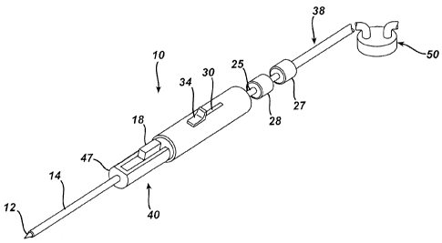
[0020] Figure 1 illustrates a biopsy device 10 useful in the biopsy method
according to one
embodiment of the present invention. Biopsy device 10 can include an outer
sheath
assembly 40 and an inner cannula assembly 116. The outer sheath assembly 40
can
include an outer cannula 14, a body 47, and a handle 45. A proximal opening 36
can be
provided in the proximal end of handle 45.
[0021] The inner cannula assembly 116 can include an inner cannula 16
extending distally from
an inner cannula locking hub 28. A proximal opening 136 can be provided in the
CA 02580181 2007-03-02
-4-
proximal end of inner cannula assembly 116. Referring to Figure 1, a vacuum
tube 38, a
syringe 32, a sample / fluid capture container 50, or other suitable device
may be
releasably attached to the proximal end of the inner cannula assembly 116,
such as by
using a locking hub 27 associated with the device.
[0022] Inner cannula 16 can include a closed, distal tissue penetrating tip 12
adapted for piercing
tissue. Inner cannula 16 can also include a side tissue sample port 26
disposed
proximally of tip 12. Sample port 26 can communicate with a central lumen
extending
the length of cannula 16 to the proximal opening 136.
[0023] The outer cannula 14 can be supported to extend distally through at
least a portion of
body 47. Body 47 can extend distally from handle 45. A biopsy method selection
button
34 can be provided on handle 45, and a release button 18 can be provided on
body 47.
The biopsy method selection button can be used to select a fine needle
aspiration mode of
operation or a core sample mode of operation. Release button 18 can be used to
release
the position of outer cannula 14, as described more fully below.
[0024] Proximal opening 36 allows for insertion of inner cannula 16 into outer
sheath assembly
40. The relative position of inner cannula 16 to outer sheath assembly 40 can
be
maintained in a plurality of positions to provide a desired biopsy sampling
mode. For
instance, the relative position of inner cannula 16 to outer sheath assembly
40 can be
maintained by a locking outer hub 28 disposed a proximal end of inner cannula
16.
Locking hub 28 can be shaped or otherwise configured to releasably engage an
inner hub
25 associated with a proximal end of the handle 45. For instance, hub 25 can
be received
in a distal end of hub 28 to provide releasable attachment using any suitable
latching or
locking mechanism, including without limitation Leur type fittings, bayonet
fittings, and
the like.
[0025] Figure 2a illustrates a cross section of biopsy device 10 in position
for insertion into
tissue. In Figure 2a, the inner cannula 16 is shown inserted within the outer
sheath
assembly 40, and with outer hub 28 releasably coupled to the inner hub 25.
Outer
CA 02580181 2007-03-02
-5-
cannula 14 can extend distally from a cannula carrier 42. Cannula carrier 42
is shown
disposed within handle 45 in Figure 2a and can be biased distally relative to
the handle
45, such as by a resilient member disposed intermediate carrier 42 and an
inner surface of
handle 45. In Figure 2a, the resilient member comprises a coil spring 35
seated in a
proximal facing recess in carrier 42 and a distal facing recess in the inner
surface of
handle 45. As shown in Figure 2a, the inner cannula 16 can extend through
spring 35
when the inner cannula 16 is inserted into outer sheath assembly 40.
[0026] The proximal end of cannula 14 can be disposed in a central opening in
the distal face of
the carrier 42, with the outer cannula 14 extending from the distal face of
cannula carrier
42, The cannula 14 can be attached to carrier 42 by any suitable means,
including
without limitation by adhesives or interference fit. As shown in Figure 2a, a
tab 5 can be
provided on cannula 14. Tab 5 can be formed from a section of wall of outer
cannula 14,
such as by milling or otherwise cutting or forming a slot in the wall of
cannula 14 and
bending a portion of the wall back to form a resilient tab 5. Spring 35 biases
tab 5
distally against a shoulder 33 formed in a passageway extending through body
47.
Various alternatives to a cut tab 5 can be employed, such as a separate
resilient tab joined
to the outer surface of cannual 14, or a resilient rib or projection formed to
extend from
the outer surface of cannula 14.
[0027] Referring to Figures 2a, 2b, and 2c, the body 47 can include slots 21a
and 21b, with slot
21b being positioned proximally of slot 21 a. In the embodiment shown, the
slots 21 a and
21b extend through the thickness of the wall of body 47. Tab 5 of outer
cannula 14 is
positionable in slot 21a for fine needle aspiration, and is positionable in
slot 21b for core
biopsy sampling. Tab 5 is shown positioned in slot 21a in Figure 2b, and tab 5
is shown
positioned in slot 21b in Figure 2c.
CA 02580181 2007-03-02
-6-
[0028] In the insertion position illustrated in Figure 2a, tab 5 of outer
cannula 14 is biased
against shoulder 33 of body 47, and the outer cannula 14 completely covers
sampling
port 26 preventing tissue from entering the sample port during insertion or
removal of the
device. Outer cannula 14 can have an open distal end with a sharped distal
edge 13. The
sharpened distal edge 13 of outer cannula 14 can be disposed just proximal of
tip 12
when inner cannula 16 is inserted fully into outer cannula 14. The distal edge
13 can
include a generally conical, tapered surface which can serve to provide an
extension of
the sloped surface of tip 12 when the inner cannula 16 is inserted fully into
outer cannula
14. The outer diameter of inner cannula 16 can be selected to slide freely
within the
lumen of outer cannula 14. Outer cannula 14 can be provided with an outer
diameter
corresponding to any size of biopsy needle. Common biopsy needle sizes range
from 8
gauge to 25 gauge.
[0029] Figure 2b illustrates the biopsy device 10 in position for obtaining a
fine needle
aspiration (FNA) sample. In the FNA position illustrated in Figure 2b, outer
cannula 14 is
retracted proximally relative to inner cannula 16 a distance less than the
longitudinal
length of sample port 26, in order to expose a portion, but not all of, the
longitudinal
length of sample port 26. This position is accomplished by pushing selection
button 34
(mounted on the outer surface of handle 45) proximally in button slot 30 (
such as with an
operator's thumb) formed in the outer surface of handle 45, until tab 5
resiliently snaps
into slot 21 a.
[0030] Selection button 34 can include a ring 22 that slip fits over an outer
surface of a circular
section of the distal end of cannula carrier 42, such that ring 22 can slide
freely with
respect to the carrier 42 and the handle 45. A coil spring 54 can be provided
to resiliently
bias the ring 22 and button 34 in a distal direction. The coil spring 54 can
be disposed
about an outer surface of the proximal portion of carrier 42. As shown in
Figures 2a, 2b,
and 2c, the proximal end of spring 54 can be seated against a shoulder formed
on an inner
surface of the handle 45, and the coil spring 54 can bear against a proximal
face of ring
22, such that removal of a thumb force on selection button 34 permits spring
54 to urge
CA 02580181 2007-03-02
-7-
ring 22 distally to its default position at a proximal end surface 51 of body
47. The
carrier 42 and outer cannula 14 will remain in the position shown in Figure 2a
until
release button 18 is depressed and releases tab 5 from slot 21a. Release of
tab 5 enables
main spring 35 to push cannula carrier 42 and outer cannula 14 to close sample
port 26.
[0031] A Fine Needle Aspiration (FNA) sample is obtained by withdrawing a
sample of cells
(as distinguished from a solid tissue sample) from a lump, cyst, fluid filled
sac, or other
suspicious lesion. To collect an FNA sample, the device can be positioned as
shown in
Figure 2b, and the user can reciprocate the exposed portion of sample port 26
(such as in
a back and forth motion) within the tissue mass, to thereby scrape cells from
the target
tissue mass. If desired, the tissue cells received in sample port 26 can be
drawn into port
26 by vacuum communicated through cannula 16. For example, vacuum can be
provided
by a syringe 32 (Figure 1) which may be releasably attached to opening 136.
[0032] Figure 2c illustrates the biopsy device 10 in position for obtaining a
core biopsy sample.
In the core biopsy position, outer cannula 14 can be retracted to expose the
entire sample
port 26, as shown in Figure 2c. To retract outer cannula 14, the selection
button 34 can
be moved proximally (by a finger of the hand holding the handpiece 40) within
button
slot 20 until tab 5 snaps into slot 21b. Moving selection button 34 proximally
(against
the biasing force of spring 54 ) within button slot 20 causes outer cannula 14
to move
proximally, against the biasing force of spring 35, to fully expose sample
port 26. With
the sample port 26 fully exposed, a vacuum can be applied through inner
cannula 16
drawing the tissue into sample port 26. In order to sever a core sample of
tissue, the
operator can depress release button 18 to release tab 5 from slot 21b.
Releasing tab 5
from slot 21b enables spring 35 to push cannula carrier 42 and outer cannula
14 distally,
thereby closing sample port 26. As outer cannula 14 moves over sample port 26,
the
distal cutting edge 13 of outer cannula 14 cuts through the tissue mass,
severing a core
tissue sample disposed in sample port 26.
CA 02580181 2007-03-02
-g-
[0033] Figure 3 discloses various components according one embodiment of
biopsy device 10.
Biopsy device 10 is shown with vacuum tube 38 connected at a proximal end of
the
device to provide vacuum to inner cannula 16 through opening 136. Vacuum tube
38
allows for application of a vacuum through inner cannula 16 to assist in
drawing cells (in
FNA procedure) or tissue (in core procedure) into sample port 26.
[0034] With biopsy device 10 in the insertion position shown in Figure 2a, the
surgeon inserts
the distal end of the biopsy device 10 into the tissue mass to be sampled. The
surgeon
can, depending on the preferred method of biopsy, use the same device to
obtain either
an FNA sample, or a core biopsy sample. For an FNA sample, selection button 34
is
pushed proximally in button slot 30 until tab 5 snaps into slot 21a. The
surgeon moves
tip 12 over tissue to scrape cells from the tissue mass. If desired, the
surgeon can employ
the device 10 under any suitable visualization method, including without
limitation, X-
ray, ultrasound, or Magnetic Resonance Imaging (MRI). For instance the
components of
the biopsy device can be formed of suitable MRI compatible materials for use
with MRI
devices. The cells are pulled into partially covered sample port 26 by vacuum
(such as
vacuum generated by a syringe 32.
[0035] After the FNA sample is taken from the tissue mass, the surgeon may
desire to obtain
either another FNA sample, or alternatively, a core biopsy sample, such as
from the same
tissue mass from which the FNA sample was taken. The surgeon can obtain a
second
sample, such as a core biopsy sample, without removing the device from the
tissue mass,
and without employing a different or additional biopsy device. For instance,
to retrieve a
second sample, the surgeon can push selection button 34 proximally into button
slot 20
until tab 5 snaps into slot 21b, so that the sample port 26 is fully open.
Release button 18
can then be depressed so that outer cannula 14 is biased distally to slide
over sample port
26, thereby cutting through the tissue mass and severing a core tissue sample
disposed in
CA 02580181 2007-03-02
-9-
sample port 26. Upon completion of the biopsy, outer cannula 14 can be
returned to
insertion position and the surgeon removes biopsy device 10 from patient.
[0036] In one embodiment, the sample port 26 can have an uncovered length, as
measured along
the length of cannula 16, of at least about 10 millimeters, and more
particularly, at least
about 20 millimeters. In the FNA position shown in Figure 2b, the sample port
can be
uncovered to provide a side tissue inlet port having a length of no more than
about 5
millimeters, and more particularly, no more than about 3 millimeters. In the
core biopsy
position shown in Figure 2c, the sample port 26 can be fully uncovered to
provide a side
tissue inlet port having a length of at least about 10 millimeters, and more
particularly at
least about 20 millimeters.
[0037] The biopsy device shown in Figures 2a-2c provides two distinct,
predetermined positions
of the outer cannula 14 relative to sample port 26, one position corresponding
to FNA
sampling, and one position corresponding to core tissue sampling. While the
biopsy
device shown in Figures 2a-2c employs two slots 21a and 21b, it will be
understood that
more than two slots can be provided to accommodate three or more positions of
the outer
cannula 14 relative to the sample port 26, so that graduated exposure of the
sample port
26 is obtained. Additionally, mechanisms other than slots 21 can be employed
to
provide positioning of cannula 14 relative to sample port 26.
[0038] The devices disclosed herein can be designed to be disposed of after a
single use, or they
can be designed to be used multiple times. In either case, however, the device
can be
reconditioned for reuse after at least one use. Reconditioning can include any
combination of the steps of disassembly of the device, followed by cleaning or
replacement of particular pieces, and subsequent reassembly. In particular,
the device
can be disassembled, and any number of the particular pieces or parts of the
device can
be selectively replaced or removed in any combination. Upon cleaning andlor
replacement of particular parts, the device can be reassembled for subsequent
use either
CA 02580181 2007-03-02
-10-
at a reconditioning facility, or by a surgical team immediately prior to a
surgical
procedure. Those skilled in the art will appreciate that reconditioning of a
device can
utilize a variety of techniques for disassembly, cleaning/replacement, and
reassembly.
Use of such techniques, and the resulting reconditioned device, are all within
the scope of
the present application.
[0039] The various components and subassemblies disclosed herein can be
described in the
alternative as a means for providing the function performed by the particular
component
or subassembly. While the present invention has been described in terms of the
embodiments disclosed in the figures, it will be understood that those skilled
in the art
may make various changes and modifications without departing from the spirit
and scope
of the present invention. Accordingly, the above description is not intended
to limit the
scope of the present invention, and it will be understood that the scope of
the present
invention is defined in terms of the claims set forth below.