Note: Descriptions are shown in the official language in which they were submitted.
CA 02580620 2007-03-15
DESCRIPTION
METHOD OF DETECTING HEPATITIS B VIRUS s ANTIGEN
Technical Field
The present invention relates to a probe recognizing a
novel epitope of hepatitis B virus (HBV) s antigen (HBs antigen)
and a method of detecting HBV, or HBs antigen, by using the probe.
Background Art
Diagnosis of viral infection is carried out mainly by a
method of detecting a virus or a virus-related component (protein
or nucleic acid) or by a method of detecting a specific antibody
produced by the living body upon viral infection.
Usually, infection with HBV can be known by confirming
the presence of HBs antigen or HBc antibody. As an indicator
not only for infection with HBV but also for a clinical state
of an HBV carrier or judgment of prognosis and treatment efficacy,
the amount of hepatitis B virus e antigen (HBe antigen) , an
antibody to the antigen, or DNA of HBV (HBV-DNA) is measured.
Among antigens constituting HBV viral particles (HBV
particles) , HBs antigen is a major constitutional envelope
protein on the surface of infectious HBV particle and is anchored
in a hepatocyte-derived lipid bilayer in which a core particle
containing HBV-DNA is enveloped. In blood from a patient
infected with HBV, there are noninfectious small spherical
particles or tubular particles consisting of HBs antigens. The
small spherical particles are present most abundantly in blood,
1
CA 02580620 2007-03-15
and about 1000 small spherical particles are observed per one
or several HBV particles. A majority of HBs antigen test agents
commercially available at present mainly detects HBs antigen
in the form of small spherical particles.
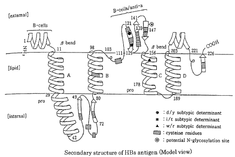
HBs antigen is a membrane protein consisting of 226 amino
acid residues (amino acid numbers 1 to 226) and penetrating 4
times through a lipid bilayer. Although a model of the
transmembrane structure of HBs antigen is not fully elucidated,
Howard et al. (Howard et al., Viral Hepatitis and Liver Disease
(ed by Zuckerman AJ, Alan R) , pp. 1094-1101, Liss Inc., New York,
1988) have proposed that the HBs antigen is composed of a (ER
lumen side) region, outside a lipid bilayer, consisting of
positions 1 to 11 from the N-terminal of HBs antigen, a hydrophobic
transmembrane region penetrating through a lipid bilayer
consisting of positions 12 to 28, a region inside the lipid bilayer
consisting of positions 29 to 80, a hydrophobic transmembrane
region consisting of positions 81 to 97, a hydrophilic ER lumen
region consisting of positions 98 to 156, and two hydrophobic
transmembrane regions consisting of positions 157 to 226 (FIG.
1) .
A main common "a" determinant used in detection of HBs
antigen in conventional methods is positioned in the amino acid
positions 110 to 156, contained in amino acids in the positions
98 to 156 localized at the ER lumen side, that is, on the surface
of viral particle. This common "a" determinant is reported to
consist of a complicated higher-order structure wherein at least
4 epitopes are present (Hiroaki Okamoto, "Nippon Rinsho, Bunshi
2
CA 02580620 2007-03-15
Kan-En Uirusubyogaku, Kiso-Rinsho-Yobo" (Japanese Clinic,
MolecularHepatitisVirology, Fundamental-Clinic-Prophylaxis),
LowerVolume,HepatitisA, B, D, EViruses, pp. 212-222, published
on October 26, 1995).
HBV is DNA virus, but HBV is known to undergo mutation
comparable with RNA virus because during viral proliferation,
its DNA is replicated into RNA, and from this RNA, DNA is
synthesized by reverse transcriptase. Accordingly, it is
estimated that mutants having various kinds of mutations occur
in an individual infected with HBV. When external selective
stress such as neutralizing antibody is applied to the HBV in
such individual, there occurs the phenomenon in which HBV strains
sensitive to the stress are decreased, while mutants resistant
or insensitive to the stress are increased. The so-called
"escape mutant" coming to be problematic in recent years is a
mutant which has undergone substitution, deletion or insertion
of amino acid(s) in the main common "a" determinant, thereby
being endowed with an ability to maintain its infection by
escaping from an antibody recognizing the "a" determinant before
mutation.
One problem associated with occurrence of the escape mutant
is that this mutant can keep persistent infection because it
can escape from an antibody induced by inoculation with a vaccine
utilizing the "a" determinant before mutation.
Another problem is that this escape mutant cannot be
detected in conventional HBs antigen examination methods.
Generally, the escape mutant having a mutation on the common
3
CA 02580620 2007-03-15
"a" determinant has lower reactivity with a monoclonal antibody
against the common "a" determinant in the wild-type HBV, and
thus a monoclonal antibody against the wild-type common "a"
determinant, used in conventional HBs antigen examination
methods, cannot recognize the HBs antigen in the escape mutant
type HBV, thus failing to find actually occurring HBV infection.
For example, it is reported that a 145Arg mutant, that is, the
mutant wherein amino acid at position 145 was changed from Gly
in the wild type to Arg, has significantly lower reactivity with
a monoclonal antibody against the common "a" determinant (Hiroaki
Okamoto, "Nippon Rinsho, Bunshi Kan-En Uirusubyogaku,
Kiso-Rinsho-Yobo" (Japanese Clinic, Molecular Hepatitis
Virology, Fundamental-Clinic-Prophylaxis) , Lower Volume,
Hepatitis A, B, D, E Viruses, pp. 212-222, published on October
26, 1995) . Actually, it is reported that transfusion of blood,
shown to be HBs antigen-negative in a screening test of HBV by
using the conventional HBs antigen measurement reagent, caused
infection with HBV (Thiers et al. Lancet, ii, 1273-1276, 1988) .
In acute infection with HBV, there is a reported phenomenon
in which an HBV-infected patient is HBs antigen-positive in an
initial stage of infection and then turns HBs antigen-negative
and simultaneously becomes HBs antibody-positive. The reason
that the patient becomes HBs antibody-positive is that an
antibody against the common "a" determinant of HBs antigen is
produced in the body of the patient. The patient's antibody
against the common "a" determinant binds to the same region as
in the common "a" determinant recognized by a monoclonal antibody
4
CA 02580620 2007-03-15
used in the HBs antigen test reagent, thus leading to competition
between both the antibodies, and by the competition, the
sensitivity of the HBs antigen test reagent is reduced so that
the detection of HBV by the test reagent is prevented.
Disclosure of the Invention
Problem to be Solved by the Invention
The conventional HBs antigen test reagent using a
monoclonal antibody to the common "a" determinant in the
wild-type HBV cannot detect an escape mutant having a mutation
on the common "a" determinant, andwhenblood j udged tobe negative
by this test reagent is used in blood transfusion, infection
with HBV may be caused. An object of the present invention is
to develop a probe capable of detecting such escape mutant of
HBV and a method of measuring HBs antigen by using the probe.
Another object of the present invention is to develop a
probe which can measure HBs antigen without being prevented by
a patient's antibody to the common "a" determinant even in an
HBs antibody-positive sample from an infected patient, as well
as a method of measuring HBs antigen by using the probe.
Means to Solve the Problem
The present inventors succeeded in solving the problem
described above by using, as a probe, an antibody recognizing
an epitope located on a peptide consisting of an amino acid
sequence set forth in SEQ ID NO: 1.
That is, the present invention relates to a probe
CA 02580620 2007-03-15
recognizing an epitope located on a peptide consisting of an
amino acid sequence corresponding to positions 26 to 80 in
hepatitis B virus s antigen and in particular to a probe
recognizing an epitope located on a peptide consisting of an
amino acid sequence in SEQ ID NO: 1.
The present invention also relates to a method of detecting
hepatitis B virus or hepatitis B virus s antigen, which comprises
using the probe described above.
In this specification, the positions of partial amino acid
sequences in the HBs antigen composed of 226 amino acid residues
are indicated by assigning number 1 to the N-terminal amino acid
residue of the antigen. In an S region of HBV gene, there are
Pre-S1, Pre-S2 and S genes coding for a large S protein composed
of 389 to 400 amino acid residues governed by the Pre-S1 gene
+ Pre-S2 gene + S gene, a middle S protein composed of 281 amino
acid residues governed by the Pre-S2 gene+S gene, and a small
S protein composed of 226 amino acid residues governed by the
S gene (Keiji Mitamura, "Nippon Rinsho, Bunshi Kan-En
Uirusubyogaku, Kiso-Rinsho-Yobo" (Japanese Clinic, Molecular
Hepatitis Virology, Fundamental-Clinic-Prophylaxis) , Lower
Volume, pp. 13-27, published on October 26, 1995) . The term
"FiBs antigen" used herein generally means the small S protein
unless otherwise specified. However, the detection method of
the present invention can detect all of large S protein, middle
S protein and small S protein, and thus the hepatitis B virus
s antigen (HBs antigen) detected by the method contains the above
3 proteins.
6
CA 02580620 2007-03-15
The probe of the present invention is a probe capable of
recognizing an epitope on a peptide consisting of the amino acid
sequence set forth in SEQ ID NO: 1 and is typically a polyclonal
or monoclonal antibody capable of specifically recognizing the
epitope. Specific examples of the antibody are monoclonal
antibodies produced by any of hybridoma cell strains 1C10, 4A3
and 6G6 deposited under Accession Nos . FERN ABP-10115, ABP-10116
and ABP-10117 since September 9, 2004, with International Patent
Organism Depositary (IPOD) , National Institute of Advanced
Industrial Science and Technology (AIST) at Central 6, 1-1-1,
Higashi, Tsukuba City, Ibaraki Pref . , Japan.
The amino acid sequence set forth in SEQ IDNO: 1 corresponds
to an amino acid sequence in the positions 26 to 80 in the HBs
antigen, that is, a hydrophilic region present in the inside
of a lipid bilayer of HBs antigen. This epitope is positioned
in the inside of HBV viral particle, small spherical particle
or tubular particle, and unlike a region positioned in the side
of ER lumen containing the common "a" determinant of HBs antigen,
will not be subject to selective stress such as an external
neutralizing antibody capable of inducing the escape mutant.
Accordingly, a mutation on the above epitope, as compared with
the common "a" determinant in the conventional method, will
hardly undergo selective stress, and with the epitope of the
present invention given, there would seldom or never occur the
phenomenon wherein specific mutants only dominate such that only
mutants not reacting with the HBs antigen measurement reagent
are increased.
7
CA 02580620 2007-03-15
The probe of the present invention, when used to detect
HBs antigen, hardly undergoes interference by a patient's
antibody to HBs antigen. This is probably because the epitope
recognized by the probe of the present invention is located in
the inside of HBV particle, small spherical particle and tubular
particle, so that as compared with the common "a" determinant
of HBs antigen, this epitope is less likely to act as an immunogen
in the body of the HBV-infected patient, and thus the production
of patient's antibody to the epitope is suppressed.
By using the probe of the present invention, it is thus
possible to reliably detect HBs antigen even in the escape mutant
having a mutation on the common "a" determinant.
For detecting HBs antigen in HBV particle, small spherical
particle or tubular particle in a sample with the probe of the
present invention, an epitope on the amino acid sequence in
positions 26 to 80 in HBs antigen, localized inside a lipid bilayer
or in a spherical or tubular particle, should be in such a state
as to be contacted with the probe.
Another aspect of the present invention is a method of
detecting HBV or HBs antigen in a sample, which comprises adding
to a sample a denaturant capable of denaturing a lipid bilayer
or a protein aggregate, typically a protein denaturant such as
a surfactant, chaotropic ion etc., particularly a surfactant
in order to detect HBs antigen in the sample with the probe of
the invention.
In the present invention, HBV particles, small spherical
particles and tubular particles are denatured by using the
8
CA 02580620 2007-03-15
denaturant, whereby their inside region consisting of the amino
acid sequence in positions 26 to 80 in HBs antigen, that is,
a hydrophilic region present in the inside of a lipid bilayer
of HBs antigen, is exposed to the outside. The denaturant usable
herein is a denaturant destroying lipid bilayers of HBV particles
and breaking bonds (aggregation) among HBs antigens in small
spherical particles and tubular particles but not inactivating
the probe of the invention, and typically a surfactant such as
sodium dodecyl sulfate can be used.
The present invention also provides a method of detecting
HBV virus, which comprises using the probe of the present
invention, a denaturant, and a probe capable of specifically
recognizing HBV antigen other than HBs antigen, as well as a
reagent for detection of HBV, having such a constitution, that
is, comprising the probe of the present invention, a denaturant,
and a probe capable of specifically recognizing HBV antigen other
than HBs antigen.
By measuring HBV antigen other than HBs antigen, for
example HBcr antigen (W002/14871) with a probe used in
combination with the probe of the present invention, a HBV patient
sample which may be judged erroneously as being HBV-negative
by detection of only HBs antigen can be grasped reliably as
HBV-positive.
As described above, FiBs antigen is known to undergo
mutation at high frequency comparative to that of RNA virus.
Accordingly, there is also a mutant HBs antigen consisting of
a sequence which is different from the amino acid sequence of
9
CA 02580620 2007-03-15
SEQ ID NO: 1 in the amino acid sequence in positions 26 to 80
in HBs antigen.
However, even HBs antigen having such a mutation can, if
its amino acid sequence is specified, be expressed in Escherichia
coli and purified according to the disclosure of this
specification. A probe directed to such purified mutant HBs
antigen is obtained and can be used to detect HBs antigen. In
the present invention, therefore, the sequence corresponding
to the amino acid sequence in positions 26 to 80 in HBs antigen
is limited neither to a probe recognizing an epitope on the amino
acid sequence shown in SEQ ID NO: 1 nor to the detection method
of using said probe.
Effect of the Invention
By the probe of the present invention and a method of
detecting HBV or HBs antigen by using the same, an escape mutant
etc. having a mutation on common "a" determinant of HBs antigen,
which cannot be detected by the conventional HBs antigen
detection method, can be highly sensitively detected thereby
highly reliably judging HBV infection. Even if a patient's
antibody to the common "a" determinant of HBs antigen inhibits
detection of HBs antigen, HBV infection can be reliably judged
by the detection method of the present invention.
Even if a patient's antibody competing with the probe of
the present invention occurs and inhibits detection of HBs
antigen, HBV infection can be reliably judged by pretreatment
with a combination of an acidifying agent or an alkalifying agent
CA 02580620 2007-03-15
and a denaturant.
By using a combination of the probe of the invention and
a probe recognizing another antigen of HBV to measure HBs antigen
and another antigen of HBV simultaneously, HBV infection can
be detected more reliably.
Brief Description of Drawing
FIG. 1 shows an illustration of the secondary structure
of HBs antigen.
Best Mode for Carrying Out the Invention
The probe of the present invention can be any probe capable
of specifically recognizing an epitope on a peptide consisting
of an amino acid sequence corresponding to the positions 26 to
80 in HBs antigen, for example the amino acid sequence set forth
in SEQ ID NO: 1, and typically an antibody, particularly a
monoclonal antibody, raised against an antigen such as the above
peptide or HBs antigen, is useful.
A peptide consisting of the amino acid sequence set forth
in SEQ ID NO: 1 can be prepared by recombinant gene technology
using a gene encoding the peptide or by chemical synthesis, and
such preparation procedures can be attained by using various
methods or instruments etc. known per se.
A gene fragment containing a nucleotide sequence encoding
the amino acid sequence of SEQ ID NO: 1 can be prepared by
separating virus genes from HBV patient serum and amplifying
the objective gene by PCR. By using restriction enzyme sites
11
CA 02580620 2007-03-15
derived from a linker added at the time of PCR, or restriction
enzyme sites derived from a plasmid into which the gene fragment
was inserted, the gene can be cloned into an expression vector.
This expression vector is transformed into a host such
as Escherichia coil, and the Escherichia coli can be cultured
to give HBs (26 to 80) antigen positioned in the inside of a
lipid bilayer. Methods of collecting and purifying the
objective protein from the microorganism thus obtained through
culture can be achieved by conventional techniques, for example
procedures such as sonicating disruption of cells,
centrifugation, and various chromatographic techniques. That
is, when the objective protein is efficiently expressed by the
method described above, many proteins have formed inclusion
bodies in the microorganism. By utilizing this feature, the
microorganisms are suspended in a buffer under physiological
conditions, such as physiological saline, then the cells are
disrupted by sonication, and the disrupted microbial material
is centrifuged to recover an insoluble fraction. The recovered
insoluble fraction is extracted with 6 M urea and subjected to
gel filtration to give high-purity trpE-HBs (26 to 80) antigen
which can then be used as an immunogen.
The probe of the present invention, for example, the
polyclonal antibody can be produced by periodically immunizing
an animal such as rat, rabbit, goat or sheep, with the
above-mentioned HBs (26 to 80) antigen or polypeptide (referred
to hereinafter as the present antigen) alone or the present
antigen conjugated to BSA, KLH or the like, as a mixture with
12
CA 02580620 2007-03-15
an adjuvant such as Freund's complete adjuvant, and then
collecting its serum. To obtain the polyclonal antibody having
a specific recognition site, there is a method of using, as an
immunogen, a partial peptide in the objective region.
Production of a monoclonal antibody by a hybridoma is
well-known. For example, an animal such as BALB/c mouse is
immunized periodically with the present antigen alone or a
conjugate thereof with BSA, KLH or the like, as a mixture thereof
with an adjuvant such as Freund's complete adjuvant. When the
antibody titer in blood is increased, the present antigen is
administered in final immunization to a caudal vein, and the
spleen is aseptically excised, and the spleen cells are fused
with suitable mouse myeloma cells to give hybridomas. This
method can be carried out by the method of Kohler and Milstein
(Nature 256:495-497, 1975).
The hybridoma obtained by the method described above is
cultured in a suitable culture medium, and thereafter, a
hybridoma cell producing an antibody showing specific reaction
to the present antigen is selected and cloned. For cloning the
antibody-producing hybridoma, not only limiting dilution but
also a soft agar method (Eur J Immunol. 6:511-519, 1976) can
be utilized. This hybridoma can be cultured in a medium or a
mouse abdominal cavity to produce a monoclonal antibody in the
medium or ascites.
The polyclonal antibody in serum or the monoclonal antibody
produced in the medium or ascites can be purified by methods
such as column chromatography on protein A. The polyclonal
13
CA 02580620 2010-02-09
antibody can be subjected to methods such as affinity
chromatography using a carrier-immobilized antigen, whereby
only the antibody reacting with the specific antigen can be
purified, and in a similar manner, the antibody not reacting
with the specific antigen can also be obtained.
Besides the monoclonal antibody and polyclonal antibody,
molecules used as the probe can be produced. For example, a
recombinant antibody is described in detail in a review of
Hoogenboon (Trends in Biotechnology, 15:62-70, 1997) .
The denaturant used in the present invention may be any
denaturant which can destroy the structure of a lipid bilayer
of HBV particle or break bonds (aggregation) among HBs antigens
in a small spherical particle and tubular particle consisting
of HBs antigens. For example, urea, an acidifying agent and
an alkalifying agent can be used, and particularly a surfactant
is effective. The surfactant includes a nonionic surfactant,
a cationic surfactant, an amphoteric surfactant and an anionic
surfactant, any of which can be utilized insofar as it can destroy
the structure of a lipid bilayer. For example, nonionic
surfactants such as Tween 20 and Nonidet P-40 can sufficiently
destroy the structure of a lipid bilayer, to expose the epitope
in the present invention, although their surface activity is
not so strong.
Anionic surfactants such as SDS and Sarcosyl are considered
to have a strong surface activity, and such surfactants can also
expose the epitope in the present invention. Treatment with
the strong surfactant may destroy the conformational epitope
14
CA 02580620 2007-03-15
of the protein so that an antibody recognizing the conformational
epitope cannot bind to the antigen in a certain case; in this
case, the antigen can be measured by using a probe recognizing
the linear epitope of HBs antigen.
The denaturant plays a role not only of efficiently
releasing HBs antigens present in a sample but also of binding
the monoclonal antibody easily to HBs antigen.
In the present invention, use of a probe binding
specifically to the antigen denatured with a surfactant as
described above is particularly preferable. When an antibody
is used as the probe, the antibody should be a probe capable
of binding to the epitope in the invention exposed and denatured
by the denaturation treatment described above.
For example, when a specific surfactant having a strong
surface activity is used, it is necessary to select a monoclonal
antibody against the epitope exposed and denatured by the
surfactant. Accordingly, it is desired that a peptide
consisting of the amino acid corresponding to positions 26 to
80 in HBs antigen (for example, a peptide consisting of the amino
acid sequence set forth in SEQ ID NO: 1) subjected previously
to denaturation treatment with the surfactant is used to immunize
an animal, and also that the antibody is selected by using said
peptide.
For screening of the antibody, the peptide antigen
subjected to denaturation treatment is immobilized onto a solid
phase and used in screening of a monoclonal antibody reacting
with the antigen in a solution containing a surfactant, whereby
CA 02580620 2007-03-15
the antibody of the invention suitable for immunoassay can be
obtained. Since the screening solution contains a surfactant,
a monoclonal antibody resistant to the denaturation action of
the surfactant can be obtained.
The denaturant-treated HBs antigen in a sample can be
detected with immunoassays such as enzyme-linked immunosorbent
assay (ELISA), enzyme immunodot assay, radioimmunoassay, and
assay based on agglutination or other well-known immunoassays.
When a labeled antibody is used in detection, a label such as
a fluorescence substance, chemiluminescent substance,
radioactive substance or enzyme is used.
For example, when a method based on the principle of ELISA
sandwich reaction is used in detecting HBs antigen in a sample,
the method comprises the following steps. First, an antibody
or the like recognizing the epitope located in the inside of
a lipid bilayer is bound to a solid support (for example, an
inner wall of a microtiter well). Then, blocking with bovine
serum albumin or the like is carried out to prevent nonspecific
reaction. A sample treated with a surfactant or the like is
added to this support, to allow HBs antigen to be captured by
the antibody immobilized thereon. A labeled antibody or the
like to the captured HBs antigen can be reacted with the HBs
antigen to detect it. The antibody to be bound to a solid support
may be any antibody binding to the epitope positioned in the
inside of a lipid bilayer. The labeled antibody may be any
antibodybinding toHBs antigen. Their combination is arbitrary,
andacombinationachievinghighsensitivityandhighspecificity
16
CA 02580620 2007-03-15
can be selected.
The usable solid support described above includes
polystyrene, polycarbonate, polypropylene, a polyvinyl
microtiter plate, a test tube, a capillary, beads (latex
particles, erythrocytes, metal compounds etc.), a membrane
(liposome etc.) and a filter and the like. The sample in which
HBs antigen in the present invention can be measured includes
biological body fluids such as whole blood, plasma, serum, urine,
saliva and cerebrospinal fluid, as well as tissues such as hepatic
tissues.
A method of treating HBs antigen in a sample in such a
state as to be suitable for binding reaction with the probe,
for example the monoclonal antibody, without involving
complicated procedures is important in the present invention.
That is, it is important that a lipid bilayer of HBs antigen
contained in a sample is solubilized so that the epitope
originally not exposed to the surfaces of virus particles becomes
exposed.
Examples
The following examples are illustrative of the present
invention, but are not intended to limit the scope of the present
invention.
Example 1
Expression and purification of trpE-HBs (26 to 80) antigen
(A) Construction of TrpE-HBs (26 to 80) antigen-expressing
plasmid
17
CA 02580620 2007-03-15
An expression plasmid for HBs (26 to 80) region was
constructed by the following method. 100 1 serum from an HBV
patient was mixed with 100 1 DNA extract [10 1 of 1M Tris-HC1
(pH 8.4), 8 1 of 250 mM EDTA, 40 1 of 10% SDS, 8 1 of 5 M
NaC1, 10 1 of 20 mg/ml Proteinase K, 1 1 tRNA (5 g/ 1), and
23 1 sterilized water] and incubated at 54 C for 30 minutes.
The sample was mixed with 200 .1 phenol/chloroform (1/1) solution
and then centrifuged at 15 Krpm for 5 minutes to give a supernatant,
and 150 1 isopropanol and 7 1 of 5 M NaC1 were added to the
supernatant and left at -20 C for 1 hour. After centrifugation
at 15 Krpm at 4 C for 5 minutes, the precipitates were rinsed
with 70% ethanol and then centrifuged again at 15 Krpm at 4 C
for 5 minutes. The precipitates were air-dried and dissolved
in 20 1 sterilized water to give an HBV DNA solution.
1 of this HBV DNA solution was subjected to PCR with
2 primers (that is, 5'-GAATTCCTCACAATACCACAGAGTCTA-3' (SEQ ID
NO: 2) and 5' -GGATCCTTAAAAACGCCGCAGACACATCCAGCG-3' (SEQ ID NO:
3)). PCR was carried out with GeneAmpTM (DNA Amplification
Reagent Kit manufactured by Perkin Elmer Cetus) under the
conditions of DNA denaturation at 95 C for 1 minute, annealing
at 55 C for 1 minute, and DNA synthesis at 72 C for 1 minute,
and the resulting DNA fragment was separated by 0.8% agarose
gel electrophoresis and purified by a glass powder method
(GeneClean). 0.5 g of this amplified HBs (26 to 80) gene
fragment was digested with 20 1 restriction enzyme reaction
solution [50mMTris-HC1 (pH 7 . 5) , 10mMMgC12, 1mMdithiothreitol,
100 mM NaCl, 15 U EcoRI enzyme and 15 U BamHI enzyme] at 37 C
18
CA 02580620 2007-03-15
for 1 hour and then subjected to 0.8% agarose gel electrophoresis
to purify an about 180-bp EcoRI-BamHI fragment.
Then, 0.5 g of DNA, that is, an expression vector pATtrpE,
was digested with 20 1 restriction enzyme reaction solution
[50 mM Tris-HC1 (pH 7.5) , 10 mM MgC12, 1 mM dithiothreitol, 100
mM NaC1, 15 U EcoRI enzyme and 15 U BamHI enzyme] at 37 C for
1 hour, then 39 p,1 water was added to the reaction solution which
was then heat-treated at 70 C for 5 minutes, and 1 1 (250 U/ 1)
of bacteria alkaline phosphatase (BAP) was added thereto and
incubated at 37 C for 1 hour.
This reaction solution was subjected to extraction with
phenol, and the resulting aqueous phase was precipitated with
ethanol, and the precipitates were dried. 0.5 tug of the resulting
EcoRI-BamHI-treated vector DNA and the above-mentioned 180-bp
HBs (26 to 80) fragment were added to a mixture prepared by 1
1 (350 U/ 1) T4 ligase to 5 1 of a 10xligase buffer [660 mM
Tris-HC1 (pH 7.5) , 66 mM MgC12, 100 mM dithiothreitol, 1 mM ATP]
and then adjusting it to 50 1 with water, and then incubated
at 16 C overnight to effect ligation reaction. To obtain the
expression plasmidpATtrpE-HBs (26 to 80) , this ligation reaction
solution was used to transform Escherichia coli HB101.
The competent Escherichia coli strain used in
transformation is produced by a calcium chloride method [Mandel,
M. and Higa, A., J. Mol. Biol., 53, 159-162 (1970) ] . The
transformed Escherichia coli was plated onto an LB plate (1%
tryptone, 0.5% NaC1, 1.5% agar) containing 25 g/m1 ampicillin
and incubated at 37 C overnight. A transformed bacterial colony
19
CA 02580620 2007-03-15
occurring on the plate was transferred via a platinum loop to
an LB medium containing 25 g/ml ampicillin and cultured
overnight at 37 C.
1. 5m1 of the transformed bacterial culture was centrifuged
to collect the bacteria, and mini-preparation of plasmid DNA
was carried out by the alkali method [Manniatis et al., Molecular
Cloning: A Laboratory Manual, (1982) ] . 1 jig of the resulting
plasmid DNA was digested with 20 jtl restriction enzyme reaction
solution [50 mM Tris-HC1 (pH 7 . 5) , 10 mMMgC12, 1 mM dithiothreitol,
100 mM NaCl, 15 U EcoRI enzyme and 15 U BamHI enzyme] at 37 C
for 1 hour and then subjected to agarose gel electrophoresis
to separate pATtrpE-HBs (26 to 80) expression plasmid generating
about 180-bp EcoRI-BamHI fragment.
(B) Expression and purification of TrpE-HBs (26 to 80) antigen
The Escherichia coli HB101 strain harboring the expression
plasmid pATtrpE-HBs (26 to 80) was inoculated onto 3 ml of 2YT
medium (1.6% tryptone, 1% yeast extract, 0.5% NaC1) containing
50 vtg/m1 ampicillin, and then cultured at 37 C for 9 hours. 1
ml of this culture was inoculated into 100 ml M9-CA medium (0.6%
Na2HPO4, 0.5% KH2PO4, 0.5% NaC1, 0.1% NH4C1, 0.1 mM CaCl2, 2 mM
MgSO4, 0.5% casamino acid, 0.2% glucose) containing 50 jig/m1
ampicillin, and then cultured at 37 C. Indol-acrylic acid was
added to a final concentration of 40 mg/1 when 0D600 reached 0.3,
and further cultured for additional 16 hours. This culture was
centrifuged at 5 Krpm for 10 minutes to collect the microorganism.
The microorganism was suspended in 20 ml buffer A [50 mM
Tris-HC1 (pH 8.0), 1 mM EDTA, 30 mM NaC1] and then centrifuged
CA 02580620 2010-02-09
again to give 2.6 g expression microorganism. The resulting
microorganism was suspended in 10 ml buffer A and then the E.
coli membrane was disrupted by sonication, followed by
centrifugation to give an insoluble fraction containing a
trpE-HBs (26 to 80) fusion antigen.
This insoluble fraction was dissolved in 3 ml PBS
containing 8 M urea, 10 mM dithiothreitol and 1 mM EDTA and
subjected to gel filtration through a Sephacryl S300HR column
in the presence of 6 M urea, whereby the trpE-HBs (26 to 80)
fusion antigen to almost homogeneity.
Example 2
Preparation of hybridoma
The polypeptide [trpE-HBs (26 to 80)] prepared by the
methoddescribedabovewas dissolvedwith 6 Mureaandthendiluted
at a final concentration of 0.2 to 1.0 mg/ml in 10 mM phosphate
buffer (pH 7.3) containing 0.15 M NaCl(PBS), then mixed with
an equal volume of Freund's adjuvant, and administered
intraperitoneally in a dose of 10 to 20 1.tg to a 4- to 6-week-old
BALB/c mouse.
Booster was carried out every 2 to 4 weeks in the same
manner as above, and for final immunization, 10 lig HBs dissolved
in PBS was administered to the caudal vein.
At three days after the final immunization, the spleen
was aseptically removed from the mouse, then broken into
individual cells with scissors and a metallic mesh and washed
3 times with RPMI-1640 medium. Mouse myeloma cell strain
21
CA 02580620 2007-03-15
Sp2/0Ag14 at the logarithmic growth phase was washed 3 times
with RPMI-1640 medium, and the cells were mixed with the spleen
cells at a ratio of 1 : 5. After centrifugation at 200 x g for
minutes, the supernatant was removed, and 1 ml RPMI-1640 medium
containing 50% polyethylene glycol (PEG) 4000 (Merck) was added
slowly to the cell mass under gentle mixing, and 10 ml RPMI-1640
medium was further added thereby effecting cell fusion.
The resultant fusion cells were centrifuged (200 x g, 5
minutes) to remove PEG and then suspended in RPMI-1640 medium
containing 10% fetal bovine serum and hypoxanthine, aminopterin
and thymidine (HAT) and plated onto a 96-well cell culture plate.
After hybridomas only were proliferated by culture for about
days, clones producing the objective antibody were selected
by the ELISA method to give hybridomas producing the monoclonal
antibody having desired reaction specificity.
The resulting hybridomas were made monoclonal by limiting
dilution to establish antibody-producing hybridomas. The
resulting hybridomas were designated 6G6, 4A3, and 1C10,
respectively. These hybridoma cells have been deposited since
September 9, 2004, with International Patent Organism Depositary
(IPOD) , National Institute of Advanced Industrial Science and
Technology (AIST) , Japan.
Example 3
Preparation and analysis of monoclonal antibody
Each of the hybridomas obtained by the method described
in Example 2 was transplanted in a BALB/c mouse abdominal cavity
22
CA 02580620 2010-02-09
previously administered with pristane, and the monoclonal
antibody produced in the ascites was obtained.
The IgG fraction containing the monoclonal antibody was
purified by affinity chromatography on a protein A Sepharose
column.
The respective obtained monoclonal antibodies were
analyzed for their target epitope by using the TrpE-HBs (26 to
80) antigen and synthetic peptides each consisting of 20 amino
acids synthesized on the basis of a sequence derived from the
HBs region, and as a result, it was found that as shown in Table
1, these monoclonal antibodies recognize an epitope (amino acid
numbers: 26 to 80) of the HBs antigen, which is located in the
inside of a lipid bilayer.
23
CA 02580620 2007-03-15
[Table 1]
Table 1
Monoclonal antibody name
(Poly)peptide Amino acid
4A3 6G6 1010
name number
HBS-1 1-20
HBS-2 11-30
HBS-3 21-40
HBS-4 31-50
HBS-5 41-60
HBS-6 51-70
HBS-7 61-80
HBS-8 71-90
TrpE-HBs(26-80) 26-80
By an isotyping kit (Zymed) using anti-mouse Ig isotype
antibodies, (sub)classes of the respective monoclonal
antibodies were identified. As a result, the subtype of 6G6
and 4A3 was IgGl, k, and the subtype of 1010 was IgG2a, K., as
shown in Table 2.
There is no report on an antibody recognizing an antigen
epitope present in the region of amino acid numbers 31 to 70
in the present invention, and it was found that the monoclonal
antibodies 6G6, 4A3, and 1C10 recognize the novel epitope
respectively.
24
CA 02580620 2007-03-15
[Table 2]
Table 2
Clone name Subclass Estimated recognition site (amino =
acid number)
4A3 IgGl, K 31-50
6G6 IgGl, ic 51-60
1C10 IgG2a, K 51-70
Example 4
Examination of the detection method using a surfactant
The anti-HBs antigen monoclonal antibody 6G6 was diluted
to a final concentration of 6 jig/m1 with 10 mM sodium phosphate
buffer (pH 7.3) containing 0.15 M NaCl and then pipetted onto
a 96-well microtiter plate (Nunc) in a volume of 80 jil per well.
The plate was left at 4 C overnight and then washed twice with
0.35 ml of 10 mM sodium phosphate buffer (pH 7.3) containing
0.15 M NaC1, followed by adding 0.35 ml of 10 mM sodium phosphate
buffer (pH 7.3) containing 0.5% casein-Na (referred to
hereinafter as blocking solution) and further incubated at room
temperature for 2 hours.
After the blocking solution was removed, 40 111 of 100 mM
sodium phosphate buffer (pH 7.3) containing 0.15 M NaC1, 1% BSA,
and 0.5% casein-Na, to which various surfactants had been added
at a final concentration of 4% or 8%, and 40 1_11 of measurement
sample, were added to each well, then reacted at room temperature
for 1 hour, washed 5 times with 0.35 ml washing solution, followed
by adding 80 ill monoclonal antibody (5C3) labeled with peroxidase
CA 02580620 2007-03-15
(POD) and reacting the mixture at room temperature for 30 minutes.
Each well was washed 6 times with 0. 35 ml of the washing solution,
and after reaction thereof with 80 1 of a substrate
(orthophenylene diamine referred to hereinafter as OPD) solution
at room temperature for 30 minutes, 80 111 of 2 N sulfuric acid
solution was added to each well which was then measured for its
absorbance at a wavelength of 492 nm (0E1492) with its absorbance
at a wavelength of 630 nm as the reference.
The results of measurement of HBs-positive serum with
various surfactants are shown in Table 3. When the
surfactant-free buffer was used to measure HBs antigen-positive
serum, HBs antigen could not be detected, but when the buffers
containing various kinds of surfactants (anionic, cationic,
amphoteric and nonionic surfactants) were used in themeasurement,
a sufficient signal could be obtained to clearly detect HBs
antigen. It was thereby revealed that a novel epitope present
in the inside of a lipid bilayer of HBs antigen could be detected
by exposing the epitope to the outside with various surfactants.
5C3, the monoclonal antibody labeled with peroxidase, is
a monoclonal antibody obtained by expressing an antigen
consisting of an amino acid sequence in the positions 1 to 226,
that is, the full-length HBs antigen, then purifying this
recombinant antigen and immunizing a mouse with it. It was
confirmed that the antibody 503 thus obtained binds to the above
recombinant HBs antigen. However, when synthetic peptides each
consisting of 20 amino acids overlapping with each other by 10
amino acids were synthesized on the basis of the amino acid
26
CA 02580620 2007-03-15
sequence in positions 1 to 226 in the HBs antigen and examined
for their binding to the antibody 5C3 by the same method as in
Example 3, the antibody 503 didnot react with any of the synthetic
peptides. Accordingly, it is estimated that the antibody 503
recognizes not a linear epitope of an amino acid sequence of
HBs antigen, but a conformational epitope thereof.
27
CA 02580620 2010-02-09
[Table 3]
Table 3
Reagent name Concentra- Negative Positive Positive
Polarity
tion (%) serum serum 1 serum 2
No addition
- - 0.004 0.005 0.010
(control)
ClOTAC cationic 8 0.008 0.579 0.451
C14TAC cationic 8 0.025 2.222 1.466
Lauryl
pyridinium cationic 8 0.026 0.145 0.121
chloride
Tween20 nonionic 8 0.006 0.552 0.541
Triton X100 nonionic 8 0.004 0.895 0.629
NP40 nonionic 8 0.005 0.640 0.528
MEGA10 nonionic 8 0.012 1.051 0.738
Brij35 nonionic 8 0.011 1.003 0.811
CHAPS amphoteric 8 0.015 0.053 0.040
C12APS amphoteric 4 0.007 1.523 1.232
Sarcosyl anionic 4 0.010 1.990 1.833
SDS anionic 4 0.029 1.401 1.198
No addition
- - 0.014 0.019 0.021
(control)
C12TAB cationic 4 0.020 0.949 0.689
C14APS amphoteric 4 0.014 2.857 2.911
C16APS amphoteric 4 0.013 2.829 2.870
C18APS amphoteric 4 0.013 2.806 2.698
C8S03 anionic 4 0.016 0.740 0.223
C11S03 anionic 4 0.048 2.912 2.922
Sodium
hexadecyl- anionic 4 0.010 0.397 0.139
sulfate
Example 5
Measurement of HBs antigen-negative sample
The HBs antigen in a sample which was HBs antigen-negative
but was suspected of infection with HBV was measured by a
modification to the method in Example 4.
The anti-HBs antigen monoclonal antibody 6G6 was diluted
to a final concentration of 6 pg/ml with 10 mM sodium phosphate
buffer (pH 7.3) containing 0.15 M NaC1 and then pipetted onto
a 96-well microtiter plate (Nunc) in a volume of 100 ill per well .
The plate was left at 4 C overnight and then washed twice with
28
CA 02580620 2007-03-15
0.35 ml of 10 mM sodium phosphate buffer (pH 7.3) containing
0.15 M NaC1, followed by adding 0.35 ml of 10 mM sodium phosphate
buffer (pH 7.3) containing 0.5% casein sodium and 3% sucrose
(blocking solution) and leaving the mixture at room temperature
for 2 hours.
After the blocking solution was removed, 50 p1 of 100 mM
sodium phosphate buffer (pH 7.0) containing 0.15 M NaCl, 10 mM
EDTA-2Na, 0.2% proclin, 1% BSA, 0.1% casein sodium, 3% horse
serum, 2% mouse serum and 10% Brij 35, and 50 1 measurement
sample, were added to each well, reacted at room temperature
for 1 hour, washed 5 times with 0.35 ml washing solution, followed
by adding 100 pl of the monoclonal antibody (503) labeled with
peroxidase (POD) and reacting the mixture at room temperature
for 30 minutes.
After the reaction, each well was washed 6 times with 0.35
ml of the washing solution, and after reaction thereof with 100
pl solution of a substrate (orthophenylene diamine referred to
hereinafter as OPD) at room temperature for 30 minutes, 2 N
sulfuric acid solution was added to the sample which was then
measured for its absorbance at a wavelength of 492 nm (0D492)
with its absorbance at a wavelength of 630 nm as the reference.
The sample used was a sample purchased from IIC Japan;
the HBs antigen and anti-HBs antibody were measured by the CLIA
method of Abbott Laboratories; and HBN-DNA was measured by the
TMA method of Gen-Probe Incorporated.
29
[Table 4]
Table 4
HBsAg
(measurement
HBV-DNA HBsAg HBsAb
HBcrAg
method of the HBeAg
HBeAb HBcAb
No.
invention)
0
TMA 6G6/5C3 CLIA CLIA
HB44+/92
0
1.)
ul
LGE/ml Mean Judgment IU/ml Judgment mIU/m1 Judgment
RLI Judgment co
0
m
1.)
1 6.1 2.772 + 2.28 + 115.7 + - +
+ 965,534 + 0
1.)
0
0
2 6.7 2.819 + 0.02 - 541.8 + - +
+ 152,531 +
1
0
w
3 6.6 0.005 - 0.01 - 773.0 + - +
+ 252,891 + H1
In
4 6.8 2.646 + 1.42 + 174.1 + - +
+ 337,807 +
6.4 2.682 + 0.01 - 545.6 + - +
+ 312,615 +
CA 02580620 2007-03-15
The 5 samples shown in Table 4 are HBV-DNA-positive by
the TMA method. The samples are also HBs antibody-positive by
the CLIA method of Abbott Laboratories and considered to be serum
from patients infected with HBV. However, the samples Nos. 2,
3 and 5 were judged to be negative by the HBsAg CLIA method of
Abbott Laboratories, that is, the conventional HBs antigen
measuring method.
When these three HBs antigen-negative samples were judged
by the method of the present invention, HBs antigen could be
detected in the samples Nos. 2 and 5. The sample No. 3 judged
to be HBs antigen-negative by both the measurement method of
the present invention and the HBsAg CLIA measurement method of
Abbott Laboratories could be detected by the HBcrAg measurement
method, and the simultaneous measurement of HBs antigen and HBcr
antigen is useful in more accurate detection of HBV antigen.
Example 6
1) Concentration of an acidifying agent
501.11 aqueous hydrochloric acid at various concentrations
was added to 50 1.1.1, of an HBV antigen-negative sample or three
anti-HBs antibody-containing HBV antigen-positive samples
(#990493, #990640, #990650) and then incubated at room
temperature for 10 minutes, and 50 !IL solution of the mixture
was examined as a measurement sample by the following method.
The anti-HBs antigen monoclonal antibody 6G6 was diluted
to a final concentration of 6 pg/m1 with 10 mM phosphate buffer
(pH 7.3) containing 0.15 M NaCl and then pipetted onto a 96-well
31
CA 02580620 2007-03-15
microtiter plate (Nunc) in a volume of 100 1 per well. The
plate was incubated at 4 C overnight.
The plate was washed twice with 10 mM phosphate buffer
(pH 7.3) containing 0.15 M NaC1, followed by adding 350 1 of
mM phosphate buffer, pH 7.1, containing 0.5% casein sodium
and incubating the plate for 2 hours. After the blocking solution
was removed, 100 41 reaction buffer containing a neutralizing
agent and each of the various measurement samples obtained by
the sample treatment method were added to the respective wells,
reacted at room temperature for 2 hours under shaking, washed
6 times with 350 ill of 10 mM phosphate buffer, pH 7.3, containing
0.05% Tween 20 (washing solution) , followed by adding 100 [1.1,
of the monoclonal antibody (5C3) labeled with peroxidase (POD)
and reacting the mixture at room temperature for 30 minutes.
Each well was washed 6 times with the washing solution and then
incubated with 100 1 solution of a substrate (orthophenylene
diamine referred to hereinafter as OPD) for 30 minutes, and then
100 tl of 2 N sulfuric acid solution was added to each well which
was then measured for its absorbance at a wavelength of 492 nm
(0D492) with its absorbance at a wavelength of 630 nm as the
reference. The hydrochloric acid concentration shown in the
table is the concentration during treatment after mixing the
sample with the treatment agent.
Even by incubation of anti-HBs antibody-containing HBs
antigen-positive samples (#990493, #990640, #990650) at room
temperature for 10 minutes with the hydrochloric acid-free
solution, HBs antigen activity could hardly be detected. HBs
32
CA 02580620 2007-03-15
antigen activity was recognized at a concentration from 0.05
N hydrochloric acid at the time of treatment and reached a peak
at a concentration of 0.25 to 1.0 N (Table 5).
[Table 5]
Table 5
HBV negative
HBV positive sample
HC1 sample
Concentration Serum from
#990493 #990640 #990650
(N) healthy person
0 0.002 0.002 0.002 0.005
0.05 0.003 0.210 0.045 0.163
0.1 0.003 0.425 0.055 0.223
0.25 0.001 0.565 0.079 0.369
0.5 0.001 0.541 0.097 0.301
0.75 0.000 0.550 0.100 0.393
1 0.003 0.450 0.085 0.333
1.5 0.003 0.550 0.084 0.281
Example 7
2) Various surfactants in the presence of an acidifying agent
30 L of each of various surfactants dissolved in 1.0 N
aqueous hydrochloric acid was added to 30 L of an HBV
antigen-negative samples or HBs antigen-positive samples
(#990493, #990640, #990650) and then incubated at room
temperature for 10 minutes, and 50 L solution of the mixture
was examined as a measurement sample by the method described
33
CA 02580620 2007-03-15
=
in 1) (Tables 6 to 9) . The hydrochloric acid concentration and
surfactant concentration shown in the tables are the
concentrations during treatment after mixing the sample with
the treating agent.
As shown in Tables 6 to 9, the surfactant with which at
least 1 of the 3 samples had shown higher reactivity than the
judgment criteria of each sample was judged to be an effective
surfactant. As a result, it was found that when various
surfactants were added together with an acidifying agent such
as hydrochloric acid or sulfuric acid, there was a surfactant
with which the immunoreactivity of the HBs antigen in the HBs
antigen-positive sample was increased. The surfactant judged
to be effective was an amphoteric or cationic surfactant having,
in its molecule, a straight-chain alkyl group and a tertiary
amine or quaternary ammonium salt.
Nonionic surfactants such as Triton X100 and Bridj 35 are
also recognized to be effective. A surfactant having a steroid
skeleton, such as CHAPS, did not show improvement in reactivity.
In addition, anionic surfactants such as SDS and sodium N-lauroyl
sarcosinate, and deoxycholic acid, were also examined, but these
were poor in solubility in the presence of an acidifying agent,
thus making their examination infeasible.
An increase in measurement sensitivity was recognized by
adding an amphoteric or cationic surfactant having, in its
molecule, a straight-chain alkyl group and a tertiary amine or
a quaternary ammonium salt to an acidifying agent. Such
surfactant effective in the presence of an acidifying agent in
34
CA 02580620 2007-03-15
the treatment solution, when used in the treatment solution
without the acidifying agent, reduced the measurement
sensitivity significantly. From the foregoing, the reason for
the increase in measurement sensitivity would be that the
anti-HBs antibody acting as a factor inhibiting detection of
HBs antigen is inactivated by the acidifying agent, while the
epitope located inside a lipid bilayer of HBs antigen in a sample
is exposed to the outside by adding the surfactant, thus
significantly improving the reactivity thereof with 6G6.
CA 02580620 2007-03-15
. .
[Table 6]
Table 6
Cationic (TAO type)
HBV
negative HBV positive sample
sample
Serum
from
#990493 #990640 #990650
Concen- healthy
tration(%) person
No addition 0 0.006 0.541 0.097 0.254
Criteria for judging
the effect of 0.812 0.146 0.381
surfactant
Surfactant added to
0.5N HC1
Octyltrimethyl- 0.5 0.014 0.648 0.146 0.268
ammonium Chloride 1 0.018 0.783 0.194 0.327
[CH3 (CH2)7N (CH3)3] Cl 2 0.019 0.898 0.272 0.348
0.012 1.285 0.419 0.624
Decyltrimethyl- 0.5 0.020 0.727 0.231 0.288
ammonium Chloride 1 0.022 0.976 0.346 0.422
[CH3 (CH2) 9N (CH3)3] Cl 2 0.011 1.232 0.391 0.525
5 0.004 1.602 0.419 0.675
Dodecyltrimethyl- 0.5 0.034 0.797 0.261 0.324
ammonium Chloride 1 0.030 0.941 0.303 0.386
[CH3 (CH2) liN (CH3)3] Cl 2 0.024 0.972 0.259 0.415
5 0.006 0.990 0.234 0.392
Tetradecyltri- 0.5 0.029 0.924 0.261 0.357
methylammonium 1 0.035 1.002 0.306 0.436
Chloride 2 0.015 1.032 0.216 0.409
[CH3 (CH2)13N (CH3)31 Cl 5 0.005 0.804 0.136 0.281
Hexadecyltri- 0.5 0.032 0.933 0.254 0.425
methylammonium 1 0.031 0.974 0.271 0.458
Chloride 2 0.021 0.977 0.206 0.402
[CH3 (CH2)15N (CH3)3] Cl 5 0.005 0.811 0.164 0.279
Lauryl pyridinium 0.5 0.021 0.587 0.190 0.228
Chloride 1 0.013 0.716 0.236 0.309
[C5H5NCH2 (CH2) ioCH3] Cl 2 0.001 0.896 0.211 0.312
5 0.001 0.847 0.168 0.249
36
CA 02580620 2007-03-15
. .
[Table 7]
Table 7
Cationic (TAB type)
HBV
negative HBV positive sample
sample
Concen- Serum
tration from
#990493 #990640 #990650
(%) healthy
person
No addition 0 0.013 0.569 0.097 0.286
Criteria for judging the
0.854 0.146 0.429
effect of surfactant
Surfactant added to 0.5N
HC1
Octyltrimethylammonium 0.5 0.019
0.713 0.128 0.301
Bromide 1 0.022 0.836 0.165 0.347
[CH3(CH2)7N(CH3)3lBr 2 0.025 0.968 0.202 0.361
0.012 1.381 0.314 0.639
Decyltrimethylammonium 0.5 0.025
0.788 0.183 0.306
Bromide 1 0.026 1.051 0.260 0.462
[CH3(CH2)9N(CH3)3]Br 2 0.008 1.535 0.320 0.588
5 0.005 1.784 0.465 0.800
Dodecyltrimethyl- 0.5 0.029 0.938 0.205 0.353
ammonium Bromide 1 0.037 1.153 0.303 0.445
[CH3(CH2)11N(CH3)3]Br 2 0.028 1.343 0.309 0.544
5 0.011 1.402 0.317 0.496
Tetradecyltrimethyl- 0.5 0.034 0.994 0.210 0.366
ammonium Bromide 1 0.041 1.181 0.284 0.467
[CH3 (CH2)33N (CH3) 31Br 2 0.020 1.272 0.237 0.443
5 0.007 1.201 0.289 0.443
Hexadecyltrimethyl- 0.5 0.034 1.080 0.208 0.429
ammonium Bromide 1 0.037 1.196 0.236 0.498
[CH3(CH2)15N(CH3)3]Br 2 0.037 1.321 0.226 0.472
5 0.005 1.017 0.179 0.414
37
CA 02580620 2007-03-15
. .
[Table 8]
Table 8
Amphoteric
HBV nega-
HBV positive sample
tive sample
Serum from
Concen- healthy #990493 #990640 #990650
tration person
( % )
No addition 0 0.008 0.533 0.097
0.240
Criteria for judging the
0.799 0.146
0.359
effect of surfactant
Surfactant added to 0.5N
HC1
3-[3-(Cholamidopropyl) 0.5 0.009 0.606 0.115
0.248
dimethyl-ammonio]- 1 0.007 0.635 0.125
0.302
1-propanesulfonate 2 0.001 0.547 0.076
0.246
0.000 0.456 0.040 0.184
N-Dodecyl-N,N-di- 0.5 0.013 0.807 0.189
0.379
methyl-3-ammonio- 1 0.009 1.073 0.246
0.455
1-propanesulfonate 2 0.002 1.296 0.302
0.651
CH3(CH2) iiii (CH3) 2 [ (CH2) 3S03] 5 0.000 1.365
0.410 0.695
N-Tetradecyl-N,N-di- 0.5 0.012 0.873 0.181
0.386
methyl-3-ammonio- 1 0.010 1.076 0.245
0.477
1-propanesulfonate 2 0.005 1.267 0.268
0.554
CH3 (CH2)13N (CH3) 2 [ (CH2) 3S03] 5 0.000 1.362 0.356
0.558
N-Hexadecyl-N,N-di- 0.5 0.015 1.013 0.209
0.502
methyl-3-ammonio- 1 0.016 1.233 0.287
0.581
1-propanesulfonate 2 0.014 1.290 0.256
0.575
CH3 (CH2)151\1 (CH3) 2 [ (CH2) 3S033 5 0.002 1.286
0.276 0.519
38
CA 02580620 2007-03-15
. .
[Table 9]
Table 9
Nonionic
HBV
negative HBV positive sample
sample
Serum from
Concen- healthy #990493 #990640 #990650
tration person
(%)
No addition 0 0.005 0.459 0.070
0.210
Criteria for judging the
0.689 0.105
0.315
effect of surfactant
Surfactant added to 0.5N
HC1
Triton X-100 0.5 0.011 0.545 0.117
0.284
1 0.009 0.675 0.163
0.356
2 0.007 0.790 0.193
0.344
0.003 0.827 0.201 0.382
Triton X-114 0.5 0.006 0.470 0.112
0.283
1 0.005 0.554 0.149
0.372
2 0.003 0.678 0.160
0.370
5 0.001 0.489 0.118
0.258
Tween 20 0.5 0.009 0.437 0.086
0.251
1 0.007 0.468 0.110
0.278
2 0.008 0.647 0.118
0.341
5 0.007 0.605 0.147
0.307
Tween 80 0.5 0.007 0.339 0.063
0.209
1 0.007 0.312 0.068
0.220
2 0.009 0.451 0.051
0.245
5 0.007 0.498 0.063
0.240
Bridj 35 0.5 0.010 0.496 0.076
0.241
1 0.010 0.526 0.097
0.291
2 0.011 0.704 0.108
0.374
5 0.020 0.907 0.173
0.434
39
CA 02580620 2007-03-15
Example 8
3) Protein denaturant in the presence of an acidifying agent
30 L protein denaturant (urea or guanidine hydrochloride)
dissolved in 1.0 N aqueous hydrochloric acid was added to 30
I, of an HBV-negative sample or three HBs antigen positive samples
(#990493, #990640, #990650) and then incubated at room
temperature for 10 minutes, and 50 I, solution of the mixture
was examined as a measurement sample by the method described
in 1) . The immunoreactivity of each HBs antigen-positive sample
is shown in Table 10. The hydrochloric acid concentration and
protein denaturant concentration shown in Table 10 are the
concentrations during treatment after mixing the sample with
the treatment agent.
The samples showed higher immunoreactivity with the
protein denaturant in the presence of the acidifying agent than
with the acidifying agent only; that is, the immunoreactivity
was increased about 1.5- to 3-fold with urea or about 2- to 3-fold
with guanidine hydrochloride. At the time of treatment with
the acidifying agent, serum protein or the like may be denatured
to cause precipitation or to become turbid in some cases so that
the pipetting procedure is hindered and precipitates are often
a major cause of giving a false-positive result. There may also
occur a reduction in sensitivity attributable to incorporation
of the objective antigen into such precipitates . It was revealed
that formation of such precipitates can be significantly reduced
by adding urea or guanidine hydrochloride at a concentration
of 0.5 M or more at the time of treatment, and this effect is
CA 02580620 2007-03-15
. .
made particularly higher by adding urea at a concentration of
1.5 to 4 M and guanidine hydrochloride at a concentration of
2 to 3.5 M at the time of treatment.
41
CA 02580620 2007-03-15
. .
[Table 10]
Table 10
HBV
negative HBV positive sample
sample
Serum from
healthy #990493 #990640 #990650
Concen- person
tration (M)
No addition 0 0.018 0.483 0.076 0.175
Protein denaturant
added to 0.5 N HC1
Urea 0.5 0.015 0.555 0.085 0.204
1 0.010 0.619 0.076 0.236
1.5 0.006 0.636 0.090 0.248
2 0.005 0.686 0.081 0.293
2.5 0.005 0.725 0.100 0.335
3 0.005 0.771 0.088 0.382
3.5 0.003 0.830 0.116 0.443
4 0.008 1.041 0.143 0.578
Guanidine-HC1 0.5 0.027 0.706 0.116 0.235
1 0.024 0.802 0.146 0.270
1.5 0.020 0.820 0.140 0.307
2 0.014 0.943 0.179 0.385
2.5 0.008 1.039 0.183 0.455
3 0.005 1.113 0.235 0.504
3.5 0.003 0.970 0.248 0.528
42
CA 02580620 2007-03-15
Example 9
4) Examination of a reducing agent in the presence of an acidifying
agent
30 pL mixed solution consisting of dithiothreitol,
2-mercaptoethylamine hydrochloride or
2-diethylaminoethanethiol hydrochloride as reducing agents
dissolved in 1.0 N aqueous hydrochloric acid was added to 30
pL of an HBV antigen-negative sample (normal plasma) or three
HBs antigen-positive samples (#990493, #990640, #990650) and
then incubated at room temperature for 10 minutes, and 50 pL
solution of the mixture was examined as a measurement sample
by the method described in 1) (Table 11).
The reducing agent concentration used herein is the
concentration thereof in the sample during treatment. Even when
the reducing agent was added to the HBV antigen-negative sample,
no change in its signal was recognized, but in one HBs
antigen-positive sample (#990640), an increase of 30% or more
was recognized with dithiothreitol at a concentration of 1 to
mM.
43
,
[Table 11]
Table 11
Reducing agent
HBV
negative HBV positive sample
sample
Serum from
healthy #990493 #990640
#990650
person
% %
% 0
Concen-
relative
relative relative 0
tration
I.)
to to
to in
co
(mM)
0
control
control control m
"
Control 0 0.012 0.446 100 0.073 100
0.159 100 0
I.)
0
0
-.3
Reducing agent
1
0
addedto O. 5NHC1
w
1
Dithiothreitol 0.25 0.016 0.497 111 0.080 110
0.177 111 H
Iri
0.5 0.014 0.487 109 0.083 114
0.184 116
1 0.013 0.500 112 0.101 138
0.184 116
2 0.012 0.453 102 0.124 170
0.174 109
0.011 0.324 73 0.116 159 0.092 58
0.007 0.082 18 0.040 55 0.033 21
0.008 0.029 7 0.011 15 0.023 14
44
4
2-Mercaptoethyl 0.25 0.014 0.432 97 0.056 77
0.154 97
amine 0.5 0.009 0.429 96 0.064 88
0.144 91
Hydrochloride 1 0.009 0.426 96 0.060 82
0.148 93
2 0.008 0.411 92 0.069 95
0.129 81
0.004 0.350 78 0.068 93 0.109 69
0.001 0.278 62 0.075 103 0.074 47
0.002 0.217 49 0.058 79 0.063 40
50 0.000 0.140 31 0.037 51
0.033 21
2-Diethylamino- 0.25 0.015 0.429 96 0.066 90
0.165 104
ethanethiol 0.5 0.012 0.429 96 0.067 92
0.156 98 n
Hydrochloride 1 0.013 0.456 102 0.066 90
0.166 104 0
I.)
2 0.008 0.436 98 0.083 114
0.151 95 in
co
0
5 0.008 0.397 89 0.081 111
0.129 81 m
I.)
0
10 0.004 0.298 67 0.085
116 0.090 57 I.)
20 0.008 0.259 58 0.079 108
0.078 49 0
0
-.3
1
50 0.006 0.145 33 0.065 89
0.049 31 0
w
I
H
In
CA 02580620 2007-03-15
Example 10
5) Concentration of an alkalifying agent
50 1_11, aqueous sodium hydroxide solution at various
concentrations was added to 50 1.11, of an HBV antigen-negative
sample or three anti-HBs antibody-containing HBV
antigen-positive samples (#990493, #990640, #990650) and then
incubated at room temperature for 10 minutes, and 501AL solution
of the mixture was examined as a measurement sample by the
following measurement method.
The anti-HBs antigen monoclonal antibody 6G6 was diluted
to a final concentration of 6 Ag/m1 with 10 mM phosphate buffer
(pH 7.3) containing 0.15 M NaC1 and then pipetted onto a 96-well
microtiter plate (Nunc) in a volume of 100 Iii per well. The
plate was incubated at 4 C overnight.
The plate was washed twice with 10 mM phosphate buffer,
pH 7.3, containing 0.15 M NaC1, followed by adding 350 !Al of
mM phosphate buffer, pH 7.1 containing 0.5% casein sodium
and incubating the plate for 2 hours . After the blocking solution
was removed, 100 111., reaction buffer containing a neutralizing
agent, and the measurement samples obtained by each of the sample
treatment method, were added to each well and reacted at room
temperature for 2 hours under shaking, and washed 6 times with
350 ill of 10 mM phosphate buffer, pH 7.3, containing 0.05% Tween
(washing solution) , followed by adding 100 of a
biotin-labeled monoclonal antibody (HBs124) . The mixture was
reacted at room temperature for 30 minutes. Each well was then
washed 6 times with the washing solution, then 100 Li of
46
CA 02580620 2007-03-15
POD-labeled avidin D was added thereto, and the mixture was
reacted at room temperature for 30 minutes. After each well
was washed 6 times with the washing solution, 100 1 solution
of a substrate (orthophenylenediamine, referred to hereinafter
as OPD) was added thereto and incubated for 30 minutes, then
1001.11 of 2 N sulfuric acid solution was added to each well which
was then measured for its absorbance at a wavelength of 492 nm
(0D492) with its absorbance at a wavelength of 630 nm as the
reference. The sodium hydroxide concentration shown in the
table is the concentration during treatment after mixing the
sample with the treatment agent.
The biotin-labeled monoclonal antibody HBs124 is a
monoclonal antibody obtained by expressing and purifying a
full-length HBs antigen (that is, an antigen consisting of the
amino acid sequence in positions 1 to 22 6 ) as described in Example
1 and immunizing a mouse with the recombinant antigen. It was
confirmed that the antibody HBs124 binds to the above recombinant
HBs antigen. However, when synthetic peptides each consisting
of 20 amino acids overlapping with each other by 10 amino acids
were synthesized on the basis of the amino acid sequence in
positions 1 to 226 in the HBs antigen and examined for their
binding to the antibody HBs124 by the same method as in Example
3, the antibody HBs124 did not react with any of the synthetic
peptides. Accordingly, it is estimated that the antibodyHBs124
recognizes not a linear epitope of an amino acid sequence of
HBs antigen, but a conformational epitope thereof.
HBs antigen activity could not be detected even by
47
CA 02580620 2007-03-15
incubating the anti-HBs antibody-containing HBV-positive
samples (#990493, #990640, #990650) ma solution not containing
sodium hydroxide at room temperature for 10 minutes, but an
increase in signal for HBs antigen was recognized in treatment
with sodium hydroxide at a concentration of 0.25 to 1 N (Table
12).
48
CA 02580620 2007-03-15
4
[Table 12]
Table 12
HBV negative
HBV positive sample
NaOH sample
concen- Serum fromhealthy #990493 #990640 #990950
tration (N) person
0 0.006 0.005 0.006 0.006
0.05 0.006 0.010 0.004 0.002
0.1 0.010 0.014 0.010 0.008
0.25 0.006 0.034 0.006 0.012
0.5 0.008 0.058 0.006 0.030
0.75 0.004 0.070 0.006 0.039
1 0.010 0.044 0.018 0.042
1.5 0.008 0.012 0.014 0.029
Example 11
6) Various surfactant concentrations in the presence of an
alkalifying agent
30 L of various surfactants dissolved in 1.0 N aqueous
sodium hydroxide solution was added to 30 L of an HBV
antigen-negative sample or three HBs antigen-positive samples
(#990493, #990640, #990650) and then incubated at room
temperature for 10 minutes, and 50 L solution of the mixture
was examined as a measurement sample by the method described
in 5) (Tables 13 to 17). The sodium hydroxide concentration
and surfactant concentration shown in the tables are the
concentrations during treatment after mixing the sample with
49
CA 02580620 2007-03-15
the treatment agent.
As shown in Tables 13 to 17, the surfactant with which
at least 1 of the 3 samples had shown higher reactivity than
the judgment criteria of each sample was judged to be an effective
surfactant. As a result, it was found that when various
surfactants were added together with an alkalifying agent such
as sodium hydroxide, there was a surfactant with which the
immunoreactivity of the HBs antigen in the HBs antigen-positive
sample was significantly increased. The surfactant judged to
be effective includes an anionic surfactant such as sodium
dodecyl sulfate or N-lauroyl sarcosine Na and an amphoteric or
cationic surfactant having, in its molecule, a straight-chain
alkyl group and a tertiary amine or quaternary ammonium salt.
Nonionic surfactants such as Triton X100, Tween 20 and
Bridj 35 and a surfactant having a steroid skeleton, such as
CHAPS, are also recognized to be effective.
An increase in measurable sensitivity was recognized by
adding an anionic surfactant or an amphoteric or cationic
surfactant having, in its molecule, a straight-chain alkyl group
and a tertiary amine or a quaternary ammonium salt to an
alkalifying agent. Such surfactant effective in the presence
of an alkalifying agent in the treatment solution, when used
in the treatment solution without the alkalifying agent, was
not recognized to increase the measurable sensitivity. It was
considered that by a combination of the alkalifying agent and
the surfactant, the anti-HBs antibody acting as a factor
inhibiting detection of HBs antigen is inactivated, and the
CA 02580620 2007-03-15
epitope located inside a lipid bilayer of HBs antigen in a sample
is exposed to the outside, thus significantly improving the
reactivity thereof with 6G6.
51
CA 02580620 2007-03-15
,
[Table 13]
Table 13
Anionic
HBV
negative HBV positive sample
sample
Serum from
healthy #990493 #990640 #990650
person
Concen-
tration
(%)
No addition 0 0.015 0.032 0.002 0.009
Criteria for judging the
0.160 0.010 0.045
effect of surfactant
Surfactant added to 0.5
N NaOH
Sodium Dodecyl Sulfate 0.5 0.014 0.225 0.137 0.152
CH3(CH2)110S03Na 1 0.015 0.340 0.241 0.195
2 0.016 0.457 0.371 0.308
0.015 0.967 0.430 0.472
Lithium Dodecyl Sulfate 0.5 0.009 0.239 0.168 0.132
CH3 (CH2) ii0S03Li 1 0.015 0.275 0.205 0.141
2 0.007 0.468 0.416 0.272
5 0.016 1.055 0.385 0.325
N-Lauroylsarcosine 0.5 0.008 0.254 0.280 0.143
sodium salt 1 0.004 0.391 0.354 0.274
2 0.007 0.540 0.434 0.361
5 0.007 0.769 0.618 0.482
52
CA 02580620 2007-03-15
4 ,
[Table 14]
Table 14
Cationic (TAC type)
HBV negative
HBV positive sample
Concen- sample
tration Serum from
#990493 #990640 #990650
( % ) healthyperson
No addition 0 0.021 0.041 0.013
0.021
Criteria for
judging the effect 0.205 0.065
0.105
of surfactant
Surfactant added to
0.5 N HC1
Octyltrimethyl- 0.5 0.020 0.163 0.120
0.209
ammonium Chloride 1 0.024 0.277 0.200
0.249
[CH3 (CH2)7N (CH3) 3] Cl 2 0.021 0.409 0.147
0.167
0.030 0.299 0.032 0.028
Decyltrimethyl- 0.5 0.022 0.402 0.287
0.342
ammonium Chloride 1 0.022 0.418 0.151
0.131
[CH3 (CH2) 9N (CI-13) 3] Ci 2 0.018 0.220 0.041
0.031
5 0.025 0.067 0.023
0.019
Dodecyltrimethyl- 0.5 0.021
0.513 0.351 0.341
ammonium Chloride 1 0.013 0.302 0.111
0.075
[CH3 (CH2) iiii (CH3) 3)C1 2 0.019 0.069 0.028
0.041
5 0.014 0.029 0.015
0.019
Tetradecyltri- 0.5 0.016 0.550 0.402
0.426
methylammonium 1 0.013 0.359 0.184
0.100
Chloride 2 0.016 0.091 0.041
0.029
[CH3 (CH2)13N (CH3)31C1 5 0.015 0.061 0.017
0.024
Hexadecyltri- 0.5 0.020 0.566 0.326
0.466
methylammonium 1 0.017 0.418 0.204
0.161
Chloride 2 0.021 0.179 0.041
0.036
[CH3 (CH2) 2.5N (CH3) 3] Cl 5 0.017 0.208 0.023
0.032
Lauryl pyridinium 0.5 0.011 0.029 0.043
0.094
Chloride 1 0.010 0.022 0.049
0.062
[C5H5NcH2 ( CH2) 10CF13] Cl 2 0.011 0.028 0.030
0.029
5 0.049 0.043 0.039
0.036
53
CA 02580620 2007-03-15
[Table 15]
Table 15
Cationic (TAB type)
HBV
negative HBV positive sample
sample
Serum from
Concen- healthy #990493 #990640 #990650
tration person
(%)
No addition 0 0.007 0.031 0.005 0.011
Criteria for judging the
0.155 0.025 0.055
effect of surfactant
Surfactant added to 0.5 N
NaOH
Octyltrimethylammonium 0.5 0.008 0.134 0.094 0.180
Bromide 1 0.008 0.229 0.173 0.202
[CH3(CH2)7N(CH3)3]Br 2 0.009 0.403 0.152 0.135
0.011 0.256 0.025 0.019
Decyltrimethylammonium 0.5 0.011 0.385 0.290 0.332
Bromide 1 0.015 0.379 0.121 0.068
[CH3 (CH2) 9N (CH3)3] Br 2 0.010 0.250 0.025 0.021
5 0.014 0.037 0.012 0.012
Dodecyltrimethyl- 0.5 0.009 0.521 0.343 0.364
ammonium Bromide 1 0.007 0.346 0.118 0.063
[CH3 (CH2) (CH3)31 Br 2 0.010 0.077 0.023 0.026
5 0.009 0.041 0.008 0.015
Tetradecyltrimethyl- 0.5 0.008 0.577 0.381 0.438
ammonium Bromide 1 0.008_ 0.419 0.190 0.099
[CH3 (CH2) 13N (CH3)31Br 2 0.009 0.109 0.031 0.028
5 0.010 0.126 0.013 0.017
Hexadecyltrimethyl- 0.5 0.011 0.611 0.335 0.440
ammonium Bromide 1 0.011 0.437 0.234 0.183
[CH3 (CH2) 15N (CH3) 31 Br 2 0.011 0.253 0.079 0.061
5 0.011 0.067 0.013 0.019
54
CA 02580620 2007-03-15
4 .
[Table 16]
Table 16
Amphoteric
HBV
negativ HBV positive sample
esample
Serum
Concen- from
#990493 #990640 #990650
tration healthy
( %) person
No addition 0 0.008 0.027 0.004
0.010
Criteria for judging the
0.135 0.020 0.050
effect of surfactant
Surfactant added to 0.5 N
NaOH
3-[3-(Cholamidopropyl) 0.5 0.006 0.396 0.183
0.200
dimethyl-ammonio]-1- 1 0.007 0.496 0.201
0.267
propanesulfonate 2 0.008 0.660 0.204
0.359
0.007 0.709 0.139 0.290
N-Dodecyl-N,N-dimethy1-3- 0.5
0.010 0.251 0.247 0.122
ammonio-1-propanesulfonate 1 0.008 0.292 0.288
0.140
CH3 (CH2) iiN (CH3) 2 [ (CH2) 3S03] 2 0.012 0.330 0.188
0.147
5 0.006 0.268 0.105
0.119
N-Tetradecyl-N,N-dimethyl- 0.5 0.008 0.339 0.308
0.230
3-ammonia-I- 1 0.007 0.419 0.347
0.187
propanesulfonate 2 0.009 0.522 0.357
0.185
CH3 (CH2)13N (CH3) 2 [ (CH2 ) 3S03] 5 0.008 1.037 0.451
0.218
N-Hexadecyl-N,N-dimethyl- 0.5
0.010 0.370 0.254 0.341
3-ammonia-I- 1 0.008 0.527 0.324
0.278
propanesulfonate 2 0.010 0.834 0.551
0.336
CH3 (CH2)15N (CH3) 2 [ (CH2) 3S03] 5 0.005 1.110 0.451
0.257
CA 02580620 2007-03-15
4 4
[Table 17]
Table 17
Nonionic
HBV
negative HBV positive sample
sample
Serum from
Concen- healthy #990493 #990640 #990650
tration person
(96)
No addition 0 0.022 0.044 0.013
0.025
Criteria for judging the
0.220 0.065
0.125
effect of surfactant
Surfactant added to 0.5 N
NaOH
Triton X-100 0.5 0.021 0.305 0.174
0.108
1 0.021 0.344 0.168
0.121
2 0.019 0.367 0.185
0.166
0.018 0.299 0.188 0.164
Triton X-114 0.5 0.023 0.293 0.128
0.111
1 0.023 0.356 0.172
0.190
2 0.020 0.404 0.173
0.250
5 0.024 0.528 0.271
0.287
Tween 20 0.5 0.020 0.094 0.033
0.063
1 0.017 0.215 0.070
0.093
2 0.021 0.345 0.187
0.250
5 0.017 0.274 0.117
0.173
Tween 80 0.5 0.017 0.104 0.038
0.081
1 0.016 0.266 0.116
0.169
2 0.020 0.379 0.212
0.215
5 0.015 0.271 0.163
0.141
Bridj 35 0.5 0.021 0.317 0.190
0.143
1 0.016 0.260 0.193
0.150
2 0.020 0.301 0.255
0.151
5 0.017 0.365 0.278
0.207
56
CA 02580620 2007-03-15
4 .
Example 12
7) Protein denaturant in the presence of an alkalifying agent
30 111, of a protein denaturant (urea or guanidine
hydrochloride) dissolved in 1.0 N aqueous sodium hydroxide
solution was added to 30 1AL of an FIBs antigen-negative sample
or three HBs antigen-positive samples (#990493, #990640,
#990650) and then incubated at room temperature for 10 minutes,
and 50 1.11, solution of the mixture was examined as a measurement
sample by the method described in 5) . The immunoreactivity of
each HBs antigen-positive sample is shown in Table 18 . The sodium
hydroxide concentration and protein denaturant concentration
shown in Table 18 are the concentrations during treatment after
mixing the sample with the treatment agent.
The three liBs antigen-positive samples showed higher
immunoreactivity with the protein denaturant in the presence
of the alkalifying agent than with the alkalifying agent only;
that is, the immunoreactivity was increased at least about 8-fold
with urea or at least about 4.5-fold with guanidine hydrochloride.
In the case of treatment with the alkalifying agent only, serum
protein or the like may be denatured at the time of neutralization
to cause precipitation or to become turbid in some cases so that
the pipetting procedure is hindered and precipitates are often
a major cause of giving a false-positive result. There may also
occur a reduction in sensitivity attributable to incorporation
of the objective antigen into such precipitates . It was revealed
that formation of such precipitates can be significantly reduced
by adding urea or guanidine hydrochloride at a concentration
57
CA 02580620 2007-03-15
4
of 1M or more at the time of treatment, and this effect is made
particularly higher by adding urea at a concentration of 2 to
4 M and guanidine hydrochloride at a concentration of 2 to 3
M at the time of treatment.
58
CA 02580620 2007-03-15
=
[Table 18]
Table 18
HBV
negative HBV positive sample
sample
Serum
Concen- from
#990493 #990640 #990650
tration healthy
(N) person
No addition 0 0.015 0.032 0.002
0.009
Protein denaturant added
to 0.5 N NaOH
Urea 1 0.006 0.072 0.017
0.074
2 0.005 0.168 0.048
0.186
3 0.007 0.263 0.133
0.305
4 0.004 0.267 0.179
0.334
Guanidine-HC1 0.5 0.008 0.037 0.017
0.070
1 0.006 0.052 0.025
0.096
2 0.006 0.121 0.065
0.147
3 0.015 0.152 0.111
0.144
59
CA 02580620 2007-03-15
Example 13
8) Examination of a reducing agent in the presence of an
alkalifying agent
30 L of a dithiothreitol, 2-mercaptoethylamine
hydrochloride, diethylaminoethanethiol hydrochloride,
2-mercaptoethanol or tri(2-carboxyethyl)phosphine
hydrochloride as reducing agents dissolved in 1.0 N sodium
hydroxide was added to 30 L of an HBV antigen-negative samples
or three HBs antigen-positive samples (#990493, #990640,
#990650) and then incubated at room temperature for 10 minutes,
and 50 L solution of the mixture was examined as a measurement
sample by the method described in 5) (Table 19).
The reducing agent concentration used herein is the
concentration thereof in the sample during treatment. The HBV
antigen-negative sample hardly showed a change in signal even
by adding the reducing agent, but all the three HBs
antigen-positive samples showed an about 2- to 3-fold increase
in signal with 2-mercaptoethylamine hydrochloride,
diethylaminoethanethiol hydrochloride and 2-mercaptoethanol at
a concentration of 20 mM respectively.
Tri(2-carboxyethyl)phosphine hydrochloride is more effective
by which a 1.5-fold increase in signal was recognized at a
concentration of 2 mM and a 15-fold or more increase in signal
was recognized at a concentration of 10 mM.
CA 02580620 2007-03-15
= .
[Table 19]
Table 19
Reducing agent
HBV
negative HBV positive sample
sample
'
Serum
from
#990493 #990640 #990650
Concen- healthy
tration person
(mM)
No addition 0 0.017 0.053 0.020 0.032
Reducing agent added
to 0.5 N NaOH
Dithiothreitol 2 0.017 0.063 0.023 0.039
0.016 0.058 0.019 0.041
0.019 0.067 0.024 0.051
0.019 0.089 0.025 0.050
2-Mercaptoethylamine 2 0.027 0.074 0.028 0.043
Hydrochloride 5 0.016 0.066 0.023 0.036
10 0.017 0.086 0.038 0.058
20 0.016 0.154 0.078 0.114
2-Diethylamino- 2 0.027 0.066 0.029 0.035
ethanethiol 5 0.023 0.052 0.031 0.043
Hydrochloride 10 0.029 0.065 0.067 0.075
20 0.022 0.094 0.307 0.197
2-Mercaptoethanol 2 0.027 0.082 0.033 0.047
5 0.023 0.084 0.030 0.046
10 0.025 0.109 0.041 0.058
20 0.024 0.160 0.064 0.104
Tri(2-carboxyethyl) 2 0.031 0.085 0.036 0.070
phosphine 5 0.028 0.141 0.042 0.104
Hydrochloride 10 0.066 0.861 0.437 0.689
20 0.131 0.495 0.534 0.479
61
CA 02580620 2010-02-09
SEQUENCE LISTING
<110> Advanced Life Science Institute, INC.
<120> A partial sequence of Hepatitis B virus s-antigen
<130> SAP-729-PCT
<160> 3
<170> PatentIn version 3.1
<210> 1
<211> 55
<212> PRT
<213> Hepatitis B virus
<400> 1
Leu Thr Ile Pro Gin Ser Leu Asp Ser Trp Trp Thr Ser Leu Asn Phe
1 5 10 15
Leu Gly Gly Ala Pro Thr Cys Pro Gly Gin Asn Ser Gin Ser Pro Thr
20 25 30
Ser Asn His Ser Pro Thr Ser Cys Pro Pro Ile Cys Pro Gly Tyr Arg
35 40 45
Trp Met Cys Leu Arg Arg Phe
50 55
<210> 2
<211> 27
<212> DNA
<213> Hepatitis B virus
<400> 2
gaattcctca caataccaca gagtcta 27
62
CA 02580620 2010-02-09
<210> 3
<211> 33
<212> DNA
<213> Hepatitis B virus
<400> 3
ggatccttaa aaacgccgca gacacatcca gcg 33
63