Note: Descriptions are shown in the official language in which they were submitted.
CA 02580714 2007-03-08
WO 2006/028382 PCT/N02005/000322
1
Prosthetic element
Field of the invention
This invention relates generally to a prosthetic element. The prosthetic
element
according to the invention is applicable in joint-replacement surgery, wherein
methods for the anchorage of the prosthetic element to living tissue like bone
to be
utilized. The configuration of the prosthetic element, according to the
invention,
facilitates the removal of the prosthetic element if deemed necessary.
Background of the invention
Prosthetic elements, like joint-replacement surgery in larger synovial joints
like the
hip, knee or the shoulder, can be anchored to the bone either by bone cement,
or by
cementless fixation like press fit, or anchorage by fibrous and bone ongrowth,
or
ingrowth in pores. As for now, fixation with bone cement has given the best
long-
term results both in the elderly as well in younger patients. Consequently,
more
cementless elements than cemented ones have been removed and subsequently the
need for adequate replacement surgery is essential.
For successful replacement surgery it is deemed necessary to preserve as much
bone
stock as possible. Thus, ideally, a cementless prosthetic element should
permit
suitable, rigid anchorage to living tissue, like bone, and in addition, be
designed so
that it can be retrieved without waste of the living tissue stock needed for
successful
replacement surgery. The object of the present invention is to fulfil these
requirements.
Cementless fixation with prosthetic elements having a plurality of indented or
raised portions, provided bone ongrowth, may give good fixation to the bone
like
the femur. Consequently, such elements may be quite difficult to retrieve.
However,
such indented or raised portions cannot hinder micro-motion at the
eleinent/tissue
interface. As the micro-motions causes the patients pain on walking, there is
a
present need for eliminating or at least minimizing these inicro-motions.
Moreover,
such instability leads to undesirable bone remodelling. Such designs have
therefore
given poor results in patients.
Replacement of the prosthetic element is necessary in many occasions.
Statistics
have shown that such revisions have been made in a number of 1 to 6 in
relation to
primary operations. This illustrates the demand of finding good solutions for
removing such prosthetic elements.
Precision press fit of prosthetic elements, like a hip stem, may give good
anchorage
to bone provided bone tissue ongrowth to a rough surface. The more rough the
surface, the better the anchorage. But the more rough the surface, the more
difficult
CA 02580714 2007-03-08
WO 2006/028382 PCT/N02005/000322
2
the retrieval. Thus, the retrieval of a rough-surfaced element can be very
cumbersome. And loss of essential bone stock is inevitable.
Several means have been proposed for the anchorage of the prosthetic element
to
bone by bone ingrowth. One of these methods involves the application to a
portion
of the surface of the implant, such as a hip implant, of a coating of sintered
balls.
The aim of this method is to provide surface porosity to encourage bone
ingrowth.
Such a method has several disadvantages like adversely affecting the
properties of
the material, difficulties to control the surface geometry of the ball coated
implant,
and surface balls can migrate from the implant causing severe third body wear
in
the artificial joint proper. Further, the removal of a totally ball coated hip
or
shoulder joint stem can be disastrous; even if a major part of the bone is
temporarily
removed, there is the risk of breaking the bone in several pieces.
Mesh pads on a portion of the implant have also been used for anchorage in
inventions like US 4406023 of September 2. 1983, William H. Harris inventor,
and
US 4536894 of August 27. 1985, Jorge O. Galante et al. inventors. Like
sintered
balls, these mesh pads give good anchorage. Although a collar obstructing the
access to the stem below is provided only medially in the above inventions,
removing such implants can be very difficult. And loss of essential bone stock
is
inevitable.
Austin More designed his cementless hemi prosthetic implant for fractured neck
of
the femur with a smooth surface facilitating both insertion and possible
retrieval. To
obtain proximal load transfer a collar was provided. And there are two quite
large
openings in the stem for bone block anchorage. But such anchorage cannot
hinder
undesirable micro-motion at the bone/tissue interface. Moreover, to retrieve
such
prosthesis the anchoring bone blocks have to be removed either by a saw or
using a
chisel. A collar naturally obstructs the access to the stern below. Thus, to
remove
the anchorage a quite large access may be needed, the larger the more loss of
bone
stock.
To obtain more bony anchorage than the above Austin More design patent US
5,330,536 Femur portion of a hip, July 19, 1994, Karl H. Tager and Hans E.
Harder
inventors, adds multiple Austin More-type-holes to a hollow prosthesis.
Naturally,
the more anchoring holes the better the fixation, but the more difficult the
retrieval
if deemed necessary. Provided the interior of the stem is packed with bone
tissue,
anchorage by bone ingrowth through these quite large holes may be induced. The
hollow space, extending from the upper to lower part of the prosthesis, is
conical
tapering distally so to aid compressing, from above, to the spongy tissue
filled
therein. The object of this invention is to "design an effective configuration
for the
CA 02580714 2007-03-08
WO 2006/028382 PCT/N02005/000322
3
stem of a hip joint prosthesis by which the charging with spongy material is
facilitated". Although some of this spongy material might be removed from the
hollow space of the prostheses, this procedure alone does not loosen the
prosthesis
sufficiently to be able to remove the prosthesis from the femur. The removal
of such
prosthesis involves cutting bone material loose froin prosthesis by an
extensive
approach to the outside of the prosthesis. This operation has features similar
to the
method used for the totally ball coated stem described above, and results in a
relatively extensive loss of bone stock.
For proximal load transfer a collar may be desirable. US 4,623,353 discloses a
stem-type femoral prosthesis including a collar provided with access slots for
resectioning means, November 18. 1986, Fredrick F. Buechel and Michael J.
Pappas
inventors, have therefore provided one access slot on each side of a collar.
This
access slot gives access to the proximal part of the stem, but using a saw or
chisel
onto the outside of a prosthetic element will inevitably lead to loss of bone
stock.
Moreover, the slots above give access to the uppermost part of the stem only.
The invention EP 0078 888 Gerader, blattartiger Schaft ftir eine
Gelenkendoprothese, July 24. 1982, Maurice E. Muller inventor, was designed
without a collar obstructing the access to the quite large Austin-More-type-
holes in
the stem for anchoring bone blocks. To reduce the problems of retrieval
somewhat
this invention also includes a shallow, longitudinal gutter partly guiding the
chisel
along the implant when hammering off the anchorage. However, as outlined
above,
such large bone block anchorage cannot hinder undesirable micro-mobility at
the
implant/bone interface. Second, on retrieval, the chisel (or saw), inevitable
destroys
bone stock. The more distal the cutting, the more bone stock loss. If not, the
femur
will crack.
US 6,187,012 B 1 a prosthetic element removal apparatus and methods, Feb. 13,
2001, Michael A. Masini inventor, incorporates a "guide means direct a cutting
tool
into the interface between a prosthesis and surrounding bone to bring about a
more
controlled separation thereof for revision or other purposes." This guide
means
might be on the outside of the prosthesis, or located in the vicinity of the
outside of
the prosthesis as externally open, semicircular, parallel gutters with an
opening in
the outside surface of the prosthesis. These guides must be parallel and
straight, or
the external cutting tool will be stuck. So these guides can only be used in
the upper
part of a curved prosthetic element. And as outlined above, using a cutting
tool into
the interface between prosthesis and the surrounding bone will inevitably lead
to
loss of bone stock, or the bone will crack.
EP 181586 concerns a prosthetic element comprising a prosthetic main part
provided with projecting ribs distributed around the circumference of the
prosthetic
CA 02580714 2007-03-08
WO 2006/028382 PCT/N02005/000322
4
main part. The ribs extend in the longitudinal direction along an upper
portion of
the prosthetic main part. Several through holes are formed in the ribs for the
ingrowth of bone tissue. A possible removal of the prosthetic element would
cause
considerable loss of bone. The space between the longitudinal ribs may provide
some guidance for the cutting tool, but the shape of these spaces does not
provide
protection so as to minimize the loss of bone stock. Further, each outer edge
of the
ribs is cogged and the distal portion of the prosthetic element has plural
structures
to ensure the fixation of the prosthetic element. As the overall design
indicates the
intention of the prosthetic element is to provide a prosthesis which ensures a
reliable fixation to the femur, while the easy removal of the prosthesis has
been no
object of the invention of EP 181566.
Several means have been proposed for anchoring cementless prosthetic elements,
and, as outlined above, some few for facilitating their retrieval. But the
better the
anchorage, the more difficult the retrieval. And retrieval of the above
prostlletic
designs inevitable lead to loss of bone stock, or the bone will break in two
or more
pieces, or both. Such alternatives are undesirable. In contrast, the present
invention
seeks to provide iinplants avoiding such serious complications on retrieval.
SZimmary of the invention
The object of present invention is to provide a prosthetic element having a
configuration which permits anchorage to living tissue like bone and which
minimizes the problems associated with the retrieval of the prosthetic
element. This
object is achieved in accordance with the independent claim 1 and the
embodiments
of the invention defined in the following dependent claims.
The prosthetic element according to the invention may be used for various
orthopaedic replacements within the body of man or animal. The prosthetic
element
may be utilized for reconstructions in the skeleton, for larger synovial
joints of the
lower and upper extremity for instance the hip, knee, shoulder and elbow etc.
Alternatively the prosthetic element may function as
an implaiit in the oral cavity.
Although primarily designed for cementless fixation of artificial joints in
man or in
animals like dogs, or as anchoring devices for artificial teeth, the implants
may, if
deemed necessary. for other reasons, be fixed by bone cement, except when
designed
for the ingrowth of fibrous tissue only (Fig. 3):
The prosthetic element has an outer surface defining an interface to the
surrounding
bone or fibrous tissue, wherein the prosthetic element is provided with at
least one
CA 02580714 2007-03-08
WO 2006/028382 PCT/N02005/000322
internal anchoring cavity for the growing of tissue and a least one guide
means for a
cutting tool.
Guide elements for a cutting tool are shown in US 6,187,012. As opposed to the
5 invention these guide elements are provided for guiding the cutting tool to
perform
the cutting operation on the outside of the prosthetic element, whereas the
cutting
operation for the loosening of the prosthetic element according to the current
invention is performed inside the prosthetic element as described above.
The spaces between the ribs of the prosthetic element disclosed in EP 181 586
may
be perceived as guide elements for the cutting tool. As for the prosthetic
element of
US 6187012, the prosthetic element of EP 181 586 requires access from the
outside
of the prosthetic element to be removed. The spaces between the ribs of EP
181 586 and guide elements are not positioned within the prosthetic element.
The outer surface of the prosthetic element in accordance with the invention
is
preferably smooth to facilitate the retrieval of the prosthetic element if
deemed
necessary.
The anchorage of the prosthetic element is provided by ingrowth of fibrous or
bone
tissue, or both, in the anchoring cavities that may be constituted by
conventional
holes or pores, of any geometrical shape inside the prosthetic element. In
contrast to
conventional holes or pores, however, the anchoring cavities are all oriented
and
connected to one or more of the guide means so that the anchorage to
periprosthetic
tissue, on retrieval, can be removed by a suitable cutting tool.
Anchoring cavities may be constituted by holes, slits, gutters, channels,
pores or micro-pores or other cavities enabling ingrowth of tissue into the
prosthetic element. Bone anchorage is best obtained when the diameter of the
holes or pores are greater than 0.25 mm, and the prosthetic element is made
of a material favouring bone ongrowth like titanium. To exclude bone in favour
of fibrous tissue ingrowth the maximum diameter of pores should
preferably be less than 0.1 mm, and the prosthetic element made of a
material favouring fibrous tissue encapsulation like stainless steel.
The guide means and the anchoring cavities are positioned essentially within
the
perimeter/circumference of the prosthetic element defined by the outer surface
of
the prosthetic element. The anchoring cavities and the guide means are
interconnected and at least one of the anchoring cavities and/or the guide
means has
an opening in the outer surface for growing of tissue into the element. Thus,
the
guide elements and/or anchoring cavities may be positioned proximate or deep
to
the outer surface.
CA 02580714 2007-03-08
WO 2006/028382 PCT/N02005/000322
6
By positioning the guide means within the prosthetic element, and
interconnecting
the guide means with the anchorage cavities, the cutting of tissues to be able
to
remove the prosthetic element, takes place essentially within the prosthetic
element.
The bone stock surrounding the prosthetic element is thereby given an improved
protection due to the positioning of the guide means. Compared to prior art as
cited
above, the need for removing bone stock surrounding the prosthetic element to
get
access to the outer surface is eliminated or minimized. Consequently, the loss
of
bone stock is to be reduced when removing a prosthetic element in accordance
with
the invention.
The positioning of the guide elements and the anchorage cavities within the
prosthetic element, and the interconnection between these two, produce a
synergistic effect of good anchorage and the possibility of easy removal of
the
prosthetic element.
The guide means may assume various shapes to fulfil the purpose of enabling a
cutting tool to remove essentially ingrown, anchoring tissue in pores or holes
without compromising periprosthetic tissue stock. The guide means may be
shaped
like grooves, recesses, hollows, gutters, channels or tunnels etc for the
insertion of
the cutting tools. Further, the cross section of the guide means may be
spherical,
semi-spherical or assume any other suitable form. The shape and/or the
dimension
of cross sections of the guide means may be uniform or vary along the
longitudinal
direction of guide means. The guide means may for instance have a circular
cross
section in the proximal part of the prosthesis element, whereas the cross
section in
the distal part is slightly more than semicircular (or semicircular).
In the particular case of a stemmed prosthesis such as a proximal femoral
component, the invention preferably provides one or more guide means
longitudinally all along, or to a portion of, the polished stem of the
implant. These
guide means are preferably designed so that the cutting tool like a stiff, or
particularly a flexible drill bit, cannot go astray. In this respect also the
collar of the
stem may be provided with holes guiding a bore exactly to the guide means of
the
stem.
The anchoring cavities are oriented with an angle to the guide means,
preferably
perpendicular to the guide means. Preferably, the guide means extend in the
longitudinal direction of the element and the anchoring cavities extend in the
transverse direction of the element. In one embodiment the anchoring cavities
extend both in the longitudinal and the transverse direction of the element
and have
a longitudinal opening in the outer surface of the prosthesis element. The
number of
guide means and anchoring cavities and their extension in the longitudinal and
transverse direction respectively, is to be varied dependent on the conditions
of the
CA 02580714 2007-03-08
WO 2006/028382 PCT/N02005/000322
7
various fields of appliance. The cavities do not necessarily extend from one
guide
means to the opposite positioned guide means.
In one embodiment of invention the guide means are positioned close to the
outer
surface of the element, each guide means having one longitudinal opening in
the
outer surface, for instance coincidently with the circumference of the
prosthetic
element. When guiding the cutting tool in the guide means for cutting purposes
this
may to some extent effect the bone stock surrounding the prosthetic element,
as the
cutting tool is not entirely separated from the bone stock in accordance with
this
embodiment. Even so, the loss of bone stock when applying this embodiment is
considerably reduced compared with prior art. If necessary the impact of the
cutting
tool on bone stock may be reduced by positioning the guide elements further
away
from the circumference.
The upper end of these guide means may be threaded so that a suitable, short
screw
can close them. The threaded part may also be used for anchorage of a suitable
instrument for the insertion or the removal of the prosthesis. According to a
special
embodiment the whole guide means may be formed as a thread in order to
functions
as a fastening surface.
According to an alternative embodiment no cavities are provided between the
guide
means. Instead the surface or a part of the surface inside the guide means may
be
provided with a layer of a porous material for ingrowth of fibrous or bone
tissue.
The prosthetic elements can be made by conventional means or by a Rapid
Prototyping and Rapid Manufacturing technique like The Metal Printing Project
(MPP), The Electron Beam Melting (EBM) process, or Laser Engineered Net
Shaping (LENS). The stem of the prosthetic element may also be curved. This
does
not lead to any problem when using a flexible bore.
Brief description of the drawings
Fig. 1 is a schematic side-view drawing of a proximal femoral endoprostllesis,
with
a medial collar, for ingrowth of fibrous or bone tissue in pores or holes
transversely
oriented to longitudinal, externally open, groves or gutters for a cutting
tool.
Figs. 1 A and B are transverse sections of Fig. 1.
Fig. 2 is a schematic side-view drawing of a modification of the proximal
femoral
endoprosthesis shown in Fig. 1, for ingrowth of fibrous or bone tissue in
pores or
holes extending right tlirough the implant, open to guide means for a cutting
tool on
opposite sides of the implant.
Figs. 2A and B are transverse sections of Fig. 2.
CA 02580714 2007-03-08
WO 2006/028382 PCT/N02005/000322
8
Fig. 3 is a schematic side-view drawing of a proximal femoral endoprosthesis.
In
the particular case that anchorage is provided by fibrous tissue ingrowth
only, the
circular guide means for a cutting tool may be located within the stem.
Figs. 3A, B and C are transverse sections of Fig. 3.
Figs 4 A-D - four views illustrating a further embodiment of the invention,
figures
4C-D showing the positioning of above.
Fig.5 Diagrams illustrating the effect of the invention when using the
embodiment
according to a test stem, in principle, designed as figure 4.
Fig. 6 - A sectional view of another embodiment in which the guide gutters are
covered by a porous layer (not drawn to scale).
Fig. 7 is a schematic side-view drawing of the outer shell of an acetabular
cup.
Fig. 7A is a schematic transverse section of Fig. 7.
Figs. 8-9 are schematic transverse drawings of a dental implant serving as
anchorage for an artificial tooth.
Figure 10 is an einbodiment with a curved femoral stem.
Detailed description of the invention
Although this description focuses on the application of the invention to a
proximal
femoral prosthesis, the invention is equally applicable to other types of
ceinentless
or cemented prosthesis. That is, the cutting guide means may be applied to
other
types of devices having different geometries.
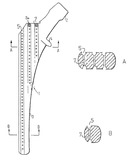
Fig. 1. illustrates a proximal modular type femoral endoprosthesis (1), with a
conventional Morse tapered upper end (2) for the ball and a tapered, polished,
conical distal end (3). Longitudinally along the stem, several anchorage
cavities
shown as slits or gutters (6) provide access for fibrous or bone tissue
ingrowth. As
illustrated in the figure the anchorage cavities may have a longitudinal
extension all
along the length of the prosthesis or along a portion of the length.
Longitudinal guide means for,a cutting tool (5) are located somewhat deep in
the
stem, outward open by the slit (6) as illustrated in Figs. lA-B.
CA 02580714 2007-03-08
WO 2006/028382 PCT/N02005/000322
9
The guide means may also be located quite superficial as illustrated in Fig. 2
in
which the shape of the cross section of the guide means (5) preferably is
slightly
more than semicircular so to hinder a cutting tool to go astray.
A medial collar only (4) is indicated in the drawing. In the case that a full
circumferential collar is provided, for proximal load transfer, the guide
means goes
right through the collar, and such a collar will thus not obstruct a
controlled
retrieval of the prosthesis.
Tissue growing into the guide means (5) is possible directly as shown in Fig.
2A-B
or via the slits (6) illustrated in Fig 1A. Further anchorage, and the most
important
one, is provided by tissue growth into pores, holes or small channels (7)
transversely interconnecting the guide means (5), or holes or pores extending
right
through the implant from one guide means to one on the other side of the
implant
(Figs. 2A-B and 4), or both.
The uppermost end of the guide means may be threaded so that a suitable, short
screw can close the guide. An example of the threaded part (5a) is illustrated
in Fig.
2. The threaded part (5a) may also be used for anchorage of a suitable
instrument
for the insertion or the removal of the prosthesis, or a drill guide.
To promote bone ingrowth in the guide means (5), and in particular into the
anchoring holes or pores (7), they may be coated by a tissue ongrowth
promoting
material like hydroxyapatite, or sustained drug release substances as
described in
US 4,913,903, 1990, inventors Einar Sudmann et al.
As outlined above a prosthetic element may by be anchored to living tissue by
tissue ingrowth. Whether anchorage by bone or by fibrous tissue only will give
the
best long-term results in patients has yet to be decided. However, the very
best
anchored "implant" in man and animals, the tooth, is anchored by fibrous
tissue, by
multiple small collagen fibrils. And such anchorage by fibrous tissue only has
stood
the test of millions of years.
Fig. 3 illustrates a prosthetic implant for fibrous tissue anchorage. As
outlined
above, to hinder bone tissue ingrowth, pores for fibrous tissue anchorage
should
preferably have a maximal diameter of less than 0.1 mm. The pores (8) in Fig.
3 are
thus not drawn to scale. The essential part is that the pores are oriented
perpendicular (Figs. 3A-B), or at any angle, to the guide means (5) for the
cutting
tool. The external opening of each pore, or hole (8) may be pointed at the
distal
perimeter (9) acting as a miniature cutting device, cutting the ingrown tissue
therein
on retrieval (Fig. 3C, 8-9). Fig 3A shows an example of plural pores/holes
connected to each guide means. In the left portion of the prosthesis element
the
CA 02580714 2007-03-08
WO 2006/028382 PCT/N02005/000322
guide means (5) are connected to through holes (8) running from one side to
the
other of the prosthesis element, whereas in the right portion of the
prosthesis
element additional holes are provided in a direction perpendicular to the
thickness
direction. As the skilled person will understand the configuration of the
holes (8)
5 may be performed in various ways.
Figures 4 A-D describe a further embodiment of the invention. This schematic
drawing of a femoral hemiprosthsis (for animals)is in principle of the same
type as
the embodiment of figure 2, but have only one guide means on each side.
Between
10 the guide means there are provided cavities or through holes 7. The collar
4 of the
prosthesis element has a special design. The upper part has a certain
thickness and
is provided with holes 9 ending in the respective guide means or channels 5.
When
the element 1 shall be removed a bore 10 is inserted in the holes 9 and the
bore is
cutting the anchoring tissues in the cavities 7 as illustrated on figure 4D.
Then the
element can be withdrawn.
Figure 5 illustrates the effect of the invention. The upper curve (A) in the
graphs
shows the load necessary for push-out/withdrawal of a test prosthetic element,
while the lower curve (B) illustrates the markedly reduced load needed after a
treatment according to the invention.
Another embodiment is proposed in figure 6. Configurations, for instance as
shown
in figures 2 or 4, may be constructed without the cavities 7. Instead, at
least a part
of the inner surface of the guide, means 5 is provided with a layer 11 that is
suitable
for bone or fibrous tissue ingrowth. The layer may have a thickness of 500
with
communicating pores. The dimension of the pores should be small, for instance
less
than 100 , if only ingrowth of fibrous tissue is wanted.
In this way it is possible to achieve a good anchorage by bone and/or fibrous
tissue
without having through holes, from one side to the other, in a thick element.
Fig. 7 illustrates how an acetabular shell for the hip joint liner may be
designed.
The guide means (5) for a flexible cutting instrument radiate out from the
dome, and
the anchoring pores or holes (7) interconnect these guide means, or goes right
through the shell.
Fig. 8 illustrates a dental implant serving as a base for an artificial tooth.
As above
anchorage is secured by bone or fibrous tissue, or both, in holes or pores (7)
open to
longitudinal guide means (5) for a cutting device. The implant may be circular
as
illustrated, or any desirable shape. The surface is polished, and for primary
fixation
in the jaw, a circular implant may be formed like a screw.
CA 02580714 2007-03-08
WO 2006/028382 PCT/N02005/000322
11
Fig. 9 illustrates a dental iinplant serving as a base for an artificial
tooth. Anchorage
is here, however, provided by fibrous tissue ingrowth in pores (8) as in Fig.
3. The
implant may be circular as illustrated, or any desirable shape. The surface is
polished, and for primary fixation in the jaw a circular implant may be formed
like a
screw.
In figure 10 there is shown an embodiment with a curved prosthetic element
(3).
The upper part of the guide means is threaded (5a), like the ones shown in
figure
2. In this embodiment a flexible bore must be used.
Anchoring pegs in an implant like knee or ankle prosthesis may be designed as
illustrated in Figs. 8-9. These pegs may be conical, circular, oval, or any
shape in
between, in transverse section. To gain access to the guide means (5) in the
peg of a
convex component, like a femoral one, it might be necessary to remove a
preformed
weakened part of the implant.
Many modifications are possible within the scope of the invention. The
configuration of guide means and cavities may be modified, as well as their
relationship. However, longitudinal threaded guide means, or gutter, 5
mentioned
above, will be suitable for anchorage by bone tissue ongrowth only, not by
fibrous
tissue. The stem may be straight, or conical as shown but the invention will
also be
suitable by curved stems, using flexible bores.