Note: Descriptions are shown in the official language in which they were submitted.
CA 02580778 2007-03-19
WO 2006/034301 PCT/US2005/033721
THIN FILM MEDICAL DEVICE AND DELIVERY SYSTEM
FIELD OF THE INVENTION
The present invention relates to a thin film medical device, and in particular
to an
intraiuminal thin film medical device and delivery system. This medical device
and delivery
system are particularly well suited for ocolusion of an aneurysm, vessel side
branch or
dissection of a body lumen or duct, such as an artery or vein.
BACKGROUND OF THE INVENTION
There are many instances when it may be desirable to permanently occlude a
vessel in
the human body. Examples of when permanent occlusion of a vessel might be
desirable
include: occlusion of an aneurysm or side branch vessel; therapeutic
occlusion, or embolization,
of the renal artery; occlusion of a Blalock-Taussig Shunt; pulmonary
arteriovenous fistulae and
transjugular intrahepatic stent shunt occlusion; some non-vascular
applications, such as
therapeutic ureteric occlusion; and the occlusion of vessels feeding large
cancerous tumors.
In the past, certain coiled stents, stent grafts or detachable balloons have
been utilized
for providing permanent occlusion of vessels. Stent-grafts are essentially
endoluminal stents
with a discrete covering on either or both of the luminal and abluminal
surfaces of the stent that
occludes the open spaces, or interstices, between adjacent structural members
of the
endoluminal stent. It is known in the art to fabricate stent-grafts by
covering the stent with
endogenous vein or a synthetic material, such as woven polyester known as
DACRON, or with
expanded polytetrafluoroethylene. Additionally, it is known in the art to
cover the stent with a
biological material, such as a xenograft or collagen.
There are certain problems associated with coiled stents, including, migration
of the
coiled stent within the vessel to be occluded, perforation of the vessel by
the coiled stent, and
failure to completely thrombose, or occlude, the vessel. Another disadvantage
associated with
such coiled stents is that the vessel may not be immediately occluded
following placement in
the vessel. Disadvantages associated with detachable occlusion balloons
include premature
detachment with distal embolization, or occlusion, and they are believed to
require a longer
period of time for the user of the device to learn how to properly use such
detachable occlusion
balloons.
In addition to vessel occlusion, conventional graft type intraluminal medical
devices are
frequently used post-angioplasty in order to provide a structural support for
a blood vessel and
reduce the incidence of restenosis following percutaneous balloon angioplasty.
A principal
example are endovascular stents which are introduced to a site of disease or
trauma within the
body's vasculature from an introductory location remote from the disease or
trauma site using
an introductory catheter, passed through the vasculature communicating between
the remote
introductory location and the disease or trauma site, and released from the
introductory catheter
at the disease or trauma site to maintain patency of the blood vessel at the
site of disease or
trauma. Stent-grafts are delivered and deployed under similar circumstances
and are utilized to
1
CA 02580778 2007-03-19
WO 2006/034301 PCT/US2005/033721
maintain patency of an anatomic passageway, for example, by reducing
restenosis following
angioplasty, or when used to exclude an aneurysm, such as in aortic aneurysm
exclusion
applications.
While these medical devices have specific advantages, their overall size, in
particular
the diameter and delivery profile, are significant disadvantages that render
these devices
prohibitive for certain uses. Another significant disadvantage is the limited
flexibility these
devices have for navigating paths through small and/or tortuous vessels. As
such, they may not
be desirable for many small diameter vessel applications, for example
neurovascular vessels.
What is needed is a medical device capable of occluding various parts of a
vessel that
can assume a reduced diameter and delivery profile.
SUMMARY OF THE INVENTION
The present invention relates to an intraluminal thin film medical device
particularly well
suited for occlusion of an aneurysm, vessel side branch or dissection of a
body lumen or duct,
such as an artery or vein. In one embodiment of the invention, the medical
device comprises a
thin film tube capable of being longitudinally stretched by the application of
mechanical energy
to achieve a smaller circumferential profile. Once the mechanical energy is
released, the thin
film tube is capable of self-expanding to the pre-stretched length and
diameter. The medical
device further comprises a plurality of slots incised in the tube wall. The
slots are arranges such
that they open and assist the thin film tube to longitudinally stretch, and
substantially close when
the thin film tube self-expands to the pre-stretched length and diameter.
Another embodiment of the present medical device for occluding a body vessel
comprises a thin film tube capable of being longitudinally stretched by the
application of
mechanical energy to achieve a smaller circumferential profile, and self-
expand to the pre-
stretched length and diameter upon release of the mechanical energy. A
plurality of apertures
are incised in the thin film tube wall such the apertures assist the thin film
tube to longitudinally
stretch.
Still another embodiment of the medical device for occluding a body vessel
comprises a
thin film tube capable of being longitudinally stretched by the application of
mechanical energy
to achieve a smaller circumferential profile, and self-expand to the pre-
stretched length and
diameter upon release of the mechanical energy. The medical device further
comprises a stent
attached to the interior surface of the thin metallic film.
BRIEF DESCRIPTION OF THE DRAWINGS
Figure 1A show a perspective view of medical device fabricated from a thin
film tube in
the deployed or "pre-stretched" configuration according to one embodiment of
the present
invention.
Figure 1 B shows a perspective view of a medical device fabricated from a thin
film tube
in the stretched reduced profile and restrained position according to one
embodiment of the
present invention.
2
CA 02580778 2007-03-19
WO 2006/034301 PCT/US2005/033721
Figure 1 C illustrates a perspective view of a medical device according to one
embodiment of the present invention where only a portion of the radial slots
along the proximal
end and distal end are open, while the radial slots in the intermediate
section remain
substantially closed.
Figure 2 is a perspective partial section view showing a medical device
deployed in a
vessel according to one embodiment of the present invention.
Figure 3A is a perspective partial section view showing a medical device
according to
an embodiment of the present invention deployed over an aneurysm in a vessel
wall, where the
medical device has a proximal stent attaching the thin film tube to the vessel
wall.
Figure 3B is a perspective partial section view showing a medical device
according to
an embodiment of the present invention deployed over an aneurysm in a vessel
wall, where the
medical device has a proximal stent attaching the thin film tube to the vessel
wall along the
proximal end, as well as a distal stent attaching the distal end of the thin
film tube to the vessel
wall along the distal end.
Figure 3C is a perspective partial section view showing a medical device
according to
an embodiment of the present invention deployed over an aneurysm in a vessel
wall, where the
medical device has a stent structure having multiple hoop sections arranged
axially along a
central longitudinal axis.
Figure 4 is a longitudinal section view illustrating a medical device having a
self-
supporting metallic thin film tube loaded on a delivery catheter according to
one embodiment of
the present invention.
Figure 5 is a longitudinal section view illustrating a medical device having a
self-
expanding stent for additional radial support according to one embodiment of
the present
invention.
Figure 6 is a longitudinal section view illustrating a medical device having a
balloon
expandable stent for additional radial support according to one embodiment of
the present
invention.
DETAILED DESCRIPTION OF A PREFERRED EMBODIMENT
The present invention discloses a thin film medical device particularly well
suited for
occlusion of an aneurysm or vessel side branch, or dissection of body lumen or
duct, such as
an artery or vein. One advantage of the present invention is that is provides
a biocompatible
graft material that enables a less invasive delivery of the medical device to
a vascular site for
occluding blood flow while sill allowing blood flow through the main vessel at
the implant
location.
Although this specification provides detailed description for implantation of
the medical
device in a artery or vein, one of skill in the art would understand that
modifications of the
disclosed invention would also be well suited for use on other body lumens and
anatomical
passageways, such as, for example those found in the cardiovascular,
lymphatic, endocrine,
renal, gastrointestinal and or reproductive systems.
3
CA 02580778 2007-03-19
WO 2006/034301 PCT/US2005/033721
The primary component of the present invention is a thin film made primarily
of a
substantially self-supporting biocompatible metal or psuedometal. The thin
film may be
fabricated either as single layer, or a plurality of layers. The terms "thin
film", "metal film", "thin
metallic film", and "metallic thin film" are used synonymously in this
application to refer to a
single or plural layer film fabricated of biocompatible metal or biocompatible
pseudometals
having a thickness greater than 0.1 pm but less than 250 pm, preferably
between 1 and 50 pm.
In some particular embodiments of the invention, such as where the thin film
is used as a
structural support component, the thin film may have a thickness greater than
approximately 25
pm. In other embodiments, for example, where the thin film is used as a cover
member with
additional structurai support, the thin film may have a thickness of between
approximately 0.1
pm and 30 pm, most preferably between 0.1 pm and 10 pm.
In a preferred embodiment, the medical device is fabricated from a shape
memory thin
metallic film or pseudometallic film having super elastic characteristics. One
example of a
shape memory metallic thin film is Nickel Titanium (Nitinol) formed into a
tubular structure.
Nitinol is utilized in a wide variety of applications, including medical
device applications
as described above. Nitinol or NiTi alloys are widely utilized in the
fabrication or construction of
medical devices for a number of reasons, including its biomechanical
compatibility, its bio-
compatibility, its fatigue resistance, its kink resistance, its uniform
plastic deformation, its
magnetic resonance imaging compatibility, its ability to exert constant and
gentle outward
pressure, its dynamic interference, its thermal deployment capability, its
elastic deployment
capability, its hysteresis characteristics, and is moderately radiopacity.
Nitinol, as described above, exhibits shape memory and/or super elastic
characteristics.
Shape memory characteristics may be simplistically described as follows. A
metallic structure,
for example, a Nitinol tube that is in an Austenitic phase may be cooled to a
temperature such
that it is in the Martensitic phase. Once in the Martensitic phase, the
Nitinol tube may be
deformed into a particular configuration or shape by the application of
stress. As long as the
Nitinol tube is maintained in the Martensitic phase, the Nitinol tube will
remain in its deformed
shape. If the Nitinol tube is heated to a temperature sufficient to cause the
Nitinol tube to reach
the Austenitic phase, the Nitinol tube will return to its original or
programmed shape. The
original shape is programmed to be a particular shape by well-known techniques
as briefly
described above.
Super elastic characteristics may be simplistically described as follows. A
metallic
structure for example, a Nitinol tube that is in an Austenitic phase may be
deformed to a
particular shape or configuration by the application of mechanical energy. The
application of
mechanical energy causes a stress induced Martensitic phase transformation. In
other words,
the mechanical energy causes the Nitinol tube to transform from the Austenitic
phase to the
Martensitic phase. By utilizing the appropriate measuring instruments, one can
determined that
the stress from the mechanical energy causes a temperature drop in the Nitinol
tube. Once the
mechanical energy or stress is released, the Nitinol tube undergoes another
mechanical phase
transformation back to the Austenitic phase and thus its original or
programmed shape. As
4
CA 02580778 2007-03-19
WO 2006/034301 PCT/US2005/033721
described above, the original shape is programmed by well know techniques. The
Martensitic
and Austenitic phases are common phases in many metals.
Medical devices constructed from Nitinol are typically utilized in both the
Martensitic
phase and/or the Austenitic phase. The Martensitic phase is the low
temperature phase. A
material is in the Martensitic phase is typically very soft and malleable.
These properties make
it easier to shape or configure the Nitinol into complicated or complex
structures. The Austenitic
phase is the high temperature phase. A material in the Austenitic phase is
generally much
stronger than the materiel in the Martensitic phase. Typically, many medical
devices are cooled
to the Martensitic phase for manipulation and loading into delivery systems.
When the device is
deployed at body temperature, they return to the Austenitic phase.
Although Nitinol is described in this embodiment, it should not be understood
to limit the
scope of the invention. One of skill in the art would understand that other
materials, both
metallic and pseudo-metallic exhibiting similar shape memory and super-elastic
characteristics
may be used.
The tubular thin film structure is sized to match or be slightly greater than
the diameter
of the inner lumen of the body vessel when the tube is in the unrestrained ("
self-expanded")
configuration. The inherent properties of the thin Nitinol tube are such that
the tube is capable
of being longitudinally stretched, which decreases the tube's diameter.
Reducing the diameter
allows the medical device to maintain a compact profile for insertion into a
body lumen via a
catheter during a percutaneous, endoluminal procedure. Accordingly, the
inherent shape
memory and super-elastic characteristics allow the thin metallic tube to be
stretched and
restrained in a reduced profile configuration, and then self-expand back to
its original "pre-
stretched" diameter once the restraint is removed. As the tube diametrically
expands, it
longitudinally contracts or foreshortens to its pre-stretched length and
diameter:
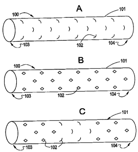
Figures 1 A and 1 B show a medical device fabricated from a Nitinol thin film
tube
according to one embodiment of the present invention. Figure 1A shows the thin
film medical
device 100 in the deployed or "pre-stretched" configuration, while Figure 1 B
shows the thin film
medical device 100 in the stretched reduced profile and restrained position.
To facilitate the ability for the thin film medical device 100 to stretch in
the longitudinal
direction, the tubular structure 101 has a plurality of radial slots 102
incised or formed
circumferentially through the tube 101 wall. In one embodiment, the slots are
in the form of slits
made completely through the thin film tube wall 101. Alternatively, where the
thin film is
manufactured in layers, the radial slots 102 may be through one or more layers
of the thin film
tube 101 wall. As the thin film tube 101 is longitudinally stretched, the
slots 102 open, creating
an opening in the tube 101 wall. When the thin film tube 101 is allowed to
return to the pre-
stretched (radially expanded) configuration, the radial slots 102 close,
excluding blood flow in
the circumferential direction.
The terms exclude, excluding and variations thereof, should not be construed
as having
zero porosity and completely preventing fluid flow. Instead, the closed slits
and apertures in the
thin film that exclude fluid flow may have openings that are small enough to
substantially
5
CA 02580778 2007-03-19
WO 2006/034301 PCT/US2005/033721
occlude blood flow through the thin film tube 101 wall. A medical device 100
illustrating all the
radial slots 102 in the open position is illustrated in Figure 1 B.
The medical device 100 may also be designed so that some of the radial slots
102 can
open, while other radial slots 102 remain substantially closed. Figure 1
C.illustrates a medical
device 100 where only a portion of the radial slots 102 along the proximal end
103 and distal
end104 are open, while the radial slots 102 in the intermediate section remain
closed.
In another embodiment of the present invention, the medical device 100 may
also has
apertures 102 incised or formed through the tube wall in various shapes. The
shapes may be
chosen to facilitate longitudinal stretching and/or radial expansion of the
thin film tube.
Essentially, the apertures 102 in the thin film have longitudinal and
latitudinal dimensions,
thereby forming an opening in the thin film having a net free open area.
The above-described medical device 100 can be used, for example, across an
aneurysm, side-branch vessel, or any vessel wall defect to exclude blood flow.
In one
embodiment of the invention, the tubular thin film 101 may be fabricated to a
thickness that can
support itself circumferentially. Alternatively, thinner films could be
supported by a balloon or
self-expanding stent or stents if additional radial support is needed.
Figure 2 is a perspective partial section view showing a medical device 200
deployed in
a vessel 205 according to one embodiment of the present invention. The vessel
205 has a
weakened vessel wall causing an aneurysm 206, and the medical device 200 is
deployed over
the aneurysm 206. The medical device 200 is self-supporting, and does not
require additional
stent(s) for support. As described earlier, the medical device 200 comprises a
thin metallic film
tube 201 having a proximal end 203 and a distal end 204. The thin film tube
201 has a series of
radial slots 202 arranged circumferentially along the thin film tube 201
longitudinal axis. Upon
deployment from a catheter system, the radial slots 202 incised in the thin
film tube 201
substantially close, excluding blood flow in the circumferential direction.
This relieves pressure
in the aneurysm 206, and mitigates potential medical conditions associated
with the aneurysm
206 bursting. Reducing the pressure in the aneurysm 206 may also allow the
vessel 205 wall to
contract.
The medical device may also include one or more stents to assist in securing
the thin
film tube into the vessel wall. Figure 3A shows a medical device 300 according
to another
embodiment of the present invention deployed over an aneurysm 306 in a vessel
wall 305.
Similar to the medical devices described above, the medical device 300
comprises a thin
metallic film formed into a tube 301, having a proximal end 303 and distal end
304. The thin
film tube 301 has a series of radial slots 302 incised circumferentially
through the tube 301 wall.
The medical device 300 additionally comprises a stent 307 along the proximal
end 303.
The stent 307 disclosed comprises at least one hoop structure extending
between the
stent 307 proximal and distal ends, 303, 304 respectively. The hoop structure
includes a
plurality of longitudinally arranged strut members and a plurality of loop
members connecting
adjacent struts. Adjacent struts are connected at opposite ends in a
substantially S or Z shaped
sinusoidal pattern so as to form a plurality of cells. However, one of
ordinary skill in the art
6
CA 02580778 2007-03-19
WO 2006/034301 PCT/US2005/033721
would recognize that the pattern shaped by the struts is not a limiting
factor, and other shaped
patterns or radially expandable structures may be used.
As previously described, the stent 307 assists in anchoring the medical device
300 to
the vessel 305 wall. The thin film tube 301 may be affixed to the stent 307 at
anchor point 308.
Attachment may be by any suitable attachment means, including adhesion
resulting from radial
pressure of the stent 307 against the thin metallic film tube 301, adhesion by
means of a binder,
heat, or chemical bond, and/or adhesion by mechanical means, such as welding
or suturing
between the stent 307 and the thin metallic film tube 301. It should be noted
that the stent 307
does not necessarily have to be fixedly attached to the metallic film tube
301. Instead, the
radially outward force that stent 307 exerts against the vessel wall may be
adequate to hold the
metallic thin film 301 in place.
In an alternate embodiment, the thin metallic film tube 301 may be anchored to
the
vessel 305 wall by a plurality of anchors. Figure 3B shows a medical device
300 having a
proximal stent 307 attaching the thin film tube 301 to the vessel 305 wall
along the proximal end
303, as well as a distal stent 309 attaching the distal end of the thin film
tube 301 to the vessel
305 wall along the distal end 304. Still one of skill in the art would
understand that additional
stents may be used to anchor the medical device 300 to the vessel 305 wall,
such as additional
proximal or distal anchors placed longitudinally along the thin film tube 301.
In a further alternate embodiment, stents having multiple hoop structures or
longer
hoop structures may be used to fully support the thin metallic film along all
or substantially all of
the film's length. Figure 3C shows a medical device 300 having a multi-hoop
stent 307
supporting the metallic thin film 301 substantially along the entire length of
the thin metallic film
301.
The multiple hoop stent 307 illustrated in Figure 3C comprises three hoop
structures
311A through 311C connected by a plurality of bridge members 314. Each bridge
member 314
comprises two ends 316A, 316B. One end 316A, 316B of each bridge 314 is.
attached to one
hoop. Using hoop sections 311A and 311 B for example, each bridge member 314
is connected
at end 316A to the proximal end of hoop 311A, and at end 316B the distal end
of hoop section
311B.
The various embodiments of the medical device described above are preferably
delivered to the target area and subsequently deployed by a catheter system.
Figure 4 is a
longitudinal section view illustrating a medical device 400 having a self-
supporting metallic thin
film tube 401 loaded on a delivery catheter 420 according to one embodiment of
the present
invention. The catheter 420 comprises an outer sheath 421 and an inner lumen
422. The outer
sheath 421 serves to hold the thin film tube 401 in the longitudinally
stretched position. The
inner lumen 422 is substantially coaxial to the outer sheath 421 and provides
a conduit for a
guide wire.
To be deployed, the medical device 400 is mounted on the delivery catheter
420. A
guide wire (not shown) is steered to the target area through well know means,
and the delivery
catheter 420/medical device 400 is loaded onto the guide wire using inner
lumen 422. The
7
CA 02580778 2007-03-19
WO 2006/034301 PCT/US2005/033721
catheter 420/medical device 400 is then pushed over the guide wire to the
target site. Once
properly located, the outer sheath 421 is retracted, allowing the thin film
tube 401 to expand and
longitudinally foreshorten to its unconstrained diameter. As previously
described, this will allow
the slots 402 (not shown) incised through the thin film tube 401 wall to
substantially close and
eliminate blood flow to the vessel wall defects.
The illustrated embodiment describes an over-the-wire delivery catheter.
However, one
of skill in the art would understand that other types of delivery catheters
may also be used,
include catheter utilizing a monorail design as are known in the art.
As previously described, very thin films may require extra radial support to
adequately
anchor the thin film in the vessel. In one embodiment, extra radial support
could be supplied by
radially expandable devices, such as radially expandable stents. Figure 5 is a
longitudinal
section view illustrating a medical device 500 having a self-expanding stent
507 for additional
radial support according to one embodiment of the present invention.
The catheter 520 for restraining and delivering the medical device 500 having
a self-
expanding stent 507 has three main components. Similar to the embodiment
described above,
the catheter 520 comprises an outer sheath 521 that serves to hold the thin
film tube 501 in the
longitudinally stretched position. Coaxial to the outer sheath 521 is a
secondary sheath 523 of
smaller diameter that serves to hold the self-expanding stent in a constrained
position. As
earlier described, the medical device 500 may have more than one stent for
added radial
support, i.e. may have stent 507 and 509 (not shown) as earlier described. In
each case,
secondary sheath 523 may serve to hold each radially expandable stent in the
constrained
position.
The third component of the medical device 500 is an inner lumen 522. The inner
lumen
522 is substantially coaxial to the outer sheath 521 and the secondary sheath
523, and provides
a conduit for a guide wire. The thin film tube 501 is affixed to the stent 507
at anchor point 508.
As earlier described, attachment may be by any suitable attachment means,
including adhesion
resulting from radial pressure of the stent 507 against the thin metallic film
tube 501, adhesion
by means of a binder, heat, or chemical bond, and/or adhesion by mechanical
means, such as
welding or suturing between the stent 507 and the thin metallic film tube 501.
To be deployed, the medical device 500 is mounted on the delivery catheter
520. A
guide wire (not shown) is steered to the target area through well-known means,
and the delivery
catheter 520/medical device 500 is loaded onto the guide wire using inner
lumen 522.
Alternatively, the delivery catheter 520/medical device 500 may be loaded onto
the guide wire in
a monorail fashion as is known in the art. The catheter 520/medical.device 500
is then pushed
over the guide wire to the target site. Once properly located, the outer
sheath 521 is retracted,
first allowing the thin film tube 501 to expand and longitudinally foreshorten
to its unconstrained
diameter. As previously described, this will allow the slots 502 (not shown)
incised through the
thin film tube 501 wall to substantially close and exclude blood flow to the
vessel wall defects.
The secondary sheath 523 may then be retracted, aiiowing the stent 507, and
any other stents
(not shown) to self-expand into the vessel wall (not shown). The radial
pressure exerted by the
8
CA 02580778 2007-03-19
WO 2006/034301 PCT/US2005/033721
stent 507 into the vessel wall anchors the stent 507 in place. As a result,
the thin film tube 501
is further supported and anchored to the vessel wall.
In an alternate embodiment, the self-expanding stent may be replace with a
balloon
expandable stent. Figure 6 is a longitudinal section view illustrating a
medical device 600
having a balloon expandable stent 607 for additional radial support according
to one
embodiment of the present invention.
The catheter 620 for restraining and delivering the medical device 600 having
a balloon
expandable stent 607 has three main components. Similar to the embodiment
described
above, the catheter 620 comprises an outer sheath 621 that serves to hold the
thin film tube '
601 in the longitudinally stretched position. Coaxial to the outer sheath 621
is balloon catheter
625 having a balloon 624 mounted thereto. The balloon expandable stent 607 is
mounted or
crimped in a low profile configuration to the balloon catheter 625 over the
expansion balloon
624. As earlier described, the medical device 600 may have more than one stent
for added
radial support, i.e. may have stent 607 and 609 (not shown), and possible
others, as earlier
described. In each case, each balloon 624 or balloons 624, on the balloon
catheter625 may
serve to hold and deliver each radially expandable stent in the constrained
position.
The third component of the medical device 600 is an inner lumen 622. The inner
lumen
622 is substantially coaxial to the outer sheath 621 and the balloon catheter
625, and provides a
conduit for a guide wire. In a preferred embodiment, the inner lumen 622 is an
integral part of
the balloon catheter 625. Alternatively, the catheter 620 may be a loop or
similar capture device
along the distal end to accept the guide wire in a monorail fashion. Monorail
type catheters are
known in the art.
The thin film tube 601 is preferably affixed to the stent 607 at anchor point
608. As
earlier described, attachment may be by any suitable attachment means,
including adhesion
resulting from radial pressure of the stent 607 against the thin metallic film
tube 601, adhesion
by means of a binder, heat, or chemical bond, and/or adhesion by mechanical
means, such as
welding or suturing between the stent 607 and the thin metallic film tube 601.
To be deployed, the medical device 600 is mounted on the balloon catheter 625.
A
guide wire (not shown) is steered to the target area through well know means,
and the balloon
catheter 625/medical device 600 is loaded onto the guide wire using inner
lumen 622. The
catheter 625/medical device 500 is then pushed over the guide wire to the
target site. Once
properly located, the outer sheath 621 is retracted, first allowing the thin
film tube 601 to expand
and longitudinally foreshorten to its unconstrained diameter. As previously
described, this will
allow the slots 602 (not shown) incised through the thin film tube 601 wall to
close and exclude
blood flow to the vessel wall defects. The balloon 624 is then inflated
(expanded), expanding
the stent 607, and any other stents (not shown) into the vessel wall (not
shown). The radial
pressure exerted by the stent 607 into the vessel wall anchors the stent 607
in place. As a
result, the thin film tube 601 is further supported and anchored to the vessel
wall.
While a number of variations of the invention have been shown and described in
detail,
other modifications and methods of use contemplated within the scope of this
invention will be
9
CA 02580778 2007-03-19
WO 2006/034301 PCT/US2005/033721
readilyapparent to those of skill in the art based upon this disciosure. It is
contemplated that
various combinations or sub combinations of the specific embodiments may be
made and still
fall within the scope of the invention. Moreover, all assemblies described are
believed useful
when modified to treat other vessels or lumens in the body, in particular
other regions of the
body where fluid flow in a body vessel or lumen needs to be excluded or
regulated. This may
include, for example, the coronary, vascular, non-vascular and peripheral
vessels and ducts.
Accordingly, it should be understood that various applications, modifications
and substitutions
may be made of equivalents without departing from the spirit of the invention
or the scope of the
following claims.
The following claims are provided to illustrate examples of some beneficial
aspects of
the subject matter disclosed herein which are within the scope of the present
invention.