Note: Descriptions are shown in the official language in which they were submitted.
CA 02580880 2007-03-06
TISSUE COAGULATION METHOD AND DEVICE USING INERT GAS
Technical Field
100011 The present disclosure relates to gas-enhanced electrosurgical methods
and
devices. More particularly, the present disclosure relates to a gas-enhanced
electrosurgical
device and method for supplying gas to and removing gas from a surgical site.
Back-yrozrnd of Related Ar=t
100021 Over the last several decades, more and more surgeons are abandoning
traditional open methods of gaining access to vital organs and body cavities
in favor of
endoscopes and endoscopic instruments that access organs through small
puncture-like
incisions. Endoscopic instruments are inserted into the patient through a
cannula, or a port
that has been made with a trocar. Typical sizes for cannulas range from about
three
millimeters to about twelve millimeters. Smaller cannulas are usually
preferred, and this
presents a design challenge to instrument manufacturers who must find ways to
make surgical
instruments that fit through the cannulas and operate in a safe and effective
manner.
[0003] Devices for arresting blood loss and coagulating tissue are well known
in the
art. For example, several prior art instruments employ thermic coagulation
(heated probes) to
arrest bleeding. However, due to space limitations, surgeons can have
difficultly
manipulating an instrument to coagulate, desiccate, fulgurate and/or cut
tissue. Other
instruments direct high frequency electric current through the tissue to stop
the bleeding.
I
CA 02580880 2007-03-06
Eschar adherence may also be a problem with these instruments. In both types
of
instruments, the depth of the coagulation is difficult to control.
[0004] Using these instruments to treat certain more sensitive tissue sites
may be
impractical since the constant and/or direct emission of ionized gas/plasma at
the tissue may
cause unintended results. Moreover, simply controlling the pressure of the gas
from the
source may not be effective or yield a desired result.
SUMMARY
100051 The present disclosure relates to an electrosurgical apparatus and
method for
coagulating tissue. An electrosurgical apparatus includes a first tube with a
proximal end and
a distal end. The proximal end is configured to receive pressurized ionizable
gas and the
distal end is configured to deliver ionized gas towards a treatment area. The
electrosurgical
apparatus also includes at least one electrode positioned to selectively
ionize the pressurized
ionizable gas prior to the pressurized ionizable gas exiting the distal end of
the first tube. The
electrode is adapted to be operatively coupled to an electrical energy source.
The
electrosurgical apparatus also includes a second tube with proximal and distal
ends. The
second tube is configured to selectively evacuate the ionized gas and
dislodged tissue material
from the treatment area.
100061 In one embodiment, the first tube is concentrically disposed within the
second
tube.
[00071 In an exeniplary embodiment, the distal end of the first tube extends
distally
relative to the distal end of the second tube.
2
CA 02580880 2007-03-06
100081 The electrode may be activated with a first electrical potential and
the
electrical energy source may include a remote patient pad that is energized to
a second
electrical potential. 100091 In an embodiment of the disclosure, the
electrosurgical apparatus is configured
for use in a bipolar mode wherein the first tube is activated with a first
electrical potential and
the second tube is activated with a second electrical potential.
1000101 In an exemplary embodiment, the electrosurgical apparatus includes a
regulator which regulates the flow of pressurized argon through the first
tube. The regulator
is disposed between a gas supply of the pressurized argon and the proximal end
of the first
tube.
[00011] In another embodiment of the disclosure, the electrosurgical apparatus
includes a fluid agitator, which may be disposed within the first tube, to
impart non-laminar
flow characteristics to the pressurized ionizable gas. Here, the pressurized
ionizable gas may
be used to cool tissue.
1000121 The present disclosure also relates to an electrosurgical apparatus
for
coagulating tissue that is configured to use in a bipolar mode. In this
embodiment, an
electrode control mechanism that controls the current intensity to the
electrode is disclosed.
[00013] The present disclosure also relates to a method for coagulating
tissue. The
method includes the steps of providing an electrosurgical apparatus including
a first tube
configured to receive pressurized ionizable gas and to deliver ionized gas
towards a treatment
area, at least one electrode positioned to selectively ionize pressurized
ionizable gas prior to
the pressurized ionizable gas exiting the first tube, and a second tube being
configured to
3
CA 02580880 2007-03-06
selectively evacuate the ionized gas and dislodged tissue material from the
treatment area.
The remaining steps include inserting the electrosurgical apparatus into
tissue; delivering
ionizable gas to the first tube; ionizing pressurized ionizable gas;
delivering pressurized
ionized gas through the first tube towards the treatment area; and removing
pressurized
ionized gas from the treatment area via the second tube. Additionally, a step
of inserting an
introducer into the tissue is disclosed.
BRIEF DESCRIPTION OF THE DRAWINGS
1000141 Fig. 1 is a perspective view of an electrosurgical instrument
according to an
embodiment of the present disclosure;
[00015] Fig. 2 is an enlarged, side sectional view of one embodiment of the
present
disclosure showing a first tube and a second tube inserted into tissue;
[00016] Fig. 3 is an enlarged, side sectional view of the area of detail shown
in Fig. 2;
[00017] Fig. 4 is an end cross-sectional view of the first tube and the second
tube
according to one embodiment of the present disclosure;
[00018] Fig. 5 is an enlarged, schematic sectional view of the first tube and
the second
tube illustrating ionized gas treating a tissue surface;
1000191 Fig. 6 is an enlarged, schematic sectional view of the first tube and
the second
tube illustrating a helically-shaped baffle located with the first tube for
causing ionizable gas
and/or ionized gas to exit the first tube with predetermined flow
characteristics;
4
CA 02580880 2007-03-06
[00020] Fig. 7A is an enlarged, schematic sectional view of the first tube and
the
second tube wherein the first tube includes a rotating plenum having an
aperture therein for
causing ionizable gas and/or ionized gas to exit the first tube with
predetermined flow
characteristics;
[00021] Fig. 7B is a cross-sectional view of the embodiment of Fig. 7A taken
along
line 7B-7B;
1000221 Fig. 8A is an enlarged, schematic sectional view of the first tube and
the
second tube wherein the first tube includes a pair of elongated flaps therein
for causing
ionizable gas and/or ionized gas to exit the first tube with predetermined
flow characteristics;
and
[00023) Fig. 8B is a cross-sectional view of the embodiment of Fig. 8A taken
along
line 8B-8B.
DETAILED DESCRIPTION
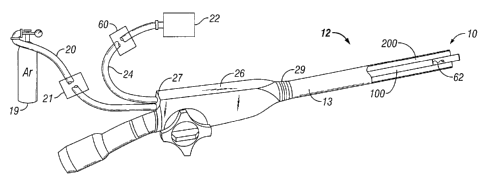
[00024] Referring to Fig. 1, a gas-enhanced tissue coagulator generally
identified by
reference numeral 10 is shown extending through a working channel of an
endoscope 12.
The coagulator 10 may be employed with a variety of suitable endoscopes, such
as those
manufactured by Olympus, Pentax and Fujinon. As such, only the basic operating
features of
the endoscope 12 that work in combination with the present disclosure need to
be described
herein.
CA 02580880 2007-03-06
1000251 Generally, the endoscope 12 includes a hand piece 26 having a proximal
end
27 and a distal end 29. The proximal end 27 is mechanically coupled to a
supply 19 of
pressurized ionizable gas, e.g., inert gas, via hose 20 and electrically
coupled to an
electrosurgical generator 22 by way of cable 24 to supply electrosurgical
energy, e.g., high
frequency coagulation current, to the endoscope 12. Tthe electrosurgical
generator 22 may be
configured to selectively control the amount of electrosurgical energy
transmitted to an
electrode during a surgical procedure. The supply 19 of pressurized ionizable
gas may be
configured to selectively control the rate of flow of gas, which is typically
greater than I liter
per minute.
1000261 As shown in Figs. I and 2, a long, generally flexible tubular member
13
having a first tube 100 located within a second concentric tube 200 is
mechanically coupled
to the distal end 29 of the hand piece 26. First tube 100 includes a proximal
end 110 and a
distal end 120 and second tube 200 includes a proximal end 210 and a distal
end 220. As best
illustrated in Fig. 4, first tube 100 and second tube 200 are concentrically
oriented, such that
first tube 100 is disposed within second tube 200. First tube 100 and second
tube 200 may
include insulation coatings 102, 202, respectively, to electrically isolate
tubes 100 and 200
from one another. Distal end 120 of the first tube 100 extends distally from
the distal end 220
of the second tube 200, the purposes of which are explained in more detail
below.
[000271 Turning now to Fig. 2, an enlarged, side sectional view of one
embodiment of
the coagulator 10 is shown. First tube 100 and second tube 200 are shown
inserted into
tissue, generally designated as "N." The first tube 100 is configured to
deliver ionizable gas
towards a treatment area "T" out of its distal end 120. The proximal end 110
of the first tube
100 is configured to receive ionizable gas from the supply 19. Second tube 200
is configured
6
CA 02580880 2007-03-06
to remove or evacuate gas and/or waste from the treatment area "T" through
distal end 220.
The gas and/or waste exits through proximal end 210 and is typically collected
in a known
manner such as a suitable medical waste container or a waste containment
system. An
introducer 300 may be utilized to facilitate the insertion of the coagulator
10 into the tissue
N"
[000281 With continued reference to Figs. I and 2, ionizable gas, e.g., argon,
is
supplied to the proximal end 110 of the first tube 100 by a gas conduit (not
explicitly shown)
located inside tubular member 13. Ionizable gas 19 may be supplied to the
first tube 100 at a
selectable, predetermined flow rate. The flow rate of the ionizable gas inay
be selectively
adjustable and/or regulated via a pressure regulator 21 depending upon a
particular purpose or
a particular surgical condition.
[000291 As mentioned above, the ionizable gas is supplied under pressure to
the
proximal end 110 of the first tube 100 and flows generally within the first
tube 100 towards
distal portion 120. An electrode 48 (see Fig. 5) discharges an electrosurgical
current, e.g.,
radio frequency (RF), which ionizes the gas prior to the gas being expelled
from the distal end
110 of the first tube 100 towards tissue "N." (Ionizable gas is illustrated as
dashed arrows 18
in Fig. 5 and the resulting ionized gas is illustrated by the area designated
as reference
numeral 46.) The stream of ionized gas 46 conducts current to the tissue 50
while effectively
scattering blood away fronl the treatment site allowing the tissue 50 to
readily coagulate and
arrest bleeding. The ionized gas 46 along with any vaporized material 52 is
then suctioned
away from the tissue (in the direction indicated by arrows A) through distal
end 220 of second
tube 200 via a suitable suctioning device (not explicitly shown). As best
shown in Fig. 5, the
7
CA 02580880 2007-03-06
generally wide ionized gas area allows a surgeon to effectively coagulate a
wide tissue area.
This is commonly referred to as a "coagulative painting."
1000301 Electrode 48 is connected by way of an electrical conduit disposed
within the
first tube 100, which is ultimately connected to the electrosurgical generator
22. The
electrode 48 may be ring- or pin-type and is spaced from the distal opening
110 of the first
tube 100 such that the electrode 48 does not come into contact with the tissue
"N" or tissue 50
during the surgical procedure. In one embodiment of the present disclosure, an
electrode
control mechanism 60 allows an operator to control the current intensity to
the electrode 48
during surgical procedures.
100031] Ionizable gas 18 is controlled/manipulated such that it flows through
the first
tube 100 in a generally non-laminar or turbulent manner. However, various
systems may be
employed to cause the ionizable gas 18 to flow more or less turbulently or
with other
predetermined flow characteristics through the first tube 100. The gas flow
may be used to
cool tissue, thus reducing thermal margins or areas of ablated tissue during
coagulation.
1000321 A fluid agitator, for example, such as a ribbon 62 (see Fig. 1), may
be
positioned within the first tube 100 to cause ionizable gas 18 and/or ionized
gas 46 to swirl
therewithin prior to the ionizable gas 18 and/or ionized gas 46 exiting the
distal end 110 of
the first tube 100. Additionally, with reference to Fig. 6, a generally
helically-shaped baffle
64 may be positioned within the first tube 100 to cause ionizable gas 18
and/or ionized gas 46
to swirl within first tube 100 prior to the gas 18 or 46 exiting distal end
120 of first tube 100.
1000331 A rotatable plenum 66 is illustrated in Figs. 7A and 7B, which
includes at least
one aperture 68 located therethrough. In this embodiment, the force of the
ionizable gas 18
8
CA 02580880 2007-03-06
and/or ionized gas 46 flowing through aperture 68 causes the plenum 66 to
rotate, which in
turn causes the ionizable gas 18 and/or ionized gas 46 to swirl with
predetermined flow
characteristics. It is envisioned that the user can control the rotational
speed of the plenum 66
by varying the pressure of ionizable gas 18 and/or ionized gas 46 flowing
through first tube
100. It is also envisioned that the rotational speed of the plenum 66 is
controlled by a
separate mechanism that is independent of the ionizable gas 18 and/or ionized
gas 46, e.g., a
regulator (not explicitly shown).
[000341 Figs. 8A and 8B illustrate a flow system that includes a pair of rods
70
disposed within first tube 100 for supporting a pair of elongated flaps 72.
Under flow
conditions, flaps 72 attenuate/extend from rods 70 and flutter within the
stream of ionizable
gas 18 and/or ionized gas 46. It is envisioned that the force of ionizable gas
18 and/or ionized
gas 46 flowing through first tube 100 causes each flap 72 to flutter, which in
turn causes
ionizable gas 18 and/or ionized gas 46 to move in a more turbulent manner. It
is also
envisioned that the rate/frequency of the flutter is directly related to the
pressure of ionizable
gas 18 and/or ionized gas 46 flowing through first tube 100. Any suitable
number of flaps 72
can be employed to create certain flow conditions, e.g., a series of flaps 72
can be positioned
at various positions along first tube 100 to create a more turbulent flow of
ionizable gas 18
and/or ionized gas 46. Moreover, the length of each flap 72 may be varied to
create
additional flow effects.
1000351 CoagLilator 10 may be configured for monopolar and/or bipolar modes.
In the
monopolar mode, the first tube 100 may be the active electrode and a patient
pad 17 (Fig. 5)
may be the return electrode. In the monopolar mode, an arcing pattern 410
(Fig. 3) may
radiate out from the distal end 120 of the first tube 100. In the bipolar
mode, the first tube
9
CA 02580880 2007-03-06
100 may be the active electrode and the second tube 200 may be the return
electrode. In the
bipolar mode, the conductive path, represented by dashed lines 420, would be
relatively self-
contained at the distal end 120 of the first tube 100 due to the proximity of
the active
electrode and the return electrode. In one embodiment, monopolar and bipolar
modes may be
alternated a plurality of times per second during use, which would enable the
conductive path
in lnonopolar mode to arc into the surrounding tissue 50 causing desiccation
and vaporization
of the tissue 50 in close proximity to the distal end 120 of the first tube
100. The conductive
path in bipolar mode further desiccates material that has been separated from
the tissue 50 as
the conductive path enters the second tube 200.
1000361 In operation, the introducer 300 may be inserted through the body and
placed
into tissue "N." A stylet (not shown) may facilitate the insertion of the
introducer 300 into
the tissue "N" by taking impedance readings. The stylet may then be removed
upon
confirmation of a desired impedance reading. Tubular member 13 of the
coagulator 10 may
then be inserted into the introducer 300, providing free access to the tissue
"N." Once tubular
member 13 is place in the tissue "N," the gas flow may be selectively
initiated and the
electrode 48 is thereafter selectively activated. A corona electrode may be
used for inducing
ignition of the ionizable gas 18. Ionized gas 46 flows out of the first tube
100 and is
suctioned back into the second tube 200. When argon gas is used, the argon
restricts the
amount of tissue affected to the material that is adjacent the distal end 120
of the first tube
100. Nuclear material near the distal end 120 of the first tube 100 is thus
vaporized and
removed via the second tube 200.
[000371 From the foregoing and with reference to the various figures, those
skilled in
the art will appreciate that not only can the coagulator 10 of the present
disclosure be used to
CA 02580880 2007-03-06
arrest bleeding tissue, but the present disclosure can also be employed for
desiccating and/or
removing the surface tissue, eradicating cysts, fonning eschars on tumors or
thermically
marking tissue. Those skilled in the art will also appreciate that certain
modifications can be
made to the present disclosure without departing from the scope of the present
disclosure.
[000381 For example, the coagulator 10 of the present disclosure may include
articulating qualities. In addition, tubular member 13, or at least a portion
thereof, may have
an arcuate shape. Moreover, the coagLilator 10 of the present disclosure may
be used while
perfonning liposuction and/or for treating tumors. In such tumor-treating
embodiments, a
level of coagulation may be achieved and the second tube 200 may remove
material, as
opposed to coagulating the tissue and leaving it in the body. Furthermore,
certain aspects of
the present disclosure may be utilized with a portable device and a portable
argon supply.
[000391 There is described and illustrated herein several embodiments of a gas-
enhanced electrosurgical device that supplies gas to and removes gas from a
treatment area.
While particular embodiments of the disclosure have been described, it is not
intended that
the disclosure be limited thereto, as it is intended that the disclosure be as
broad in scope as
the art will allow and that the specification be read likewise. Therefore, the
above description
should not be construed as limiting, but merely as exemplifications of various
embodiments.
Those skilled in the art will envision other modifications within the scope
and spirit of the
claims appended hereto.
11