Note: Descriptions are shown in the official language in which they were submitted.
CA 02581200 2007-03-21
WO 2006/033006 PCT/IB2005/002900
THERAPEUTIC COMBINATIONS COMPRISING POLY(ADP-RIBOSE) POLYMERASES INHIBITOR
This application claims the benefit of U. S. Provisional Application No.
60/612,458 filed on
September 22, 2004, and U. S. Provisional Application No. 60/683,006 filed on
May 19, 2005, the
contents of which are hereby incorporated by reference in their entireties.
Field of the Invention
This invention generally relates to use of 8-fluoro-2-{4-
[(methylamino)methyl]phenyl}-1,3,4,5-
tetrahydro-6H-azepino[5,4,3-cd]indol-6-one as a chemosensitizer that enhances
the efficacy of cytotoxic
drugs or radiotherapy. This invention provides pharmaceutical combinations of
8-fluoro-2-{4-
[(methylamino)methyl]phenyl}-1,3,4,5-tetrahydro-6H-azepino[5,4,3-cd]indol-6-
one, or a pharmaceutically
acceptable salt thereof and at least one additional therapeutic agent, kits
containing such combinations
and methods of using such combinations to treat subjects suffering from
diseases such as cancer.
Background of the Invention
The compound 8-fluoro-2-{4-[(methylamino)methyl]phenyl}-1,3,4,5-tetrahydro-6H-
azepino[5,4,3-
cd]indol-6-one represented by formula I
H
O N
~ ~ -
F H N-CH3
H
is a small molecule inhibitor of poly(ADP-ribose) polymerase (PARP). The
compound of formula 1 and
salts thereof, can be prepared as described in U.S. Patent No. 6,495,541; PCT
Application No.
PCT/IB2004/000915, International Publication No. WO 2004/087713; U.S.
Provisional Patent Application
Nos. 60/612,457, 60/612,459 and 60/679,296, the disclosures of which are
incorporated herein by
reference in their entireties.
To date, eighteen enzymes have been identified by DNA sequence homology in the
PARP family
and the biochemical and enzymatic properties of seven have been investigated:
PARP-1, and PARP-2
are stimulated by DNA strand breaks, PARP-3 interacts with PARP-1 and the
centrosome, PARP-4 also
known as vault PARP (VPARP) is the largest PARP and is associated with
cytoplasmic vaults, tankyrase
1 and 2 (PARP-5a and 5b) are associated with telombric proteins and the
function of PARP-7 (TiPARP) is
not clear at present but it may be involved in T-cell function and it can
poly(ADP-ribosylate) histones (Ame
JC, Splenlehauer C and de Murcia G. The PARP Superfamily. Bioessays 26 882-893
(2004)).
Pharmacology studies have shown that the compound of formula 1 is an inhibitor
of PARP-1 (Ki = 1.4 nM)
and PARP-2 (K; = 0.17 nM). Based on structural similarities in the amino acid
sequences among the
PARP enzymes, the compound of formula 1 likely binds with high affinity to the
other members of the
family as well.
CA 02581200 2007-03-21
WO 2006/033006 PCT/IB2005/002900
-2-
Enzyme-mediated repair of single- or double-strand breaks in DNA is a
potential mechanism of
resistance to radiotherapy or cytotoxic drugs whose mechanism depends on DNA
damage. Inhibition of
DNA repair enzymes is thus a strategy for the potentiation of these agents.
PARP-1, the best-
characterized member of the PARP family, is a nuclear enzyme that upon
activation by DNA damage
mediates the transfer of ADP-ribose fragments from NAD+ to a number of
acceptor proteins. Depending
on the extent of DNA damage incurred, PARP-1 activation and subsequent
poly(ADP-ribosyl)ation
mediate the repair of the damaged DNA or induce cell death. When DNA damage is
moderate, PARP-1
plays a significant role in the DNA repair process. Conversely, in the event
of massive DNA damage,
excessive activation of PARP-1 depletes ATP pools (in an effort to replenish
NAD), which ultimately
leads to cell mortality by necrosis (Tentori L, Portarena I, Graziani G.
Potential applications of poly(ADP-
ribose) polymerase (PARP) inhibitors. Pharmacol Res 2002, 45, 73-85). This
activation of PARP can also
lead to release of AIF (apoptosis-inducing factor) triggering a caspase-
independent apoptotic pathway.
(Hong SJ, Dawson TM and Dawson VL. Nuclear and mitochondrial conversations in
cell death: PARP-1
and AIF. Trends in Pharmacological Sciences 25 259-264 (2004)).
As the result of the dual role of PARP-1, inhibitors of this enzyme, such as 8-
fluoro-2-{4-
[(methylamino)methyl]phenyl}-1,3,4,5-tetrahydro-6H-azepino[5,4,3-cd]indol-6-
one represented by formula
1, may have a role as chemosensitizing agents (by preventing DNA repair, for
example, after anticancer
therapy), or as treatments for a variety of disease and toxic states that
involve oxidative or nitric oxide
induced stress and subsequent PARP hyperactivation. Such conditions include
neurologic and
neurodegenerative disorders (e.g., Parkinson's disease, Alzheimer's disease)
(Love S, Barber R, Wilcock
GK. Increased poly(ADP-ribosyl)ation of nuclear proteins in Alzheimer's
disease. Brain 1999;122:247-53;
Mandir AS, Przedborski S, Jackson-Lewis V, et al. Poly(ADP-ribose) polymerase
activation mediates 1-
methyl-4-phenyl-1,2,3,6-tetrahydropyridine (MPTP)-induced parkinsonism. Proc
Natl Acad Sci USA
1999;96:5774-9); cardiovascular disorders (e.g., myocardial infarction,
ischemia-reperfusion injury)
(Pieper AA, Walles T, Wei G, et al. Myocardial postischemic injury is reduced
by poly(ADP-ribose)
polymerase-1 gene disruption. J Mol Med 2000;6:271-82; Szab6 G, Bahrle S,
Stumpf N, et al. Poly(ADP-
ribose) polymerase inhibition reduces reperfusion injury after heart
transplantation. Circ Res 2002;90:100-
6; U.S. Patent 6,423,705); inflammatory diseases, (Szab6 C, Dawson V. Role of
poly(ADP-ribose)
synthetase in inflammation and ischaemia-reperfusion. TIPS 1998;19:287-98);
diabetic vascular
dysfunction (Soriano FG, Virag L, Szab6 C. Diabetic endothelial dysfunction:
role of reactive oxygen and
nitrogen species production and poly(ADP-ribose) polymerase activation. J Mol
Med 2001;79:437-48);
arthritis (Szab6 C, Virag L, Cuzzocrea S, et al. Protection against
peroxynitrite-induced fibroblast injury
and arthritis development by inhibition of poly(ADP-ribose) synthase. Proc
Nati Acad Sci USA 1998,
vol.95, pp. 3867-72); and cisplatin-induced nephrotoxicity (Racz et al. "BGP-
15 - a novel poly(ADP-ribose)
polymerase inhibitor - protects against nephrotoxicity of cisplatin without
compromising its antitumor
activity." Biochem Pharmaco12002;63:1099-111). Furthermore, it was shown that
BRCA2 deficient tumor
cells are acutely sensitive to PARP inhibitors alone (Bryant et al. "Specific
killing of BRCA2 deficient
tumors with inhibitors of poly(ADP-ribose)polymerase," Nature, 2005, vol. 434,
pp. 913-917; Farmer et al.
"Targeting the DNA repair defect in BRCA mutant cells as a therapeutic
strategy," Nature, 2005, vol. 434,
pp. 917-921). PARP inhibitors are also involved in enhancing the induction of
the expression of Reg gene
CA 02581200 2007-03-21
WO 2006/033006 PCT/IB2005/002900
-3-
in R cells and HGF gene and, accordingly, promote the proliferation of
pancreatic R-cells of Langerhans'
islets and suppress apoptosis of the cells (U.S. Patent Application
Publication 2004/0091453; PCT
Publication No. WO 02/00665). In addition, PARP inhibitors are also used in
cosmetic preparations,
especially in after-sun lotions (PCT Publication No. WO 01/82877). There are
no marketed PARP
inhibitors presently.
Cancer remains a disease with high unmet medical need. Cytotoxic chemotherapy
remains the
mainstay of systemic therapy for the majority of cancers, particularly late-
stage disease. However, for
patients with advanced or metastatic disease, few of the cytotoxic
chemotherapy agents or regimens have
been effective in increasing overall survival. Furthermore, the small
therapeutic window associated with
cytotoxic agents results in significant toxicity in conjunction with
suboptimal efficacy. Therefore, a
chemosensitizer that enhances the efficacy of cytotoxic drugs at well-
tolerated doses would fulfill a critical
need for cancer patients.
Radiotherapy is an effective form of cancer treatment used in most tumor types
for localized
disease control. Over 50% of all cancer patients will receive radiotherapy
during the course of their illness
(Foroudi F. et al. An evidence-based estimate of appropriate radiotherapy
utilization rate for breast
cancer. Int J Radiat Oncol Biol Phys. 2002, 53:1240-53; Foroudi F. et al. An
evidence-based estimate of
the appropriate radiotherapy utilization rate for colorectal cancer. Int J
Radiat Oncol Biol Phys. 2003,
56:1295-307; Foroudi F. et al. Evidence-based estimate of appropriate
radiotherapy utilization rate for
prostate cancer. Int J Radiat Oncol Biol Phys. 2003, 55:51-63; Barbera L. et
al. Estimating the benefit and
cost of radiotherapy for lung cancer. Int J Technol Assess Health Care. 2004,
20:545-5 1). However, even
in front-line treatment of cancers, in which radiotherapy is administered with
curative intent (for example,
head and neck cancer, soft tissue sarcoma and carcinoma of the cervix), not
all patients respond well.
There is, therefore, a need for strategies that will enhance the overall
patient response. Often standard
chemotherapy will be administered prior to or post-radiotherapy. An
alternative approach is to combine
radiation treatment with novel anti-cancer agents that are specifically
designed to enhance the efficacy of
radiation treatment. Such agents impact upon the five key factors that govern
tumor radiation response
("Cell survival as a determinant of tumor response." Basic clinical
radiobiology 3rd Edition. Steel GG (Ed.).
Arnold Press UK, pp. 52-63, 2002). These are the capacity to repair the DNA-
damage caused by
radiation treatment; the redistribution of cells through the cell cycle
following radiation treatment (such that
tumor cells that were in a resistant phase at the first radiation dose may
have progressed to a more
sensitive phase by the next radiation fraction); repopulation, whereby
surviving cells continue to divide
thereby increasing the tumor burden between radiation fractions; reoxygenation
of cells that survived the
initial round of radiation treatment as a consequence of being more poorly
oxygenated and finally, the
inherent radiosensitivity of the particular tissue. Of these factors, enhanced
repair and repopulation
results in radioresistance whereas redistribution, reoxygenation and inherent
radiosensitivity can render
the tumor more responsive to radiation treatment. Clearly the use of agents
that reduce the capacity for
DNA-repair in combination with radiotherapy have potential to enhance
radiotherapeutic outcome. PARP-
1 activation and subsequent poly-(ADP-ribosylation) is seen in response to
radiation-induced DNA-
damage (Satoh MS & Lindahl T. "Role of poly(ADP-ribose) formation in DNA
repair." Nature. 1992,
356:356-358). Further, cell lines and knock-out mice generated to lack PARP-1
expression and activity
CA 02581200 2007-03-21
WO 2006/033006 PCT/IB2005/002900
-4-
show exquisite radiosensitivity supporting PARP-1 as an attractive target for
radiopotentiation (Wang et
al. "Mice lacking ADPRT and poly(ADP-ribosyl)ation develop normally but are
susceptible to skin
disease." Genes Dev. 1995, 9:509-20; de Murcia et al. "Requirement of poly(ADP-
ribose) polymerase in
recovery from DNA damage in mice and in cells." Proc Natl Acad Sci U S A.
1997, 94:7303-7; Masutani et
al. "Function of poly(ADP-ribose) polymerase in response to DNA damage: gene-
disruption study in
mice." Mol Cell Biochem. 1999,193:149-52). In addition to direct affects on
DNA-repair the class of
PARP-1 inhibitors detailed are vasoactive and as such increase the potential
for tumor reoxygentaion
between radiation fractions that can further contribute to enhanced radiation
response (Calabrese et al.
"Anticancer chemo- and radio-sensitisation in vitro and in vivo by a potent
novel poly(ADP-ribose)
polymerase-1 (PARP-1) inhibitor, AG14361." J. Natl. Cancerlnst. 2004, 96: 56-
67).
Summary of the Invention
In one embodiment, the present invention provides a dosage form for
administration to a mammal,
the dosage form comprising a compound of formula 1:
H
O N
~ ~ -
F H N-CH3
H
a pharmaceutically acceptable salt or solvate, or a mixture thereof, in an
amount effective to provide a
sustained plasma concentration value of at least 5.9 ng/mL of the compound of
formula 1 for at least 24
hours after administration to the mammal.
In another embodiment, the invention provides a dosage form for administration
to a mammal, the
dosage form comprising a compound of formula 1, a pharmaceutically acceptable
salt or solvate, or a
mixture thereof, in an amount effective to provide a sustained plasma
concentration value of at least 10
ng/mL of the compound of formula 1 for at least 24 hours after administration
to the mammal.
In another embodiment, the invention provides a dosage form for administration
to a mammal, the
dosage form comprising a compound of formula 1, a pharmaceutically acceptable
salt or solvate, or a
mixture thereof, in an amount effective to provide a sustained plasma
concentration value of at least 5.9
ng/mL of the compound of formula 1 for at least 24 hours after administration
to the mammal, wherein the
dosage form is a lyophilized powder for injection.
In another embodiment, the invention provides a dosage form for administration
to a mammal, the
dosage form comprising a compound of formula 1, a pharmaceutically acceptable
salt or solvate, or a
mixture thereof, in an amount effective to inhibit a poly(ADP-ribose)
polymerase enzyme by at least 50%
for at least 24 hours in peripheral blood lymphocytes after administration to
the mammal.
In another embodiment, the invention provides a dosage form for administration
to a mammal, the
dosage form comprising a compound of formula 1, a pharmaceutically acceptable
salt or solvate, or a
mixture thereof, in an amount effective to inhibit a poly(ADP-ribose)
polymerase enzyme by at least 50%
CA 02581200 2007-03-21
WO 2006/033006 PCT/IB2005/002900
-5-
for at least 24 hours in peripheral blood lymphocytes after administration to
the mammal, wherein the
dosage form is a lyophilized powder for injection.
In another embodiment, the invention provides a dosage form for administration
to a mammal, the
dosage form comprising a compound of formula 1, a pharmaceutically acceptable
salt or solvate, or a
mixture thereof, in an amount of from I to 48 mg/m2 expressed as free base
equivalent mass of the
compound of formula 1.
In another embodiment, the invention provides a dosage form for administration
to a mammal, the
dosage form comprising a compound of formula 1, a pharmaceutically acceptable
salt or solvate, or a
mixture thereof, in an amount of from I to 48 mg/m2 expressed as free base
equivalent mass of the
compound of formula 1, wherein the dosage form is a lyophilized powder for
injection.
In another embodiment, the invention provides a dosage form for administration
to a mammal, the
dosage form comprising a compound of formula 1, a pharmaceutically acceptable
salt or solvate, or a
mixture thereof, in an amount of from 2 to 96 mg expressed as free base
equivalent mass of the
compound of formula 1.
In another embodiment, the invention provides a dosage form for administration
to a mammal, the
dosage form comprising a compound of formula 1, a pharmaceutically acceptable
salt or solvate, or a
mixture thereof, in an amount of from 2 to 96 mg expressed as free base
equivalent mass of the
compound of formula 1, wherein the dosage form is a lyophilized powder for
injection.
In another embodiment, the invention provides a method of treating cancer in a
mammal, the
method comprising administering to the mammal
(a) a compound of formula 1, a pharmaceutically acceptable salt or solvate, or
a mixture thereof in
an amount effective to provide a sustained plasma concentration value of at
least 5.9 ng/mL of the
compound of formula 1 for at least 24 hours after administration to the
mammal; and
(b) a therapeutically effective amount of at least one anti-cancer agent.
In another embodiment, the invention provides a method of treating cancer in a
mammal, the
method comprising administering to the mammal
(a) a compound of formula 1, a pharmaceutically acceptable salt or solvate, or
a mixture thereof in
an amount effective to provide a sustained plasma concentration value of at
least 5.9 ng/mL of the
compound of formula 1 for at least 24 hours after administration to the
mammal; and
(b) a therapeutically effective amount of at least one anti-cancer agent,
wherein the anti-cancer agent is administrated within 1 hour after
administration of the compound of
formula 1.
In another embodiment, the invention provides a method of treating cancer in a
mammal, the
method comprising administering to the mammal
(a) a compound of formula 1, a pharmaceutically acceptable salt or solvate, or
a mixture thereof in
an amount effective to provide a sustained plasma concentration value of at
least 5.9 ng/mL of the
compound of formula I for at least 24 hours after administration to the
mammal; and
(b) a therapeutically effective amount of at least one anti-cancer agent,
wherein the cancer is selected from lung cancer, bone cancer, pancreatic
cancer, skin cancer, cancer of the
head or neck, cutaneous or intraocular melanoma, uterine cancer, ovarian
cancer, rectal cancer, cancer of
CA 02581200 2007-03-21
WO 2006/033006 PCT/IB2005/002900
-6-
the anal region, stomach cancer, colon cancer, breast cancer, carcinoma of the
fallopian tubes, carcinoma of
the endometrium, carcinoma of the cervix, carcinoma of the vagina, carcinoma
of the vulva, Hodgkin's
Disease, cancer of the esophagus, cancer of the small intestine, cancer of the
endocrine system, cancer of
the thyroid gland, cancer of the parathyroid gland, cancer of the adrenal
gland, sarcoma of soft tissue,
cancer of the urethra, cancer of the penis, prostate cancer, chronic or acute
leukemia, lymphocytic
lymphomas, cancer of the bladder, cancer of the kidney or ureter, renal cell
carcinoma, carcinoma of the
renal pelvis, neoplasms of the central nervous system (CNS), primary CNS
lymphoma, spinal axis tumors,
brain stem glioma, pituitary adenoma, and combinations thereof.
In another embodiment, the invention provides a kit for treating cancer in a
mammal, the kit
comprising:
(a) an amount of a compound of formula 1, a pharmaceutically acceptable sait
or solvate, or a
mixture thereof, and a pharmaceutically acceptable carrier or diluent in a
first unit dosage form;
(b) an amount of at least one anti-cancer agent and a pharmaceutically
acceptable carrier or
diluent in at least a second unit dosage form; and
(c) container for containing the first and at least the second dosage forms;
wherein the amount of the compound of formula 1 is effective to provide a
sustained plasma concentration
value of at least 5.9 ng/mL of the compound of formula 1 for at least 24 hours
after administration to the
mammal.
In another embodiment, the invention provides a method of treating cancer in a
mammal, the
method comprising administering to the mammal
(a) a compound of formula 1, a pharmaceutically acceptable salt or solvate, or
a mixture thereof in
an amount effective to provide a sustained plasma concentration value of at
least 5.9 ng/mL of the
compound of formula I for at least 24 hours after administration to the
mammal; and
(b) a combination of irinotecan, 5-flourouracil and leucovorin.
In another embodiment, the invention provides a method of treating cancer in a
mammal, the
method comprising administering to the mammal
(a) a compound of formula 1, a pharmaceutically acceptable salt or solvate, or
a mixture thereof in
an amount effective to provide a sustained plasma concentration value of at
least 5.9 ng/mL of the
compound of formula 1 for at least 24 hours after administration to the
mammal; and
(b) a dose of radiation effective to destroy the cancer.
Definitions and Abbreviations of Terms
The term "Compound I" refers to the phosphate salt of 8-fluoro-2-{4-
[(methylamino)methyl]phenyl}-1,3,4,5-tetrahydro-6H-azepino[5,4,3-cd]indol-6-
one. The term "the
compound of formula 1" refers to 8-fluoro-2-{4-[(methylamino)methyl]phenyl}-
1,3,4,5-tetrahydro-6H-
azepino[5,4,3-cd]indol-6-one, free base.
"Abnormal cell growth", as used herein, unless otherwise indicated, refers to
cell growth that is
independent of normal regulatory mechanisms (e.g., loss of contact
inhibition).
The term "treating", as used herein, unless otherwise indicated, means
reversing, alleviating,
inhibiting the progress of, or preventing the disorder or condition to which
such term applies, or one or more
CA 02581200 2007-03-21
WO 2006/033006 PCT/IB2005/002900
-7-
symptoms of such disorder or condition. The term "treatment", as used herein,
unless otherwise indicated,
refers to the act of treating as "treating" is defined immediately above.
The term " radiosensitizer", as used herein, means a drug that makes tumor
cells more sensitive to
radiation therapy.
The term "radiotherapy", as used herein, includes external beam radiotherapy
(XBRT) or
teletherapy, brachytherapy or sealed source radiotherapy and unsealed source
radiotherapy. The
differences between these three main divisions of radiotherapy relate to the
position of the radiation
source; external is outside the body, while sealed and unsealed source
radiotherapy has radioactive
material delivered internally. External beam radiotherapy is the most common
form of radiotherapy where
a patient lies on a couch and an external source of X-rays is pointed at a
particular part of the body. The
radiation interacts with tissues and is absorbed, damaging the DNA of the
cell. Brachytherapy is the
delivery of radiation therapy using sealed sources which are placed as close
as possible to the site to be
treated. It is applicable for the treatment of tumors where a radiation source
can be placed within a body
cavity such as the oesophagus or bronchus or where the tumor is accessible to
needle or catheter
sources being placed within it, such as the head and neck and skin.
Brachytherapy has potential
applications to most tumor sites. It can be used as primary treatment or in
combination with external
beam radiotherapy. Unsealed source radiotherapy relates to the use of soluble
forms of radioactive
substances which are injected into the body. There is one common feature to
all these substances, and
that is the biological role of the non-radioactive parent substance. Proton
therapy is a special case of
external beam radiotherapy where the particles are protons.
The term "radio-immunotherapy", as used herein, means radiotherapy where
cytotoxic
radionuclides are linked to antibodies in order to deliver toxins directly to
tumor targets. Therapy with
targeted radiation rather than antibody-targeted toxins (immunotoxins) has the
advantage that adjacent
tumor cells, which lack the appropriate antigenic determinants, can be
destroyed by radiation cross-fire.
Radioimmunotherapy is sometimes called targeted radiotherapy, but this latter
term can also refer to
radionuclides linked to non-immune molecules (radiotherapy).
The phrase "pharmaceutically acceptable salt(s)", as used herein, unless
otherwise indicated,
includes salts of acidic or basic groups which may be present in a compound.
Compounds that are basic in
nature are capable of forming a wide variety of salts with various inorganic
and organic acids. The acids that
may be used to prepare pharmaceutically acceptable acid addition salts of such
basic compounds are those
that form non-toxic acid addition salts, i.e., salts containing
pharmacologically acceptable anions, such as
the acetate, benzenesulfonate, benzoate, bicarbonate, bisulfate, bitartrate,
borate, bromide, calcium edetate,
camsylate, carbonate, chloride, clavulanate, citrate, dihydrochloride,
edetate, edislyate, estolate, esylate,
ethylsuccinate, fumarate, gluceptate, gluconate, glutamate,
glycollylarsanilate, hexyiresorcinate,
hydrabamine, hydrobromide, hydrochloride, iodide, isothionate, lactate,
lactobionate, laurate, malate,
maleate, mandelate, mesylate, methylsulfate, mucate, napsylate, nitrate,
oleate, oxalate, pamoate
(embonate), palmitate, pantothenate, phospate/diphosphate, polygalacturonate,
salicylate, stearate,
subacetate, succinate, tannate, tartrate, teoclate, tosylate, triethiodode,
and valerate salts. Particularly
preferred salts include phosphate and gluconate salts.
CA 02581200 2007-03-21
WO 2006/033006 PCT/IB2005/002900
-8-
The invention also includes isotopically-labeled compounds, which are
identical to this recited in
Formula 1, but for the fact that one or more atoms are replaced by an atom
having an atomic mass or
mass number different from the atomic mass or mass number usually found in
nature. Examples of
isotopes that can be incorporated into compounds of the invention include
isotopes of hydrogen, carbon,
nitrogen, oxygen, phosphorus, sulfur, fluorine and chlorine, such as 2H, 3H,
11C 13C 14C 15N, 1e0, 170,
31P 32P' 35S, 18F, and 36CI, respectively. Compounds of the present invention
and pharmaceutically
acceptable salts of said compounds, which contain the aforementioned isotopes
and/or other isotopes of
other atoms, are within the scope of this invention. Certain isotopically-
labeled compounds of the present
invention, for example those into which radioactive isotopes such as 3H, 14C
11C or 18F are incorporated,
are useful in drug and/or substrate tissue distribution assays. Tritiated,
i.e., 3H, and carbon-14, i.e., 14C,
isotopes are particularly preferred for their ease of preparation and
detectability and 11C or 18F for use in
positron emission tomography. Further, substitution with heavier isotopes such
as deuterium, i.e., 2H, can
afford certain therapeutic advantages resulting from greater metabolic
stability, for example increased in
vivo half-life or reduced dosage requirements and, hence, may be preferred in
some circumstances. An
isotopically labeled compound of Formula 1 of this invention can generally be
prepared by carrying out the
procedures described for the non-labeled compound, substituting a readily
available isotopically labeled
reagent for a non-isotopically labeled reagent.
ADP adenosine diphosphate
AE adverse event
ALT alanine aminotransferase
ANC absolute neutrophil count
AST aspartate aminotransferase
AUC area under the plasma concentration-time curve
AUC(o-24) area under the plasma concentration-time curve from 0 to 24 hours
AUC(o-uast) area under the plasma concentration-time curve from time 0 to the
last recorded observation
BLD below limit of detection
BSA Body surface area
BUN blood urea nitrogen
Co initial concentration
CL clearance
Cmax maximum plasma concentration
CRC colorectal cancer
CTCAEv3 Common Terminology Criteria for Adverse Events version 3
CV cardiovascular
DLT dose-limiting toxicities
DNA deoxyribonucleic acid
EC50 concentration producing 50% of maximum effect
ECG electrocardiogram
FcR Fc receptor
5-FU 5-fluorouracil
GI gastrointestinal
GIST gastrointestinal stromal tumor
GLP good laboratory practice
HCT hematocrit
hERG human ether-a-go-go-related gene
hERG-lKr human ether-a-go-go-related gene channel blockade
HGB hemoglobin
G150 50% cell growth inhibitory concentration
IC50 50% enzyme activity inhibitory concentration
IGF insulin-like growth factor
CA 02581200 2007-03-21
WO 2006/033006 PCT/IB2005/002900
-9-
IGF-1 R insulin-like growth factor receptor, Type 1
IL interleukin
IP intraperitoneal
IV intravenous
LLN lower limit of normal
LLOQ lower limit of quantitation
LV leucovorin
MMNG N-methyl-N'-nitro-N-nitrosoguanidine
MTD maximum tolerated dose
NAD nicotinamide adenine dinucleotide
NOAEL no-observed-adverse-effect level
PARP poly(ADP-ribose) polymerase
PBMCs peripheral blood monocytes
PD pharmacodynamic
PID PARP-inhibitory dose
PK pharmacokinetic
PO orally
RBC red blood cells
RECIST Response Evaluation Criteria in Solid Tumors
QC Quality control
SAE serious adverse event
SWFI/SWI sterile water for injection
tl/ apparent terminal half-life
Tmax time of occurrence of Cmx
ULN upper limit of normal
Vdss volume of distribution at steady-state
WFI water for injection
Brief Description of the Drawings
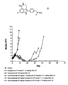
Figure 1 represents the data on efficacy of temozolomide in combination with 8-
fluoro-2-{4-
[(methylamino)methyl]phenyl}-1,3,4,5-tetrahydro-6H-azepino[5,4,3-cd]indol-6-
one as the phosphate
salt against the SW620 xenograft.
Figure 2 represents the data on efficacy of temozolomide in combination with 8-
fluoro-2-{4-
[(methylamino)methyl]phenyl}-1,3,4,5-tetrahydro- 6H-azepino[5,4,3-cd]indol-6-
one as the glucuronate salt
against the SW620 xenograft.
Figure 3 represents the mean 8-fluoro-2-{4-[(methylamino)methyl]phenyl}-
1,3,4,5-tetrahydro-6H-
azepino[5,4,3-cd]indol-6-one plasma concentration-time profiles for Day -7
(the phosphate salt of 8-fluoro-
2-{4-[(methylamino)methyl]phenyl}-1,3,4,5-tetrahydro-6H-azepino[5,4,3-cd]indol-
6-one alone) and Days 1
and 4 (the phosphate salt of 8-fluoro-2-{4-[(methylamino)methyl]phenyl}-
1,3,4,5-tetrahydro-6H-
azepino[5,4,3-cd]indol-6-one plus temozolomide) when the phosphate salt was
given as a 30-minute IV
Infusion and oral temozolomide was given as 100 mg/mZ.
Figure 4 represents the Median PARP activity in peripheral blood lymphocytes
following
administration of the phosphate salt of 8-fluoro-2-{4-
[(methylamino)methyl]phenyl}-1,3,4,5-tetrahydro-6H-
azepino[5,4,3-cd]indoi-6-one.
Detailed Description of the Invention
I Pharmaceutical Formulations of 8-fluoro-2-{4-f(methylamino)methyllphenyl}-
1.3,4.5-tetrahvdro-6H-
azepinof5,4,3-cdlindoi-6-one
CA 02581200 2007-03-21
WO 2006/033006 PCT/IB2005/002900
-10-
The compound of formula I and salts thereof, can be prepared as described in
U.S. Patent No.
6,495,541; PCT application No. PCT/IB2004/000915; U.S. Provisional Patent
Application No. 60/612,457;
and U.S. Provisional Patent Application No. 60/612,459, the disclosures of
which are incorporated herein
by reference in their entireties. Certain starting materials may be prepared
according to methods familiar to
those skilled in the art and certain synthetic modifications may be done
according to methods familiar to
those skilled in the art.
The compound of formula I is capable of forming a wide variety of different
salts with various
inorganic and organic acids. Although such salts must be pharmaceutically
acceptable for administration to
mammals, it is often desirable in practice to initially isolate the compound
of formula 1 from the reaction
mixture as a pharmaceutically unacceptable salt and then simply convert the
latter back to the free base
compound by treatment with an alkaline reagent and subsequently convert the
latter free base to a
pharmaceutically acceptable acid addition salt. The acid addition salts of the
base compounds of this
invention are readily prepared by treating the base compound with a
substantially equivalent amount of the
chosen mineral or organic acid in an aqueous solvent medium or in a suitable
organic solvent, such as
methanol or ethanol. Upon careful evaporation of the solvent, the desired
solid salt is readily obtained. The
desired acid salt can also be precipitated from a solution of the free base in
an organic solvent by adding to
the solution an appropriate mineral or organic acid. Specific examples of
preparation of a preferred salt, the
phosphate salt, can be found in PCT application No. PCT/1B2004/000915; U.S.
Provisional Patent
Application No. 60/612,457; and U.S. Provisional Patent Application No.
60/612,459, the disclosures of
which are incorporated herein by reference in their entireties.
Administration of the compound of formula I can be effected by any method that
enables delivery
of the compound to the site of action. These methods include oral routes,
intraduodenal routes,
parenteral injection (including intravenous, subcutaneous, intramuscular,
intravascular or infusion),
topical, and rectal administration.
The compound may, for example, be provided in a form suitable for oral
administration as a tablet,
capsule, pill, powder, sustained release formulation, solution, suspension,
for parenteral injection as a sterile
solution, suspension or emulsion, for topical administration as an ointment or
cream or for rectal
administration as a suppository.
The compound may be in unit dosage forms suitable for single administration of
precise dosages.
Preferably, dosage forms include a conventional pharmaceutical carrier or
excipient and the compound of
formula I as an active ingredient. In addition, dosage forms may include other
medicinal or pharmaceutical
agents, carriers, adjuvants, etc.
Exemplary parenteral administration forms include solutions or suspensions in
sterile aqueous
solutions, for example, aqueous propylene glycol or dextrose solutions. Such
dosage forms can be suitably
buffered, if desired.
Suitable pharmaceutical carriers include inert diluents or fillers, water and
various organic solvents.
The pharmaceutical composition may, if desired, contain additional ingredients
such as flavorings, binders,
excipients and the like. Thus for oral administration, tablets containing
various excipients, such as citric acid
may be employed together with various disintegrants such as starch, alginic
acid and certain complex
silicates and with binding agents such as sucrose, gelatin and acacia.
Additionally, lubricating agents such
CA 02581200 2007-03-21
WO 2006/033006 PCT/IB2005/002900
-11-
as magnesium stearate, sodium lauryl sulfate and talc are often useful for
tableting purposes. Solid
compositions of a similar type may also be employed in soft and hard filled
gelatin capsules. Preferred
materials therefor include lactose or milk sugar and high molecular weight
polyethylene glycols. When
aqueous suspensions or elixirs are desired for oral administration the active
compound therein may be
combined with various sweetening or flavoring agents, coloring matters or dyes
and, if desired, emulsifying
agents or suspending agents, together with diluents such as water, ethanol,
propylene glycol, glycerin, or
combinations thereof.
In preferred embodiments of the dosage forms of the invention, the dosage form
is an oral dosage form,
more preferably, a tablet or a capsule.
In preferred embodiments of the methods of the invention, the compound of
formula I is
parenterally administered, for example, using a lyophilized powder.
Preparation of the lyophilized powder
for injection for clinical use is described in U.S. Provisional Patent
Application No. 60/612,459, the
disclosure of which is incorporated herein by reference in its entirety.
For example, the phosphate salt of the compound of formula I may be formulated
and supplied as
a lyophilized powder for injection, 12 mg/vial (as free base), in 10 mU20 mm,
Type I, amber glass vials.
The composition of the phosphate salt of the compound of formula 1 drug
product may consist of the
phosphate salt of the compound of formula 1, mannitol, water for injection,
and nitrogen. The resulting
drug product may be an off-white to yellow cake. Each drug product vial may be
reconstituted with 6 mL
sterile water for injection to yield a 2.02 mg/mL (rounded to 2 mg/mL), as
free base of the compound of
formula 1.
In preferred embodiments of the invention, plasma concentrations of the
compound of formula 1 is
maintained at or above 5.9 ng/mL. This value was determined from the target
effect (IC89) for inhibition
of cellular NAD+ depletion and poly-ADP-ribose polymer formation and adjusted
for protein binding.
Specifically, as shown in Example 4, the compound of formula I at 5 nM
(temozolomide PF50 = 1.3),
greatly reduced the MNNG-induced cellular NAD+ consumption and inhibited
cellular poly-ADP-ribose
formation by 89% in A549 cells. Correcting the 5 nM target effect for human
protein binding (27.4% mean
unbound for the compound of formula I concentrations between 0.05 to 25 nM)
yielded a plasma
concentration of 5.9 ng/mL:
5 nM x 323.37 = 5.9 ng/mL
0.274 x 1000
II. Pharmaceutical Combinations of the Present Invention and Their Use
In one embodiment of the present invention the compound of formula I is used
to enhance the
efficacy of cytotoxic drugs whose mechanism depends on DNA damage. These drugs
include but not
limited to temozolomide (SCHERING), irinotecan (PFIZER), topotecan (GLAXO
SMITHKLINE), cisplatin
(BRISTOL MEYERS SQUIBB; AM PHARM PARTNERS; BEDFORD; GENSIA SICOR PHARMS;
PHARMACHEMIE), and doxorubicin hydrochloride (AM PHARM PARTNERS; BEDFORD;
GENSIA;
SICOR PHARMS; PHARMACHEMIE; ADRIA; ALZA).
Therapeutically effective amounts of the agents of the invention may be
administered, typically in
the form of a pharmaceutical composition, to treat diseases mediated by
modulation or regulation of
CA 02581200 2007-03-21
WO 2006/033006 PCT/IB2005/002900
-12-
PARP. An "effective amount" is intended to mean that amount of an agent that,
when administered to a
mammal, including a human, in need of such treatment, is sufficient to effect
treatment for a disease
mediated by the activity of one or more PARP enzyme. Thus, a therapeutically
effective amount of a
compound of the invention is a quantity sufficient to modulate, regulate, or
inhibit the activity of one or
more PARP enzyme such that a disease condition that is mediated by that
activity is reduced or
alleviated. The effective amount of a given compound will vary depending upon
factors such as the
disease condition and its severity and the identity and condition (e.g.,
weight) of the mammal in need of
treatment, but can nevertheless be routinely determined by one skilled in the
art. "Treating" is intended to
mean at least the mitigation of a disease condition in a mammal, including a
human, that is affected, at
least in part, by the activity of one or more PARP enzymes and includes:
preventing the disease condition
from occurring in a mammal, particularly when the mammal is found to be
predisposed to having the
disease condition but has not yet been diagnosed as having it; modulating
and/or inhibiting the disease
condition; and/or alleviating the disease condition. Exemplary disease
condition includes cancer.
The activity of the compound of formula 1 as a modulator of PARP activity may
be measured by
any of the methods available to those skilled in the art, including in vivo
and/or in vitro assays. Examples
of suitable assays for activity measurements include those described in U.S.
Patent No. 6,495,541 and
the specific examples of the present invention.
The present invention is directed to therapeutic methods of treating a disease
condition mediated
by PARP activity, for example, cancer and a variety of disease and toxic
states that involve oxidative or
nitric oxide induced stress and subsequent PARP hyperactivation. Such
conditions include, but not
limited to, neurologic and neurodegenerative disorders (eg, Parkinson's
disease, Alzheimer's disease),
cardiovascular disorders (eg, myocardial infarction, ischemia-reperfusion
injury), diabetic vascular
dysfunction, cisplatin-induced nephrotoxicity. The therapeutic methods of the
present invention comprise
administering to a mammal in need thereof a therapeutically effective amount
of a pharmaceutical
composition which comprises any of the polymorphic forms, or pharmaceutical
compositions discussed
above.
This invention also relates to a method for the treatment of abnormal cell
growth in a mammal,
including a human, comprising administering to said mammal an amount of the
compound of formula 1, as
defined above, or a pharmaceutically acceptable salt or solvate thereof, that
is effective in treating abnormal
cell growth.
In one embodiment of this method, the abnormal cell growth is cancer,
including, but not limited to,
mesothelioma, hepatobilliary (hepatic and billiary duct), a primary or
secondary CNS tumor, a primary or
secondary brain tumor, lung cancer (NSCLC and SCLC), bone cancer, pancreatic
cancer, skin cancer,
cancer of the head or neck, cutaneous or intraocular melanoma, ovarian cancer,
colon cancer, rectal
cancer, cancer of the anal region, stomach cancer, gastrointestinal (gastric,
colorectal, and duodenal),
breast cancer, uterine cancer, carcinoma of the fallopian tubes, carcinoma of
the endometrium, carcinoma
of the cervix, carcinoma of the vagina, carcinoma of the vulva, Hodgkin's
Disease, cancer of the
esophagus, cancer of the small intestine, cancer of the endocrine system,
cancer of the thyroid gland,
cancer of the parathyroid gland, cancer of the adrenal gland, sarcoma of soft
tissue, cancer of the urethra,
cancer of the penis, prostate cancer, testicular cancer, chronic or acute
leukemia, chronic myeloid
CA 02581200 2007-03-21
WO 2006/033006 PCT/IB2005/002900
-13-
leukemia, lymphocytic lymphomas, cancer of the bladder, cancer of the kidney
or ureter, renal cell
carcinoma, carcinoma of the renal pelvis, neoplasms of the central nervous
system (CNS), primary CNS
lymphoma, non hodgkins's lymphoma, spinal axis tumors, brain stem glioma,
pituitary adenoma,
adrenocortical cancer, gall bladder cancer, multiple myeloma,
cholangiocarcinoma, fibrosarcoma,
neuroblastoma, retinoblastoma, or a combination of one or more of the
foregoing cancers.
In another embodiment of said method, said abnormal cell growth is a benign
proliferative disease,
including, but not limited to, psoriasis, benign prostatic hypertrophy or
restinosis.
This invention also relates to a method for the treatment of abnormal cell
growth in a mammal which
comprises administering to said mammal an amount of the compound of formula 1,
or a pharmaceutically
acceptable salt or solvate thereof, that is effective in treating abnormal
cell growth in combination with an
anti-tumor agent selected from the group consisting of mitotic inhibitors,
alkylating agents, anti-metabolites,
intercalating antibiotics, growth factor inhibitors, cell cycle inhibitors,
enzymes, topoisomerase inhibitors,
biological response modifiers, antibodies, cytotoxics, anti-hormones, and anti-
androgens.
This invention also relates to a pharmaceutical composition for the treatment
of abnormal cell
growth in a mammal, including a human, comprising an amount of the compound of
formula 1, as defined
above, or a pharmaceutically acceptable salt or solvate thereof, that is
effective in treating abnormal cell
growth, and a pharmaceutically acceptable carrier. In one embodiment of said
composition, said abnormal
cell growth is cancer, including, but not limited to, mesothelioma,
hepatobilliary (hepatic and billiary duct), a
primary or secondary CNS tumor, a primary or secondary brain tumor, lung
cancer (NSCLC and SCLC),
bone cancer, pancreatic cancer, skin cancer, cancer of the head or neck,
cutaneous or intraocular
melanoma, ovarian cancer, colon cancer, rectal cancer, cancer of the anal
region, stomach cancer,
gastrointestinal (gastric, colorectal, and duodenal), breast cancer, uterine
cancer, carcinoma of the
fallopian tubes, carcinoma of the endometrium, carcinoma of the cervix,
carcinoma of the vagina,
carcinoma of the vulva, Hodgkin's Disease, cancer of the esophagus, cancer of
the small intestine, cancer
of the endocrine system, cancer of the thyroid gland, cancer of the
parathyroid gland, cancer of the
adrenal gland, sarcoma of soft tissue, cancer of the urethra, cancer of the
penis, prostate cancer,
testicular cancer, chronic or acute leukemia, chronic myeloid leukemia,
lymphocytic lymphomas, cancer of
the bladder, cancer of the kidney or ureter, renal cell carcinoma, carcinoma
of the renal pelvis, neoplasms
of the central nervous system (CNS), primary CNS lymphoma, non hodgkins's
lymphoma, spinal axis
tumors, brain stem glioma, pituitary adenoma, adrenocortical cancer, gall
bladder cancer, multiple
myeloma, cholangiocarcinoma, fibrosarcoma, neuroblastoma, retinoblastoma, or a
combination of one or
more of the foregoing cancers. In another embodiment of said pharmaceutical
composition, said abnormal
cell growth is a benign proliferative disease, including, but not limited to,
psoriasis, benign prostatic
hypertrophy or restinosis.
The invention also relates to a pharmaceutical composition for the treatment
of abnormal cell growth
in a mammal, including a human, which comprises an amount of the compound of
formula 1, as defined
above, or a pharmaceutically acceptable salt or solvate thereof, that is
effective in treating abnormal cell
growth in combination with a pharmaceutically acceptable carrier and an anti-
tumor agent selected from the
group consisting of mitotic inhibitors, alkylating agents, anti-metabolites,
intercalating antibiotics, growth
CA 02581200 2007-03-21
WO 2006/033006 PCT/IB2005/002900
-14-
factor inhibitors, cell cycle inhibitors, enzymes, topoisomerase inhibitors,
biological response modifiers, anti-
hormones, and anti-androgens.
The invention also relates to a method for the treatment of a
hyperproliferative disorder in a mammal
which comprises administering to said mammal a therapeutically effective
amount of the compound of
formula 1, or a pharmaceutically acceptable salt or hydrate thereof, in
combination with an anti-tumor agent
selected from the group consisting antiproliferative agents, kinase
inhibitors, angiogenesis inhibitors,
growth factor inhibitors, cox-I inhibitors, cox-II inhibitors, mitotic
inhibitors, alkylating agents, anti-
metabolites, intercalating antibiotics, growth factor inhibitors, radiation,
cell cycle inhibitors, enzymes,
topoisomerase inhibitors, biological response modifiers, antibodies,
cytotoxics, anti-hormones, statins,
and anti-androgens.
The present invention is also directed to combination therapeutic methods of
treating a disease
condition mediated by PARP activity, which comprises administering to a mammal
in need thereof a
therapeutically effective amount of a pharmaceutical composition which
comprises any of the polymorphic
forms, or pharmaceutical compositions discussed above, in combination with a
therapeutically effective
amount of one or more substances selected from anti-tumor agents, anti-
angiogenesis agents, signal
transduction inhibitors, and antiproliferative agents. Such substances include
those disclosed in PCT
Publication Nos. WO 00/38715, WO 00/38716, WO 00/38717, WO 00/38718, WO
00/38719, WO
00/38730, WO 00/38665, WO 00/37107 and WO 00/38786, the disclosures of which
are incorporated
herein by reference in their entireties.
Examples of anti-tumor agents include temozolomide (SCHERING), irinotecan
(PFIZER),
topotecan (GLAXO SMITHKLINE), cisplatin (BRISTOL MEYERS SQUIBB; AM PHARM
PARTNERS;
BEDFORD; GENSIA SICOR PHARMS; PHARMACHEMIE), and doxorubicin hydrochloride (AM
PHARM
PARTNERS; BEDFORD; GENSIA; SICOR PHARMS; PHARMACHEMIE; ADRIA; ALZA).
The combination therapeutic methods include administering the compound of
formula 1 and an
anti-tumor agent using any desire dosage regimen. For example, the regimens
can be dependent on the
combination agent as follows:
(a) the compound of formula 1, a pharmaceutically acceptable salt or solvate,
or a mixture
thereof, can be administered in an amount of from 1 to 48 mg/m2 expressed as
free base equivalent mass
of the compound of formula 1, daily x 5 days every 28 days 1 hour before 25-
200 mg/m2 temozolomide,
preferably, 100-200 mg/m2 temozolomide;
(b) the compound of formula 1, a pharmaceutically acceptable salt or solvate,
or a mixture
thereof, can be administered in an amount of from I to 48 mg/mZ expressed as
free base equivalent mass
of the compound of formula 1, 1 hour before the irinotecan dose and 24 hours
later.
Dose ranges for irinotecan:
62-125 mg/m2 weekly x 4 weeks every 6 weeks
175-350 mg/mZ every 3 weeks
90-180 mg/m2 every 2 weeks.
(c) the compound of formula 1, a pharmaceutically acceptable salt or solvate,
or a mixture
thereof, can be administered in an amount of from 1 to 48 mg/m2 expressed as
free base equivalent mass
of the compound of formula 1, daily x 5 days every 21 days, 1 hour before the
topotecan dose.
CA 02581200 2007-03-21
WO 2006/033006 PCT/IB2005/002900
-15-
Dose range for topotecan:
0.75-1.5 mg/m2 daily x 5 days every 21 days
(d) the compound of formula 1, a pharmaceutically acceptable salt or solvate,
or a mixture
thereof, can be administered in an amount of from I to 48 mg/m2 expressed as
free base equivalent mass
of the compound of formula 1, either once every 3-4 weeks or daily x 3-5 days
every 3-4 weeks, 1 hour
before the cisplatin dose.
Dose ranges for cisplatin:
10-100 mg/m2 every 3-4 weeks
10-40 mg/m2 daily x 3-5 days every 3-4 weeks.
(e) the compound of formula 1, a pharmaceutically acceptable salt or solvate,
or a mixture
thereof, can be administered in an amount of from 1 to 48 mg/mZ expressed as
free base equivalent mass
of the compound of formula 1, 1 hour before the doxorubicin dose and 24 hours
later.
Dose range for doxorubicin:
20-75 mg/m2 every 21-28 days.
The combination therapeutic methods of the present invention may include
administering the
compound of formula 1 a pharmaceutically acceptable salt or solvate, or a
mixture thereof, in an amount
of from 1 to 48 mg/m2 expressed as free base equivalent mass of the compound
of formula 1, and an anti-
tumor agent(s) using, for example, dosage regimens presented in Table 1.
Table 1
Name Regimen Reference
125 mg/m over 90 minutes, days 1, 8, 15, 22 Saltz et al. N Engi J Med.
Irinotecan Repeat every 6 weeks 2000;343:905-914.
300 or 350 mg/m IV over 90 minutes, day 1 Cunningham et al. Lancet.
Irinotecan Repeat every 3 weeks 1998;352:1413-1418.
Irinotecan 125 mg/m IV over 90 minutes, days 1, 8, 15, 22
IFL LV 20 mg/m2 IV bolus, days 1, 8, 15, 22 Saltz et al. N Engl J
Saltz regimen 5-FU 500 mg/m2 IV bolus, days 1, 8, 15, 22 Med.2000;343:905-914.
Repeat every 6 weeks
Irinotecan 180 mg/m over 2 hours, day 1
Irinotecan + LV 200 mg/m2 IV over 2 hours prior to 5-FU, days 1 and 2
Douillard et al. Lancet.
5-FU/LV 5-FU 400 mg/m IV bolus, then 600 Mg/M2 continuous infusion
2000;355:1041-1047.
Douillard regimen over 22 hours, days 1 and 2
Repeat eve 2 weeks
Irinotecan 180 mg/m over 90 minutes, day 1
Z Andre et aI. EurJ
LV 200 mg/m over 2-hour infusion during irinotecan
FOLFIRI 5-FU bolus 400 mg/m2, then 2.4-3 g/m2 continuous infusion
Cancer.1999;35:1343-1347.
over 46 hours, days 1 and 2 Tournigand et al. J Clin
Repeat every 2 weeks Oncol. 2004;23:229-237.
Capecitabine 1,000 mg/m PO bid, days 1-14 Grothey et al. Proc Am Soc
Caplri Irinotecan 100 mg/ma, days I and 8 Clin Oncol. 2003;22:255.
Repeat every 22 days Abstract 1022.
Irinotecan 250 mg/m IV, day 1
Capecitabine 1,000 mg/m2 PO bid, evening day 1-morning day Patt et al. Proc Am
Soc Clin
XELIRI Oncol. 2004;23;271.
Repeat every 3 weeks Abstract 3602.
Irinotecan 200 mg/m IV over 90 minutes, day I
IROX Oxaliplatin 85 mg/m2 IV over 2 hours, day 1 Goldberg et al. J Clin Oncol.
11 Repeat every 3 weeks 2004;22:23-30.
CA 02581200 2007-03-21
WO 2006/033006 PCT/IB2005/002900
-16-
Name Regimen Reference
Irinotecan 125 mg/m IV over 90 minutes, days 1, 8, 15, 22
LV 20 mg/m2 IV, days 1, 8, 15, 22
5-FU 500 mg/m2 IV, days 1, 8, 15, 22 Hurwitz et al. N Engl J Med.
IFL + Bevacizumab Repeat every 6 weeks 2004;350:2335-2342.
Bevacizumab 5 mg/kg IV over 90 minutes* following
chemotherapy, day 1
Re eat eve 2 weeks
CRC = colorectal cancer; 5-FU = 5-fluorouracil; LV = leucovorin.
*If first infusion is well tolerated, subsequent infusions may be administered
over 60 minutes and then 30 minutes.
The dosing schemes listed in Table I can be modified. For example, irinotecan
may be given at a
dose of 50-350 mg/mZ; 5-FU may be given at a dose of 370 mg/mZ - 3.0g. LV may
be given at 20-500
mg/m2.
The combination therapeutic methods of the present invention which include
administering the
compound of formula 1 a pharmaceutically acceptable salt or solvate, or a
mixture thereof, in an amount
of from 1 to 48 mg/mz expressed as free base equivalent mass of the compound
of formula 1, and an anti-
tumor agent(s), may be used, for example, in treatment patients who, for
example, failed treatment with
the regimens presented in Table 2.
Table 2
Name Regimen Reference
Oxaliplatin 85 mg/m IV over 2 hours, day 1 de Gramont et al. J Clin
LV 200 mg/m2 IV over 2 hours, days 1 and 2 Onco1.2000;18:2938 2947.
FOLFOX4 5-FU 400 mg/m2 IV bolus, then 600 mg/m2 IV over 22 hours, Rothenberg
et al. J Clin
days 1 and 2
Repeat eve 2 weeks Oncol. 2003;21:2059-2069.
Oxaliplatin 100 mg/m IV over 2 hours, day 1 Maindrault-Goebel et al. Eur
LV 200 mg/m2 IV over 2 hours, day 1 J Cancer. 1999;35:1338-
FOLFOX6 5-FU 400 mg/m2 IV bolus, then 2.4-3 g/m2 over 46 hours, 1342.
continuous infusion Tournigand et al. J Clin
Repeat every 2 weeks Oncol. 2004;23:229-237.
Oxaliplatin 85 mg/m IV over 2 hours, day 1
LV 175 mg/mZ IV over 2 hours, day 1 Cheeseman et al. Br J
mFOLFOX6 5-FU 400 mg/mZ IV bolus, then 2.4-3 g/m2 over 46 hours, Cancer.
2002;87:393-399.
continuous infusion
Repeat every 2 weeks
Oxaliplatin 130 mg/m IV over 2 hours, day I
Andre et al. Proc Am Soc
LV 400 mg/m2 IV over 2 hours Clin OncoL 2003;22:253.
FOLFOX7 5-FU 2,400 mg/m2 IV over 46 hours, continuous infusion Abstract 1016.
Repeat every 2 weeks for 6 cycles
Oxaliplatin 85 mg/m IV over 2 hours, days 1, 15, 29
LV 500 mg /m2 IV over 2 hours, days 1, 8, 15, 22, 29, 36 Smith et al. Proc Am
Soc
FLOX 5-FU 500 mg/ma IV bolus 1 hour after start of LV, days 1, 8, 15, Clin
Oncol. 2003;22:294.
22, 29, 36 Abstract 1181.
Repeat every 8 weeks for 3 cycles
Oxaliplatin 60 mg/m IV over 2 hours, days 1, 8, 15, 22
LV 500 mg/mZ IV over 2 hours, days 1, 8, 15, 22 Moehler et al. Z
FUFOX 5-FU 2.6 g/m2 IV over 24 hours, continuous infusion, Gastroenterol.
2002;40:957-
days 1, 8, 15, 22 964.
Repeat every 36 days
Oxaliplatin 85 mg/m IV over 2 hours, every 2 weeks
LV 20 mg/m2 IV over 10-20 minutes, days 1, 8, 15 Hochester et al. J Clin
bFOL 5-FU 500 mg/mZ IV bolus, days 1, 8, 15 Oncol. 2003;21:2703-2707.
Repeat every 28 days
CA 02581200 2007-03-21
WO 2006/033006 PCT/IB2005/002900
-17-
Name Regimen Reference
Oxaliplatin 85 mg/m IV over 2 hours, day 1
LV 200 mg/ma IV over 2 hours, days 1 and 2 Benson et al. Proc Am Soc
FOLFOX 4 + 5-FU 400 mg/m IV bolus, then 600 Mg/M2 IV over 22 hours, Clin
Oncol. 2003;22:243.
Bevacizumab days 1 and 2 Abstract 975.
Bevacizumab 10 mg/kg IV over 90 minutes,* day 1
Repeat eve 2 weeks
Oxaliplatin 85 mg/m IV over 2 hours, day 1
LV 200 mg/m2 IV over 2 hours, days 1 and 2
FOLFOX4 + 5-FU 400 mg/ma IV bolus, then 600 mg/m2 IV over 22 hours, Tabernero
et al. Proc Am
Cetuximab days 1 and 2 Soc Clin Oncol.
Repeat every 2 weeks 2004;23:248. Abstract 3512.
Cetuximab 400 mg/m? IV over 2 hours week I followed by
250 mg/m2 IV over 60 minutes weekly
The dosage units are represented in mg per m 2 of BSA. For example, the
Mosteller formula, the
DuBois and DuBois formula, the Haycock formula, the Gehan and George formula,
the Boyd formula are
applicable for measuring BSA (Mosteller RD: Simplified Calculation of Body
Surface Area. N Engl J Med
1987 Oct 22;317(17):1098; DuBois D; DuBois EF: A formula to estimate the
approximate surface area if
height and weight be known. Arch lnt Med 1916 17:863-71; Haycock G.B.,
Schwartz G.J.,Wisotsky D.H.
Geometric method for measuring body surface area: A height weight formula
validated in infants, children
and adults. The Journal of Pediatrics 1978 93:1:62-66; Gehan EA, George SL,
Estimation of human
body surface area from height and weight. Cancer Chemother Rep 1970 54:225-35;
Boyd E, The growth
of the surface area of the human body. Minneapolis: university of Minnesota
Press, 1935; Lam TK, Leung
DT: More on simplified calculation of body-surface area. N Engl J Med 1988 Apr
28;318(17):1130).
Additional examples of anti-tumor agents include antiproliferative agents,
kinase inhibitors,
angiogenesis inhibitors, growth factor inhibitors, cox-I inhibitors, cox-II
inhibitors, mitotic inhibitors,
alkylating agents, anti-metabolites, intercalating antibiotics, growth factor
inhibitors, radiation, cell cycle
inhibitors, enzymes, topoisomerase inhibitors, biological response modifiers,
antibodies, cytotoxics, anti-
hormones, statins, and anti-androgens.
In one embodiment of the present invention the anti-tumor agent used in
conjunction with the
compound of formula 1 and pharmaceutical compositions described herein is an
anti-angiogenesis agent,
kinase inhibitor, pan kinase inhibitor or growth factor inhibitor.
Preferred pan kinase inhibitors include SU-11248, described in U.S. Patent No.
6,573,293 (Pfizer,
Inc, NY, USA).
Anti-angiogenesis agents, include but are not limited to the following agents,
such as EGF
inhibitor, EGFR inhibitors, VEGF inhibitors, VEGFR inhibitors, TIE2
inhibitors, IGF1R inhibitors, COX-II
(cyclooxygenase II) inhibitors, MMP-2 (matrix-metalloprotienase 2) inhibitors,
and MMP-9 (matrix-
metalloprotienase 9) inhibitors.
Preferred VEGF inhibitors, include for example, Avastin (bevacizumab), an anti-
VEGF
monoclonal antibody of Genentech, Inc. of South San Francisco, California.
Additional VEGF inhibitors include CP-547,632 (Pfizer Inc., NY, USA), AG13736
(Pfizer Inc.), ZD-
6474 (AstraZeneca), AEE788 (Novartis), AZD-2171), VEGF Trap
(Regeneron,/Aventis), Vatalanib (also
known as PTK-787, ZK-222584: Novartis & Schering AG), Macugen (pegaptanib
octasodium, NX-1838,
EYE-001, Pfizer Inc./Gilead/Eyetech), IM862 (Cytran Inc. of Kirkland,
Washington, USA); and angiozyme,
CA 02581200 2007-03-21
WO 2006/033006 PCT/IB2005/002900
-18-
a synthetic ribozyme from Ribozyme (Boulder, Colorado) and Chiron (Emeryville,
California) and
combinations thereof. VEGF inhibitors useful in the practice of the present
invention are disclosed in US
Patent No. 6,534,524 and 6,235,764, both of which are incorporated in their
entirety for all purposed.
Particularly preferred VEGF inhibitors include CP-547,632, AG13736, Vatalanib,
Macugen and
combinations thereof.
Additional VEGF inhibitors are described in, for example in WO 99/24440
(published May 20,
1999), PCT International Application PCT/IB99/00797 (filed May 3, 1999), in WO
95/21613 (published
August 17, 1995), WO 99/61422 (published December 2, 1999), United States
Patent 6, 534,524 (discloses
AG13736), United States Patent 5,834,504 (issued November 10, 1998), WO
98/50356 (published
November 12, 1998), United States Patent 5,883,113 (issued March 16, 1999),
United States Patent
5,886,020 (issued March 23, 1999), United States Patent 5,792,783 (issued
August 11, 1998), U.S. Patent
No. US 6,653,308 (issued November 25, 2003), WO 99/10349 (published March 4,
1999), WO 97/32856
(published September 12, 1997), WO 97/22596 (pubiished June 26, 1997), WO
98/54093 (published
December 3, 1998), WO 98/02438 (published January 22, 1998), WO 99/16755
(published April 8, 1999),
and WO 98/02437 (published January 22, 1998), all of which are herein
incorporated by reference in their
entirety.
Other antiproliferative agents that may be used with the compounds of the
present invention
include inhibitors of the enzyme famesyl protein transferase and inhibitors of
the receptor tyrosine kinase
PDGFr, including the compounds disclosed and claimed in the following United
States patent applications:
09/221946 (filed December 28, 1998); 09/454058 (filed December 2, 1999);
09/501163 (filed February 9,
2000); 09/539930 (filed March 31, 2000); 09/202796 (filed May 22, 1997);
09/384339 (filed August 26,
1999); and 09/383755 (filed August 26, 1999); and the compounds disclosed and
claimed in the following
United States provisional patent applications: 60/168207 (filed November 30,
1999); 60/170119 (filed
December 10, 1999); 60/177718 (filed January 21, 2000); 60/168217 (filed
November 30, 1999), and
60/200834 (filed May 1, 2000). Each of the foregoing patent applications and
provisional patent
applications is herein incorporated by reference in their entirety.
PDGRr inhibitors include but not limited to those disclosed international
patent application
publication number W001/40217, published July 7, 2001 and international patent
application publication
number W02004/020431, published March 11, 2004, the contents of which are
incorporated in their
entirety for all purposes.
Preferred PDGFr inhibitors include Pfizer's CP-673,451 and CP-868,596 and its
pharmaceutically
acceptable salts.
Preferred GARF inhibitors include Pfizer's AG-2037 (pelitrexol and its
pharmaceutically
acceptable salts. GARF inhibitors useful in the practice of the present
invention are disclosed in US
Patent No. 5,608,082 which is incorporated in its entirety for all purposed.
Examples of useful COX-II inhibitors which can be used in conjunction with the
compound of
formula land pharmaceutical compositions described herein include CELEBREXTM
(celecoxib),
parecoxib, deracoxib, ABT-963, MK-663 (etoricoxib), COX-189 (Lumiracoxib), BMS
347070, RS 57067,
NS-398, Bextra (vaidecoxib), paracoxib, Vioxx (rofecoxib), SD-8381, 4-Methyl-2-
(3,4-dimethylphenyl)-1-
(4-sulfamoyl-phenyl)-1 H-pyrrole, 2-(4-Ethoxyphenyl)-4-methyl-l-(4-
sulfamoylphenyl)-1 H-pyrrole, T-614,
CA 02581200 2007-03-21
WO 2006/033006 PCT/IB2005/002900
-19-
JTE-522, S-2474, SVT-2016, CT-3, SC-58125 and Arcoxia (etoricoxib).
Additonally, COX-II inhibitors are
disclosed in U.S. Patent Application Nos. 10/801,446 and 10/801,429, the
contents of which are
incorporated in their entirety for all purposes.
In one preferred embodiment the anti-tumor agent is celecoxib as disclosed in
U.S. Patent No.
5,466,823, the contents of which are incorporated by reference in its entirety
for all purposes. The
structure for Celecoxib is shown below:
O vC~
H CF3
2 NN
celecoxib
CAS No. 169590-42-5
5,466,823
C-2779
SC-58635
H3C
In one preferred embodiment the anti-tumor agent is valecoxib as disclosed in
U.S. Patent No.
5,633,272, the contents of which are incorporated by reference in its entirety
for all purposes. The
structure for vaidecoxib is shown below:
I 3
H N S/O ~ CH
2
0
_ /
N valdecoxib
CAS No. 181695-72-7
5,633,272
C-2865
SC-65872
In one preferred embodiment the anti-tumor agent is parecoxib as disclosed in
U.S. Patent No.
5,932,598, the contents of which are incorporated by reference in its entirety
for all purposes. The
structure for paracoxib is shown below:
3
HN~S/O \ CH
s / o
N
r~o
O \ parecoxib
CAS No. 198470-84-7
~ 5,932,598
C-2931
In one preferred embodiment the anti-tumor agent is deracoxib as disclosed in
U.S. Patent No.
5,521,207, the contents of which are incorporated by reference in its entirety
for all purposes. The
structure for deracoxib is shown below:
CA 02581200 2007-03-21
WO 2006/033006 PCT/IB2005/002900
-20-
H N 5~0aN 2 CHF2
deracoxib
F CAS No. 169590-41-4
5,521,207
C-2779
H3C-0
In one preferred embodiment the anti-tumor agent is SD-8381 as disclosed in
U.S. Patent No.
6,034,256, the contents of which are incorporated by reference in its entirety
for all purposes. The
structure for SD-8381 is shown below:
0
C1
ONa
C F3 SD-8381
ci 6,034,256
Ex. 175
In one preferred embodiment the anti-tumor agent is ABT-963 as disclosed in
International
Publication Number WO 2002/24719, the contents of which are incorporated by
reference in its entirety
for all purposes. The structure for ABT-963 is shown below:
/ F
0 I
HO/~0 I N ~ F
N ABT-963
WO 00/24719
H3CO 2S
In one preferred embodiment the anti-tumor agent is rofecoxib as shown below:
O\\ S ~ ~
H3C 1 / 0
~
/ \ rof0ecoxib
- CAS No. 162011-90-7
In one preferred embodiment the anti-tumor agent is MK-663 (etoricoxib) as
disclosed in
International Publication Number WO 1998/03484, the contents of which are
incorporated by reference in
its entirety for all purposes. The structure for etoricoxib is shown below:
CA 02581200 2007-03-21
WO 2006/033006 PCT/IB2005/002900
-21 -
0~1 iio
S'CH3
C 1 / \ MK-663
etoricoxib
N I\ CAS No. 202409-33-4
WO 98/03484 110 N CH 3 SC-86218
In one preferred embodiment the anti-tumor agent is COX-189 (Lumiracoxib) as
disclosed in
International Publication Number WO 1 999/1 1 605, the contents of which are
incorporated by reference in
its entirety for all purposes. The structure for Lumiracoxib is shown below:
CO2H
/
\ NH
F Cl
COX-189
Lumiracoxib
CAS No. 220991-20-8
Novartis
WO 99/11605
In one preferred embodiment the anti-tumor agent is BMS-347070 as disclosed in
United States
Patent No. 6,180,651, the contents of which are incorporated by reference in
its entirety for all purposes.
The structure for BMS-347070 is shown below:
S02CH3
Cl
/
0 0
BMS 347070
CAS No. 197438-48-5
6,180,651
In one preferred embodiment the anti-tumor agent is NS-398 (CAS 123653-11-2).
The structure
for NS-398 (CAS 123653-11-2) is shown below:
CA 02581200 2007-03-21
WO 2006/033006 PCT/IB2005/002900
-22-
02N
0/ 0
CH3
3
HN-II ~0
0
NS-398
CAS No. 123653-11-2
In one preferred embodiment the anti-tumor agent is RS 57067 (CAS 17932-91-3).
The structure
for RS-57067 (CAS 17932-91-3) is shown below:
0
N
HN N\ ~ h
0 Cl
RS 57067
CAS No. 17932-91-3
In one preferred embodiment the anti-tumor agent is 4-Methyl-2-(3,4-
dimethylphenyl)-1-(4-
sulfamoyl-phenyl)-1H-pyrrole. The structure for 4-Methyl-2-(3,4-
dimethylphenyl)-1-(4-sulfamoyl-phenyl)-
1 H-pyrrole is shown below:
CH
I N
H3C \ /
H3C
SOZNHZ
In one preferred embodiment the anti-tumor agent is 2-(4-Ethoxyphenyl)-4-
methyl-l-(4-
sulfamoylphenyl)-1 H-pyrrole. The structure for 2-(4-Ethoxyphenyl)-4-methyl-l-
(4-sulfamoylphenyl)-1 H-
pyrrole is shown below:
CH3
~ ~ ~
C2H50 (N
~
SO2NHZ
In one preferred embodiment the anti-tumor agent is meloxicam. The structure
for meloxicam is
shown below:
CA 02581200 2007-03-21
WO 2006/033006 PCT/IB2005/002900
-23-
OH 0 N'
NS
H
~ ~\ N\ Meloxicam
0 0
Other useful inhibitors as anti-tumor agents used in conjunction with the
compound of formula
land pharmaceutical compositions described herein include aspirin, and non-
steroidal anti-inflammatory
drugs (NSAIDs) which inhibit the enzyme that makes prostaglandins
(cyclooxygenase I and II), resulting in
lower levels of prostaglandins, include but are not limited to the following,
Salsalate (Amigesic), Diflunisal
(Dolobid), lbuprofen (Motrin), Ketoprofen (Orudis), Nabumetone (Relafen),
Piroxicam (Feldene),
Naproxen (Aleve, Naprosyn), Diclofenac (Voltaren), Indomethacin (Indocin),
Sulindac (Clinoril), Tolmetin
(Tolectin), Etodolac (Lodine), Ketorolac (Toradol), Oxaprozin (Daypro) and
combinations thereof.
Preferred COX-I inhibitors include ibuprofen (Motrin), nuprin, naproxen
(Aleve), indomethacin
(Indocin), nabumetone (Relafen) and combinations thereof.
Targeted agents used in conjunction with the compound of formula land
pharmaceutical
compositions described herein include EGFr inhibitors such as Iressa
(gefitinib, AstraZeneca), Tarceva
(erlotinib or OSI-774, OSI Pharmaceuticals Inc.), Erbitux (cetuximab, Imclone
Pharmaceuticals, Inc.), EMD-
7200 (Merck AG), ABX-EGF (Amgen Inc. and Abgenix Inc.), HR3 (Cuban
Government), IgA antibodies
(University of Erlangen-Nuremberg), TP-38 (IVAX), EGFR fusion protein, EGF-
vaccine, anti-EGFr
immunoliposomes (Hermes Biosciences Inc.) and combinations thereof
Preferred EGFr inhibitors include Iressa, Erbitux, Tarceva and combinations
thereof.
The present invention also relates to anti-tumor agents selected from pan erb
receptor inhibitors
or ErbB2 receptor inhibitors, such as CP-724,714 (Pfizer, Inc.), CI-1033
(canertinib, Pfizer, Inc.),
Herceptin (trastuzumab, Genentech Inc.), Omitarg (2C4, pertuzumab, Genentech
Inc.), TAK-165
(Takeda), GW-572016 (lonafarnib, GlaxoSmithKline), GW-282974
(GlaxoSmithKline), EKB-569 (Wyeth),
PKI-166 (Novartis), dHER2 (HER2 Vaccine, Corixa and GlaxoSmithKline), APC8024
(HER2 Vaccine,
Dendreon), anti-HER2/neu bispecific antibody (Decof Cancer Center),
B7.her2.IgG3 (Agensys), AS HER2
(Research Institute for Rad Biology & Medicine), trifuntional bispecific
antibodies (University of Munich)
and mAB AR-209 (Aronex Pharmaceuticals Inc) and mAB 28-1 (Chiron) and
combinations thereof.
Preferred erb selective anti-tumor agents include Herceptin, TAK-165, CP-
724,714, ABX-EGF, HER3 and
combinations thereof.
Preferred pan erbb receptor inhibitors include GW572016, CI-1033, EKB-569, and
Omitarg and
combinations thereof.
Additional erbB2 inhibitors include those described in WO 98/02434 (published
January 22,
1998), WO 99/35146 (published July 15, 1999), WO 99/35132 (published July 15,
1999), WO 98/02437
(published January 22, 1998), WO 97/13760 (published April 17, 1997), WO
95/19970 (published July 27,
1995), United States Patent 5,587,458 (issued December 24, 1996), and United
States Patent 5,877,305
(issued March 2, 1999), each of which is herein incorporated by reference in
its entirety. ErbB2 receptor
inhibitors useful in the present invention are also described in United States
Patent Nos. 6,465,449, and
CA 02581200 2007-03-21
WO 2006/033006 PCT/IB2005/002900
-24-
6,284,764, and International Application No. WO 2001/98277 each of which are
herein incorporated by
reference in their entirety.
Additionally, other anti-tumor agents may be selected from the following
agents, BAY-43-9006
(Onyx Pharmaceuticals Inc.), Genasense (augmerosen, Genta), Panitumumab
(Abgenix/Amgen), Zevalin
(Schering), Bexxar (Corixa/GlaxoSmithKline), Abarelix, Alimta, EPO 906
(Novartis), discodermolide (XAA-
296), ABT-510 (Abbott), Neovastat (Aeterna), enzastaurin (Eli Lilly),
Combrestatin A4P (Oxigene), ZD-6126
(AstraZeneca), flavopiridol (Aventis), CYC-202 (Cyclacel), AVE-8062 (Aventis),
DMXAA (Roche/Antisoma),
Thymitaq (Eximias), Temodar (temozolomide, Schering Plough) and Revilimd
(Celegene) and combinations
thereof.
Other anti-tumor agents may be selected from the following agents, CyPat
(cyproterone acetate),
Histerelin (histrelin acetate), Plenaixis (abarelix depot), Atrasentan (ABT-
627), Satraplatin (JM-216),
thalomid (Thalidomide), Theratope, Temilifene (DPPE), ABI-007 (paclitaxel),
Evista (raloxifene), Atamestane
(Biomed-777), Xyotax (polyglutamate paclitaxel), Targetin (bexarotine) and
combinations thereof.
Additionally, other anti-tumor agents may be selected from the following
agents, Trizaone (tirapazamine),
Aposyn (exisulind), Nevastat (AE-941), Ceplene (histamine dihydrochloride),
Orathecin (rubitecan), Virulizin,
Gastrimmune (G17DT), DX-8951f (exatecan mesylate), Onconase (ranpirnase), BEC2
(mitumoab), Xcytrin
(motexafin gadolinium) and combinations thereof.
Further anti-tumor agents may selected from the following agents, CeaVac
(CEA), NeuTrexin
(trimetresate glucuronate) and combinations thereof.
Additional anti-tumor agents may selected from the following agents, OvaRex
(oregovomab),
Osidem (IDM-1), and combinations thereof.
Additional anti-tumor agents may selected from the following agents, Advexin
(ING 201), Tirazone
(tirapazamine), and combinations thereof.
Additional anti-tumor agents may selected from the following agents, RSR13
(efaproxiral), Cotara
(1311 chTNT 1/b), NBI-3001 (IL-4) and combinations thereof.
Additional anti-tumor agents may selected from the following agents, Canvaxin,
GMK vaccine, PEG Interon
A, Taxoprexin (DHA/paciltaxel) and combinations thereof.
Other preferred anti-tumor agents include Pfizer's MEK1/2 inhibitor PD325901,
Array Biopharm's
MEK inhibitor ARRY-142886, Bristol Myers' CDK2 inhibitor BMS-387,032, Pfizer's
CDK inhibitor PD0332991
and AstraZeneca's AXD-5438 and combinations thereof.
Additionally, mTOR inhibitors may also be utilized such as CCI-779 (Wyeth) and
rapamycin
derivatives RAD001 (Novartis) and AP-23573 (Ariad), HDAC inhibitors SAHA
(Merck Inc./Aton
Pharmaceuticals) and combinations thereof.
Additional anti-tumor agents include aurora 2 inhibitor VX-680 (Vertex),
Chk1/2 inhibitor XL844
(Exilixis).
The following cytotoxic agents, , e.g., one or more selected from the group
consisting of epirubicin
(Ellence), docetaxel (Taxotere), paclitaxel, Zinecard (dexrazoxane), rituximab
(Rituxan) imatinib mesylate
(Gleevec), and combinations thereof, may be used in conjunction with the
compound of formula land
pharmaceutical compositions described herein.
CA 02581200 2007-03-21
WO 2006/033006 PCT/IB2005/002900
-25-
The invention also contemplates the use of the compounds of the present
invention together with
hormonal therapy, including but not limited to, exemestane (Aromasin, Pfizer
Inc.), leuprorelin (Lupron or
Leuplin, TAP/Abbott/Takeda), anastrozole (Arimidex, Astrazeneca), gosrelin
(Zoladex, AstraZeneca),
doxercalciferol, fadrozole, formestane, tamoxifen citrate (tamoxifen,
Nolvadex, AstraZeneca), Casodex
(AstraZeneca), Abarelix (Praecis), Trelstar, and combinations thereof.
The invention also relates to hormonal therapy agents such as anti-estrogens
including, but not
limited to fulvestrant, toremifene, raloxifene, lasofoxifene, letrozole
(Femara, Novartis), anti-androgens
such as bicalutamide, flutamide, mifepristone, nilutamide, CasodexO(4'-cyano-3-
(4-
fluorophenylsulphonyl)-2-hydroxy-2-methyl-3'-(trifluoromethyl) propionanilide,
bicalutamide) and
combinations thereof.
Further, the invention provides a compound of the present invention alone or
in combination with
one or more supportive care products, e.g., a product selected from the group
consisting of Filgrastim
(Neupogen), ondansetron (Zofran), Fragmin, Procrit, Aloxi, Emend, or
combinations thereof.
Particularly preferred cytotoxic agents include Camptosar, Erbitux, Iressa,
Gleevec, Taxotere and
combinations thereof.
The following topoisomerase I inhibitors may be utilized as anti-tumor agents
camptothecin,
irinotecan HCI (Camptosar), edotecarin, orathecin (Supergen), exatecan
(Daiichi), BN-80915 (Roche) and
combinations thereof.
Particularly preferred toposimerase II inhibitors include epirubicin
(Ellence).
The compounds of the invention may be used with antitumor agents, alkylating
agents,
antimetabolites, antibiotics, plant-derived antitumor agents, camptothecin
derivatives, tyrosine kinase
inhibitors, antibodies, interferons, and/or biological response modifiers.
Alkylating agents include, but are not limited to, nitrogen mustard N-oxide,
cyclophosphamide,
ifosfamide, melphalan, busulfan, mitobronitol, carboquone, thiotepa,
ranimustine, nimustine,
temozolomide, AMD-473, altretamine, AP-5280, apaziquone, brostallicin,
bendamustine, carmustine,
estramustine, fotemustine, glufosfamide, ifosfamide, {<W-2170, mafosfamide,
and mitolactol; platinum-
coordinated alkylating compounds include but are not limited to, cisplatin,
Paraplatin (carboplatin),
eptaplatin, lobaplatin, nedaplatin, Eloxatin (oxaliplatin, Sanofi) or
satrplatin and combinations thereof.
Particularly preferred alkylating agents include Eloxatin (oxaliplatin).
Antimetabolites include but are not limited to, methotrexate, 6-mercaptopurine
riboside,
mercaptopurine, 5-fluorouracil (5-FU) alone or in combination with leucovorin,
tegafur, UFT, doxifluridine,
carmofur, cytarabine, cytarabine ocfosfate, enocitabine, S-1, Alimta
(premetrexed disodium, LY231514,
MTA), Gemzar (gemcitabine, Eli Lilly), fludarabin, 5-azacitidine,
capecitabine, cladribine, clofarabine,
decitabine, eflornithine, ethynylcytidine, cytosine arabinoside, hydroxyurea,
TS-1, melphalan, nelarabine,
nolatrexed, ocfosfate, disodium premetrexed, pentostatin, pelitrexol,
raltitrexed, triapine, trimetrexate,
vidarabine, vincristine, vinorelbine; or for example, one of the preferred
anti-metabolites disclosed in
European Patent Application No. 239362 such as N-(5-[N-(3,4-dihydro-2-methyl-4-
oxoquinazolin-6-
ylmethyl)-N-methylamino]-2-thenoyl)-L-glutamic acid and combinations thereof.
Antibiotics include intercalating antibiotics but are not limited to:
aclarubicin, actinomycin D,
amrubicin, annamycin, adriamycin, bleomycin, daunorubicin, doxorubicin,
elsamitrucin, epirubicin,
CA 02581200 2007-03-21
WO 2006/033006 PCT/IB2005/002900
-26-
galarubicin, idarubicin, mitomycin C, nemorubicin, neocarzinostatin,
peplomycin, pirarubicin,
rebeccamycin, stimalamer, streptozocin, valrubicin, zinostatin and
combinations thereof.
Plant derived anti-tumor substances include for example those selected from
mitotic inhibitors, for
example vinblastine, docetaxel (Taxotere), paclitaxel and combinations
thereof.
Cytotoxic topoisomerase inhibiting agents include one or more agents selected
from the group
consisting of aclarubicn, amonafide, belotecan, camptothecin, 10-
hydroxycamptothecin, 9-
aminocamptothecin, diflomotecan, irinotecan HCI (Camptosar), edotecarin,
epirubicin (Ellence),
etoposide, exatecan, gimatecan, lurtotecan, mitoxantrone, pirarubicin,
pixantrone, rubitecan, sobuzoxane,
SN-38, tafluposide, topotecan, and combinations thereof.
Preferred cytotoxic topoisomerase inhibiting agents include one or more agents
selected from the
group consisting of camptothecin, 10-hydroxycamptothecin, 9-aminocamptothecin,
irinotecan HCI
(Camptosar), edotecarin, epirubicin (Ellence), etoposide, SN-38, topotecan,
and combinations thereof.
Immunologicals include interferons and numerous other immune enhancing agents.
Interferons
include interferon alpha, interferon alpha-2a, interferon, alpha-2b,
interferon beta, interferon gamma-la,
interferon gamma-lb (Actimmune), or interferon gamma-n1 and combinations
thereof. Other agents
include filgrastim, lentinan, sizofilan, TheraCys, ubenimex, WF-10,
aldesleukin, alemtuzumab, BAM-002,
dacarbazine, daclizumab, denileukin, gemtuzumab ozogamicin, ibritumomab,
imiquimod, lenograstim,
lentinan, melanoma vaccine (Corixa), molgramostim, OncoVAX-CL, sargramostim,
tasonermin, tecieukin,
thymalasin, tositumomab, Virulizin, Z-100, epratuzumab, mitumomab, oregovomab,
pemtumomab (Y-
muHMFG1), Provenge (Dendreon) and combinations thereof.
Biological response modifiers are agents that modify defense mechanisms of
living organisms or
biological responses, such as survival, growth, or differentiation of tissue
cells to direct them to have anti-
tumor activity. Such agents include krestin, lentinan, sizofiran, picibanil,
ubenimex and combinations
thereof.
Other anticancer agents include alitretinoin, ampligen, atrasentan bexarotene,
bortezomib.
Bosentan, calcitriol, exisulind, finasteride,fotemustine, ibandronic acid,
miltefosine, mitoxantrone, I-
asparaginase, procarbazine, dacarbazine, hydroxycarbamide, pegaspargase,
pentostatin, tazarotne,
Telcyta (TLK-286, Telik Inc.), Velcade (bortemazib, Millenium), tretinoin, and
combinations thereof.
Other anti-angiogenic compounds include acitretin, fenretinide, thalidomide,
zoledronic acid,
angiostatin, aplidine, cilengtide, combretastatin A-4, endostatin,
halofuginone, rebimastat, removab,
Revlimid, squalamine, ukrain, Vitaxin and combinations thereof.
Platinum-coordinated compounds include but are not limited to, cisplatin,
carboplatin, nedaplatin,
oxaliplatin, and combinations thereof.
Camptothecin derivatives include but are not limited to camptothecin, 10-
hydroxycamptothecin, 9-
aminocamptothecin, irinotecan, SN-38, edotecarin, topotecan and combinations
thereof.
Other antitumor agents include mitoxantrone, I-asparaginase, procarbazine,
dacarbazine,
hydroxycarbamide, pentostatin, tretinoin and combinations thereof.
Anti-tumor agents capable of enhancing antitumor immune responses, such as
CTLA4 (cytotoxic
lymphocyte antigen 4) antibodies, and other agents capable of blocking CTLA4
may also be utilized, such
as MDX-010 (Medarex) and CTLA4 compounds disclosed in United States Patent No.
6,682,736; and
CA 02581200 2007-03-21
WO 2006/033006 PCT/IB2005/002900
-27-
anti-proliferative agents such as other farnesyl protein transferase
inhibitors, for example the farnesyl
protein transferase inhibitors. Additional, specific CTLA4 antibodies that can
be used in the present
invention include those described in United States Provisional Application
60/113,647 (filed December 23,
1998), United States Patent No. 6, 682,736 both of which are herein
incorporated by reference in their
entirety.
Specific IGF1 R antibodies that can be used in the present invention include
those described in
International Patent Application No. WO 2002/053596, which is herein
incorporated by reference in its
entirety.
Specific CD40 antibodies that can be used in the present invention include
those described in
International Patent Application No. WO 2003/040170 which is herein
incorporated by reference in its
entirety.
Gene therapy agents may also be employed as anti-tumor agents such as TNFerade
(GeneVec),
which express TNFalpha in response to radiotherapy.
In one embodiment of the present invention statins may be used in conjunction
with the compound
of formula land pharmaceutical compositions. Statins (HMG-CoA reducatase
inhibitors) may be selected
from the group consisting of Atorvastatin (Lipitor, Pfizer Inc.), Provastatin
(Pravachol, Bristol-Myers
Squibb), Lovastatin (Mevacor, Merck Inc.), Simvastatin (Zocor, Merck Inc.),
Fluvastatin (Lescol, Novartis),
Cerivastatin (Baycol, Bayer), Rosuvastatin (Crestor, AstraZeneca), Lovostatin
and Niacin (Advicor, Kos
Pharmaceuticals), derivatives and combinations thereof.
In a preferred embodiment the statin is selected from the group consisting of
Atovorstatin and
Lovastatin, derivatives and combinations thereof.
Other agents useful as anti-tumor agents include Caduet.
The methods include administering the compound of formula 1 using any desire
dosage regimen.
In one specific embodiment, the compound is administered once per day,
although more or less frequent
administration is within the scope of the invention. The compound of formula I
can be administered on the
same schedule as the cytotoxic with which it is being co-administered. In
cases where the half-life of the
cytotoxic agent is long (ie, >10 hours) consideration can be given to
administering the compound of
formula I alone on the day after the cytotoxic is administered as well. The
compound of formula I can be
administered to the mammal, including a human, preferably by intravenous
injection over a period of 30
minutes.
In another embodiment of the present invention the compound of formula I is
used as a
radiosensitizer that enhances the efficacy of radiotherapy. In according to
the present invention, the
compound of formula 1 can be used in combination with any kind of radiotherapy
including external beam
radiotherapy (XBRT) or teletherapy, brachytherapy or sealed source
radiotherapy, unsealed source
radiotherapy and radio-immunotherapy. In according to the present invention,
to maximize clinical tumor
response, radiation is usually given as daily fractions of 2-4Gy to a total
dose of 50-60 Gy. One with the
ordinary skills in the art will appreciate that precise protocols will differ
dependent on the disease site and
whether radiation is administered with curative intent or as palliative
treatment. Further information
regarding different kinds of radiotherapy can be found, for example, in
"Absorbed Dose Determination in
External Beam Radiotherapy," International Atomic Energy Agency, Vienna, 2000,
Technical Reports
CA 02581200 2007-03-21
WO 2006/033006 PCT/IB2005/002900
-28-
Series No. 398; "Principles and Practice of Brachytherapy: Using Afterloading
Systems," Joslin et al.
(Eds.), Arnold Publishers, 1st Edition, 2001; "Proton Therapy and
Radiosurgery," Smit et al. (Eds.),
Springer-Verlag Telos, 1st Edition, 2000; Greig et al. "Treatment with
unsealed radioisotopes," Br. Med.
Bull., 1973, 29(1):63-68; "Radioimmunotherapy of Cancer," Abrams et al.
(Eds.), Marcel Dekker, 1st
Edition, 2000. U.S. Patent No. 6,649,645 teaches combination therapy of
radiation and cyclooxygenase-2
inhibitor for treatment of neoplasia disorders.
In another embodiment of the present invention the compound of formula 1 is
used in combination
with radiotherapy and at least one anti-tumor agent.
In another embodiment of the present invention the compound of formula 1 is
used in combination
with radiotherapy and at least one radiopotentiator such as, for example,
growth factor receptor
antagonists.
The methods and compositions of the present invention provide one or more
benefits. A
combination of the compound of formula I with chemotherapy or radiation
therapy of the present invention
may be administered at a low dose, that is, at a dose lower than has been
conventionally used in clinical
situations for each of the individual components administered alone. A benefit
of lowering the dose of the
chemotherapies or radiation therapies of the present invention administered to
a mammal includes a
decrease in the incidence of adverse effects associated with higher dosages.
By lowering the incidence
of adverse effects, an improvement in the quality of life of a patient
undergoing treatment for cancer is
contemplated. Further benefits of lowering the incidence of adverse effects
include an improvement in
patient compliance, and a reduction in the number of hospitalizations needed
for the treatment of adverse
effects.
Alternatively, the methods and combination of the present invention can also
maximize the
therapeutic effect at higher doses.
Examples
The examples and preparations provided below further illustrate and exemplify
the combinations,
dosage forms and methods of the present invention. It is to be understood that
the scope of the present
invention is not limited in any way by the scope of the following examples.
Materials
All chemicals were obtained from Sigma (Poole, Dorset, UK) unless stated
otherwise. Dulbecco's
phosphate buffer saline (PBS) was obtained from Gibco (Paisley, UK), sucrose,
sodium hydroxide and
potassium chloride were supplied by BDH (Lutterworth, UK) and digitonin by
Boehringer Mannheim (Roche
Diagnostics, Lewes, UK). The BCA protein assay kit (Pierce, Perbio Science,
Rockford, IL, USA) was
used for protein concentration determinations. Milk powder was obtained from
Marvel Premier Brands UK
Ltd (Spalding, UK), and ECL Western Blot Detection kits from Amersham (Little
Chalfont, UK). Nycomed
Lymphoprep was obtained from Axis-Shield (Oslo, Norway) and EDTA blood
collection tubes from BD
Vacutainer (Plymouth, UK). The 10H mouse monoclonal primary antibody was
generously supplied by
Professor Alexander Burkle, and the goat anti-mouse secondary antibody (HRP-
conjugated) was obtained
from DAKO (Ely, UK). The oligonucleotide used to stimulate PARP activity was
initially synthesised by Dr
J Lunec (Northern Institute for Cancer Research, Newcastle), and subsequent
supplies were obtained
CA 02581200 2007-03-21
WO 2006/033006 PCT/IB2005/002900
-29-
from Invitrogen (Glasgow, UK). Purified poly(ADP ribose) (PAR) polymer was
obtained from BIOMOL
Research Lab (Plymouth, PA, USA). _
Tissue culture of SW620 and L1210 (quality control) cells
Cells were maintained in RPMI 1640 medium (Sigma) supplemented with 10% (v/v)
foetal calf
serum (Invitrogen) and I U/ml penicillin-streptomycin solution (Sigma), in a
Hereus incubator (Fischer
Scientific, Manchester, UK) maintained at 37 C in a humidified atmosphere of
5% CO2 in air. L1210 cells
used were obtained from ATCC (American Type Culture Collection, Manassas, VA)
and grown as a
suspension to a density of approximately 6x10⁵/mI at harvesting, to ensure
exponential growth. Aliquots of
1x10⁶ cells for use as quality control samples were resuspended in 1 ml of
medium plus 10% (v/v) DMSO and
10% (v/v) foetal calf serum and frozen at -80 C.
Preparation of tumour xenograft samples
Tumours were excised and were snap frozen in liquid nitrogen and stored at -80
C until
homogenised for analysis. The specimen was defrosted on ice and the wet weight
documented. The
tissue was homogenised using a Pro 2000 instrument (Pro Scientific Inc,
Monroe, CT, USA) in 3 volumes
(i.e. 1 mg plus 3 I isotonic buffer - 7 mM Hepes, 26 mM KCI, 0.1 mM dextran,
0.4 mM EGTA, 0.5 mM
MgCIa, 45 mM sucrose, pH 7.8), giving a homogenate with an overall dilution of
1 in 4. The homogenate
was kept on ice throughout the process, and homogenisation was performed in 10
second bursts to
prevent undue warming of the sample. Prior to assay the samples were further
diluted with isotonic buffer
where necessary to give a final dilution of I in 40 for [32P]NAD incorporation
assay or 1 in 1000 for
immunoblot aaasy.
Preparation of PBL and tumour samples.
Whole blood was collected into EDTA vacutainers and human PBLs were obtained
by
lymphopreparation according to the manufacturers instructions. Tumour biopsies
were collected from the
operating theatre in a sterile container and placed immediately on ice. Within
30 minutes tumour samples
were snap frozen in liquid nitrogen and stored at -80 C until homogenised for
analysis. The specimen
was defrosted on ice and the wet weight documented. For weights over 100 mg
the tissue was
homogenised using a Pro 2000 instrument (Pro Scientific Inc, Monroe, CT, USA)
in 3 volumes (i.e. 1 mg
plus 3 l isotonic buffer - 7 mM Hepes, 26 mM KCI, 0.1 mM dextran, 0.4 mM
EGTA, 0.5 mM MgC12, 45
mM sucrose, pH 7.8), giving a homogenate with an overall dilution of I in 4.
Where smaller samples had
been obtained they were homogenised in 99 or 999 volumes, giving final
dilutions of I in 100 and 1 in
1000, respectively. The homogenate was kept on ice throughout the process, and
homogenisation was
performed in 10 second bursts to prevent undue warming of the sample. Unless
assayed on the day of
homogenisation, samples were re-frozen to -80 C and stored at this temperature
until analysed. Prior to
assay the samples were further diluted with isotonic buffer where necessary to
give a final dilution of 1 in
1000.
PARP assay using f32P1NAD incorporation
As described in: Calabrese CR, Almassy R, Barton S, Batey MA, Calvert AH,
Canan-Koch S,
Durkacz BW, Hostomsky Z, Kumpf RA, Kyle S, Li J, Maegley K, Newell DR, North
M, Notarianni E,
Stratford IJ, Skalitzky D, Thomas HD , Wang L-Z, Webber SE, Williams KJ and
Curtin NJ. Preclinical
evaluation of a novel poly(ADP-ribose) polymerase-1 (PARP-1) inhibitor,
AG14361, with significant
CA 02581200 2007-03-21
WO 2006/033006 PCT/IB2005/002900
-30-
anticancer chemo- and radio-sensitization activity. JNCI 96 56-67 (2004) and
Bowman KJ, Newell DR,
Calvert AH and Curtin NJ. Differential effects of the poly(ADP-ribose)
polymerase (PARP) inhibitor
NU1025 on topoisomerase I and II inhibitor cytotoxicity. Br. J Cancer 84 106-
112 (2001). based on
previously published methods (Halldorsson H., Gray D. A., and Shall S. (1978).
Poly(ADP-
ribose)polymerase activity in nucleotide permeable cells. FEBS Letters 85: 349-
352, Grube, K., Kopper,
J.H. & Burkle, A. Direct stimulation of poly(ADP-ribose) polymerase in
permeabilised cells by double-
stranded DNA oligomers. Anal. Biochemistry. 1991; 193: 236-239)
PARP inhibition was determined in digitonin (0.15 mg/ml)-permeabilised cells
(8x 10⁵-
1x10⁶/reaction), stimulated with exogenously added 12-mer blunt-ended DNA
double stranded
oligonucleotide (2.5 g/mi), by measuring inhibition of 75 M NAD+ +[32 P]NAD+
(Amersham),
incorporation into cellular macromolecules during a 6 min incubation at 25 C
then precipitated by ice-cold
10% TCA, 10% NaPPi (w/v) as described previously. Briefly, cells were
suspended in hypotonic buffer (9
mM HEPES pH 7.8, 4.5% (v/v) dextran, 4.5 mM MgCi2 and 5 mM DTT) at 1.5 x 10⁷
/ml on ice for 30
minutes then 9 vol of isotonic buffer (40 mM HEPES pH 7.8, 130 mM KCI, 4%
(v/v) dextran, 2 mM EGTA,
2.3 mM MgCI2 , 225 mM sucrose and 2.5 mM DTT) was added. The reaction was
started by adding 300
l cells to 100 l 300 M NAD+ containing [32P]-NAD+ (Amersham, UK), and
terminated by the addition of
2 ml ice cold 10% (w/v) TCA + 10% (w/v) sodium pyrophosphate. After 30 min on
ice the precipitated 32P
- labelled ADP-ribose polymers were filtered on Whatman GC/C filters (Whatman
International Ltd, Kent,
UK), washed 5 times with 1% (vlv) TCA / 1% (v/v) sodium pyrophosphate, dried
and counted. PARP
inhibitory IC50 values were calculated from computer-fitted curves (GraphPad
Software, Inc., San Diego,
CA).
Tumour homogenates were assayed in a similar manner; however, the
homogenisation process
introduces sufficient DNA damage to maximally stimulate PARP activity and
oligonucleotide was not
therefore required. Results were expressed in terms of pmol PAR former per mg
tumour.
PARP assay using monoclonal antibodies
As described in Plummer ER, Middleton MR, Jones C, Olsen A, Hickson I, McHugh
P, Margison
G, McGown G, Thorncroft M, Watson AJ, Boddy AV, Calvert AH, Harris AL, Newell
DR, Curtin NJ.
Temozolomide pharmacodynamics in patients with metastatic melanoma: DNA damage
and activity of
repair enzymes ATase and PARP-1. Clinical Cancer Research. 11 3402-3409 (2005)
based on
modification of previously published methods (Pfieffer R, Brabeck C, Burkle A:
Quantitative nonisotopic
immuno-dot-blot method for the assessment of cellular poly(ADP-ribosyl)ation
capacity. Analytical
Biochemistry 1999; 275:118-122).
Cultured cells or rapidly defrosted lymphocyte preparations were washed twice
in ice cold PBS.
The cell pellets were resuspended in 0.15 mg/mi digitonin to a density of
approximately 1-2 x10⁶ cells per ml
for 5 minutes to permeabilise the cells, following which 9 volumes of ice-cold
buffer (7 mM HEPES, 26 mM
KCI, 0.1 mM dextran, 0.4 mM EGTA, 0.5 mM MgCIzi 45 mM sucrose, pH 7.8) were
added and the sample
placed on ice. The permeabilised (i.e. trypan blue stained) cell density was
counted and the cell suspension
was diluted if necessary with the above buffer to achieve a cell density that
allowed 20,000 permeabilised
cells to be added to each reaction tube. In the assay maximally stimulated
PARP activity is measured by
exposure to a blunt ended oligonucleotide in the presence of NAD+ substrate
[25], at 26 C in an oscillating
CA 02581200 2007-03-21
WO 2006/033006 PCT/IB2005/002900
-31-
water bath. Five pl of 7 mM NAD+ and 5 pl 200 pg/mI pallindromic
oligonucleotide (CGGAATTCCG) were
mixed with permeabilised cells and reaction buffer (100 mM Tris HCI, 120 mM
MgCI2, pH 7.8) to a final
volume of 100 pl. The reaction was stopped after 6 minutes by the addition of
excess PARP inhibitor (400 pI
of 12.5 pM Compound I) and the cells blotted onto a nitrocellulose membrane
(Hybond N, Amersham)
using a 24-well manifold. A purified PAR standard curve was loaded onto each
membrane (0-25 pmol
monomer equivalent) to allow quantification. Overnight incubation with the
primary antibody (1 in 500 in
PBS-MT (PBS plus 0.05% Tween 20 Plus 5% Milk powder) at 4 C was followed by 2
washes in PBS-T
(PBS plus 0.05%Tween 20) and then incubation in secondary antibody (1 in 1000
in PBS-MT) for 1 hour
at room temperature. The incubated membrane was washed frequently with PBS
over the course of one
hour then exposed for one minute to ECL reaction solution as supplied by the
manufacturer.
Chemiluminesence detected during a 5 minute exposure was measured using a Fuji
LAS3000 UV
Illuminator (Raytek, Sheffield, UK) and digitised using the imaging software
(Fuji LAS Image version 1.1,
Raytek). The acquired image was analysed using Aida Image Analyser (version
3.28.001), and results
expressed in LAU/mmZ. Three background areas on the exposed blot were measured
and the mean of
the background signal from the membrane subtracted from all results. The PAR
polymer standard curve
was analysed using an un-weighted one site binding non-linear regression model
and unknowns read off
the standard curve so generated. Results were then expressed relative to the
number of cells loaded.
Triplicate QC samples of 5000 L1210 cells were run with each assay, all
samples from one patient being
analysed on the same blot.
Tumour homogenates were assayed in a similar manner; however, the
homogenisation process
introduces sufficient DNA damage to maximally stimulate PARP activity and
oligonucleotide was not
therefore required. The protein concentration of the homogenate was measured
using the BCA protein
assay and Titertek Multiscan MCC/340 plate reader. Results can be expressed in
terms of pmol PAR
former per mg protein or per mg tumour.
The PARP activity assay in peripheral blood monocytes (PBMCs) is based on the
method of
Boulton et al. ("Potentiation of temozolomide-induced cytotoxicity: a
comparative study of the biological
effects of poly(ADP-ribose) polymerase inhibitors." 1995. British J. Cancer
72, 849-856).
All processes to be carried out at 0-4 C.
Preparation of PBMC's
1. Collect 5 ml blood into a Lithium Heparin tube and mix gently.
2. Dilute heparinised blood 1:1 with PBS in 30 ml disposable universal tube
(final volume 10 ml).
3. Carefully layer the diluted blood over 8-10 ml of pre-chilled Lymphoprep in
a 30 ml disposable
universal tube. Take care not to mix the blood with the separation fluid.
4. Centrifuge the samples for 15 minutes in a swing out rotor (Mistral
centrifuge) at 800xG, at 4 C, brake
rate 0.
5. After centrifugation, a leukocyte band should be visible at the interface.
This cell band should be
harvested using a glass Pasteur pipette and put in a 30 ml disposable
Universal tube.
6. Dilute the lymphocyte suspension with 20 ml ice cold PBS and centrifuge the
cells for 10 minutes at
500xG,4 C.
CA 02581200 2007-03-21
WO 2006/033006 PCT/IB2005/002900
-32-
7. Remove the supernatant.
8. Resuspend the pellet in 20 ml ice cold PBS and centrifuge at 333xG / 4 C
for 5 minutes.
9. Remove the supernatant and resuspend the cells in 500 pl pre-chilled medium
(RPMI plus 10% foetal
calf serum) supplemented with 10% DMSO
10. Transfer to a labelled screw capped Eppendorf tube and freeze.
11. Store at -70 C.
PARP Assay of PBMC's
1. 32P 600 pM NAD solution is prepared fresh on the day of experiment as
detailed above. Oligonucleotide
stock is removed from storage and defrosted.
2. Water bath is warmed to 26 C and set to agitate at 70 oscillations per
minute.
3. Reaction test tubes are set up as follows.
Reagent TO + oligo - oligo Final
concentration
Oligonucleotide 5 pl 5 I 2.5 /ml
P 600 pM NAD stock 50 pi 50 I 50 I 75 pM
Water 45 I 45 I 50 pl
Running total 100 NI 100 NI 100 NI
Cell suspension 300 1 300 pl 300 pl
Reaction Total 400 NI 400 NI 400 NI
4. Each PBMC sample and a QC standard is assayed in triplicate, with TO, +
oligo and - oligo samples x 3.
(Total of 9 tubes per sample.)
5. The cell density in each suspension is calculated. A 10 pl sample of each
cell suspension is diluted 1:1
with Trypan Blue and the number of permeabilised cells per ml counted on a
haemocytometer.
6. Reaction test tubes and permeabilised cell suspension are warmed in the
water bath to 26 C for 7
minutes.
7. The permeabilised cell suspension is vortexed briefly and the reaction is
started by adding 300 pI
(approx. 1x10⁶cells) of this to each reaction tube.
8. The reaction is stopped exactly 6 minutes after addition of cells by adding
2 ml of ice cold 10% TCA +
10% NaPPi and vortexing.
9. The tube is then incubated on ice for at least one hour (at this stage of
the assay precipitation must occur
for at least one hour, the reaction tubes may be left overnight if the
temperature is maintained at <_4 C)
prior to filtration.
10. 2 ml ice cold 10% TCA + 10% NaPPi is added to the TO tubes prior to
addition of permeabilised cells, to
correct for non-specific binding of radio-label to the filter.
Preparation of tumour/tissue samples
1. The frozen tumour samples are weighed.
2. 3 volumes (i.e. 3 pl solution added for each 1 mg tissue) of isotonic
buffer plus DTT is added to the
tumour sample. This is stored on ice until and during homogenisation.
3. The sample is homogenised on ice within a Class II cabinet for 10-second
bursts until no detectable
macroscopic pieces of tissue are visible.
CA 02581200 2007-03-21
WO 2006/033006 PCT/IB2005/002900
-33-
4. Sufficient volume of the homogenate is diluted 1 in 10 with isotonic buffer
plus DTT to provide an overall
dilution of 1 in 40 from the original sample. A final volume of 3 ml is
sufficient for triplicate sampling and a
subsequent protein assay.
5. The diluted homogenate is stored on ice and assayed within one hour as
below.
PARP Assay of tumour/tissue samples
1. 32P 600 pM NAD solution is prepared fresh on the day of experiment as
detailed above. Oligonucleotide
stock is removed from storage and defrosted.
2. Water bath is warmed to 26 C and set to agitate at 70 oscillations per
minute.
3. Reaction test tubes are set up as per table A for the QC samples and as per
table B for the homogenate.
Table A
Reagent TO + oligo - oligo Final concentration
Oligonucleotide 5 pi 5 I 2.5 /ml
600 pM P NAD stock 50 pi 50 I 50 I 75 pM
Water 45 pi 45 I 50 I
Running total 100 NI 100 pi 100 pi
Cell suspension 300 I 300 I 300 pi
Reaction Total 400 pi 400 pi 400 pi
Table B
Reagent TO Reaction Final concentration
600 pM P NAD stock 50 pi 50 I 75 pM
Water 50 I 50 I
Running total 100 I 100 pi
Homogenate 300 I 300 pi
Reaction Total 400 pi 400 pi
4. Each QC sample is assayed in triplicate, with TO, + oligo and - oligo
samples x 3. (Total of 9 tubes per
sample, if low cell counts -oligo samples are omitted.) Homogenates are also
assayed in triplicate, with TO
and reaction samples x3. (Total 6 tubes per sample).
5. 2 ml ice cold 10% TCA + 10% NaPPi is added to the TO tubes prior to
addition of homogenate or cells, to
correct for non-specific binding of radio-label to the filter.
6. Reaction test tubes homogenates and QC cells are warmed in the water bath
to 26 C for 7 minutes.
7. Each preparation is vortexed briefly and the reaction is started by adding
300 pl of this to each reaction
tube.
8. The reaction is stopped exactly 6 minutes after addition of homogenate by
adding 2 ml of ice cold 10%
TCA + 10% NaPPi and vortexing.
9. The tube is then incubated on ice for at least one hour prior to
filtration.
10. A 10 pl sample of the QC suspension is diluted 1:1 with Trypan Blue and
the number of permeabilised
cells per ml counted on a haemocytometer.
11. The remaining homogenates are centrifuged at 500 xG for 5 minutes at 4 C,
200 pi of the supernatant is
removed and placed in a labelled screw capped microtube for protein
measurement. Supernatant samples
may be stored for at least one month at -20 C if not assayed immediately.
Example 1. Inhibition of Poly-ADP-ribose Polymerase
Crystallographic analysis of the compound of formula 1 bound to the inhibited
target enzyme
revealed that the drug binds to the active site of PARP-1, forming 3 hydrogen
bonds. The PARP enzyme
CA 02581200 2007-03-21
WO 2006/033006 PCT/IB2005/002900
-34-
inhibiting activity of the compound of formula I was assayed as described in
U.S. Patent 6,495,541. The
K; determined using 32P-NAD+ incorporation into polymer by purified full-
length human PARP-1, is 1.4 nM
(Table 3). The compound of formula I is also a potent inhibitor of PARP-2 (K;
= 0.17 nM) and, based on
strong structural similarities in the amino acid sequences among the various
PARP family enzymes
(tankyrase, V-PARP), the phosphate salt of the compound of formula 1(Compound
I) will likely bind with
high affinity to these enzymes as well.
Table 3. Kinetic Constants for the Interaction of the Compound of Formula I
with PARP
Compound PARP-1 KI PARP-2 K;
(nM SD*) (nM SD*)
compound of 1.4 0.2 0.17 0.05
formula 1
* SD = standard deviation.
Example 2. Inhibition of Cell Growth
The intrinsic growth inhibitory activity of the compound of formula 1
following 5-day continuous
exposure (Table 4) was determined in A549, LoVo and SW620 cell lines as
described in U.S. Patent
6,495,541. G150 values (the concentration required to inhibit growth by 50%)
ranged from 7 to 12 pM.
Similarly, the ability of 0.4 pM the compound of formula 1(ie, <5% of the
IC50) to increase the growth
inhibitory potency of temozolomide and topotecan was determined (Table 2). The
potentiation factor at the
IC50 concentration; PF50, is calculated as: G150 temozolomide or topotecan
alone/ GI 50 temozolomide or
topotecan + 0.4 pM the compound of formula 1. There was an 8-fold decrease in
the GI 50 of
temozolomide in the LoVo cells and a 3.5-fold decrease in the GI 50 of
temozolomide in A549 cells upon
addition of 0.4 pM the compound of formula 1. There was a 1.6-fold decrease in
the G150 of topotecan in
the LoVo cells and a 2.6-fold decrease in the IC50 of topotecan in both A549
and SW620 cells upon
addition of 0.4 pM the compound of formula 1.
Table 4. Inhibition of Cell Growth by the Compound of Formula 1 and
Potentiation of
Temozolomide and Topotecan by 0.4 pM the compound of formula 1
Cell line A549 LoVo SW620
GI50 compound of formula 1 7 12 11
( M)
Temozolomide PF50 3.5 8.1 -
Topotecan PF50 1.6 1.7 2.6
Example 3. Chemosensitization of Standard Chemotherapeutic Agents by Compound
I
In vitro studies of human tumor cells lines carried out according to the
procedure described in U.S.
Patent 6,495,541 have shown that at sub-micromolar concentrations the compound
of formula I enhances
the sensitivity of cells to temozolomide and the type-l-topoisomerase
inhibitors, topotecan and SN-38 (the
active metabolite of irinotecan) against human H460 non-small cell lung cancer
(NSCLC) cells (Table 5).
CA 02581200 2007-03-21
WO 2006/033006 PCT/IB2005/002900
-35-
Table 5. Effect of the compound of formula 1 as a Glucuronate Salt on the In
Vitro Potency of
Standard Chemotherapeutic Agents in Human H460 NSCLC Cells
Chemotherapeutic Agent Types PF50 (H460)a
Paclitaxel Microtubule antagonist 0.77
5-Fluorouracil Pyrimidine antagonist 0.92
Gemcitabine Pyrimidine antagonist 1.2
6-thioguanine Purine antagonist 1.1
Doxorubicin Anthracycline antibiotic 1.1
Oxaliplatin Platinum compound 0.98b
Cisplatin Platinum compound 1.2
Etoposide Topoisomerase 11 inhibitor 0.75 b
Topotecan Topoisomerase I inhibitor 1.6
SN-38 Topoisomerase I inhibitor 2.2
Temozolomide Monofunctional methylating agent 3.7 b
a PF50= Glso (Single Agent)/ G150 (Agent +0.4 pM the compound of formula 1) in
human H460
NSCLC cells.
b Compound I (the phosphate salt of the compound of formula 1) was substituted
for the compound
of formula I glucuronate salt in these experiments.
Example 4. Inhibition of Cellular NAD Depletion and Poly-ADP-ribose Polymer
Formation by the
compound of formula I
Poly(ADP_ribose) polymers and NAD+ were quantified as described by Abou-Ela
et.al (Anal
Biochem. (1988), 174:239-250) with minor modifications as follows. A549 cells
(ATCC, Rockville, MD.)
were seeded into 35 mm culture dishes and allowed to grow to confluence.
Medium was removed and
replaced with fresh medium containing 20-50 pCi mI-1 [3H] adenine. Cells were
labeled for 16 h at 37 C.
Medium was replaced with fresh medium for 45 min prior to experimental
manipulation. Following
experimental manipulation, the medium was removed and the cells were rinsed
with ice-cold phosphate
buffered saline, pH 7.2, and harvested by the addition of 1 ml 20% ice-cold
trichloroacetic acid. Acid
insoluble material was removed from the dishes by scraping. The dishes were
washed once with 1 ml 20%
trichloroacetic acid and the samples were subjected to centrifugation. The
supernatant was saved for NAD+
determination. The pellet was dissolved in 0.2 ml of ice-cold 98% formic acid,
then diluted to 10 ml with ice
cold deionized H20. Two hundred microliters of 10 mg/mI bovine serum albumin
was added to facilitate
precipitation. The concentration of trichloroacetic acid was adjust to 20 % by
addition of 2.55 ml 100%
trichloroacetic acid. The acid insoluble fraction was collected by
centrifugation.
NAD+ determination. The trichloroacetic acid supernatant was diluted to 10 ml
with 250 mM
ammonium formate, pH 8.6, and adjusted to pH 8.6 with concentrated ammonium
hydroxide. The sample
was applied to a 0.5 ml DHB-Sepharose column that had been pre-washed with 10
ml of 250 mM
ammonium formate, pH 8.6. The column was washed with 10 ml 250 mM ammonium
formate, pH 8.6 and
2 ml H20. NAD+ was eluted with 4 ml 250 mM ammonium formate, pH 4.5.
CA 02581200 2007-03-21
WO 2006/033006 PCT/IB2005/002900
-36-
Determination of ADP-ribose polymers. The acid-insoluble pellet was dissolved
in 1 ml guanidinium
chloride, 250 mM ammonium acetate, 10 mM EDTA, pH 6.0; and 1 ml of 1 M KOH,
100 mM EDTA. The
sample was incubated at 37 C for 2 h. The sample was diluted to 10 ml with 1
M guanidinium chloride,
250 mM ammonium acetate, 10 mM EDTA, pH 9.0 (buffer A), adjusted to pH 9.0 and
applied to a 0.5 ml
column of DHBB ( Bio Rad) that had been pre-washed with 5 ml H20 and 10 ml
buffer A. Following
application, the column was washed with 25 ml buffer A, followed by 10 ml 1 M
ammonium bicarbonate, 1
mM EDTA, pH 9Ø Poly(ADP-ribose) was eluted with 0.5 ml H20. The ample was
lyophilized to dryness,
then suspended in 2 ml of 50 mM MOPS, 5 mM McGiii, pH 7.5. The suspension was
digested by addition
of 1 unit Snake venom phophodiesterase 1(Worthington Biochemicals) and I unit
BAP for 3 h at 37 C.
HPLC analysis: Analysis was performed by HPLC on 5 pm Beckman C18 ODS-reversed-
phase
column, with a 7 mM ammonium formate, 7% methanol running phase at a flow rate
of 1 mI/min.. Each
sample was co-injected with 10 nmol each of adenosine and deoxyadenosine. One
milliliter column fraction
were collected and counted in 5 mi scintillation fluid.
In vitro potentiation of temozolomide by the compound of formula I correlates
with inhibition of
alkylating agent-induced cellular NAD depletion and blockage of poly-ADP-
ribose polymer formation with
an effective range of 5 to 400 nM. After DNA damage, cellular NAD is rapidly
incorporated into poly-ADP-
ribose polymer. The compound of formula 1, 5 nM (PF50 = 1.3), greatly reduced
MNNG-induced cellular
NAD consumption and inhibited cellular poly-ADP-ribose formation by 89% (Table
6). The compound of
formula I increases the potency of temozolomide in A549 cells by at least 2-
fold at concentrations as low
as 50 nM, corresponding to 93% inhibition of MNNG-induced NAD depletion and
95% inhibition of poly-
ADP-ribose polymer formation. The compound of formula I also inhibits PARP
catalytic activity, measured
as inhibition of cellular NAD consumption in P388 mouse leukemia cells and
mouse peripheral blood
lymphocytes following activation of PARP by MNNG, hydrogen peroxide, or gamma
irradiation (data not
shown).
Table 6. Inhibition of PARP Activity by the compound of formula I in A549
Cells In Vitro
MNNG The compound
Concentration of formula I NAD Polymer PF50 PF50
Concentration (%DMSOa) (%MNNGb) (Topotecan) (Temozolomide)
Nonea None 100a 1
25 pMb None 44 100b
25 pM 0.0005 pM 77 40 1.4 1
25 pM 0.005 NM 95 11 1.8 1.3
25 pM 0.05 pM 96 5 2.1 2.2
25 pM 0.1 pM 97 4 2.2 2.6
25 pM 0.4NM 97 4 2.4 3.5
a DMSO control.
b MNNG only control.
Example 5. In Vivo Antitumor Efficacy Studies for the Compound of Formula 1-
Temozolomide
In vivo experiments were performed as described in Calabrese et.al (JNCI
(2004), 96:56-67).
CA 02581200 2007-03-21
WO 2006/033006 PCT/IB2005/002900
-37-
For these studies, the calculation of Compound I (the phosphate salt) dose and
the glucuronate
salt dose was based on the free base.
In these studies, Compound I demonstrated no single-agent antitumor effects.
In combination
studies Compound I increased the dose potency of gamma irradiation,
irinotecan, and'temozolomide. In a
single-dose experiment the compound of formula I enhanced the antitumor
effects of 200 mg/kg
temozolomide over a 10-fold range of nontoxic dosages in the SW620 human colon
carcinoma xenograft
in mice (Table 7).
Table 7. In Vivo Activity of the Compound of Formula I in Combination with a
Single Dose of
Temozolomide Against the Human SW620 Colon Carcinoma Xenograft
Tumor model Dose Dose Regimenc Enhancement
Temozolomidea compound of (%)d
formula 1 b
SW620 200 mg/kg 0.1 mg/kg Single dose 50e
SW620 200 mg/kg 0.3 mg/kg Single dose 80e
SW620 200 mg/kg 1 mg/kg Single dose 107e
SW620 200 mg/kg 5 mg/kg Single dose 111e,f
a Temozolomide dosage was delivered by oral gavage.
b compound of formula I dosage was delivered by intraperitoneal injection.
n= 5 for all groups.
d %Enhancement = 100 x(Delay with temozolomide + compound of Formula 1- Delay
temozolomide alone)/Delay temozolomide alone. Delay is calculated as time to
RTV(relative
tumor volume)4 in treated group - time to RTV4 in controls, where RTV4 is the
tumor volume
equivalent to 4x the tumor volume at the start of treatment.
e Significantly different from single agent temozolomide (Mann-Whitney test)
f 2 deaths due to toxicity.
In a repeat-dose experiment (QD x 5 for each agent) against the SW620
xenograft, the compound
of formula 1(as the glucuronate salt) at 0.05, 0.15, and 0.5 mg/kg in
combination with 68 mg/kg
temozolomide enhanced the activity of temozolomide in all 3 combination groups
(68 mg/kg
temozolomide) + 0.05, 0.15, or 0.5 mg/kg the compound of formula 1 glucuronate
salt. A 100% complete
remission rate was observed in the combination groups with 0.15 or 0.5 mg/kg
the compound of formula I
glucuronate salt. No body weight loss was observed with the compound of
formula I glucuronate salt
dose combinations (68 mg/kg temozolomide + 0.05 or 0.15 mg/kg the compound of
formula I glucuronate
salt). One toxic mortality was observed with the high-dose combination group
(68 mg/kg temozolomide +
0.5 mg/kg the compound of formula I glucuronate salt). In a similar experiment
against LoVo xenografts,
the compound of formula I glucuronate salt (0.5 mg/kg) enhanced the antitumor
activity of temozolomide
(68 mg/kg) by 67% (Table 8, Figure 1). No body weight loss was observed in any
dose group in the LoVo
experiment. In no combination study was antagonism observed.
Table 8. Efficacy of Temozolomide in Combination With The Compound Of Formula
I As The
Glucuronate Salt Against SW620 and LoVo Xenografts
Tumor Dose Dose Regimen Enhancemente
model Temozolomidea the compound of (%)
formula 1 b,
CA 02581200 2007-03-21
WO 2006/033006 PCT/IB2005/002900
-38-
Tumor Dose Dose Regimen Enhancemente
model Temozolomidea the compound of ( la)
formula 1 b, c
SW620 68 mg/kg 0.05 mg/kg Once daily for 5 days 35
SW620 68 mg/kg 0.15 mg/kg Once daily for 5 days 270f
SW620 68 mg/kg 0.5 mg/kg Once daily for 5 days >60f' g
LoVo 68 mg/kg 0.05 mg/kg Once daily for 5 days -
LoVo 68 mg/kg 0.15 mg/kg Once daily for 5 days -
LoVo 68 mg/kg 0.5 mg/kg Once daily for 5 days 67f
a Temozolomide dosages were delivered by oral gavage.
b the compound of formula 1 dosages were delivered by intraperitoneal
injection.
the compound of formula 1(free base equivalent) dosed as glucuronate salt
d n = 5 for all groups.
e %Enhancement = 100 x(Delay with temozolomide + compound of Formula I Delay
temozolomide alone)/Delay temozolomide alone. Delay is calculated as time to
RTV(relative
tumor volume)4 in treated group - time to RTV4 in controls, where RTV4 is the
tumor volume
equivalent to 4x the tumor volume at the start of treatment.
f Significantly different from single agent temozolomide (Mann-Whitney test)
g 1 mortality due to toxicity.
Example 7. In Vivo Antitumor Efficacy Studies For The Compound Of Formula 1-
Irinotecan
In vivo experiments were performed as described in Calabrese et.al (J. Natl.
Cancer Inst. (2004),
96:56-67).
As a single agent the topoisomerase I inhibitor, irinotecan (25 mg/kg, QW x 3
IP), did not
significantly inhibit SW620 tumor growth. The combination of 25 mg/kg
irinotecan with the compound of
formula I dosed as the glucuronate salt resulted in substantial antitumor
effects and enhancement of
irinotecan activity in all combination groups (25 mg/kg irinotecan + 0.05,
0.15, or 0.5 mg/kg the compound
of formula 1(Table 9). No significant toxicity was observed in the irinotecan
single agent groups or in the
groups with irinotecan and PARP inhibitor combined. Tumor growth inhibition
(percent enhancement)
increased with increasing dosages of the compound of formula 1. In a similar
experiment against LoVo
xenografts the compound of formula 1 (0.5 mg/kg) enhanced the antitumor
activity of irinotecan (25 mg/kg)
by 86% (Table 9). In no combination study was antagonism observed.
CA 02581200 2007-03-21
WO 2006/033006 PCT/IB2005/002900
-39-
Table 9. Efficacy of Irinotecan in Combination With The Compound Of Formula 1
as the Glucuronate
Salt Against SW620 and LoVo Xenografts
Tumor Dose Dose Regimenc Enhancement ' e
model Irinotecana the compound of formula (%)
1 GSb'f
SW620 25 mg/kg 0.05 mg/kg Once weekly x 3 700
SW620 25 mg/kg 0.15 mg/kg Once weekly x 3 1100f
SW620 25 mg/kg 0.5 mg/kg Once weekly x 3 1100f
LoVo 25 mg/kg 0.0 mg/kg Once weekly x 3 0
LoVo 25 mg/kg 0.15 mg/kg Once weekly x 3 71
LoVo 25 mg/kg 0.5 mg/kg Once weekly x 3 86f
a Irinotecan dosages were delivered by intraperitoneal injection.
b the compound of formula I glucuronate salt dosages were delivered by
intraperitoneal injection.
c n= 5 for all groups.
d %Enhancement = 100 x(Delay with irinotecan + compound of Formula 1- Delay
irinotecan
alone)/Delay irinotecan alone. Delay is calculated as time to RTV(relative
tumor volume)4 in treated
group - time to RTV4 in controls, where RTV4 is the tumor volume equivalent to
4x the tumor volume
at the start of treatment.
e the compound of formula 1(free base equivalent) dosed as glucuronate salt.
f Significantly different from single agent irinotecan (Mann-Whitney test)
Example 8. Pharmacodynamics of the Compound of Formula 1- Temozolomide
Treatment of mice bearing the SW620 human colon carcinoma xenograft with 10
mg/kg the
compound of formula I alone (QD x 5) resulted in no tumor growth delay and was
not toxic. Table 10(a)
represents plasma and tumor concentration of the compound of formula I after
intraperitoneal
administration of the phosphate salt (Compound I). In a repeat-dose
combination experiment (QD x 5 for
each agent) against the SW620 xenograft, 0.1 mg/kg the compound of formula I
enhanced the antitumor
effects of 68 mg/kg temozolomide by 28% over that of 68 or 136 mg/kg
temozolomide alone (Table 10(b)).
Increasing the dosage of the compound of formula I to 1 mg/kg enhanced the
antitumor effects of
temozolomide (68 mg/kg) by 100 %. The combination of 10 mg/kg the compound of
formula I and 68
mg/kg temozolomide was toxic.
In a parallel study plasma and tumor levels of the compound of formula I were
measured by an
HPLC/MS assay. In addition the degree of inhibition of tumor PARP catalytic
activity was assessed using
P32-NAD incorporation into poly-ADP-ribose polymer in homogenates from SW620
tumors of treated
animals. At the effective dosage of the compound of formula 1(1.0 mg/kg), the
compound of formula I
plasma concentrations were barely detectable at 6 hours but tumor levels of 40
to 60 ng/mL were
detectable at 6 and 24 h after injection. PARP catalytic activity was
inhibited by 50% at 6 h and by 25% at
24 h.
At the toxic dosage of the compound of formula 1 (10 mg/kg), the compound of
formula I plasma
concentrations were 30 ng/mL at 6 h but barely detectable at 24 h. Tumor
levels of the compound of
formula 1 >200 ng/mL were detectable at all times up to 24 h after a dosage of
10 mg/kg of the compound
of formula I and PARP catalytic activity was inhibited by 90% at 6 h and by
75% at 24 h.
CA 02581200 2007-03-21
WO 2006/033006 PCT/IB2005/002900
-40-
Table 10(a). Plasma and Tumor Concentration of the Compound of Formula 1 after
Intraperitoneal
Administration of the Phosphate Salt (Compound I)
Dose Plasma Tumor
compound of formula 1 ,b Time compound of formula I compound of formula 1
(mg/kg) (h) (ng/mL SDe) (ng/gm SDe)
1.0 0.5 107 32.3 98.3 25.2
1.0 6 6.64 1.55 49.5 2.12
1.0 24 1.85 t 0.14 BLQ
0.5 1867 + 102 767 55.6
10 6 64.4 11 523 37.3
10 24 2.34 0.33 167 54.2
a the compound of formula I dosages were delivered by intraperitoneal
injection.
b the compound of formula 1(free base equivalent) dosed as phosphate salt.
5 c BLQ: Below limit of quantitiation
Table 10(b). Efficacy and Pharmacodynamics of the Compound of Formula I as the
Glucuronate Salt in
Combination with Temozolomide Against the SW620 Human Colon Carcinoma
Xenograft
Dose Plasma
compound of compound of
formula I a'"'a Time formula I PARP
(mg/kg) (h) (ng/mL SDe) Activity (%) Enhancementf (%)
Control 100
Temozolomide
alone 100
0.1 0.5 12.7 2.3 77.6 13.3 281
0.1 6 0.0 0.0 90.2 11.5
0.1 24 0.0 0.0 100.4 7.5
1.0 0.5 92.0 28.0 24.9 16.2 _1009
1.0 6 4.7 4.0 68.6 36.2
1.0 24 0.0 0.0 70.2 19.0
10 0.5 1532 + 134 3.4 1.4 N/A"
10 6 98.0 28.0 4.8 0.87
10 24 17.8 29.5 18.3 t 12.7
a the compound of formula I dosages were delivered by intraperitoneal
injection.
10 b the compound of formula 1(free base equivalent) dosed as glucuronate
salt.
Temozolomide dosages were delivered by oral gavage.
d Dosage of the compound of formula 1, delivered i.p., in combination with 68
mg/kg Temozolomide,
delivered p.o.
e SD = standard deviation.
f Enhancement calculated as ((Delay (combination) / Delay (temozolomide
alone)) x 100 - 100)
9 Significantly different from single agent temozolomide (Mann-Whitney test)
h N/A, not applicable, 5/5 mortalities due to toxicity.
Example 9. Pharmacokinetic Studies in Animals
The compound of formula 1(free base drug substance) pharmacokinetics,
following IV
administration of the compound of formula 1 salts, were evaluated in CD-1
mice, Wistar rats, Beagle dogs,
CA 02581200 2007-03-21
WO 2006/033006 PCT/IB2005/002900
-41 -
and cynomoigus monkeys and is summarized in Table 11. IV dosing to all species
resulted in moderate
to rapid clearance (34 to 136 mUmin/kg) and a large volume of distribution (7
to 15 Ukg), indicating this
compound is well distributed in the body. The terminal half-life was
relatively short to moderate (2 to 5
hours). Combination studies of Compound I (the phosphate salt) with
temozolomide were conducted in
mice and rats to investigate the potential impact of this cytotoxic agent on
the pharmacokinetics of the
compound of formula 1. For the mouse combination study, one group of 8 mice
received a single 6.5
mg/kg IV dose of Compound I (equivalent to 5 mg/kg of the compound of formula
1) while a second group
of 8 mice received a single 6.5 mg/kg IV dose of Compound I and a single 200
mg/kg oral dose of
temozolomide. Each of the dose treatment groups was split into 2 cohort groups
of 4 mice. The reason
for cohort blood sampling is due to the blood volume limitations of the mouse
species. Blood was drawn
from each cohort every other pharmacokinetic sampling time. For the rat
combination study, one group of
2 rats received a single 6.5 mg/kg IV dose of Compound I(5 mg/kg) while a
second group of 2 rats
received both the 6.5 mg/kg IV dose of Compound I and a 50 mg/kg oral dose of
temozolomide. Results
from the combination study of Compound I and temozolomide in mice and rats
showed this cytotoxic
agent to have only minor effects on the pharmacokinetic profile of the
compound of formula 1(Table 12
and Table 13). Similarly, Compound I was shown to have only minor effects on
the pharmacokinetic
profile of temozolomide (data not shown). In addition, combination studies of
Compound I and irinotecan
were conducted in male CD-1 mice and male Wistar rats. For the mouse
combination study, one group of
15 mice received a single 6.5 mg/kg IV dose of Compound I (equivalent to 5
mg/kg of the compound of
formula 1) while a second group received both a 6.5 mg/kg IV dose of Compound
I and a 45 mg/kg IV
dose of irinotecan. Three mice per dose group were euthanized at each of the
collection time points. For
the rat combination study, one group received a 6.5 mg/kg IV dose of Compound
I while the second group
received both the 6.5 mg/kg IV dose of Compound I and the 45 mg/kg dose of
irinotecan. Blood from
each rat was collected for each time point. Results from this study suggest
that at the administered doses
there are no drug-drug interactions between Compound I and irinotecan that
result in altered
pharmacokinetics (Table 14 and Table 15).
Table 11. Mean Pharmacokinetic Parameters of a Single IV Dose of the Compound
of Formula I in
Mice, Rats, Dogs, and Monkeys
Species Dose Vss CL t1i2 AUC(a,)
(mg/kg) (Ukg) (mUmin/kg) (hours) (pg.h/mL)
Mousea 5 10 136 2.3 0.62
Rata 5b 10 85 2.8 0.99
Dog 15 15.2 2.3 61.8 8.2 4.5 1.1 4.1 0.5
Monkey 15c 7.2 1.2 33.8t3.1 5.2 0.8 7.4 0.7
Group mean from n = 15 mice, all others n = 2 or 3( SD)
a Mouse and rat data are from the combination studies with temozolomide
b the compound of formula 1(free base equivalent) dosed as Compound I
(phosphate salt)
' the compound of formula 1(free base equivalent) dosed as glucuronate salt
CA 02581200 2007-03-21
WO 2006/033006 PCT/IB2005/002900
-42-
Table 12. Group Mean the Compound of Formula I Pharmacokinetic Parameters in
Mice Dosed
with Compound I (phosphate salt) Alone or in Combination with Temozolomide
Compound I Compound I (+ TEMO)
Route of Administration IV IV (Oral)
Dose (mg/kg)a 5 5 (200)
AUC(o,) (pg=h/mL) 0.62 0.95
CL (mUmin/kg) 136 88
Vss (Ukg) 10 9
tji2 (hours) 2.3 2.2
a Compound I (phosphate salt of the compound of formula 1); doses corrected
for salt.
Table 13. Mean the Compound of Formula I Pharmacokinetic Parameters in Rats
Dosed with
Compound I (phosphate salt) Alone or in Combination with Temozolomide
Compound I Compound I (+ TEMO)
Route of Administration IV IV (Oral)
Dose (mg/kg)a 5 5 (50)
AUC(o_.) (pg=h/mL) 1.0 0.7
CL (mUmin/kg) 85 123
Vss (Ukg) 10 11
tj/z (hours) 2.8 1.6
a Compound I (phosphate salt of the compound of formula 1); doses corrected
for salt.
Table 14. Group Mean the Compound of Formula I IV Pharmacokinetic Parameters
in Mice Dosed with
Compound I (phosphate salt) Alone or in Combination with Irinotecan
Compound I Compound I (+ IRINO)
Route of Administration IV IV (IV)
Dose (mg/kg)a 5 5 (45)
AUC(o_.) (pg=h/mL) 0.93 1.12
CL (mUmin/kg) 90 75
Vss (Ukg) 11 6
t112 (h) 2.1 1.6
a Compound I (phosphate salt of the compound of formula 1); doses corrected
for salt.
CA 02581200 2007-03-21
WO 2006/033006 PCT/IB2005/002900
-43-
Table 15. Mean (SD) the Compound of Formula I IV Pharmacokinetic Parameters in
Rats Dosed with
Compound I (phosphate salt) Alone or in Combination with Irinotecan
Compound I Compound I (+ IRINO)
Route of Administration IV IV (IV)
Dose (mg/kg)a 5 5 (45)
AUC(o_.) (pg=h/mL) 0.70 (0.06) 0.91 (0.03)
CL (mUmin/kg) 119 (10.99) 91 (2.87)
V. (Ukg) 16 (4.13) 14 (0.88)
t1/2 (h) 2.2 (0.28) 2.3 (0.07)
a Compound I (phosphate salt of the compound of formula 1); doses corrected
for salt.
Example 10. Effects in Humans: a Phase 1 Trial of the Intravenous PARP
Inhibitor Compound I in
Combination with Five Days of Oral Temozolomide Given Every Four Weeks
This is an open-label, multi-center, dose-escalation study being conducted in
2 parts.
Part 1 of the study was open to patients with advanced tumors. Following a
test dose of single-agent
Compound I given on Day -7, Compound I was given as a daily IV infusion for 5
days with temozolomide
(100 mg/m2/dose). In sequential cohorts of patients, doses of Compound I were
escalated until the PARP
inhibitory dose (PID, see section D below) was identified by pharmacodynamic
and pharmacokinetic data.
The PID has been determined to be 12 mg/m2. Intrapatient escalation of
Compound I was allowed after
safety of the higher dose was established in a previous cohort.
Part 2 of the study is open to patients with metastatic melanoma. Sequential
cohorts of patients
receive the PID of Compound I in addition to escalating doses of temozolomide
until the MTD of the
combined drugs is established or the temozolomide dose reaches a maximum of
200 mg/m2. Patients
entering part 2 of the study must consent to a pre- and posttreatment tumor
biopsy to measure PARP
inhibition. Intrapatient escalation of temozolomide is allowed after safety of
the higher dose is established
in a previous cohort.
Clinical results on 17 patients were consented and treated in part I of study.
Table 16 shows the
demographics of these patients.
Table 16. Patients Demographics in Part 1 of Phase 1 Study
Cohort ID Compound I Median Sex Ethnic Performance Median Body
(# Dose (mg/m2) Age (yr) % Male Origin Status (WHO) Surface Area
Patients) (Range) % White 0/1 % (m)
(Range)
1(3) 1 56(49-62) 33 100 67/33 1.74 (1.32-1.82)
2(4) 2 55 (32-72) 100 100 75/25 2.29 (1.86-2.44)
3(3) 4 55 (36-56) 67 100 0/100 1.80 (1.71-2.01)
4(4) 8 55 (31-68) 75 100 25/75 1.94 (1.82-2.10)
5(3) 12 65 (59-71) 100 100 33/67 2.06 (2.04-2.26)
Total (17) 56 (31-72) 76 100 41/59 2.01 (1.32-2.44)
Primary cancer diagnoses of these patients, all with advanced disease, were
breast (1), colon (2), kidney
(1), liver (1), pancreas (2), prostate (1), rectum (1), melanoma (3), soft
tissue sarcoma (3), and stomach
CA 02581200 2007-03-21
WO 2006/033006 PCT/IB2005/002900
-44-
(2). Twelve (71%) patients received prior chemotherapy, 3 (18%) patients did
not, and 2 (12%) have no
information.
A. Pharmacokinetics and Product Metabolism in Humans
The pharmacokinetics of the compound of formula I was evaluated in the Phase I
open-label, dose-
escalation study of IV Compound I in combination with temozolomide. In part 1
of the study (dose
escalation of Compound I), serial blood samples were collected for the
compound of formula I
determination at the following times:
Cycle 1, Day -7 (Cl D-7, Compound I single dose)
Cycle 1, Day 1(C1D1, Compound I plus temozolomide single dose)
Cycle 1, Day 4(C1 D4, Compound I plus temozolomide multiple dose)
The PK analysis was conducted on preliminary interim data using nominal
collection times.
B. PK Analysis at All Cycles up to Day 4
Determination of the compound of formula I in human plasma was performed by
using protein
precipitation extraction followed by reverse phase HPLC with tandem mass
spectrometric detection.
The following chromatographic conditions were used:
Analytical Column: Thermo Hypersil Keystone Betabasic C8, 5 pm, 100 x 2.1 mm
ID
Mobile Phase A Composition: 0.1 % formic acid in water
Mobile Phase B Composition: 0.1% formic acid in acetonitrile
Flow Rate: 200 pUmin
Injection Volume: 10 pL
Autosampler Needle Wash: Water: acetonitrile: formic acid (500: 500: 1, v:v:v)
Typical Retention Times*: Compound of formula 1: 1.5 minutes
d6-Compound of formula 1:1.5 minutes
* Retention times are approximate, and may vary between and within analytical
batches.
The following are typical mass spectrometry parameters and may vary between
instruments to obtain the
equivalent response:
Mass Spectrometer: Sciex API 365
lonisation: Sciex Turbo Ion Spray
Turbo lonspray: positive ion mode
Ion Spray Voltage: 4000 V
Turbo Heater Temperature: 450 C
Mass Transitions (nominal): Compound of formula 1: m/z = 324.4 --> m/z 293.2
d6- Compound of formula 1: m/z = 330.3--+ m/z 299.1
Dwell Time: Compound of formula 1: 350 ms
d6- Compound of formula 1:: 150 ms
Nebuliser Gas Pressure: 6
Curtain Gas Setting: 8
CA 02581200 2007-03-21
WO 2006/033006 PCT/IB2005/002900
-45-
CAD: 2
A summary of the preliminary PK parameters in all 17 patients is presented in
Table 17 and the
mean plasma concentration-time profiles of the compound of formula 1 for each
dose cohort are shown in
Figure 3.
Table 17. Summary of Preliminary Pharmacokinetic Parameters (Mean (CV%)) of
the compound of
formula 1 Following a 30-minute IV Infusion of Compound I Alone (Cl D-7), or
Compound I
Plus Oral 100 mg/ma Temozolomide (Cl Dl and Cl D4)
Com- Cmax AUCo-24 AUCinfa Vss CLa t1/2
pound I ng/mL (ng*h/mL) (ng*h/mL) (L) (L/hr) (h)
temo
cohort dose dose mean cv% mean cv% mean cv% mean cv% mean cv% mean cv%
2 day -7 24.8 23 24.2 39 24.2 39 72.9b 38 72.4b 28 1.2b 56
1 1 mg/m b b b
(n = 3) 100 day 1 26.5 7 37.5 67 39.6 74 114.3 87 54 56 3.5 123
day 4 26.9 27 46.8 85 - - 227.0b 118 49.2b 52 9.3b 148
2 m/mZ day -7 71.6 21 139.0 54 167.0 78 158.8 53 34.9 42 6.2 116
2 (n=~-4) 100 day 1 84.9 15 106.9 14 108.1 14 138.8 14 39.6 3 3.7 17
ay 4 74.1 16 159.6 39 - - 259.9 36 29.7 38 7.7 76
4 mg/mZ day -7 168.9 61 237.3 20 286.6 18 286.4 94 26.0 15 10.7 69
3 (n = 3) 100 day 1 133.8 18 212.8 29 255.9 40 241.4 68 32.1 42 9.8 69
day 4 159.0 18 284.9 24 - - 214.1 34 26.7 23 9.5 55
8 mg/m2 day -7 456.4 24 892.4 36 1118.8 45 151.7 35 16.9 54 10.5 55
4 (n=4) 100 day 1 472.8 33 883.0 29 1105.8 25 203.2 34 14.7 24 13.5 22
da 4 558.6 54 1363.9 39 - - 230.5 21 12.8 38 17.9 23
12 100- day -7 882.5 95 1017.1 38 1163.3 36 270.1 8 23.9 38 10.6 24
5 mg/m2 200 day 1 867.9 68 1159.6 39 1475.9 44 278.4 3 19.0 32 11.5 26
day 4 670.5 24 1718.0 36 - - 250.7 15 10.1 45 22.1 31
a AUCo_;nf and CL may not be reflected accurately as the extrapolation for
AUCo_;nf
was > 20% of the AUCO_24 for some patients.
b Not included in statistical analysis. The value may not be correctly
estimated due to insufficient data.
B.1. PK Analysis at Cycle 1 Day -7 (C1 D-7)
After IV infusion of Compound I alone for 30 minutes (CID-7), the plasma
concentrations of the
compound of formula 1 declined in a multi-exponential manner with a mean
terminal half-life of about 6.2 -
10.7 hours. Between 2 to 12 mg/mZ of Compound I alone given as a 30-minute IV
infusion, there was
linear dose proportionality in AUC(o_24) and Cmax. The AUCo-24 at 1 mg/mZ was
not included in the
evaluation of dose proportionality because the concentrations were below the
limit of the analytical assay
(LLOQ = 2 ng/mL) for all patients after 3 hours of dosing. The mean total body
clearance was 27 L/h
CA 02581200 2007-03-21
WO 2006/033006 PCT/IB2005/002900
-46-
(C1 D-7), which is approximately 30% of hepatic blood flow. The mean steady
state volume of distribution
was 197 L (C1 D-7).
B.2. PK Analysis at Cycle I Day 1(C1 D1)
After a single oral dose of 100 mg/m2 temozolomide and single doses of I to 12
mg/mZ of
Compound I, the compound of formula I concentrations were similar to those of
Compound I given alone.
The compound of formula I AUC(a24) on Cl D-7 (Compound I alone) was comparable
with that on Cl D1
(Compound I plus temozolomide) at all doses.
B.3. PK Analysis at Cycle 1 Day 4(C1 D4)
After 4 days of daily dosing of Compound I plus temozolomide, there was
minimal accumulation
of the compound of formula I in plasma based on visual inspection of the
individual plasma concentration-
time profiles. However, there was a trend of increasing (range: 50% to 75%)
the compound of formula I
AUC(o-24) between the dose on Cycle 1 Day 4 and the dose on Cycle 1 Day
1(Table 17).
B.4. Inter- and Intra-patient Variability
The compound of formula 1 interpatient variability of AUC(O-24) was 14% to 85%
and of Cma, was
7% to 95%. However, interpatient variability within each cohort was <60% in
general for both AUC(o-24)
and Cma,, (Table 17). The intrapatient variability of AUC(a24) and Cn,,, was
assessed by comparing
Compound I alone on C1D-7 with Compound I + temozolomide on C1D1. Intrapatient
variability ranged
from 7% to 47% for AUC(o-24) and 3% to 44% for Cma,
C. Determination of PARP Inhibitory Dose: Assay Methodology
A pharmacodynamic assay for PARP activity and inhibition uses monoclonal
antibodies to
measure the amount of PAR polymer that is formed under set conditions in
permeabilized peripheral
blood lymphocytes and homogenized tumor samples. The quantity of polymer
formed can be used as a
correlate for PARP activity, whereby decreasing polymer formation correlates
with degree of PARP
inhibition. PARP activity is expressed as a percentage of baseline, and is
calculated by dividing the
amount of PAR polymer formed after infusion by the quantity formed before
infusion. The feasibility of this
assay was successfully tested in the 12 patients Phase 2 study of single-agent
temozolomide in patients
with metastatic melanoma. This study showed that single-agent temozolomide did
not inhibit PARP
activity in either peripheral blood lymphocytes or tumor biopsy specimens.
D. Pharmacodynamics Evaluated from Phase 1 Clinical Study
In the Phase 1 open-label, dose-escalation study of IV Compound I in
combination with
temozolomide, one of the primary objectives of the study was to determine the
PID of Compound I; the
PID was defined as the dose at which PARP activity in peripheral blood
lymphocytes was reduced to less
than 50% of baseline, and there was a plateau (+/- 10% absolute) in the degree
of PARP inhibition
between 2 Compound I dose levels. The definition was based on PARP activity
observed 24 hours after
administration of Compound I on Day 1.
CA 02581200 2007-03-21
WO 2006/033006 PCT/IB2005/002900
-47-
Using a pharmacodynamic assay for PARP activity disclosed in Section C above,
PARP inhibition
in peripheral blood lymphocytes and tumor tissue was assessed in a Phase 1
clinical trial. As indicated
above, enrollment in the Phase 1 Part 2 study has been restricted to patients
having metastatic melanoma
with biopsiable disease. All patients have been required to consent to pre-
treatment and post-treatment
biopsies such that PARP activity in the tumor can be evaluated.
In the Phase 1 study, whole blood samples were collected from all patients on
the day that the
test dose of Compound I was administered (usually Day -7), and on Days 1 and
4. The timing of the
collections was before infusion of Compound I, end of infusion, 4 to 6 hours
after infusion, and 24 hours
after infusion (before infusion next day). Compound I was administered by IV
infusion over 30 minutes.
Peripheral blood lymphocytes were harvested from the blood samples, and where
possible, the samples
were analyzed in triplicate.
In Table 18 PARP activity in peripheral blood lymphocytes following
administration of Compound
I on Day 1 is summarized. Marked PARP inhibition (at least a 50% decrease in
median PARP activity)
was shown in all patients regardless of dose after completion of the 30-minute
infusion of Compound I on
Day 1. Depending on the patient, maximum inhibition was observed at the 0.5 or
4- to 6-hour time point.
Durable inhibition of PARP activity was demonstrated in all patients in
cohorts 4 and 5, where >50%
median reduction in PARP activity was observed 24 hours after their Day 1 dose
of Compound I.
As shown in Table 19, PARP activity has been inhibited 50%-93% in tumor
samples taken from 6
patients in part 2 of the study, 4-6 hours after being dosed with 12 mg/m2 of
Compound I on Day 1, 4 or 5
of their first cycle.
CA 02581200 2007-03-21
WO 2006/033006 PCT/IB2005/002900
- 48 -
Table 18. PARP Activity at 0.5, 4 to 6, and 24 Hours Following Administration
of Compound I on Day I
% Pretreatment
PARP Activity
Cohort No. of Compound I Time No. of Median Range
Patients Starting (h) Patients (%) (%)
Treated Dose with
(mg/mz) Samples
1a 3 1 0.5 2 18 17-18
4-6 2 57 BLD-114
24 2 77 40-114
2 4 2 0.5 3 10 9-21
4-6 3 27 15-62
24 2 36 22-48
3 3 4 0.5 2 42 23-60
4-6 3 28 22-47
24 3 108 50-264
4 4 8 0.5 3 1 1-22
4-6 3 16 5-30
24 3 9 7-31
5-8 12 12 0.5 12 14.5 BLD-59
4-6 12 13 BLD-83
24 12 29 3-73
9 6 18 0.5 6 6 0-21
4-6 6 7.5 0-21
24 6 14 0-34
PARP = poly (ADP-ribose) polymerase; BLD = below limit of detection.
a For the first cohort only, samples were not collected on Day 1. Data are
from samples collected after
the test dose of Compound I (usually Day -7).
10
CA 02581200 2007-03-21
WO 2006/033006 PCT/IB2005/002900
-49-
Table 19. PARP Inhibition in Tumors
Starting temozolomide Starting Compound I Day of % PARP
Patient dose2 dose2
m /m Biopsy Inhibition
m/m
A 100 4 1 79%
B 135 12 1 91%
C 135 12 4 85%
D 135 12 4 50%
E 170 12 5 93%
F 170 12 1 77%
G 170 12 1 98%
H 200 12 1 90%
1 200 12 1 88%
J 200 18 1 98%
K 200 18 1 94%
L 200 18 1 89%
M 200 18 1 86%*
N 200 18 1 97%
Post-treatment biopsies taken at 4-6 hours post-treatment on day indicated,
except *: taken at 24 hours
post-treatment on day 1 (prior to day 2 treatment).
Example 11. Effects in Humans: A Phase 1/2 Trial of the Intravenous PARP
Inhibitor Compound I in
Combination with the "FOLFIRI" Regimen in Patients with Advanced Colorectal
Cancer Who Have Failed
a"FOLFOX" Rec]imen in the First Line Metastatic Setting
A lead-in Phase I portion of the study identifies the dose of Compound I in
combination with
irinotecan, 5-FU and leucovorin to be used in a Phase 2 portion. The Phase 2
portion is an open-label
multi-centre study of Compound I given in combination with FOLFIRI for
patients who have received prior
FOLFOX chemotherapy for 1St line metastatic colorectal cancer.
The Phase 1 portion of the trial is in 2 parts. Part 1 is an open-label dose
escalation study
evaluating the safety and tolerability of the combination of Compound I with
irinotecan (see Table 20 -
Part 1). Part 2 adds 5-FU + leucovorin to the combination already established
in part 1(see Table 20 -
Part 2). Patients are dosed in 2-week cycles to facilitate transition into the
Phase 2 FOLFIRI dosing
schema. Patients have histologically or cytologically proven colorectal cancer
that is refractory to or who
have failed FOLFOX in the first line metastatic setting, be at least 18 years
of age, have good
performance status (WHO 0 or 1), have adequate bone marrow, liver, and renal
function as determined by
routine blood tests, provide informed consent, as well as meeting several
other entry criteria. Patients
receive the PARP inhibitory dose of Compound I (as determined in an earlier
Phase 1 study and part 1 of
this trial) and FOLFIRI.
In Phase 2, Cycle numbers refers to each 2-week cycle of FOLFIRI, which is
given in the
standard fashion. Irinotecan (dose based upon Phase 1) is given intravenously
over 90 minutes on Day 1.
CA 02581200 2007-03-21
WO 2006/033006 PCT/IB2005/002900
-50-
Leucovorin (LV 200 mg/m2) infusion begins concurrently with irinotecan, and
proceeds over 2 hours on
Day 1. A 5-FU bolus (400 mg/m2) and 46 hour 5-FU infusion (2400 mg/m2)
immediately follows the
leucovorin infusion. Compound I is added to irinotecan in escalating doses in
serial patient cohorts as
shown in Table 20. Compound I initially is given at a starting dose of 12
mg/m2 given by 30-minute
intravenous infusion 1 hour before each irinotecan dose and again 24 hours
later. The starting dose of
irinotecan is 150 mg/m2 (about 80% of the full dose of irinotecan used in the
FOLFIRI regimen). Blood
samples are collected in cycle 1 to determine the PK profiles of Compound I,
irinotecan, and SN-38.
Dose Limiting Toxicity (DLT) is used to determine the maximum tolerated dose
(MTD) and is
assessed in the first 4 weeks. Initially, 3 patients are entered into each
dose level. If a DLT is observed
in 1 of the first 3 patients of any cohort, an additional 3 patients are
enrolled. The MTD is defined as the
highest dose level at which _<1/6 patients experience DLT during the first 4
weeks. No dose escalations of
Compound I above 18mg/m2 are made. Once 6 patients treated at the MTD have
completed 4 weeks, the
Phase 2 portion begins. MTD is defined as a dose below that at which more than
30% (2 of up to 6
patients) of the cohort, experienced dose limiting toxicity due to the drug
combination during the first 21
days of treatment. Patients who do not complete the pre-requisite time for
evaluation of DLTs for any
reason other than a treatment-related toxicity are replaced. The MTD is the
recommended starting dose
for phase 2 trials.
Table 20 - Phase 1 doses
Cohort (Dose Level) lrinotecan Dose Level Compound I mg/m
PART 1
-1 150mg/m 8mg/m
1 - Starting dose 150mg/m 12mg/m
2 180mg/m 12mg/m
3 180mg/m 18mg/m2
PART 2
Use the dose identified as safe and tolerable in part-I to combine with 5-FU +
Leucovorin
Cohort 5-FU dose Leucovorin
4 A 5-FU bolus (400 mg/m ) and Leucovorin (LV 200 mg/m2)
46 hour 5-FU infusion infusion will begin concurrently
(2400mg/m2) will immediately with irinotecan, and proceed
follow the leucovorin infusion. over 2 hours on Day 1.
Objective response rate is the primary endpoint for the Phase 2 study.
Patients have assessments for
tumor response every 3 cycles of FOLFIRI. The objective response rate (RR) of
the combination of
Compound I with FOLFIRI is determined using Response Evaluation Criteria In
Solid Tumors (RECIST)
CA 02581200 2007-03-21
WO 2006/033006 PCT/IB2005/002900
-51-
criteria. Therasse et al. New guidelines to evaluate the response to treatment
in solid tumors. J Natl
Cancer Inst, 2000, v. 92, pp. 205-216.
Example 12. Radiosensitization by the Compound of Formula I
A contributory factor to radiation resistance in vivo is the ability of
quiescent cells to repair
potentially lethal damage (PLD) (Weichselbaum, R. R. and Little, J. B. The
differential response of human
tumors to fractionated radiation may be due to a post-irradiation repair
process. Br. J. Cancer 1982; 46:
532-537). In vitro models of PLD measure the increase in survival of
irradiated, growth arrested cells
following delayed plating for colony formation. Recovery from potentially
lethal damage was measured in
vitro using LoVo cells that had been arrested in G, phase by growing to
confluence to mimic the radio-
resistant quiescent cell population in tumors. Cells were exposed to 8 Gy of y-
irradiation (Gammacel 1000
Elite, Nordian International Inc. Canada), and either harvested and plated for
colony formation assay
immediately or maintained as growth arrested confluent cultures for a 24-hour
recovery period before
harvesting and plating for the colony formation assay. Where indicated, 0.4 M
compound Formula I was
added 30 minutes'before irradiation and was present throughout the recovery
incubation. As shown in
Table 21, cell survival was increased approximately 7-fold following 24 hr
recovery in control medium.
Incubation with the compound of formula 1 during the recovery period inhibited
PLD recovery by 64.9%.
Table 21. Inhibition of Potentially Lethal Damage Recovery (PLDR)
Treatment Survival at 0 time Survival at 24 hr % PLDRa Inhibition
of PLDRb'0
8 Gy alone 0.03 0.01 0.22 0.026 694 308
8 Gy + 0.4 M 0.02 0.01 0.057 0.038 211 184 64.9 39.4
the compound of
formula 1
8% PLDR is calculated as 100x(survival at 24hr-survival at 0 time)/survival at
0 time
b% inhibition of recovery is calculated as 100-((PLDR in presence of compound
Formula 1/PLDR of control)x100)
mean of 3 independent experiments
The in vivo efficacy of the compound of formula 1 as a radiopotentiating agent
has been
evaluated using two independent approaches: ex-vivo clonogenic assay and tumor
growth delay analysis.
For the first approach, established LoVo xenografts were treated with the
compound of formula 1 (15 or
mg/kg; parent compound) 30 minutes prior to tumor-localized radiation at a
dose of 5 Gy. 24h later
tumors were excised, disaggregated to obtain single cell suspensions and
plated for colony forming
30 assay. As shown in Table 22, the surviving fraction (SF) of tumor cells
treated with the compound of
formula I and 5 Gy was enhanced compared with radiation alone. The SF for the
15 mg/kg and 30 mg/kg
IR combinations equated to that which would have been achieved using radiation
doses of 8Gy or 9.5Gy,
giving dose modification factors (DMF) of 1.6 and 1.9 respectively.
Table 22. Ex-vivo radiopotentiation by the Compound of Formula 1
CA 02581200 2007-03-21
WO 2006/033006 PCT/IB2005/002900
-52-
Treatment CFEa SF DMF
None 7.6 1 -
5Gy 1.3 0.17 1
15mg/kg F1+ 5Gy 0.44 0.06 1.6
30mg/kg Fl + 5G 0.24 0.03 1.9
a Colony forming efficiency (% plated cells)
b Surviving fraction: colony forming efficiency (CFE) as a function of that
untreated control tumors
Dose modification factor: fold increase in radiation dose that would be
required to give the same level of clonogenic survival as the
Formula I plus radiation combination
For tumor growth delay studies, LoVo xenografts of approximately 250 mm3 in
volume were
treated with 10 Gy radiation, administered in 2 Gy fractions once daily for 5
days. In combination groups,
the compound of formula I was administered 30 minutes prior to each 2 GY
fraction at a dose of either 15
or 0.15 mg/kg (again parent compound was used). The experimental endpoint was
defined to be the time
required for relative tumor volume to increase to four times the volume
measured at the start of treatment
(RTV4). Growth delays were calculated from the difference in time taken to
achieve RTV4 (days)
between IR/Formula I treated tumors and untreated controls. As shown in Table
23, both doses of the
compound of formula 1 caused a significant (36%) enhancement in the activity
of radiation against LoVo
xenografts.
Table 23. Efficacy of X-irradiation in Combination with the Compound of
Formula 1 Against LoVo
Xenografts
Tumor Dose Dose Regimen Enhancement
model IRa the compound of formula (%)
1
LoVo 2 Gy 0.15 mg/kg Daily x 5 36e
LoVo 2 Gy 15 mg/kg Daily x5 36f
a Local tumor irradiation.
b the compound of Formula 1 dosages were delivered by intraperitoneal
injection.
n = 5 for all groups.
d Enhancement calculated as % Enhancement = 100 x(Growth Delay with IR +
Compound of Formula 1- Growth Delay IR
alone)/Growth Delay IR alone.
e Significantly different from IR alone p 0.015 (Mann-Whitney test)
f Significantly different from IR alone p 0.009 (Mann-Whitney test)
The disclosures of all cited references are incorporated herein by reference
in their entirety.