Note: Descriptions are shown in the official language in which they were submitted.
CA 02582127 2012-08-28
51915-25
SYSTEMS AND METHODS FOR POSTERIOR DYNAMIC
STABILIZATION OF THE SPINE
[0001]
Field of the Invention
[0002] The present invention is directed towards the treatment of spinal
disorders and pain.
More particularly, the present invention is directed to systems and methods of
treating the spine,
which eliminate pain and enable spinal motion, which effectivelymimics that of
a normally
functioning spine.
Background of the Invention
[0003] Fig. 1 illustrates a portion of the human spine having a superior
vertebra 2 and an
inferior vertebra 4, with an intervertebral disc 6 located in between the two
vertebral bodies.
The superior vertebra 2 has superior facet joints 8a and 8b, inferior facet
joints 10a and 10b, and
spinous process 18. Pedicles 3a and 3b interconnect the respective superior
facet joints 8a, 8b
to the vertebral body 2. Extending laterally from superior facet joints 8a, 8b
are transverse
processes 7a and 7b, respectively. Extending between each inferior facet
joints 10a and 10b
and the spinous process 18 are laminal zones 5a and 5b, respectively.
Similarly, inferior
vertebra 4 has superior facet joints 12a and 12b, superior pedicles 9a and 9b,
transverse
processes ha and 11b, inferior facet joints 14a and 14b, laminal zones 15a and
15b, and
spinous process 22.
[0004] The superior vertebra with its inferior facets, the inferior
vertebra with its superior facet
joints, the intervertebral disc, and seven spinal ligaments (not shown)
extending between the
superior and inferior vertebrae together comprise a spinal motion segment or
functional spine
unit. Each spinal motion segment enables motion along three orthogonal axes,
both in rotation
and in translation. The various spinal motions are illustrated in Figs. 2A-2C.
In particular, Fig.
2A illustrates flexion and extension motions and axial loading, Fig. 2B
illustrates lateral
CA 02582127 2007-03-27
WO 2006/045094
PCT/US2005/038026
bending motion and Fig. 2C illustrated axial rotational motion. A normally
functioning spinal
motion segment provides physiological limits and stiffness in each rotational
and translational
direction to create a stable and strong column structure to support
physiological loads.
[0005] Traumatic, inflammatory, metabolic, synovial, neoplastic and
degenerative disorders of
the spine can produce debilitating pain that can affect a spinal motion
segment's ability to
properly function. The specific location or source of spinal pain is most
often an affected
intervertebral disc or facet joint. Often, a disorder in one location or
spinal component can lead
to eventual deterioration or disorder, and ultimately, pain in the other.
[0006] Spine fusion (arthrodesis) is a procedure in which two or more
adjacent vertebral bodies
are fused together. It is one of the most common approaches to alleviating
various types of
spinal pain, particularly pain associated with one or more affected
intervertebral discs. While
spine fusion generally helps to eliminate certain types of pain, it has been
shown to decrease
function by limiting the range of motion for patients in flexion, extension,
rotation and lateral
bending. Furthermore, the fusion creates increased stresses on adjacent non-
fused motion
segments and accelerated degeneration of the motion segments. Additionally,
pseudarthrosis
(resulting from an incomplete or ineffective fusion) may not provide the
expected pain-relief for
the patient. Also, the device(s) used for fusion, whether artificial or
biological, may migrate out
of the fusion site creating significant new problems for the patient.
[0007] Various technologies and approaches have been developed to treat
spinal pain without
fusion in order to maintain or recreate the natural biomechanics of the spine.
To this end,
significant efforts are being made in the use of implantable artificial
intervertebral discs.
Artificial discs are intended to restore articulation between vertebral bodies
so as to recreate the
full range of motion normally allowed by the elastic properties of the natural
disc.
Unfortunately, the currently available artificial discs do not adequately
address all of the
mechanics of motion for the spinal column.
[0008] It has been found that the facet joints can also be a
significant source of spinal disorders
and debilitating pain. For example, a patient may suffer from arthritic facet
joints, severe facet
joint tropism, otherwise deformed facet joints, facet joint injuries, etc.
These disorders lead to
spinal stenosis, degenerative spondylolithesis, and/or isthmic
spondylotlisthesis, pinching the
nerves that extend between the affected vertebrae.
[0009] Current interventions for the treatment of facet joint disorders
have not been found to
provide completely successful results. Facetectomy (removal of the facet
joints) may provide
some pain relief; but as the facet joints help to support axial, torsional,
and shear loads that act
2
CA 02582127 2007-03-27
WO 2006/045094
PCT/US2005/038026
on the spinal column in addition to providing a sliding articulation and
mechanism for load
transmission, their removal inhibits natural spinal function. Laminectomy
(removal of the
lamina, including the spinal arch and the spinous process) may also provide
pain relief
associated with facet joint disorders; however, the spine is made less stable
and subject to
hypermobility. Problems with the facet joints can also complicate treatments
associated with
other portions of the spine. In fact, contraindications for disc replacement
include arthritic facet
joints, absent facet joints, severe facet joint tropism, or otherwise deformed
facet joints due to
the inability of the artificial disc (when used with compromised or missing
facet joints) to
properly restore the natural biomechanics of the spinal motion segment.
[0010] While various attempts have been made at facet joint
replacement, they have been
inadequate. This is due to the fact that prosthetic facet joints preserve
existing bony structures
and therefore do not address pathologies that affect facet joints themselves.
Certain facet joint
prostheses, such as those disclosed in U.S. Pat. No. 6,132,464, are intended
to be supported on
the lamina or the posterior arch. As the lamina is a very complex and highly
variable
anatomical structure, it is very difficult to design a prosthesis that
provides reproducible
positioning against the lamina to correctly locate the prosthetic facet
joints. In addition, when
facet joint replacement involves complete removal and replacement of the
natural facet joint, as
disclosed in U.S. Patent No. 6,579,319, the prosthesis is unlikely to endure
the loads and
cycling experienced by the vertebra. Thus, the facet joint replacement may be
subject to long-
term displacement. Furthermore, when facet joint disorders are accompanied by
disease or
trauma to other structures of a vertebra (such as the lamina, spinous process,
and/or transverse
processes) facet joint replacement is insufficient to treat the problem(s).
[0011] Most recently, surgical-based technologies, referred to as
"dynamic posterior
stabilization," have been developed to address spinal pain resulting from more
than one
disorder, when more than one structure of the spine have been compromised. An
objective of
such technologies is to provide the support of fusion-based implants while
maximizing the
natural biomechanics of the spine. Dynamic posterior stabilization systems
typically fall into
one of two general categories: posterior pedicle screw-based systems and
interspinous spacers.
[0012] Examples of pedicle screw-based systems are disclosed in U.S.
Patent Nos. 5,015,247,
5,484,437, 5,489,308, 5,609,636 and 5,658,337, 5,741,253, 6,080,155,
6,096,038, 6,264,656
and 6,270,498. These types of systems involve the use of screws that are
positioned in the
vertebral body through the pedicle. Certain types of these pedicle screw-based
systems may be
used to augment compromised facet joints, while others require removal of the
spinous process
3
CA 02582127 2007-03-27
WO 2006/045094
PCT/US2005/038026
and/or the facet joints for implantation. One such system, the Zimmer Spine
Dynesys
employs a cord which is extended between the pedicle screws and a fairly rigid
spacer which is
passed over the cord and positioned between the screws. While this system is
able to provide
load sharing and restoration of disc height, because it is so rigid, it does
not effective in
preserving the natural motion of the spinal segment into which it is
implanted. Other pedicle
screw-based systems employ articulating joints between the pedicle screws.
Because these
types of systems require the use of pedicle screws, implantation of the
systems are often more
invasive to implant than interspinous spacers.
[0013] Where the level of disability or pain to the affected spinal
motion segments is not that
severe or where the condition, such as an injury, is not chronic, the use of
interspinous spacers
are preferred over pedicle based systems as they require a less invasive
implantation approach
and less dissection of the surrounding tissue and ligaments. Examples of
interspinous spacers
are disclosed in U.S. Patent Nos. Re. 36,211, 5,645,599, 6,149,642, 6,500178,
6,695,842,
6,716,245 and 6,761,720. The spacers, which are made of either a hard or
compliant material,
are placed in between adjacent spinous processes. The harder material spacers
are fixed in
place by means of the opposing force caused by distracting the affected spinal
segment and/or
by use of keels or screws that anchor into the spinous process. While slightly
less invasive than
the procedures required for implanting a pedicle screw-based dynamic
stabilization system,
implantation of hard or solid interspinous spacers still requires dissection
of muscle tissue and
of the supraspinous and interspinous ligaments. Additionally, these tend to
facilitate spinal
motion that is less analogous to the natural spinal motion than do the more
compliant and
flexible interspinous spacers. Another advantage of the compliant/flexible
interspinous spacers
is the ability to deliver them somewhat less invasively than those that are
not compliant or
flexible; however, their compliancy makes them more susceptible to
displacement or migration
over time. To obviate this risk, many of these spacers employ straps or the
like that are
wrapped around the spinous processes of the vertebrae above and below the
level where the
spacer is implanted. Of course, this requires some additional tissue and
ligament dissection
superior and inferior to the implant site, i.e., at least within the adjacent
interspinous spaces.
100141 With the limitations of current spine stabilization
technologies, there is clearly a need
for an improved means and method for dynamic posterior stabilization of the
spine that address
the drawbacks of prior devices. In particular, it would be highly beneficial
to have a dynamic
stabilization system that involves a minimally invasive implantation
procedure, where the
extent of distraction between the affected vertebrae is adjustable upon
implantation and at a
4
CA 02582127 2012-08-28
' 51915-25
later time if necessary. It would be additionally advantageous if the system
or device was also
removable in a minimally invasive manner.
atuatunary of the Invention
[0015] Some embodiments of the present invention provide devices, systems
and methods for stabilizing
at least one spinal motion segment. The stabilizing devices include an
expandable spacer or member
having an unexpanded configuration and an expanded configuration, wherein the
expandable
member in an expanded configuration has a size, volume, diameter, length,
cross-section and/or
shape configured for positioning between the spinous processes of adjacent
vertebrae in order to
distract the vertebrae relative to each other.
[0016] In certain embodiments, the expandable member is a balloon made of
either non-
compliant or compliant material which may be porous or non-porous, or may
include a mesh
material which may be coated or lined with a porous or non-porous material.
The material may
define a cavity which is fillable with an inflation and/or expansion medium
for inflating and/or
expanding the expandable member. The device may further include a port for
coupling to a
source of inflation/expansion medium. in certain einbodiments, the pOrt may be
used to deflate
or evacuate the expandable member.
[0017] In other embodiments, the expandable members are cages, struts,
wires or solid objects
having a first or unexpanded shape (having a lower profile) which facilitates
delivery to the
implant site and a second or expanded shape (having a larger profile) which
facilitates
distraction between vertebrae; The devices may have annular, spherical,
cylindrical, cross, "X", .
star or elliptical shapes when in an expanded condition and/or unexpanded
condition. The
expandable members may be self-expanding or adjustably expandable depending on
the extent
of distraction required.
[0018] The stabilizing devices may be configured such that the
transformation from the low-
profile state to the high-profile state is immediate or gradual, where the
extent of expansion is
controllable. The transformation may occur in one-step Or evolve in continuous
fashion where
at least one of volume, shape, size, diameter, length, etc. is continually
changing until the
desired expansion end point is achieved. This transformation may be reversible
such that after
implantation, the stabilizing device may be partially or completely
unexpanded, collapsed,
deflated Or at least reduced in size, volume, etc. in order to facilitate
removal of the member
from the implant site or to facilitate adjusluient or repositioning of the
member in vivo.
[0019] The stabilizing devices may be configured to stay stationary in the
implant site on their
own (or "float") or may be further fixed or anchored to surrounding tissue,
e.g., bone (e.g.,
CA 02582127 2012-08-28
51915-25
spinous processes, vertebrae), muscle, ligaments or other soft tissue, to
ensure against
migration of the implant. In their final deployed state, the stabilizing
devices may be flexible
to allow some degree of extension of the spine or may otherwise be rigid so as
prevent
extension altogether. Optionally, the devices may include one or more markers
on a surface
of the expandable member to facilitate fluoroscopic imaging.
[0020] Some embodiments of the invention include systems for
stabilizing at least
one spinal motion segment which include one or more of the expandable members
as
described above. For spacers having a balloon configuration, the systems may
further include
an expansion medium for injection within or for filling the interior of the
expandable member
via the port. For expandable members which are expandable by mechanical means
or
actuation, the systems may further include delivery mechanisms to which the
stabilizing
spacers are attached which, when actuated or released from the stabilizing
device, cause the
device to expand. The subject systems may further include at least one means
for anchoring
or securing the expandable member to the spinal motion segment.
[0021] Some embodiments of the invention include methods for stabilizing at
least
one spinal motion segment which involve the implantation of one or more
devices or
expandable spacers of the present invention, in which the expandable member is
positioned
between the spinous processes of adjacent vertebrae in an unexpanded or
undeployed
condition and then subsequently expanded or deployed to a size and/or shape
for selectively
distracting the adjacent vertebrae. The invention also contemplates the
temporary
implantation of the subject devices which may be subsequently removed from the
patient
once the intended treatment is complete. The methods may also include
adjustment of the
implants in vivo.
[0022] Many of the methods involve the percutaneous implantation of
the subject
devices from either an ipsolateral approach or a mid-line approach into the
interspinous space.
Certain methods involve the delivery of certain components by a lateral
approach and other
6
CA 02582127 2013-07-03
51915-25
=
components by a mid-line approach. The implantation methods may involve the
use of
cannulas through which the stabilizing devices are delivered into an implant
site, however,
such may not be required, with the stabilizing devices be configured to pass
directly through
an incision.
[0022a] Some embodiments of the invention include an interspinous device
for
stabilizing at least one spinal motion segment comprising a first vertebra
having a
first spinous process and a second vertebra having a second spinous process,
the device
comprising: a metal member having side portions, end portions and apexes
defined between
the side portions and the end portions, wherein the end portions are recessed
to provide a
narrowed central portion, wherein the metal member has a delivery
configuration and a
deployed configuration, wherein the metal member in the delivery configuration
has a narrow,
elongated generally cylindrical shape configured to be translated through a
delivery cannula,
and wherein the side portions of the metal member project from the central
portion to have a
substantially H-shaped configuration of a size and shape configured for
positioning between
and providing distraction of the first and second spinous processes in the
deployed
configuration.
10022b] Some embodiments of the invention include an interspinous
device for
stabilizing at least one spinal motion segment comprising a first vertebra
having a first
spinous process and a second vertebra having a second spinous process, the
device
comprising: an expandable member having a cross-sectional dimension and an
enclosed
volume configured to retain a fluid, the expandable member being deployable
from a first
configuration to a second configuration, wherein the cross-sectional dimension
of the
expandable member is different in the first configuration than in the second
configuration,
wherein the expandable member includes apexes configured to fold proximally of
a distal end;
wherein, in the first configuration, the expandable member has a low profile
configuration;
and wherein, in the second configuration, the expandable member has (a) a
central portion
with a first dimension configured to extend between the first and second
spinous processes
such that the central portion bears against the first and second spinous
processes, and (b)
6a
CA 02582127 2013-07-03
51915-25
lateral ends with a second dimension greater than the first dimension such
that the lateral ends
are configured to seat the first and second spinous processes at the central
portion and
configured to extend vertically along and contact opposing lateral sides of
the first spinous
process and to extend vertically along and contact opposing lateral sides of
the second spinous
process.
10022c1 Some embodiments of the invention include an interspinous
device for
stabilizing a spinal motion comprising a first vertebra having a first spinous
process and a
second vertebra having a second spinous process, the interspinous device
comprising: a
spacer configured to be positioned between the first spinous process and the
second spinous
process and expand from an undeployed configuration to a deployed
configuration, wherein a
cross-sectional dimension of the spacer is less in the undeployed
configuration than in the
deployed configuration, the spacer comprising - an enclosed volume configured
to retain a
fluid; a central portion that bears against the first and second spinal
processes when the spacer
is in the deployed configuration; and lateral end portions on opposing sides
of the central
portion, wherein the lateral end portions include a pair of first elongated
portions configured
to extend vertically along and contact opposing sides of the first spinous
process and a pair of
second elongated portions configured to extend vertically along and contact
opposing sides of
the second spinous process to hold the first and second spinous processes at
the central portion
when the spacer is in the deployed configuration.
[0022d] Some embodiments of the invention include an interspinous device
for
stabilizing a spinal motion comprising a first spinous process of a first
vertebra adjacent to a
second spinous process of a second vertebra, the interspinous device
comprising: a spacer
configured to at least partially distract the first spinous process from the
second spinous
process, wherein the spacer comprisesa body defining an enclosed volume
configured to
retain a fluid, the body having a central portion positioned between first and
second lateral
end portions, the body being expandable from an undeployed state to a deployed
state,
wherein in the undeployed state the spacer has a low profile cross-sectional
dimension for
insertion between the first spinous process and the second spinous process;
and in the
6b
CA 02582127 2013-07-03
51915-25
deployed state the spacer has an enlarged cross-sectional dimension for at
least partially
distracting the first spinous process and the second spinous process, and
wherein the central
portion bears against the first and second spinous processes and the first and
second lateral
end portions contact opposing lateral sides of the first and the second
spinous processes to
hold the first and the second spinous processes at the central portion.
[0022e] Some embodiments of the invention include an interspinous
device for
stabilizing at least one spinous motion segment comprising a first vertebra
having a first
spinous process and a second vertebra having a second spinous process, the
device
comprising: an expandable member having a cross-sectional dimension and an
enclosed
volume configured to retain a fluid, the expandable member being deployable
from a first
configuration to a second configuration, wherein the cross-sectional dimension
of the
expandable member is different in the first configuration than in the second
configuration,
wherein the expandable member includes apexes configured to fold proximally of
a distal end,
wherein the expandable member comprises a first tab coupled to one of the
apexes, a first
tether coupled to the first tab, a second tab coupled to another one of the
apexes, and a second
tether coupled to the second tab; wherein, in the first configuration, the
expandable member
has a low profile configuration; and wherein, in the second configuration, the
expandable
member has (a) a central portion with a first dimension configured to extend
between the first
and second spinous processes such that the central portion bears against the
first and second
spinous processes, and (b) lateral ends with a second dimension greater than
the first
dimension such that the lateral ends are configured to seat the first and
second spinous
processes at the central portion.
1002211 Some embodiments of the invention include a device for
stabilizing at least one
spinal motion segment comprising a first vertebra having a first spinous
process and a second
vertebra having a second spinous process, the device comprising: an undeployed
configuration having an axial dimension and a radial dimension substantially
transverse to the
axial dimension; a deployed configuration having an axial dimension and a
radial dimension
substantially transverse to the axial dimension; and wherein the radial
dimension of the
6c
CA 02582127 2013-07-03
51915-25
undeployed configuration is less than the radial dimension in the deployed
configuration, the
radial dimension is defined at least in part by a plurality of radially
expanding members, the
radially expanding members comprise linkages, and wherein the device further
comprises a
plurality of brackets and each bracket has a substantially rigid central
portion and two
substantially flexible lateral portions, wherein, when the device is in the
deployed
configuration, at least one of the brackets extends between one of the
radially expanding
members positioned on a first side of the first spinous process and one of the
radially
expanding members positioned on a second side of the first spinous process
such that the
bracket extends transversely across and holds the first spinous process.
[0022g] Some embodiments of the invention include an interspinous device
for
stabilizing at least one spinal motion segment comprising a superior vertebra
having a
superior spinous process and an inferior vertebra having an inferior spinous
process, the
interspinous device comprising: a central member having an anterior-posterior
axis; a
plurality of spinous process engagement members operatively coupled to the
central member,
wherein the spinous process engagement members are configured to move between
an
unexpanded condition and an expanded condition, wherein the plurality of
spinous process
engagement members are configured to expand concurrently, and wherein the
spinous process
engagement members are (a) parallel to the central member in the unexpanded
condition and
(b) extend outward from the central member in the expanded condition to seat
the superior
and inferior spinous; a support element comprising a bracket configured to
seat one of the
superior spinous process or the inferior spinous process, the support element
is coupled to the
spinous process engagement members such that the support element moves away
from the
central member, the support element is configured to receive and hold one of
the superior
spinous process and the inferior spinous process when the spinous process
engagement
members to which the support element is coupled move from the unexpanded
condition to the
expanded condition; and a posteriorly accessible tool interface configured to
engage a
delivery tool for moving the spinous process engagement members from the
unexpanded
condition to the expanded condition.
6d
CA 02582127 2014-04-23
5J915-25
[0022h] Some embodiments of the invention include a system for
stabilizing at least
one spinal motion segment having a first vertebra with a first spinous
process, a second
vertebra with a second spinous process, and an interspinous space between the
first and
second spinous processes, the system comprising: an interspinous device
comprising - a
central member having an anterior-posterior axis; a plurality of spinous
process engagement
members, wherein the spinous process engagement members are configured to move
between
an unexpanded condition and an expanded condition, wherein the spinous process
engagement members are configured to expand concurrently, and wherein the
spinous process
engagement members are (a) parallel to the central member in the unexpanded
condition and
(b) extend outward from the central member in the expanded condition to seat
the first and
second spinous processes; a support element comprising a bracket configured to
seat one of
the first and second spinous processes, the support element is coupled to a
pair of the spinous
process engagement members such that the support element moves away from the
central
member and is configured to bear against and hold one the first spinous
process and the
second spinous process when the pair of spinous process engagement members
move from the
unexpanded condition to the expanded condition; a posteriorly accessible tool
interface
configured to engage a delivery tool for moving the spinous process engagement
member
from the unexpanded condition to the expanded condition; and a delivery tool
for engaging
the tool interface and delivering the interspinous device in the unexpanded
condition within
the interspinous space and for expanding the interspinous device from the
unexpanded
condition to the expanded condition.
[00221] Some embodiments of the invention include a use of the
interspinous device as
described herein for stabilizing a superior vertebra having a superior spinous
process relative
to an inferior vertebra having an inferior spinous process.
[0023] These and other objects, advantages, and features of the invention
will become
apparent to those persons skilled in the art upon reading the details of the
invention as more
fully described below.
6e
CA 02582127 2007-03-27
WO 2006/045094
PCT/US2005/038026
Brief Description of the Drawings
u024] The invention is best understood from the following detailed
description when read in
conjunction with the accompanying drawings. It is emphasized that, according
to common
practice, the various features of the drawings are not to-scale. On the
contrary, the dimensions
of the various features are arbitrarily expanded or reduced for clarity.
Included in the drawings
are the following figures:
[0025] Fig. 1 illustrated s perspective view of a portion of the human
spine having two vertebral
segments.
[0026] Figs. 2A, 2B and 2C illustrate left side, dorsal and top views,
respectively, of the spinal
segments of Fig. lA under going various motions.
[0027] Fig. 3A illustrates an interspinous device of the present invention
in an unexpanded or
collapsed state coupled to a cannula of the delivery system of the present
invention. Fig. 3B is
an enlarged view of the interspinous device of Fig. 3A.
[0028] Fig. 4A illustrates an interspinous device of the present invention
in an expanded state
coupled to a cannula of the delivery system of the present invention. Fig. 4B
is an enlarged
view of the interspinous device of Fig. 4A.
[0029] Figs. 5A-5C illustrates top, dorsal and side views of an initial
step of the method of the
present invention in which a cannula is delivered to the target implant site.
[0030] Figs. 6A and 6B illustrate dorsal and side views of the step of
dissecting an opening
within the spinous ligament utilizing a cutting instrument of the system of
Figs. 3 and 4. Fig.
6C is an enlarged view of the target area within the spinous ligament.
[0031] Figs. 7A and 7B illustrate dorsal aid side views of the step of
inserting the interspinous
device of Fig. 4A into the dissected opening of the spinous ligament. Figs. 7C
and 7D are
enlarged views of the target area in Figs. 7A and 7B, respectively.
[0032] Figs. 8A and 8B illustrate dorsal aid side views of the step of
inflating or expanding the
interspinous device of Fig. 4A within the implant site. Figs. 8C and 8D are
enlarged views of
the target area in Figs. 8C and 8D, respectively.
[0033] Fig. 9A illustrates a side view of the step of filling the
interspinous device of Fig. 4A
with an expansion medium. Fig. 9B is an enlarged view of the target area in
Fig. 9A.
[0034] Fig. 10A illustrates a dorsal view of the step of further securing
the interspinous device
of Fig. 4A within the implant site. Fig. 10B is an enlarged view of the target
area in Fig. 10A.
7
CA 02582127 2007-03-27
WO 2006/045094
PCT/US2005/038026
0351 Figs. 11A and 11B illustrate dorsal aid side views of the step of
inserting another
embodiment of an interspinous device into the dissected opening of the spinous
ligament. Figs.
11C and 11D are enlarged views of the target area in Figs. 11A and 11B,
respectively.
[0036] Figs. 12A and 12B illustrate dorsal aid side views of the step of
expanding the
interspinous device of Figs. 11A-11D within the implant site. Figs. 12C and
12D are enlarged
views of the target area in Figs. 12A and 12B, respectively.
[0037] Fig. 13A illustrates a side view of the step of filling the
interspinous device of Figs.
11A-11D with an expansion medium. Fig. 13B is an enlarged view of the target
area in Fig.
13A.
[0038] Figs. 14A-14F illustrate dorsal views of another interspinous device
of the present
invention and a device for implanting the interspinous device where the
implantation device is
used initially distract the interspinous space prior to implanting the
interspinous device.
[0039] Figs. 15A and 15B illustrate dorsal views of another interspinous
device of the present
invention implanted within an interspinous space.
[0040] Figs. 16A and 16B illustrate dorsal views of another interspinous
device of the present
invention implanted within an interspinous space. Fig. 16C is a side view of
Fig. 16B.
[0041] Figs. 17A and 17B illustrate side views of another interspinous
device of the present
invention implanted within an interspinous space. Fig. 17C is a dorsal view of
Fig. 17B.
[0042] Figs. 18A and 18B illustrate another interspinous device of the
present invention in
undeployed and deployed states, respectively.
[0043] Figs. 19A and 19B illustrate the device of Fig. 18 implanted within
an interspinous
space and operably coupled to a delivery device of the present invention.
[0044] Figs. 20A and 20B illustrate cut-away views of two embodiments of
the handle portion
of the delivery device of Figs. 19A and 19B.
[0045] Fig. 21 illustrates a cut-away view of a distal portion of the
device of Fig. 18 operably
positioned over the delivery device of Fig. 20B.
[0046] Figs. 22A-22C illustrate another interspinous spacer device of the
present invention in
undeployed, partially deployed and fully deployed states, respectively.
[0047] Figs. 23A-23C illustrate another interspinous spacer device of the
present invention in
undeployed, partially deployed and fully deployed states, respectively.
[0048] Figs. 24A-24C illustrate yet another interspinous spacer device of
the present invention
in undeployed, partially deployed and fully deployed states, respectively.
8
CA 02582127 2007-03-27
WO 2006/045094
PCT/US2005/038026
0491 Figs. 25A-25C illustrate another interspinous spacer device of the
present invention in
undeployed, partially deployed and fully deployed states, respectively.
[0050] Figs. 26A and 26B illustrate perspective and front views of another
interspinous spacer
device of the present invention in a deployed state.
[0051] Fig. 27 illustrates a front view of another interspinous spacer
device of the present
invention.
[0052] Fig. 28A illustrates a step in a method of implanting the
interspinous spacer device of
Figs. 26A and 26B. Figs. 28A' and 28A" illustrate side and front views of the
interspinous
spacer device in an undeployed state in the context of the step illustrated in
Fig. 28A.
[0053] Fig. 28B illustrates a step in a method of implanting the
interspinous spacer device of
Figs. 26A and 26B. Figs. 28W and 28B" illustrate side and front views of the
interspinous
spacer device in a partially deployed state in the context of the step
illustrated in Fig. 28B.
[0054] Fig. 28C illustrates a step in a method of implanting the
interspinous spacer device of
Figs. 26A and 26B. Figs. 28C' and 28C" illustrate side and front views of the
interspinous
spacer device in a partially deployed state in the context of the step
illustrated in Fig. 28C.
[0055] Fig. 28D illustrates a step in a method of implanting the
interspinous spacer device of
Figs. 26A and 26B in which the spacer is fully deployed and being released
from a delivery
device.
[0056] Fig. 28E illustrates the interspinous spacer device of Figs. 26A and
26B operatively
implanted within an interspinous space.
[0057] Fig. 29A and 29A' illustrate perspective and front views of another
interspinous spacer
device of the present invention in an undeployed state.
[0058] Fig. 29B and 29B' illustrate perspective and front views of the
interspinous spacer
device of Fig. 29A in a partially deployed state.
[0059] Fig. 29C and 29C' illustrate perspective and front views of the
interspinous spacer
device of Fig. 29A in a partially deployed state but one which is more
deployed than depicted in
Fig. 29B.
[0060] Fig. 29D and 29D' illustrate perspective and front views of the
interspinous spacer
device of Fig. 29A in a fully deployed state.
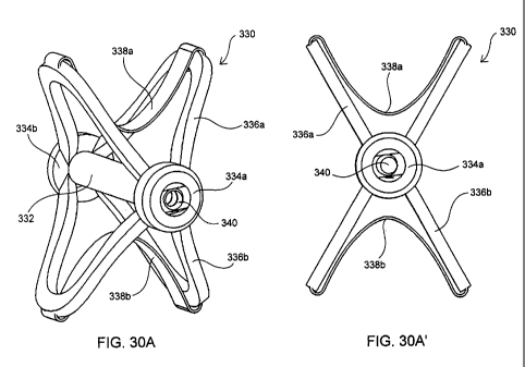
[0061] Fig. 30A and 30A' illustrate perspective and front views of another
interspinous spacer
device of the present invention in a fully deployed state.
[0062] Fig. 30B and 30W illustrate perspective and side views of the
interspinous spacer device
of Fig. 30A in an undeployed state.
9
CA 02582127 2007-03-27
WO 2006/045094
PCT/US2005/038026
0631 Fig. 30C and 30C' illustrate perspective and side views of the
interspinous spacer device
of Fig. 30A in a partially deployed state.
[0064] Figs. 31A and 31B illustrate perspective views of another
stabilizing device of the
present invention in partial and fully deployed states, respectively.
Detailed Description of the Invention
[0065] Before the subject devices, systems and methods are described, it is
to be understood
that this invention is not limited to particular embodiments described, as
such may, of course,
vary. It is also to be understood that the terminology used herein is for the
purpose of
describing particular embodiments only, and is not intended to be limiting,
since the scope of
the present invention will be limited only by the appended claims.
[0066] Unless defined otherwise, all technical and scientific terms used
herein have the same
meaning as commonly understood by one of ordinary skill in the art to which
this invention
belongs.
[0067] It must be noted that as used herein and in the appended claims, the
singular forms "a",
"an", and "the" include plural referents unless the context clearly dictates
otherwise. Thus, for
example, reference to "a spinal segment" may include a plurality of such
spinal segments and
reference to "the screw" includes reference to one or more screw and
equivalents thereof known
to those skilled in the art, and so forth.
[0068] Where a range of values is provided, it is understood that each
intervening value, to the
tenth of the unit of the lower limit unless the context clearly dictates
otherwise, between the
upper and lower limits of that range is also specifically disclosed. Each
smaller range between
any stated value or intervening value in a stated range and any other stated
or intervening value
in that stated range is encompassed within the invention. The upper and lower
limits of these
smaller ranges may independently be included or excluded in the range, and
each range where
either, neither or both limits are included in the smaller ranges is also
encompassed within the
invention, subject to any specifically excluded limit in the stated range.
Where the stated range
includes one or both of the limits, ranges excluding either or both of those
included limits are
also included in the invention.
[0069] All publications mentioned herein are incorporated herein by
reference to disclose and
describe the methods and/or materials in connection with which the
publications are cited. The
publications discussed herein are provided solely for their disclosure prior
to the filing date of
the present application. Nothing herein is to be construed as an admission
that the present
invention is not entitled to antedate such publication by virtue of prior
invention. Further, the
CA 02582127 2007-03-27
WO 2006/045094
PCT/US2005/038026
dates of publication provided may be different from the actual publication
dates which may
need to be independently confirmed.
[0070] The present invention will now be described in greater detail by
way of the following
description of exemplary embodiments and variations of the devices and methods
of the present
invention. The invention generally includes an interspinous spacer device as
well as
instruments for the percutaneous implantation of the interspinous spacer. A
key feature of the
interspinous spacer device is that it is expandable from a low profile
configuration to a higher
profile or operative configuration. This design allows the device, when in the
low profile
condition, to be delivered by percutaneous means without requiring the removal
of any portion
of the spinal motion segment into which the device is implanted.
[0071] As mentioned above, certain of the devices include balloon
embodiments or those
having expandable cavities which are expandable by the introduction of an
inflation or
expansion medium therein. Many of these are illustrated in Figs. 3-14. Certain
other devices
include those which have a more mechanical structure which is self-expandable
upon release
from a confined condition or which is actively expandable by actuation of
another instrument.
These are illustrated in Figs. 15-31.
[0072] Referring now to the drawings and to Figs. 3 and 4 in particular,
an exemplary
interspinous spacer device 24 of the present invention is illustrated in
collapsed and expanded
configurations, respectively. Interspinous device 24 includes an expandable
spacer body 4 that
has a size and shape when in the expanded condition for operative positioning
between the
spinous processes of adjacent superior and inferior vertebrae of the spinal
motion segment
being treated. Expandable body 34 is made of an expandable or inflatable
biocompatible
material such as non-porous material, e.g., latex, acrylate or a metal mesh,
e.g., a nitinol or
titanium cage.
[0073] Those spacers made of an inflatable non-porous material, i.e.,
balloon type spacers (see
Figs. 3-10), are inflated with an inflation or expansion medium, such as air,
saline, another
biologically compatible fluid, or a flowable solid material, such as
polyurethane, or a gel, which
thickens or hardens substantially upon injection into balloon 34. In one
embodiment, balloon
34 is initially inflated with air to provide some structure or rigidity to it
to facilitate its optimum
positioning and alignment between the spinous processes. Once positioned as
desired, balloon
34 is injected with a flowable solid material (the air therein being displaced
possibly via a vent
hole within port 32). In certain embodiments, the expandable body is made of a
non-compliant
or semi-compliant material so as to maintain a substantially fixed shape or
configuration and
11
CA 02582127 2007-03-27
WO 2006/045094
PCT/US2005/038026
ensure proper, long-term retention within the implant site. In other
embodiments, the
expandable member may be made of a compliant material. In any embodiment, the
compressibility and flexibility of balloon 34 can be selected to address the
indications being
treated.
[0074] Other embodiments of the subject spacers are made of an expandable
mesh or cage (see
Figs. 11-12). The mesh or cage maybe made of a super-elastic memory material
which is
compressible for delivery through a cannula and which is self-expanding upon
implantation.
Upon expansion, the mesh or cage may be self-retaining whereby its struts,
links or wires are
sufficiently rigid by themselves to maintain the expanded condition and
withstand the natural
forces exerted on it by spine. The mesh or cage may have an exterior coating
or an interior
lining made of materials similar to or the same as that used for the balloon
spacers, or may
otherwise be embedded in such material. In certain embodiments, an expansion
medium may
be used to fill the interior of the cage or mesh structure, such as with a
biologically compatible
fluid or flowable solid material used with the balloon-type embodiments.
[0075] In certain embodiments of present invention, either during the
implant procedure or in a
subsequent procedure, the size or volume of the implanted expandable spacer
may be
selectively adjusted or varied. For example, after an initial assessment upon
implant, it may be
necessary to adjust, either reduce or increase, the size or volume of the
spacer to optimize the
intended treatment. Further, it may be intended to only temporarily implant
the spacer for the
purpose of treating a temporary condition, e.g., an injured or bulging or
herniated disk. Once
the repair is achieved or the treatment completed, the spacer may be removed,
either with or ,
without substantially reducing the size or volume of the spacer. In other
embodiments, the
spacer as well as the inflation/expansion material may be made of
biodegradable materials
wherein the spacer degrades after a time in which the injury is healed or the
treatment
completed.
[0076] When unexpanded or deflated, as shown in Figs. 3A and 3B (balloon
type) and in Figs.
11C and 11D (mesh type) expandable body 34 has a low profile, such as a
narrow, elongated
shape, to be easily translated through a delivery cannula 70. The shape of
expandable body 34,
when in an expanded or inflated state, has larger profile which is generally H-
shaped.
Expandable body 34 has lateral or side portions 30, end portions 26 and apexes
28 defined
between the side portions 30 and the end portions 26. End portions 26 are
preferably recessed
or contoured to provide a narrowed central portion along the height dimension
or major axis of
expandable body 34 to readily fit between and to conform to the spinous
processes.
12
CA 02582127 2007-03-27
WO 2006/045094
PCT/US2005/038026
Accordingly, expandable body 34 has an apex-to-apex dimension (i.e., height or
major axis
dimension) from about 3 to about 5 cm and a width dimension (minor axis
dimension) from
about 2 to about 4 cm
[0077] For those embodiments of expandable bodies which comprise a balloon
configuration,
balloon 34 has an inflation or injection port 32 at a sidewall 30 for coupling
to a source of
inflation or expansion material or medium. Port 32 may consist of a one-way
valve which is
self-sealing upon release from an inflation mechanism or tube 76. Port 32 is
further configured
to releasably engage from tube 76, where such engagement may be threaded or
involve a
releasable locking mechanism. Where the expandable body comprises a mesh or
cage, port 32
simply acts as an exit port, however, where an expansion material is used, it
also functions as an
injection port for the expansion material.
[0078] Optionally, device 24 may include a pair of tabs 36 which may be
positioned on one
side of the device where the tabs 36 are preferably situated at the apexes 28
of expandable body
34. Pins or screws (not yet shown) may be used to secure the tabs against the
spinous process to
further ensure long-term retention of device 24 within the implant site. Tabs
36 are made of a
biocompatible material, such as latex, acrylate, rubber, or a metal, and may
be made of the same
material used for the expandable member 34. Shown here attached to tabs 36 are
tethers 38
which are used in part to manipulate the positioning of expandable body 34
upon implantation
into the targeted spinal motion segment. The tethers may be made of any
suitable material
including but not limited to materials used to make conventional sutures. They
may also be
made of a biodegradable material. While two tabs and associated tethers are
provided in the
illustrated embodiment, one, three or more may be employed, where the
respective tabs are
located on the expandable body so as to be adjacent a bony structure of the
vertebra suitable for
anchoring thereto. In embodiments which do not employ securing tabs 36,
tethers 38 may be
attached directly to the expandable body itself
[0079] Optionally still, device 24 may further include radiopaque markers
40 on the surface of
expandable body 34 visible under fluoroscopic imaging to facilitate
positioning of the
expandable body. Any number of markers 40 may be employed anywhere on
expandable body
34, however, as few as four markers, one at each apex, may be sufficient. With
embodiments
employing cage or mesh expandable bodies, the cage or mesh material itself may
be
radiopaque.
[0080] A system of the present invention includes a cannula device 70
having an outer sheath
72, a proximal hub 78 and preferably at least two interior lumens 74, 76 for
the percutaneous
13
CA 02582127 2007-03-27
WO 2006/045094
PCT/US2005/038026
delivery the device and other tools for implanting the device, which tools
mayinclude a cutting
instrument 62 (see Fig. 6C), a device delivery instrument 76, an endoscope,
etc., which tools
will be further discussed in the context of the description of the subject
methods with reference
to Figs. 5-10.
[0081] In Figs. 5A-5C, the spinal motion segment of Fig. 1 is illustrated
having spinal ligament
54 extending between the superior spinous process 18 and the inferior spinous
process 22. A
percutaenous puncture is made into the skin 30 adjacent the target spinal
motion segment of a
patient undergoing the implantation of the interspinous device of the present
invention, and a
cannula 70 is penetrated to the spinous ligament 54. The puncture and
subsequent penetration
may be made by way of a sharp distal tip of cannula 70 or by a trocar (not
shown) delivered
through a lumen of cannula 70.
[0082] As illustrated in Figs. 6A-6C, the spinous ligament 54 is then
dissected and an opening
58 created therein by way of a cutting instrument 60, such as a simple
scalpel, an electro surgical
device or the like, delivered through a lumen of cannula 70. Cutting
instrument 60 may then be
removed from cannula 70 and, as illustrated in Figs. 7A-7D (balloon type) and
in Figs. 11A-
11D (cage type), a delivery instrument 16 having interspinous device 24
operatively preloaded
is delivered through cannula 70.
[0083] The preloading of device 24 to delivery instrument 76 involves
providing expandable
body 34 in an unexpanded or deflated state and releasably coupled, as
described above, by way
of inflation or injection port 32 of expandable body 34 to the distal end of
delivery instrument
76. In addition to functioning as a pusher, instrument 76 may act as an
inflation lumen for
balloon type embodiments through which an inflation medium is transported to
within
expandable body 34.
[0084] Depending upon the material used to fabricate expandable body 34,
the expandable
body may have a degree of stiffness in an unexpanded or deflated state such
that it may
maintain an elongated configuration so as to be directly insertable and
pushable through
cannula 70. This may the case where the expandable member 34 is made of a cage
or mesh
material. Alternatively, a pusher or small diameter rod (not shown) may be
inserted through
inflation port 32 to within expandable body 34 to keep it in an elongated
state so as to prevent
expandable body 4 from bunching within cannula 70 and to provide some rigidity
to more
effectively position the expandable body in the target implant site. The rod
is then removed
from expandable body 34 and from delivery device 76 upon positioning the
expandable body at
the target implant site. In either case, expandable body 34 is folded or
compressed about its
14
CA 02582127 2007-03-27
WO 2006/045094
PCT/US2005/038026
minor axis with the side wall opposite the inflation port 32 defining a distal
end 25 (see Fig. 3B)
and the apexes 28 of the expandable body folded proximally of distal end 25 to
provide a
streamline, low profile configuration for delivery through cannula 70.
[0085] Once interspinous device 24 is preloaded to delivery device 76
as just described, device
24 is then inserted into a lumen of cannula 70 with tethers 38 pulled back and
trail proximally
so that the tether ends 38a extend from hub 78 of cannula 70. Expandable body
member 34 is
translated through cannula 70 to within opening 58 within spinous ligament 54
as best
illustrated in Figs. 7C and 11C. For best results, expandable body 34 is
centrally positioned
within opening 58 so that the countered ends 26 of expandable body 34 readily
engage with the
opposed spinous processes 18, 22. Fluoroscopy may be employed to visualize
markers 40 so as
to ensure that expandable body 34 centrally straddles the spinous ligament
opening 58, i.e., the
markers on the distal side 25 of the expandable body are positioned on one
side of the spine and
the markers on the proximal side of the expandable body (the side on which
port 32 is located)
are positioned on the other side of the spine.
[0086] Once centrally positioned, expandable body 34 is inflated or
expanded, as illustrated in
Figs. 8A-8D and 12A-12D. For balloon spacers, inflation occurs by allowing an
inflation or
expansion medium, as discussed above, to enter into the interior of the
expandable body via
port 32. For expandable mesh spacers, the expandable body may be configured to
expand
automatically upon exiting cannula 70. The inflation or expansion of
expandable body 34 may
also be visualized under fluoroscopy whereby markers 40, as best shown in Fig.
8C, are
observed and the position of expandable body 34 may be adjusted to ensure
optimum
positioning upon complete inflation. Adjustments of the expandable body's
position may be
accomplished by manually pulling on one or both tether ends 38a which in turn
pulls on tabs 26
to which the tethers 38 are attached at their proximal ends. The tethers 38
are selectively pulled
as necessary to center or optimally position interspinous expandable body 34
to achieve the
desired treatment of the targeted spinal motion segment.
[0087] With embodiments in which the expandable body is initially inflated
with air and then
filled with a solid or fluid medium, the latter is preferably not delivered or
injected into the
interior of the expandable body until the position of the expandable body
within the
interspinous space has been verified and optimized. This is beneficial in
situations where, upon
inflation, it is found that the expandable body is misaligned within the
interspinous space and
requires repositioning. The expandable body may simply be deflated of air to
the extent
necessary and repositioned in a less inflated or deflated state. If necessary,
for example where it
CA 02582127 2007-03-27
WO 2006/045094
PCT/US2005/038026
is found that the maximum spacer or expandable body size is insufficient for
the particular
application at hand, expandable body 34 may be completely deflated and removed
and replaced
with a more suitably sized unit.
[0088] For balloon spacers and those mesh spacers which are not by
themselves sufficiently
self-retaining, once the position and extent of inflation or expansion of
expandable body 34 are
optimized, the expansion medium, e.g., polyurethane, is allowed to flow or
injected into the
interior of the expandable body via port 32. As illustrated in Figs. 9A and
9B, expandable body
34 is caused to expand to a selected volume and in so doing forces apart (see
arrow 80) the
spinous processes 18, 22 in between which it is situated. This selective
distraction of the
spinous processes also results in distraction of the vertebral bodies 2, 4
(see arrow 82) which in
turn allows the disk, if bulging or distended, to retract to a more natural
position (see arrow 84).
Again, the extent of distraction or lordosis undergone by the subject
vertebrae can be monitored
by observing expandable body markers 40 under fluoroscopy.
[0089] The extent of possible distraction maybe limited by the capacity
of expandable body 34
and the type of expandable body material employed. In certain embodiments,
such as
expandable bodies made of non-compliant or semi-compliant balloons, the
requisite volume of
the inflation medium may be substantially fixed whereby the balloon achieves
its fully
expanded configuration upon filling it with the fixed volume of medium. In
other
embodiments, such as with balloons made of a compliant material, the extent of
expansion may
be variable and selectable intraoperatively depending on the extent of
lordosis or distraction to
be achieved between the spinous processes in which balloon 34 is now
interposed.
[0090] Upon achieving the desired distraction between the vertebrae,
inflation/expansion lumen
76 is disengaged from expandable body port 32 which then becomes sealed by
means of a one-
way valve that is closed upon disengagement of lumen 76. Inflation/expansion
lumen is then
removed from cannula 70. While the opposing compressive force exerted on
expandable body
34 by the distracted spinous processes 18, 22 may be sufficient to permanently
retain
expandable body 34 therebetween, the interspinous device may be further
secured to the
spinous processes 18, 22 to ensure that the expandable body does not slip or
migrate from its
implanted position. To this end, tabs 36 are anchored to the spinous processes
as illustrated in
Figs. 10A and 10B and in Figs. 13A and 13B. Any type of anchoring means, such
as screws,
tacks, staples, adhesive, etc. may be employed to anchor tabs 36. Here,
cannulated screws 90
are used as anchors and are delivered to the target site releasably coupled to
screw driving
instrument 88. While various screw attachment and release mechanisms may be
employed, a
16
CA 02582127 2007-03-27
WO 2006/045094
PCT/US2005/038026
simple configuration involves providing the screws 90 with a threaded inner
lumen which is
threadably engagable with the threaded distal end of instrument 88.
[0091] To ensure accurate placement of screws 90, along with instrument
88, can be tracked
and translated over respective tethers 38, which function as guide wires. By
manipulating
instrument 88, the screws are driven or screwed into the respective spinous
process.
Screwdriver 88 is then disengaged or unscrewed from screw 90. After both tabs
36 are securely
anchored to the spinous processes, the screwdriver and the cannula may be
removed from the
patient's back.
[0092] Figs. 14A-14F illustrate an alternative method for implanting the
expandable member.
In particular, the method contemplates pre-inflating or pre-expanding the
expandable member
prior to positioning the expandable member within the interspinous space. To
accomplish this,
the vertebrae 2 and 4 may be distracted prior to insertion of the pre-
expandable balloon implant.
A temporary distraction mechanism, such as another balloon or a mechanically
actuated device,
is inserted into the interspinous space. When the desired amount of
distraction is achieved, the
permanent or implantable expandable member can then be placed within the
interspinous space,
and the temporary distraction member may then be removed from the space.
[0093] While certain of the expandable spacers are intended to be
permanently implanted
within a spine, certain others may be implanted only temporarily to facilitate
the healing of an
injury or the treatment of a reversible or non-chronic condition, such as a
herniated disk. For
such temporary treatments, the expansion material most likely is a fluid, such
as saline, which
may be easily aspirated through port 32 or may be allowed to drain out via a
penetration or cut
made in the expandable member. In those embodiments in which the expansion
material is a
flowable solid, which may or may not subsequently harden within the expandable
member, the
material may be one that is reconstitutable into a liquid form which may then
be subsequently
aspirated or evacuated from the expandable member. For percutaneous removal of
the
expandable member, a cannula such as cannula 70 may be used and an aspiration
instrument
delivered therethrough and coupled to port 32. After deflation and/or
evacuation of the
expandable member, and removal of the tacks, sutures, staples, etc. if such
are used to secure
tabs 36, the expandable member may be easily removed through cannula 70. With
biodegradable spacers, removal of the spacer is obviated.
[0094] It should be noted that any of the above-described steps or
procedures, including but not
limited to cannulation of the target area, dissection of the spinous ligament,
insertion of the
expandable body within the dissected opening of the spinous ligament,
inflation and/or
17
CA 02582127 2007-03-27
WO 2006/045094
PCT/US2005/038026
expansion of the expandable body, adjustment or readjustment of the expandable
body, and
anchoring of the tabs, etc., may be facilitated by way of a scope 62 delivered
through a lumen
of cannula 70 to the open distal tip of cannula 70. Alternatively, a second
cannula delivered
through another percutaneous penetration may be employed for use of an
endoscope and any
other instruments needed to facilitate the procedure.
[0095] Fig. 14A illustrates an exemplary embodiment of a temporary
distraction mechanism
100 having an expandable strut configuration. Mechanism 100 includes bilateral
struts 102
which are hinged and foldable at hubs 104, respectively. Bridging the struts
102 at superior and
inferior ends are spinous process engagement portions 106 which are preferably
configured to
conformingly engage with the spinous processes 18, 22. Extending centrally
between hubs 104
is a distal portion of guide wire 108, which also extends proximally through
proximal hub 104a.
Guide wire 108 is in threaded engagement with both hub 104a whereby hub 104a
can be
translated both proximally and distally along guide wire 108. As such,
expandable member 100
can be provided in a low profile, compressed state upon proximally translating
hub 104a in a
proximal direction. In such a low-profile state, distraction mechanism 100 is
easily deliverable
through cannula 70, as described above, to with the interspinous space. Upon
proper
positioning, distraction mechanism 100 is expandable to a higher profile or
expanded state by
translating hub 104a toward hub 104b in a distal direction along guide wire
108, as illustrated in
Fig. 14A.
100961 After the desired amount of distraction is achieved between
vertebrae 2 and 4, an
implantable expandable member 110 of the present invention is delivered
adjacent the
distracted spinal motion segment. Expandable member 110 may be delivered from
the same
incision and side as distraction mechanism 100 (ipsolateral approach) and as
well as through the
same working channel, or may be delivered through a different incision on the
same or
opposing side of the spinal motion segment being treated (bilateral approach)
using two
different working channels. In the illustrated embodiment, expandable member
110 is delivered
from the same side of the spinous process as distraction mechanism 100.
Expandable member
110 may delivered through a separate designated lumen in cannula 70 and
translated distally of
hub 104b of distraction mechanism 100.
[0097] As shown in Fig. 14B, after deployment, expandable member 110 is
inflated or
expanded as described above with respect to expandable member 34, for example,
by way of an
inflation lumen extending through guide wire 108. Tethers 112 may be provided
on expandable
member 110 to retract and manipulate it to within the interspinous space, as
illustrated in Fig.
18
CA 02582127 2007-03-27
WO 2006/045094
PCT/US2005/038026
14C. Once expandable member 110 is properly positioned within the interspinous
space,
distraction mechanism 100 may be removed from the interspinous space
immediately or, if the
expandable member has been filled with a curable expansion medium or one that
involves
setting or hardening, the distraction mechanism may be kept in the
interspinous space until the
desired consistency, curing or hardening has been achieved by the expansion
medium. To
remove distraction mechanism 100 from the interspinous space, its profile is
reduced to a low
profile state, as illustrated in Fig. 14D. As mentioned earlier, this is
accomplished by
translating proximal hub 104a proximally along guide wire 108. Distraction
member 100 may
be retracted out through a cannula or removed directly in this low profile
state, leaving
expandable member 100 alone within the implant site as illustrated in Fig.
14E. Tethers 112
may then be cut or secured in place. Optionally, a strap 116 or the like may
be implanted to
further secure expandable member 110 within the implant site and reduce the
risk of migration.
Here, bores or holes 114 have been formed through the thickness of the spinous
processes 18,
22 and strap 116 threaded there through with its ends secured together by a
securing means 120,
such as a suture, staple or clip, as illustrated in Fig. 14F. Alternatively,
strap 116 could be
wrapped around the spinous processes 18, 22.
[0098] In addition to the expandable balloon spacers, the present
invention further provides for
mechanically expandable spacers such as those illustrated in Figs. 15-17. For
example,
= expandable spacer 130 of Fig. 15A is a cage-like structure having spaced-
apart, parallel strut
members 132 extending between and fixed to hubs 134. Like the distraction
mechanism of
Figs. 14A-14F, spacer 130 may be provided on and deliverable by way of a guide
wire 136
which is threadably engaged to and disengagable from proximal hub 134a. After
placement of
spacer 130 within the interspinous space, as illustrated in Fig. 15A, spacer
130 is expanded by
advancing proximal hub 134a distally along guide wire 136 thereby forcing
struts 132 radially
outward and away from each other whereby the expanded configuration of spacer
130 is
elliptical or, in a more advanced state of expansion, substantially spherical.
Once the desired
degree of distraction is achieved between vertebrae 2 and 4, guide wire 136
unthreaded from
hub 134a and removed from the implant region. =
[0099] Figs. 16A and 16B illustrate another embodiment of an expandable
spacer 140 which is
in the form of a coiled band 142 terminating at an outer end 144 having a
configuration for
receiving and locking onto inner end 146 upon full expansion or unwinding of
the coil. The
diameter of coil 142 in an unexpanded or fully wound state is small enough to
allow easy
insertion between spinous processes 18, 22. Upon proper positioning within the
interspinous
19
CA 02582127 2007-03-27
WO 2006/045094
PCT/US2005/038026
space, coil 142 is allowed to expand and unwind thereby distracting vertebrae
2 and 4 apart
from each other. Once the desire level of distraction is achieved, inner end
146 is coupled to
outer end 144. While the figures show band 142 inserted transversely to
spinous processes 18,
22, it may alternatively be inserted in line or in the same plan defined by
the spinous processes.
[00100] Figs. 17A-17C illustrate another interspinous spacer 150 having
interlocked nested
portions 152. Nested portions 152 are each shaped and configured to be
received within one of
its adjacent portions and to receive the other of the adjacent portions when
in a low profile state,
as illustrated in Fig. 17A. Upon expansion of spacer 150, which may be spring
loaded or be
expandable by way of an instrument (not shown) which may be inserted into the
spacer's center
and rotated to flare portions 152, vertebrae 2 and 4 are caused to distract
from each other.
Portions 152 may have a configuration or shape which allows them to bite or
dig into the
spinous process 18, 22 and become securely retained therein.
[00101] Figs. 18A and 18B illustrate another interspinous spacer 160 of
the present invention in
an undeployed or unexpanded state and a deployed or expanded state,
respectively. Spacer 160
includes an expandable tubular member 162 having end portions 164a, 164b which
are capped
by hubs 166a, 166b, respectively. As is explained in greater detail below, one
or both hubs may
be provided fixed to tubular member 162 or may be releasably coupled thereto.
A sleeve or
retaining member 168 is circumferentially positioned about tubular between end
portions 164a,
165a. Most typically, retaining member 168 is positioned substantially
centrally (as shown) on
tubular member 162, but may be positioned laterally towards one or the other
end. Retaining
member 168 has a length that covers about one third of the length of tubular
member 162, but,
may be longer or shorter depending on the application. As is explained in
greater detail below,
interspinous spacer 160 may further include a core member (shown in Fig. 21)
within the lumen
of the tubular member and which may be provided integrated with spacer 160.
Alternatively,
the core member may be provided as a detachable component of the device used
to deliver and
implant the spacer (see Figs. 19A and 19B).
[00102] In the undeployed state, as illustrated in Fig. 18A, spacer 160
has an elongated tubular
or cylindrical shape, and may have any suitable cross-sectional shape, e.g.,
circular, oval,
starred, etc., where the more angular cross-sections may allow the device to
bite or dig into the
spinous processes and for better retention. In this undeployed or lengthened
state, tubular
member 162 has a length in the range from about 20 mm to about 80 mm, and more
typically
from about 30 mm to about 50 mm, and a diameter or average thickness in the
range from about
4 mm to about 12 mm, and more typically from about 6 mm to about 9 mm. As
such, spacer
CA 02582127 2007-03-27
WO 2006/045094
PCT/US2005/038026
160 is deliverable to an implant site between adjacent spinous processes in a
minimally invasive
manner.
[00103] In the deployed state, as illustrated in Fig. 18B, spacer 160 has
a dumbbell or H-shaped
configuration, where the length of spacer 160 is less than and the diameter or
height of spacer
160 is greater than the corresponding dimensions of the spacer when in an
undeployed state. In
particular, the length dimension of the end portions 164a, 164b of tubular
member 162 has been
reduced by about 25% to about 70% while the diameter of the end portions 164a,
164b has been
increased by about 50% to about 600%, and the diameter of the central or
sleeve-covered
portion has been increased by about 200% to about 400%, where the diameter of
the portions of
the tubular member 164a, 164b not covered by retaining member 168 have a
greater diameter
than the portion of tubular member 162 which is covered by retaining member
168. The
increased diameter of covered or central portion 168 distracts the adjacent
vertebrae so as to
provide pain relief. The diameter of hubs 166a, 166b may remain constant upon
deployment of
device 160. In this deployed state, tubular member 162 has a length in the
range from about 15
mm to about 50 mm, and more typically from about 20 mm to about 40 mm, and an
end portion
diameter in the range from about 10 mm to about 60 mm, and more typically from
about 15 mm
to about 30 mm, and a central portion diameter in the range from about 5 mm to
about 30 mm,
and more typically from about 8 mm to about 15 mm. As such, when operatively
placed and
deployed within an interspinous space, the deployed spacer 160 fits snugly
within the
interspinous space and is held in place by the surrounding muscle, ligaments
and tissue.
[00104] Any suitable materials may be used to provide a spacer 160 which
is provided in a first
state or configuration, e.g., the undeployed state illustrated in Fig. 18A,
and which can be
manipulated to achieve a second state or configuration, and back again if so
desired. A polymer
based material or any other material which allows for simultaneous axial
shortening and radial
expansion is suitable for use to form tubular member 162. The end portions
164a, 164b may be
made of the same or a different material as that of the central or covered
portion. A flexible or
shaped memory material or any other material which also allows for
simultaneous axial
shortening and radial expansion, but which is less expandable, i.e., maintains
a compressive
force about tubular member 162, than the material employed for tubular member
162 may be
used to form retaining member 168. As such, retaining member 168 limits the
extent of radial
expansion as well as axial shortening that the covered portion of tubular
member 162 can
undergo. Examples of suitable materials for the retaining member include but
are not limited to
Nitinol or polyethelene in a braided or mesh form. Further, the construct of
retaining member
21
CA 02582127 2007-03-27
WO 2006/045094
PCT/US2005/038026
168 may be such that the radial force applied to the portion of tubular member
162 that it covers
is constant or consistent along its length so as to maintain a constant
diameter along its length
or, alternatively, may have a varying radial force so as to allow for
selective shaping of the
covered portion of tubular member when in a deployed state. Retaining member
168 may be
constructed so as to resist bending or flexing upon forcible contact with the
spinous processes
and, as such, does not conform to the spinous processes. Conversely, the
retaining member 168
may be constructed from a more flexible material that allows for some
compression and, as
such, may conform or be conformable to the spinous processes. Further, the
physical
properties and dimensions of the materials used for both the tubular member
and the retaining
may be selected to provide the desired amount of distraction between target
vertebrae.
[00105] Referring now to Figs. 19A and 19B, spacer 160 is shown
operatively employed within
an interspinous space and coupled to delivery device 170. Delivery device 170
includes an
outer shaft 172 and an inner shaft 178, movable relative (axially,
rotationally or both) to outer
shaft 172, both extending from a handle mechanism 174. For example, inner
shaft 178 may be
configured to be retracted proximally within outer shaft 172, or outer shaft
172 may be
configured to be advanced distally over inner shaft 178, or both
configurations may be
employed together, i.e., while outer shaft 178 is advanced, inner shaft 178 is
retracted. The
relative movement may be accomplished in any suitable manner, for example by
way of a screw
configuration, i.e., where the shaft members engage by way of corresponding
threads, as
illustrated in Fig. 20A, or by way of a ratchet configuration, as illustrated
in Fig. 20B. The
relative movement is accomplished by manual actuation of actuator 176 coupled
to handle 174.
While only mechanical embodiments of the movement actuation are illustrated,
the same can be
achieved by electrically or pneumatically-driven devices or mechanisms.
[00106] As mentioned above, spacer 160 may be provided with an integrated
core member or
the core member may be detachably provided on the distal end 182 of inner
shaft 178. In the
first embodiment, distal end 182 of inner shaft 178 is configured to
temporarily couple with a
proximal end (i.e., the end closest to handle 174) of the core member. In the
latter embodiment,
the distal end 182 of inner shaft 178 is configured.to be inserted into the
lumen of tubular
member 162, as illustrated in Fig. 21, connect to or engaged with distal hub
166b (i.e., the hub
positioned furthest from handle 174) and be detachable at a proximal end 184
from inner shaft
178 to function as a core member. An advantage of the latter embodiment is
that the end
portion 182 of the inner shaft 178 functioning as the core member may have a
length that is as
short as the length of tubular member 172 when in a deployed state, with no
extra length or
22
CA 02582127 2007-03-27
WO 2006/045094
PCT/US2005/038026
remaining portion extending laterally of the implanted device. In the
integrated embodiment,
the core length may need to be as long as tubular member 172 when in the
undeployed state.
However, the core member may be segmented to allow for selective removal of
one or more
lengths or portions from the proximal side of the core member subsequent to
implantation of the
spacer so as not to have any excess length extending from the spacer.
[00107] With either embodiment, retraction of inner shaft 178, as
described above, retracts
distal hub 166b towards proximal hub 166a and/or advancement of outer shaft
172 advances
proximal hub 166a towards distal hub 166b, thereby causing tubular member 162
to be
compressed axially, and thus expanded radially, as shown in Fig. 19B. While
distal hub 166b
may be fixed to tubular member 162, proximal hub 166a may be provided as a
separate
component having a central bore which allows it to receive and axially
translate over inner shaft
178. Proximal hub 166a may be configured to readily slide over inner shaft 178
in a distal
direction (but possibly not in a proximal direction) or may be threaded in
order to advance over
inner shaft 178. The advancement of proximal hub 166a axially compresses
tubular member
172 and causes it to radially expand. The axial compression or radial
expansion may be
continued until the desired extent of distraction occurs between vertebrae 2
and 4. When the
desired level of distraction is achieved, proximal hub 166a is secured to
either the proximal end
of tubular member 162 and/or the proximal end of the core member 182, such as
by a threaded
or snap-fit engagement or by activating a lock mechanism (not shown). Inner
shaft 178 may
then be released from the core member (or distal end 182 of inner shaft 178
may be released
from inner shaft 178 and left within tubular member 172 to function as the
core member)
which, along with the end hubs 166a and 166b, maintain the implanted spacer
160 in a deployed
state so as to maintain distraction between the vertebrae.
[00108] The reconfiguration of spacer 160 may be further facilitated by
selectively configuring
the wall of tubular member 162. For example, the interior or lurninal surface
of tubular member
162 may be contoured or incorporated with divets or spaces 180 where, upon
compression of
tubular member 162, the walls of the uncovered portions 164a, 164b of tubular
member 162
will more readily fold inward to provide the resulting configuration shown in
Fig. 18B.
[00109] Figs. 22A-22C illustrate another interspinous spacer 190 of the
present invention in an
undeployed/unexpanded state, in an intermediate state during deployment and in
a
deployed/expanded state, respectively. Spacer 190 includes expandable end
portions 192a,
192b which are capped by hubs 198a, 198b, respectively. As mentioned
previously, one or both
hubs may be provided fixed to the end members or may be releasably coupled
thereto.
23
CA 02582127 2007-03-27
WO 2006/045094
PCT/US2005/038026
Extending between end portions 192a, 192b is a central portion 194 including a
plurality of
blocks or wedges, such as side blocks 200 and end blocks 202, surrounded by a
cover, sleeve or
retaining member (not shown) which functions to hold the blocks in frictional
engagement with
each other. A core member or rod 196 extends centrally through end portions
192a, 192b and
central portion 194 where end blocks 202 are coaxially positioned on core 196
and are slidably
translatable thereon. Core member 196 or a portion thereof may be provided
integrated with
spacer 190 or may be provided as a detachable component of the device used to
deliver and
implant the spacer.
[00110]
As with the previously described spacer, end portions 192a, 192b may be made
of a
polymer based material or any other material which allows for simultaneous
axial shortening
and radial expansion when compressed. Blocks 200, 202 have a more rigid
configuration in
order to distract the adjacent spinous processes which define the interspinous
space into which
spacer 190 is positioned without substantial compression of central portion
194. As such, the
blocks may be made of a-rigid polymer material, a metal, ceramics, plastics,
or the like. In
order to effect radial expansion and axial shortening of central portion 194,
the blocks are
selectively sized, shaped and arranged such that an inwardly compressive force
on end blocks
202 along the longitudinal axis of the spacer forces end blocks 202 together
which in turn forces
side or lateral blocks 200 outward and away from each other, as illustrated in
Fig. 22B. The
inwardly tapered sides of the blocks enable slidable engagement between
adjacent blocks. The
covering (not shown) around the blocks is made of a stretchable material so as
to accommodate
the radial expansion of central portion 194. As such, the cover may be made of
a polymer ,
based material.
[00111]
When in an undeployed state, as shown in Fig. 22A, the central and end
portions of
spacer 190 have tubular or cylindrical configurations, and may have any cross-
sectional shape,
length and or diameter as provided above with respect to spacer 160 of Figs.
18A and 18B.
Deployment of spacer 190 within an interspinous space may be accomplished in
the manner
described above. In a fully deployed state, as illustrated in Fig. 22C, spacer
190 has a dumbbell
or H-shaped configuration with a change in length and height dimensions as
provided above.
The increased diameter of central portion 194 when spacer 190 is the deployed
configuration
distracts the adjacent vertebrae so as to provide pain relief. While the
respective dimensions of
the spacers change from an undeployed to a deployed state, the spacers may be
configured such
that the overall size of volume occupied by the spacer does not change.
24
CA 02582127 2007-03-27
WO 2006/045094
PCT/US2005/038026
01121 Another interspinous spacer 210 of the present invention is
illustrated in an
undeployed/unexpanded state, in an intermediate state during deployment and in
a
deployed/expanded state in Figs. 23A-23C, respectively. Spacer 210 includes
expandable end
portions 212a, 212b capped by hubs 224a, 224b, respectively. As mentioned
previously, one or
both hubs may be provided fixed to the end members or may be releasably
coupled thereto.
Extending between end portions 212a, 212b is a central portion 214 including a
plurality of
linkages 216 and blocks 220, 222, which collectively provide opposing struts.
Each linkage
216 has a length and is pivotally coupled to a side block 220 and an end block
222, where end
blocks 222 are coaxially positioned on core 218 and are slidably translatable
thereon. While the
materials and configuration of end portions 212a, 21 2b may be as described
above, linkages 216
are preferably made of a metal material. A core member or rod 218 extends
centrally through
end portions 212a, 212b and central portion 214. Core member 218 or a portion
thereof may be
provided integrated with spacer 210 or may be provided as a detachable
component of the
device used to deliver and implant the spacer.
[00113] In an undeployed state, as shown in Fig. 23A, the central and end
portions of spacer 190
have tubular or cylindrical configurations, and may have any cross-sectional
shape, length and
or diameter as provided above. As such, side blocks 220 are close together and
end blocks 222 .
are spaced apart with the lengths of linkages 216 aligned with the
longitudinal axis of core
member 218. When opposing, inwardly compressive forces are exerted on spacer
210 along its
longitudinal axis, end portions 212a, 212b axially compress and radially
expand as described
above thereby forcing end blocks 222 together which in turn force side or
lateral blocks 220 ,
outward and away from each other, as illustrated in Fig. 23B. This action
causes linkages 216
to spread apart, as shown in Fig. 23B, and move to positions where their
lengths are transverse
to the longitudinal axis of core 218, as illustrated in Fig. 23C.
[00114] Interspinous spacer 230 of Figs. 24A-24C employs the linkage
arrangement of the
central portion of spacer 190 of Figs. 23A-23C in both of its end portions
232a, 232b as well as
its central portion 234. Specifically, end portions 232a, 232b employ linkages
236, which are
longer than linkages 238 used for central portion 234, but which are arranged
in similar
engagement with side blocks 248 and end blocks 250. On each side of central
portion 234 and
in between the central portion and the end portions 232a, 232b, respectively,
are dampening
washers 244. A core member 240 extends between and through the end blocks 250
of distal
end member 232a and the end blocks 252 of central portion 234 as well as the
dampening
washers 244 positioned therebetween, all of which, except the most distal end
block, may
CA 02582127 2007-03-27
WO 2006/045094
PCT/US2005/038026
slidably translatable along core member 240. Core member 240 is releasably
attached at a
proximal end to ratcheted drive rod 242 of a delivery device as discussed
above with respect to
Figs. 19-21 which rod 242 extends through the proximal end portion 232a and
hub 246, as
illustrated in Fig. 24B.
[00115] In an undeployed state, as shown in Fig. 24A, the central and end
portions of spacer 230
have tubular or cylindrical configurations. As such, side blocks 248 and 252
of end portions
232a, 232b and central portion 234, respectively, are close together and end
blocks 250 and 252
of end portions 232a, 232b and central portion 234, respectively, are spaced
apart with the
lengths of linkages 236, 238 aligned with the longitudinal axis of core member
240. When
opposing, inwardly compressive forces are exerted on the distal block 250 and
hub 246 of
spacer 230 along its longitudinal axis, the end blocks are drawn together
thereby forcing side or
lateral blocks 220 outward and away from each other, as illustrated in Fig.
24B. This action
causes the linkages of the end and central portions to spread apart, and move
to positions where
their lengths are transverse to the longitudinal axis of core 240, as
illustrated in Fig. 24C, the
fully deployed state of spacer 230.
[00116] The end portions and central portions of the compressible spacers
described above may
be used in any combination. For example, the polymer-based central portion of
Figs. 18A and
18B and the linkage end portions of Figs. 24A-24C may be used together to form
a spacer of
the present invention. Such a spacer 260 is illustrated in Figs. 25A-25C.
Spacer 260 includes
linkage-block end portions 262a, 262b and a compressible central member 264
around which is
positioned a circumferential retaining member 278 made of a braided mesh-like
material. A
core member 274 extends between and through the end blocks 270 of distal end
member 262a
and through central portion 264, all of which, except the most distal end
block, may slidably
translatable along core member 260. Core member 260 is releasably attached at
a proximal end
to ratcheted drive rod 272 of a delivery device as discussed above with
respect to Figs. 19-21
which rod 272 extends through the proximal end portion 262a and hub 272, as
illustrated in Fig.
25B.
[00117] In an undeployed state, as shown in Fig. 25A, the central and end
portions of spacer 230
have tubular or cylindrical configurations. As such, side blocks 268 of end
portions 262a, 262b
are close together and end blocks 270 of end portions 262a, 262b are spaced
apart with the
lengths of linkages 266 aligned with the longitudinal axis of core member 274.
When
opposing, inwardly compressive forces are exerted on the distal block 270 and
hub 272 of
spacer 260 along its longitudinal axis, the end blocks are drawn together
thereby causing
26
CA 02582127 2007-03-27
WO 2006/045094
PCT/US2005/038026
linkages 266 of the end portions to spread apart thereby forcing side or
lateral blocks 268
outward and away from each other, as illustrated in Fig. 25B, until linkages
266 move to
positions where their lengths are transverse to the longitudinal axis of core
274, as illustrated in
Fig. 25C, the fully deployed state of spacer 260.
[00118] Each of the expandable and or inflatable interspinous spacers
described thus far is
particularly configured to be delivered minimally invasively, even
percutaneously, from a
single incision located laterally to one side (left or right) of the spinal
motion segment to be
treated. However, the present invention also includes interspinous spacers
which are
deliverable through a mid-line incision made directly into the interspinous
ligament. Examples
of such spacers are now described.
[00119] Figs. 26A and 26B are perspective and front views, respectively,
of interspinous spacer
280 which is configured for implantation by way of a percutaneous mid-line
approach. Spacer
280, shown in a deployed state, includes a central member or portion 282 and
four struts or legs
284 which are substantially radially expandable from central portion 282.
Central portion 282
has a cylindrical configuration having a diameter sized for delivery through a
small gauge
cannula and a length that allows placement within an interspinous space. A
lumen 285 extends
at least partially through the center of central portion 282 and is
configured, e.g., threaded, to be
releasably engaged to a delivery tool.
[00120] Each strut 284 includes one or more blocks 288. Where more than
one block 288 per ,
strut is employed, such as with spacer 280 which employs two blocks 288 per
strut 284 and
spacer 290 of Fig. 27 which employs three blocks 288 per strut 284, the blocks
are stacked and
slidably interconnected to each other in a manner that allows the to translate
linearly relative to
each other along parallel axes. A tongue and groove configuration 292 is
employed with the
illustrated embodiment to interconnect stacked blocks, but any suitable
interconnection which
enables such relative motion between the blocks may be used. Such
configuration may also be
employed to interconnect the innermost block to central member 282 where outer
ridges or
tongues 296 on central member 282 slidably interface with a corresponding
groove on inner end
of the innermost block. As such, blocks 288 are slidable relative to central
member 282 along
an axis parallel to the longitudinal axis of central member 282. Depending on
the application
and the particular anatomy of the implant site, struts 284 may be evenly
spaced apart about the
circumference of central member 282. In other embodiments the distance between
superior
struts 284a and between inferior struts 284b may vary and/or the distance
between each of those
and between struts on the same side of the central member may vary.
27
CA 02582127 2007-03-27
WO 2006/045094
PCT/US2005/038026
0121] Spanning between each strut pair 284a and 284b is a strap 286a and
286b, respectively,
affixed to the outermost blocks. Straps 286 may be made of any suitable
material which is
strong enough to maintain distraction between adjacent spinous processes and
to endure any
frictional wear which it may undergo due to natural spinal motion. The straps
may be flexible
such that they act as slings, or may be conformable to the spinous processes
once in situ.
Alternatively, the straps may be non-conforming and rigid with a planar or
curved shape
depending on the application at hand. Suitable strap materials include but are
not limited to
polyester, polyethylene, etc.
[00122] With reference to Figs. 28A-28E, various steps of a method
according to the present
invention for implanting spacer 280 as well as other spacers of the present
invention configured
for a mid-line implantation approach into a target spinal motion segment
(defined by
components of vertebral bodies 2 and 4) of a patient are described.
[00123] The initial steps of creating a percutaneous puncture and
subsequent penetration into the
skin 30 and the dissection of the spinous ligament 54 involve many of the same
instruments
(e.g., K-wire, trocar, cutting instrument, delivery cannula, etc.) and
surgical techniques used in
the ipsolateral implantation approach described above with respect to Figs. 5
and 6. Upon
creating an opening within the interspinous space extending between the
superior spinous
process 18 and the inferior spinous process 22, a delivery instrument 300
having interspinous
device 280 operatively preloaded in an undeployed state at a distal end is
delivered to within the
interspinous space. The delivery instrument 300 is provided with a mechanism
for releasably
connecting to spacer 380, such as by way of threaded screw 302 (see Fig. 28D)
which is
threadedly engaged with threaded lumens 285 of spacer 280.
[00124] As best illustrated in Figs. 28A' and 28A", when in an undeployed
state, spacer 280 has
a relatively low profile to facilitate entry into the interspinous space. Once
properly positioned
within the interspinous space, deployment of the spacer 280 is initiated, as
illustrated in Fig.
28B, by manipulation of instrument 300 which simultaneously causes outward
radial movement
of the outermost blocks of strut pairs 284a, 284b and distal linear
advancement of the proximal
portion 304 of spacer 282 (see Figs. 28B' and 28B") resulting in radial
expansion and axial
shortening of spacer 280. Spacer 280 may be configured such that deployment of
the struts is
accomplished by either or both axial rotation of internally componentry or
axial compression of
central member 282.
[00125] As the struts are radially extended, straps 286a and 286b emerge
and they become tauter
as the slack in them is gradually reduced by the extension of the struts.
Continued deployment
28
CA 02582127 2007-03-27
WO 2006/045094
PCT/US2005/038026
of spacer 280 causes straps 286a, 286b to engage with opposing surfaces of
spinous processes
18 and 22. The radial extension of the struts is continued, as illustrated in
Figs. 28C, 28C' and
28C", until the desired amount of distraction between the vertebra is
achieved. This selective
distraction of the spinous processes also results in distraction of the
vertebral bodies 2, 4 which
in turn allows the disk, if bulging or distended, to retract to a more natural
position. The extent
of distraction or lordosis undergone by the subject vertebrae can be monitored
by observing the
spacer under fluoroscopy.
[00126] At this point, the delivery instrument 300 is released from spacer
280 by unscrewing
threaded screw 302 from threaded lumen 285 and removing it from the implant
site, as
illustrated in Fig. 28D. Spacer 280 remains behind within the interspinous
space, locked in a
deployed state (see Fig. 28E).
[00127] Spacer 280 may configured such that the struts are not retractable
without active
manipulation of delivery instrument 300 to ensure that their extension, and
thus the distraction
on the spinal motion segment, is maintained. As configured, spacer 280 may be
easily
repositioned or removed by subsequent insertion of instrument 300 into the
interspinous space
and operative engagement with the spacer. Instrument 300 is then manipulated
to cause
retraction of the struts and the straps, reducing the spacer's profile to
allow repositioning or
removal of the spacer.
[00128] Figs. 29A-29D illustrate another spacer 310 of the present
invention that is implantable
through a mid-line approach to the interspinous space. Spacer 310 includes
centrally opposed
front and rear structures or blocks 312a, 32b which are pivotally
interconnected on both sides to
pairs of elongated linkages 314. The other end of each linkage 314 is
pivotally connected to a
lateral structure 318a or 318b. The resulting "X" configuration provides
interconnected strut
pairs on each side of spacer 310 which move and function similarly to the
linkages described
above with respect to the spacers illustrated in Figs. 23, 24 and 25, i.e.,
the lengths of linkages
314 extend parallel to the central axis of spacer 310 when in a fully
undeployed state (Fig. 29A)
and extend transverse to the central axis of spacer 310 in a fully deployed
state (Fig. 29D).
Extending between opposing superior lateral structures 318a and between
opposing inferior
structures 318b are straps 316a and 316b, respectively.
[00129] Spacer 310 is implantable and deployable by way of a mid-line
approach similar to that
described above with respect to the spacer of Figs. 28A-28E. Spacer 310 is
preloaded to a
delivery instrument shaft 320 which is insertable and axial translatable
through a central
opening within front block 312a. The distal end of shaft 320 is releasably
attached to an axial
29
CA 02582127 2007-03-27
WO 2006/045094
PCT/US2005/038026
member (not shown) of spacer 310. Axial member is fixed to rear block 312b and
extends
along the central axis of spacer 310, having a length which extends to front
block 312a when
spacer 210 is in a fully deployed state, as illustrated in Fig. 29D but which
extends only a
portion of the length of spacer 310 when it is in an undeployed state (Fig.
29A) or a partially
undeployed (Figs. 29B and 29C) state.
[00130] After the necessary space is created within the interspinous space
as described above,
spacer 310, which is releasably connected to delivery shaft 320 as described
above, is inserted
into the space in a fully undeployed sate (see Figs. 29A and 29A'). Deployment
of the spacer is
accomplished by proximally pulling on shaft 320 which compresses rear block
312b towards
front block 312a. This in turn causes the linkages 314 to pivot about their
respective attachment
points with superior and inferior lateral structures or blocks 318a and 318b
forced away from
each other, as illustrated in Figs. 29B and 29131. Continued pulling of
instrument 320 further
expands linkages 314 in a direction transverse to the central axis of spacer
310 and extend
straps 316a, 316b towards respective surfaces of the spinous processes. As
front and rear
blocks 312a and 312b are centrally tapered, defining a bowtie or hourglass
configuration, the
strut pairs define a centrally tapered profile as the align to their fully
deployed position, as best
shown in Figs. 29C' and 29D'. In the fully deployed state, the spacer's axial
member is
positioned within the opening of front block 312a and locked to it.
Additionally, straps 316a
and 316b are firmly engaged against the spinous processes and the contacted
vertebra are
distracted from each other. Delivery instrument 320 may then be released from
spacer 310 and
removed from the implant site.
[00131] Figs. 30A-30C illustrate yet another spacer 330 of the present
invention having an
shape in an expanded condition and which is implantable through a mid-line
approach to the
interspinous space. As best illustrated in Figs. 30A and 30A', spacer 330
includes an elongated
central member 332 extending between front and rear hubs 334a and 334b and a
plurality of
flexible or deformable struts 336 which also extend between hubs 334a, 334b.
Struts 336 are
configured to be deformable and to have a directional character to facilitate
deployment of them
radially outward from central member 332. Examples of suitable constructs of
these struts
include but are not limited to thin metal plates, e.g., flat springs, wire
bundles or a polymer
material. Extending between and affixed to each of strut pairs 336a and 336b
are straps 338a
and 338b, respectively.
[00132] The proximal end 342 of central member 332 is provided with
ratcheted grooves which
are releasably engaged within the distal end of 352 of delivery instrument 350
(see Fig. 30C').
CA 02582127 2007-03-27
WO 2006/045094
PCT/US2005/038026
Front hub 334a is provided with an opening 340 which also has a grooved
internal surface for
engaging with the grooves of central member 332.
[00133] Spacer 330 is implantable and deployable by way of a mid-line
approach similar to that
described above with respect to the spacer of Figs. 29A-2D. Spacer 330 is
preloaded in a fully
undeployed state to delivery instrument shaft 350 as illustrated in Figs. 30B
and 30B'. After the .
necessary space is created within the interspinous space as described above,
spacer 330 is
inserted into the interspinous space. Deployment of the spacer is accomplished
by proximally
pulling on shaft 350, by ratcheting as described above, which compresses rear
hub 334b
towards front hub 334a or distally pushing on front hub 334a towards rear hub
334b. This in
turn causes struts 336a, 336b to flex or bend outward, as illustrated in Figs.
30C and 30C'.
Continued pulling of instrument 350 (or pushing of hub 334a) further bends the
struts such that
they define an X-shaped structure with straps 338a and 338b forcably abutting
against the
interspinous processes. The pulling (or pushing) action advances the grooved
proximal end 342
of central member 332 into grooved opening 340 of front hub 334a. The opposing
grooves of
the central member and the opening provide a ratchet relationship between the
two whereby
central member is readily translatable in a proximal direction but not in a
distal direction,
thereby locking spacer 330 in a deployed state. Upon achieving the desired
amount of
distraction between the vertebra, delivery instrument 350 is released from
spacer 310 (such as
by unscrewing) and removed from the implant site.
[00134] Figs. 31A and 31B illustrate a stabilizing spacer 360 similar to
spacer 330 just described
but which forms the expanded "X" configuration with solid linkages rather than
struts. Spacer
360 includes an elongated central member 362 extending from and fixed to a
rear hub 364a and
slidably through a front hub 364b proximally to a delivery tool having a shaft
372. Also
extending between the front and rear hubs are four linkage pairs, where each
linkage pair 366a
and 366b are interconnected to a respective hub by a hinge 368 and are
interconnected to each
other by a hinge 370. When in a fully unexpanded condition, each linkage pair
extends parallel
to central member 362, providing a low profile for delivery. When the front
and rear hubs are
caused to approach each other, each linkage pair 366a, 366b expands
substantially radially
outward from central member 362, as illustrated in Fig. 31A. The hubs are
brought together to
the extent desired to provide an expanded "X" configuration, as illustrated in
Fig. 31B. Upon
achieving the desired expansion, central member 362 is released or detached
from delivery shaft
372. As with many of the "mechanical" type spacers discussed above, attachment
and release
of the spacer from the delivery device may be accomplished by various means,
including but
31
CA 02582127 2007-03-27
WO 2006/045094
PCT/US2005/038026
not limited to ratchet, threaded or quick-release configurations between the
spacer and the
delivery device.
[00135] Extending between and affixed to each of the top and bottom linkage
pairs are brackets
or saddles 374 for receiving the inner surfaces of opposing interspinous
processes. Brackets
374 have a substantially rigid and flat central portion 374a and relatively
flexible lateral
portions 374b which are affixed to hinges 370. The rigid, flat central portion
374a facilitates
engagement with the interspinous process. The flexible lateral portions 374b
and their hinged
connections to spacer 360 facilitate folding of the lateral portions 374b when
in an undeployed
state and allow for adjustment of spacer 360 once in a deployed state, where a
least a portion of
the adjustment may be self-adjustment by spacer 360 relative to interspinous
space into which it
is implanted.
[00136] The subject devices and systems may be provided in the form of a
kit which includes at
least one interspinous device of the present invention. A plurality of such
devices may be
provided where the devices have the same or varying sizes and shapes and are
made of the same
or varying biocompatible materials. Possible biocompatible materials include
polymers,
plastics, ceramic, metals, e.g., titanium, stainless steel, tantalum, chrome
cobalt alloys, etc. The
kits may further include instruments and tools for implanting the subject
devices, including but
not limited to, a cannula, a trocar, a scope, a device
delivery/inflation/expansion lumen, a
cutting instrument, a screw driver, etc., as well as a selection of screws or
other devices for
anchoring the spacer tabs to the spinous processes. The kits may also include
a supply of the
expandable body inflation and/or expansion medium. Instructions for implanting
the
interspinous spacers and using the above-described instrumentation may also be
provided with
the kits.
[00137] The preceding merely illustrates the principles of the invention.
It will be appreciated
that those skilled in the art will be able to devise various arrangements
which, although not
explicitly described or shown herein, embody the principles of the invention
and are included
within its spirit and scope. Furthermore, all examples and conditional
language recited herein
are principally intended to aid the reader in understanding the principles of
the invention and
the concepts contributed by the inventors to furthering the art, and are to be
construed as being
without limitation to such specifically recited examples and conditions.
Moreover, all
statements herein reciting principles, aspects, and embodiments of the
invention as well as
specific examples thereof, are intended to encompass both structural and
functional equivalents
thereof. Additionally, it is intended that such equivalents include both
currently known
32
CA 02582127 2013-07-03
51915-25
equivalents and equivalents developed in the future, i.e., any elements
developed that perform -
the same function, regardless of structure. The scope of the present
invention, therefore, is not
intended to be limited to the exemplary embodiments shown and described
herein. Rather, the
scope of present invention is embodied by the appended claims.
=
33