Note: Descriptions are shown in the official language in which they were submitted.
CA 02582149 2012-12-11
_ ANALOGS OF ShK TOXIN AND THEIR USES IN SELECTIVE
INHIBITION OF Kv1.3 POTASSIUM CHANNELS
FIELD OF THE INVENTION
The present invention provides a) novel compositions of matter, b)
methods and kits for in vivo and/or in vitro inhibition of the Kv1.3 channel
in
T- and B-lymphocytes and other cell types and c) methods for treating
autoimmune and other disorders in human or animal subjects.
BACKGROUND OF THE INVENTION
Cell plasma membranes form the outer surfaces of eukaryotic cells.
Various ions (e.g., sodium, potassium, calcium, etc.) move in and out of cells
by passive diffusion through the cells' plasma membranes. Such diffusion of
ions into and out of cells is facilitated by the presence of "ion channels"
within
the cell membranes. Ion channels are proteins embedded within the cell
membrane that control the selective flux of ions across the membrane,
thereby allowing for the formation of concentration gradients between the
intracellular contents of the cell and the surrounding extracellular fluid.
Because ion concentrations are directly involved in the electrical activity of
excitable cells (e.g., neurons), the functioning (or malfunctioning) of ion
channels can substantially control the electrical properties and behavior of
such cells. Indeed, a variety of disorders, broadly termed "channelopathies,"
are believed to be linked to ion channel insufficiencies or dysfunctions.
1
CA 02582149 2007-03-27
WO 2006/042151
PCT/US2005/036234
Ion channels are referred to as "gated" if they can be opened or closed.
The basic types of gated ion channels include a) ligand gated channels, b)
mechanically gated channels and c) voltage gated channels. In particular,
voltage gated channels are found in neurons, muscle cells and non-excitable
cells such as lymphocytes. They open or close in response to changes in the
charge across the plasma membrane.
Kv1.3 Channels and Autoimmune Diseases.
Autoimmune diseases such as multiple sclerosis (MS), type-1 diabetes
mellitus (11DM), rheumatoid arthritis (RA) and psoriasis affect several
hundred million people worldwide. In these disorders specific autoreactive T
cells - for instance myelin-specific T cells in MS patients ¨ are believed to
undergo repeated autoantigen stimulation during the course of disease and
differentiate into chronically activated memory cells that contribute to
pathogenesis by migrating to inflamed tissues and secreting cytokines
(Viglietta et al., 2002; Vissers et al., 1002; Wulff et al., 2003b). Therapies
that
preferentially target chronically activated memory T cells would have
significant value for autoimmune diseases.
Memory T cells are divided into two subsets ¨ central memory (Tcm)
and effector memory (TEO - based on the expression of the chemokine
receptor CCR7 and the phosphatase CD45RA (Geginat et al., 2001; Sallusto
et al., 1999). Naïve and Tcm cells home to the lymph node before they migrate
to sites of inflammation, whereas TEm cells home directly to sites of
inflammation where they secrete copious amounts of IFN-p and TNF-a and
exhibit immediate effector function. It has recently been shown that myelin-
specific autoreactive T cells in MS patients are predominantly activated TEM
cells (Wulff et al., 2003b), and adoptive transfer of myelin-specific
activated
rat TEm cells into naïve recipients induced severe EAE (Beeton et al., 2001a;
Beeton et al., 2001b). An exciting new therapeutic target for
immunomodulation of TEm cells is the voltage-gated Kv1.3 Kf channel. TEm
cells up-regulate Kv1.3 channels upon activation and their antigen-driven
proliferation is exquisitely sensitive to Kv1.3 blockers (Wulff et al.,
2003b).
Naïve and Tcm cells in contrast are significantly less sensitive to Kv1.3
2
CA 02582149 2007-03-27
WO 2006/042151
PCT/US2005/036234
blockers to begin with and rapidly become resistant to Kv1.3 blockade by up-
regulating the calcium-activated K+ channel IKCa1 (Ghanshani et al., 2000;
Wulff et al., 2003b).
The dominance of Kv1.3 in TEm cells provides a powerful way to
manipulate the activity of this subset with specific Kv1.3 inhibitors. The
functionally restricted tissue distribution of the channel and the fact that
in vivo
Kv1.3 blockade ameliorates TEm¨mediated EAE, bone resorption in peridontal
disease and delayed type hypersensitivity reactions in animal models without
causing obvious side effects has enhanced the attractiveness of Kv1.3 as a
therapeutic target (Beeton et al., 2001b; Koo et al., 1997; Valverde et al.,
2004). Although Kv1.3 blockers would suppress all activated TEm cells (for
example TEm cells specific for vaccine antigens), a Kv1.3-based therapy would
be a significant improvement over current therapies that broadly and
indiscriminately modulate the entire immune system. An additional advantage
of Kv1.3 blockers is that they are reversible. Thus, one could titrate the
therapeutic effect of Kv1.3 blockers when needed and stop therapy in the face
of infection, unlike chemotherapeutic agents, which take months to subside.
Kv1.3 Channels and Obesity
The Kv1.3 channel was found to play a role in energy homeostasis and
energy balance (Hum Mol Genet. 2003 12:551-9). Mice with the Kv1.3
channel genetically knocked out were able to eat fatty diets without gaining
weight, while control mice given the same diet became over-weight.
Pharmacological blockade of Kv1.3 channels recapitulated the effect of
genetic knockout of Kv1.3 channels. Consequently, Kv1.3 blockers are likely
to have use in the management of obesity.
Kv1.3 Channels and Type-2 Diabetes Mellitus.
Kv1.3 channels play a role in regulating insulin-sensitivity in peripheral
target organs such as the liver and muscle (Proc Nat! Acad Sci U S A. 2004
101:3112-7). Genetic knockout of the Kv1.3 channel in mice enhanced the
sensitivity of the liver and muscle to insulin. Consequently, Kv1.3 blockers
may have use in the treatment of type-2 diabetes mellitus by enhancing
3
CA 02582149 2007-03-27
WO 2006/042151
PCT/US2005/036234
insulin's peripheral actions and thereby decreasing blood glucose levels.
Naturally Occurring Polypeptides Known to Inhibit Ky1.3 Channels
The most potent Kv1.3 inhibitor is the peptide ShK from the Caribbean
sea anemone Stichodaciyla helianthus. ShK is a 35-residue polypeptide
cross-linked by 3 disulfide bridges. ShK blocks Kv1.3 (Kd = 11 pM) and
suppresses proliferation of TEm cells at picomolar concentrations, and
ameliorates experimental autoimmune encephalomyelitis (EAE) in rats
induced by the adoptive transfer of myelin-specific TEm cells. A potential
drawback of ShK is its low picomolar affinity for the neuronal Kv1.1 channel
(Kd 28 pM). Although no side effects were observed with ShK in EAE trials,
ingress of high concentrations of ShK into the brain, as might happen when
the blood-brain-barrier is compromised in MS, could lead to unwanted
neurotoxicity. The development of highly specific Kv1.3 inhibitors is
therefore
necessary. An extensive effort by the pharmaceutical industry and academic
groups has yielded several small molecules that inhibit Kv1.3 in the mid-
nanomolar range, but these compounds do not have the selectivity or potency
to make them viable drug candidates.
Several truncated peptidic analogs of ShK have previously been
reported. In one of these ShK analogs, the native sequence was truncated
and then stabilized by the introduction of additional covalent links (a non-
native disulfide and two lactam bridges). In others, non-native structural
scaffolds stabilized by disulfide and/or lactam bridges were modified to
include key amino acid residues from the native toxin. These ShK analogs
exhibited varying degrees of Kv1.3 inhibitory activity and specificity.
Lanigan,
M.D. et al.; Designed Peptide Analogues of the Potassium Channel Blocker
ShK Toxin; Biochemistry, 25;40(51):15528-37 (December 2001).
There remains a need in the art for the development of new analogs of
ShK that selectively inhibit Kv1.3 channels in lymphocytes with minimal or no
inhibitory effects on Kv1.1 channels or other potassium channels.
4
CA 02582149 2007-03-27
WO 2006/042151
PCT/US2005/036234
SUMMARY OF THE INVENTION
The present invention provides novel compositions (referred to herein
as "ShK analogs") comprising ShK toxin attached (e.g., bound, linked by a
linker or otherwise associated with) to an organic or inorganic chemical
entity
(e.g. an atom, molecule, group, residue, compound, moiety, etc.) that has an
anionic charge.
Further in accordance with the present invention, there are provided
methods for inhibiting potassium channels and/or treating diseases or
disorders in human or animal subjects by administering to the subject an
effective amount of an ShK analog of the present invention. In some
embodiments, the chemical entity to which the ShK toxin is attached may be
chosen to provide selective inhibition of certain potassium channels (e.g.,
Kv1.3 channels) over other potassium channels (e.g., Kv1.1 channels).
Still further in accordance with the present invention, ShK analogs of
the foregoing character may include a fluorophore tag and such fluorophore
tagged ShK analogs of the present invention may be used in flow cytometry
alone, or in conjunction with class II tetramers that can detect autoreactive
cells.
Further aspects, elements and details of the present invention will be
apparent to those of skill in the art upon reading the detailed description
and
examples set forth herebelow.
BRIEF DESCRIPTION OF THE DRAWINGS
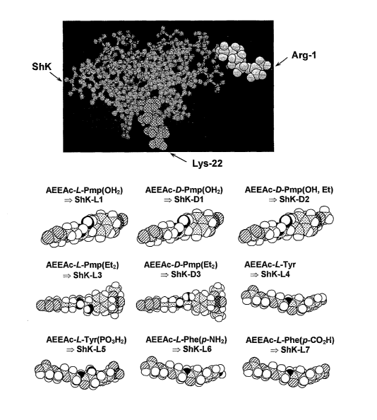
Figure 1 shows the chemical structures of a number of ShK analogs of
the present invention.
Figure 2A shows a molecular model of ShK based on the published
NMR structure wherein the Lys22, critical for channel blockade, is highlighted
in one shade of grey. L-pTyr was attached to the a-amino group of Argl of
ShK (highlighted in a second shade of grey) through an Aeea linker (right).
The structures of the linker and L-pTyr were modeled with AM1 in
5
CA 02582149 2007-03-27
WO 2006/042151
PCT/US2005/036234
Hyperchem.
Figure 2B shows the effect of ShK (top) and ShK(L5) (bottom) on Kv1.3
and Kv1.1 currents in stably transfected cells.
Figure 2C shows dose-dependent inhibition of Kv1.3 (open symbols)
and Kv1.1 (closed symbols) by ShK (dark) and ShK(L5) (light). Kds on Kv1.3
=10 1 pM (ShK) and 69 5 pM (ShK(L5)); Kds on Kv1.1 = 28 6 pM (ShK)
and 7.4 0.8 nM (ShK(L5)).
Figure 2D shows the time course of wash-in and wash-out of ShK(L5)
on Kv1.3 wherein cells were held at a holding potential of 80 mV and
depolarized for 200 msec to 40 mV every 30 secs.
Figure 2E shows Kd values for inhibition of Kv1.3 and Kv1.1 by ShK
analogs. Kds for ShK-F6CA and ShK-Dap22 based on published sources.
Figure 3A is a graph showing staining intensities of CD45RA and
CCR7 as determined by flow cytometry in the CD3-gated population of
human PBMCs stained with antibodies against CD3, CD45RA and CCR7.
Figure 3B is a graph showing staining intensities of CD45RA and
CCR7 as determined by flow cytometry in the CD3-gated population in cells
of a human TEm line stained with antibodies against CD3, CD45RA and
CCR7.
Figure 3C is a graph showing the inhibitory effects of ShK (dark grey)
and ShK(L5) (light grey) of [3F1] thymidine incorporation by PBMCs (open
symbols, a mixture of naIveffcm cells) and TEm cells (closed symbols)
stimulated for 48 hours with anti-CD3 antibody.
Figure 3D is a graphic showing of pre-activated human PBMCs
(naIve/Tcm cells) that up-regulate KCa3.1 expression become resistant to
ShK(L5) inhibition when reactivated with anti-CD3 antibody. These cells have
previously been reported to become sensitive to the Kca3.1-specific inhibitor
TRAM-34.
6
CA 02582149 2007-03-27
WO 2006/042151
PCT/US2005/036234
Figure 4A is a graph showing CD45RC staining of rat splenic T cells
(left) and PAS T cells (right) detected by flow cytometry.
Figure 4B is a graphic showing of Kv1.3 currents exhibited by
quiescent (top) and myelin antigen-activated (bottom) PAS T cells.
Figure 4C provides a graphic representation of flow cytometry profiles
of ShK-F6CA-staining in quiescent (top) and myelin antigen-activated
(bottom) PAS T cells. Unstained cells (black lines) and cells stained with
ShK-F6CA (area filled in light grey). Competition of ShK-F6CA staining by
unlabeled ShK(L5) is represented by the area filled in dark grey.
Figure 4D shows confocal images of Ky1.3 immunostaining in
quiescent (top) and myelin antigen-activated (bottom) PAS T cells. Statistical
analysis was carried out using the Mann-Whitney U-test.
Figure 4E shows dose-dependent inhibition by ShK (dark lines) and
ShK(L5) (light lines) of [3F1] thymidine incorporation by rat (left) naTve/Tcm
(open symbols) and TEm (closed symbols) cells activated with Con A (1
pg/ml).
Figure 4F shows dose-dependent inhibition by ShK (dark lines) and
ShK(L5) (light lines) of IL2 secretion by PAS T cells 7 hours after
stimulation
with MBP. G, ShK(L5)-induced inhibition of myelin-antigen triggered [31-I]
thymidine incorporation by PAS T cells (open symbols) is reversed by the
addition of 20 u/ml IL2 (closed symbols).
Figure 5A is a graph showing Kv1.3 blocking blocking activity of
ShK(L5) as determined on Kv1.3 channels stably expressed in L929 cells.
Figure 5B is a graph showing blood levels of ShK(L5) at various times
after a single subcutaneous injection of 200 mg/kg of ShK(L5) in four rats.
Blood was drawn at the indicated times and serum was tested by patch-clamp
to determine the amount of ShK(L5).
Figure 5C is a graph of the data of Figure 5B fitted to a single
exponential decay indicating a half-life of approximately 50 minutes.
7
CA 02582149 2007-03-27
WO 2006/042151
PCT/US2005/036234
Figure 5D is a graph showing blood levels of ShK(L5) in five Lewis rats
receiving single daily subcutaneous injections of 10 ig/kg/day ShK(L5) for 5
days. Blood was drawn each morning (24 hours after the previous injection)
and tested for blocking activity on Kv1.3 channels by patch-clamp.
Figure 5E is a graph showing serum levels of ShK(L5) in rats at various
times following a single dose of 10 mg/kg ShK(L5) either subcutaneously
(open bars; n = 4) or intravenously (closed bars; n = 4). Blood was drawn at
the indicated times and serum was tested by patch-clamp to determine the
amount of ShK(L5) in blood. ShK(L5) maintained a steady-state level of 300
pM in the blood almost 24 hourse after a single subcutaneous injection. This
concentration is sufficient to selctively inhibit the function of TEM cells.
Figure 5F is a graph showing the % recovery of ShK(L5) after a half-
blocking dose of ShK(L5) was added to rat plasma or PBS containing 2% rat
plasma and incubated at 37 C for varying duration. Aliquots were taken at the
indicated times and blocking activity determined on Kv1.3 channels. ShK(L5)
is extremely stable in plasma.
Figure 6A is a graph showing scored prevention of EAE. PAS T cells
were activated in vitro, washed, and injected intraperitoneally on day 0.
Clinical scoring of EAE: 0= no clinical signs, 0.5 = distal limps tail, 1 =
limp tail,
2 = mild paraparesis or ataxia, 3 = moderate paraparesis, 4 = complete hind
limb paralysis, 5 = 4 + incontinence, 6 = death. Rats (n = 6/group) were
injected subcutaneous with vehicle alone (n = 6) or ShK(L5) (n = 6;
10mg/kg/day) from day 0 to day 5.
Figure 6B is a graph showing scored treatment of EAE. PAS T cells
were activated in vitro, washed, and injected intraperitoneally on day 0.
Treatment with ShK(L5) at 10mg/kg/day was started when rats developed
clinical signs of EAE and was continued for 3 days.
Figure 60 is a graph showing ear thicknes as an indicator of DTH
reaction elicited against ovalbumin in rats. Animals (n = 6/group) were
treated
with ShK(L5) 10 mg/kg/day for 2 days, after which ear swelling was
measured. Statistical analysis was carried out using the Mann-Whitney U-test.
8
CA 02582149 2007-03-27
WO 2006/042151
PCT/US2005/036234
Figure 7A shows the ShK(L5) structure and a graph showing inhibition
of Kv1.3 channels in TEm cells as a function of ShK(L5) concentration. Each
data-point represents mean of three determinations.
Figure 7B is a diagram of Kv1.3-containing signaling complex.
Figure 7C shows co-localization of CD4, Kv1.3, Kv132, SAP97, ZIP and
p56Ick at IS.
Figure 7D shows CD4 and Kv1.3 staining in absence of visible TEM'
APC contact.
Figure 7E shows CD4 and Kv1.3 staining in GAD65-specific TEM cells
exposed to MBP-loaded APCs.
Figure 7F shows that ShK(L5) 100 nM does not prevent IS formation.
Figure 7G shows that ShK(L5) 100 nM does not disrupt the IS.
Figure 8A is a graphic showing of calcium signaling in GAD-specific
TEm cells from three TI DM patients triggered by anti-CD3 + cross-linking
secondary antibodies (arrow) in the absence (black) or presence of ShK(L5)
0.1 nM (dark grey), 1 nM (medium grey) or 100 nM (light grey).
Figure 8B is a graph showing [31-1]-thymidine incorporation by naIve/Tcm
and TEm cells (left) and naive/Tcm-effectors and TEm-effectors from patients
with TI DM and RA (right). TEm cells: GAD65-activated TEm clones from three
TI DM patients and anti-CD3 antibody activated SF-TEm cells from three RA
patients. NaIve/Tcm cells: anti-CD3 antibody-activated PB-naIve/Tcm cells
from the same three RA patients.
Figure 8C is a series of bar graphs showing Cytokine production by the
TEm and naTve/Tcm cells used in Figure 8B.
Figure 8D shows the phenotype of disease-relevant and disease-
irrelevant autoreactive T cells in MS, TI DM and RA.
Figure 8E is a diagram showing the manner in which ShK(L5) inhibits
calcium signaling, lymphocyte proliferation and cytokine production but not IS
9
CA 02582149 2007-03-27
WO 2006/042151
PCT/US2005/036234
formation.
Figure 9 is a diagram representing a rat model of delayed type
hypersensitivity (DTH) caused by effector memory T cells.
Figure 10 is a diagram showing a treatment protocol for ShK(L5) in a
rat model of delayed type hypersensitivity (DTH) caused by effector memory T
cells
Figure 11 is a diagram represneting specific suppression of effector
memory responses in vivo in rats by ShK(L5) without impairing the function of
naive and central memory T cells or B cells.
Figure 12A shows Kv1.3 currents (top) and channel number/cell
(bottom) in GAD65-, insulin and myelin-specific T cells from patients with new
onset type-1 diabetes mellitus (TI DM), health controls and patients with
multiple sclerosis..
Figure 12B shows Kv1.3 staining (top) and fluorescence intensities of
individual T cells (bottom) from these patients.
Figure 12C shows graphs of relative cell number vs. CCR7 staining
intensity. Cells expressing high levels of Kv1.3 are CCR7-negative i.e. they
are TEm-effectors. Cells expressing low levels of Kv1.3 are CCR&-positive i.e.
they are either naïve or Tcm cells
Figure 12D shows Kv1.3 number/cell in autoreactive T cells from a
patient having T1DM and MS (left), patients having T1DM for greater than 5
years duration (middle) and patients having non-autoimmune type-2 DM.
Figure 12E shows Kv1.3 numbers in CD4+GAD65-tetramer+ T cells
from a patient with new-onset TI DM.
Figure 13A shows Kv1.3 channel numbers per cellin peripheral T cells
blood and synovial fluid T cellsof RA patients and synovial fluid T cellsof OA
patients.
Figure 13B shows confocal images of Kv1.3 (light grey) and Kv62
(darker grey) staining in the cells shown in Figure 13A.
CA 02582149 2007-03-27
WO 2006/042151
PCT/US2005/036234
Figure 13C shows graphs of relative cell number vs. CCR7 staining
intensity.
Figure 13D shows micrographs (top) and bar graphs of inflammatory
index (bottom) of synovium from RA and OA patients stained with anti-CD3 or
anti-Kv1.3 antibodies and counter-stained with hematoxylin/eosin (40X).
DETAILED DESCRIPTION
The following detailed description and the accompanying drawings are
intended to describe some, but not necessarily all, examples or embodiments
of the invention only. This detailed description and the accompanying
drawings do not limit the scope of the invention in any way.
The present invention provides novel analogs of ShK, methods for
making such compositions and methods for using such compositions to inhibit
Kv1.3 channels (or other ion channels) in human or animal cells and for
treatment or prevention of diseases and disorders, such as T cell mediated
autoimmune disorders. The compositions of the present invention comprise
ShK toxin attached (e.g., bound, linked by a linker or otherwise associated
with) to an organic or inorganic, anionic-charged chemical entity (e.g. an
atom, molecule, group, residue, compound, moiety, etc.). In at least some
embodiments of the invention, the organic or inorganic, anionic-charged
chemical entity may be selected to increase or optimize the affinity of the
composition for inhibition of Kv1.3 channels over Kv1.1 channels. Examples
of organic or inorganic, anionic-charged molecules or groups that may be
linked or bound to ShK in accordance with the present invention include but
are not necessarily limited to:
amino acids;
polypeptides;
amino acid residues;
unnatural amino acid residues;
threonine;
threonine derivatives;
11
CA 02582149 2014-06-27
phospho-threonine;
serine;
serine derivatives;
phospho-serine;
glutamic acid;
glutamic acid derivatives;
gammacarboxy-glutamic acid;
aspartic acid;
aspartic acid derivatives;
inorganic compounds or groups;
organic compounds or groups;
succinic anhydride; and
phthalic anhydride.
In accordance with the present invention, some non-limiting examples
of compositions of the present invention, wherein the anionic-charged
chemical entity comprises an amino acid residue, are shown in Figures 1 and
2C and referred to herein by alphaneumeric designations, as shown in Table
1 below:
Table 1
DESIGNATION AMINO ACID
RESIDUE BOUND TO
ShK AT POSITION 2
ShK-L1 AEEAc-L-Pmp(0F12)
ShK-D1 AEEAc-D-Pmp (0H2)
ShK-D2 AEEAc-D-Pmp(OH, Et)
ShK-L3 AEEAc-L-Pmp(Et2)
ShK-D3 AEEAc-D- Pmp(Et2)
ShK-L4 AEEAc-L-Tyr
ShK-L5 AEEAc-L-Tyr(P03 H2)
ShK-L6 AEEAc-L-Phe(p-NH2)
ShK-L7 AEEAc-L-Phe(p-CO2H)
12
CA 02582149 2007-03-27
WO 2006/042151
PCT/US2005/036234
With specific reference to Figure 1, tyrosine or phenylalanine or their
charged non-natural derivatives were conjugated to ShK (top left) through a
linker attached on its N terminus (residue Arg1 shown in shaded grey). The
Lys22, required for channel blockade, is shown in a darker shade of grey. The
molecular model of ShK is based on the published NMR structure and the
structures of the linker and the new residues were modeled. These
embodiments of compositions of the present invention generally comprise the
ShK toxin, which is a polypeptide, bound to (e.g., chemically bound, linked or
otherwise associated with) at least one anionic-charged amino acid residue.
In embodiments where the aminio acid residue has a chiral center, the D
and/or L enantiomer of such amino acid residue may be used. The anionic-
charged amino acid residue may be an unnatural residue and may be
attached or linked to an N-terminus of the ShK polypeptide. In some
embodiments, the anionic-charged amino acid residue may be linked to an N
terminus of ShK through a linker, such as an aminoethyloxyethyloxy-acetyl
linker. These analogs of ShK inhibit the Kv1.3 channel more specifically than
ShK because they have reduced affinity for other potassium channels (e.g.,
Kv1.1). The ShK may be isolated from natural sources as known in the art,
or it may be synthesized.
Synthesis of ShK Toxin
ShK Toxin may be synthesized by any suitable method. In one such
method, Fmoc-amino acids (Bachem Feinchemikalien) including Arg(Pmc),
Asp(OtBu), Cys(Trt), Gln(Trt), His(Trt), Lys(Boc), Ser(tBu) and Thr(tBu) are
obtained commercially and assembled to form ShK Toxin. Stepwise assembly
of the amino acids may be carried out on an Applied Biosystems 431A peptide
synthesizer at the 0.25 mmol scale starting with Fmoc-Cys(Trt)-R. Residues 34
through 22 are single coupled. Thereafter, an aliquot (e.g., half) of the
resin is
removed to effect better mixing. The remainder of the peptide sequence is then
double coupled to the remaining resin aliquot All couplings are mediated by
dicyclohexylcarbodiimide in the presence of 2 eq of 1-hydroxybenzotriazole.
The
13
CA 02582149 2012-12-11
final two residues are also coupled via HBTU/DIEA chemistry. These residues
are Aeea (Fmoc-aminoethyloxyethyloxyacetic acid) and as the N-terminal
residue Fmoc-Tyr (PO4) monobenzyl ester. Following final removal of the
Fmoc-group, the peptide resin (2.42 g) is cleaved from the resin and
simultaneously deprotected using reagent K for 2 h at room temperature.
Reagent K is known in the art and has been described in the literature. See,
King, D.S., Fields, C.G. and Fields, G.B. (1990) Int. J. Peptide Protein Res.
36, 255-266. Following cleavage, the peptide is filtered to remove the spent
resin beads and precipitated with ice cold diethyl ether. The peptide is then
collected on a fine filter funnel, washed with ice cold ether and finally
extracted
with 20% AcOH in H20. The peptide extract is subsequently diluted into 2
liters
of H20, the pH is adjusted to 8.0 with NH4OH and allowed to air oxidize at
room
temperature for 36 hours. Following oxidation of the disulfide bonds with a
2:1
ratio of reduced to oxidized glutathione, the peptide solution is acidified to
pH
2.5 and pumped onto a RaininTM DynamaxTM C18 column (5.0 x 30 cm). The Sample
is eluted with a linear gradient from 5-30% acetonitrile into H20 containing
0.1%
TFA. The resulting fractions are analyzed using two analytical RP-HPLC
systems: TFA and TEAP. Pure fractions are pooled and lyophilized. (See,
Pennington, M.W., Byrnes, M.E., Zayden berg, I., Khaytin, I., de Chastonay,
J.,
Krafte, D., Hill, R., Mahnir, V., Volberg, W.A., Gorczyca, W. and Kern, W.R.
(1995) mt. J. Peptide Protein Res. 46, 354-358.)
Alternatively, solid-phase peptide synthesis employing a Boc-BzI
protecting group strategy may be utilized to assemble the primary structure as
well as analogs of the peptide. The peptide could then be cleaved from the
solid-phase by anhydrous HF, yielding the linear peptide ready for folding as
described above for the Fmoc synthesized peptide. (See, Stewart, J.M. and
Young J.D. (1984) Solid Phase Peptide Synthesis. 2nd Edition. Pierce
Chemical Company. Rockford, II.)
14
CA 02582149 2007-03-27
WO 2006/042151
PCT/US2005/036234
Alternatively, other synthetic methods to assemble the primary structure
of ShK or analogs could include chemical ligation technology where the peptide
is prepared as a series of designed fragments with C-terminal thioester
peptides. The the thioester peptide can react with another peptide containing
an
N-terminal Cys residue to form a peptide containing a native peptide bond. By
using this technology, one could effectively assemble the primary structure of
ShK. (See, (4) Wilken, J. and Kent S.B.H. (1998) Chemical protein synthesis.
Current Opin. Biotech. 9, 412-426.)
Alternatively, another synthetic method that may be employed to
assemble the primary structure of ShK would utilize a protected peptide
fragment convergent approgch as described in Albericio, F., Lloyd-Williams,
P.,
and Giralt, E. (1997) Convergent peptide synthesis; in Methods in Enzymol.
Ed G. Fields, Academic Press, New York, NY. pp 313-335. In this method,
linear protected fragments are assembled as fully side chain protected
fragments. These fragments can then be coupled together in a convergent
manner to assemble the primary sequence of ShK or one of its analogs.
Assembly of the fragments could also utilize a solid-phase resin to facilitate
coupling and wash steps.
Alternatively, recombinant methods may be used wherein the cDNA
coding sequence for ShK could be generated for expression in either a
prokaryotic or eucaryotic expression system. Recombinant ShK analogs
containing unnatural amino acids are also possible by utilizing preload tRNA
molecules which utilize non-standard condons. The cCNA construct can be
engineered to use one of these unused codons to add the phosphotyrosine
residue as well as the Aeea residue. Folding of the recombinantly produced ShK
analog could then be accomplished in a similar method to that used for the
synthetic peptides. (See, Pennington, M.W., Byrnes, M.E., Zaydenberg, I.,
Khaytin, I., de Chastonay, J., Krafte, D., Hill, R., Mahnir, V., Volberg,
W.A.,
Gorczyca, W. and Kern, W.R. (1995) Int J. Peptide Protein Res. 46, 354-
358.)
CA 02582149 2007-03-27
WO 2006/042151
PCT/US2005/036234
Attaching Anionic Amino Acid Residues To ShK and Optional
Modifications to ShK
Anionic amino acid residues may be attached to the N terminus of natural
or synthetic ShK Toxin by way of a linker, such as an aminoethyloxyethyloxy-
acetyl linker, or bay any other suitable means. In this example, the nine (9)
ShK analogs shown in Figure 1 are prepared. Initially, Fmoc-Aeea-OH is
coupled to the N-terminus of synthetic ShK Toxin assembled as described
above. The resin is then divided into 9 aliquots. Either Fmoc-Tyr(PO4Bz1)-
OH, Fmoc-d-Tyr(PO4Bz1)-0H, Fmoc-Tyr(PO4Me2)-0H, Fmoc-Pmp-OH, Fmoc-
d-Pmp-OH, Fmoc-Pmp(Et)-0H, Fmoc-Pmp(Et)2-0H, Fmoc-Tyr(tBu)-0H, or
Fmoc-Amp(Boc)-OH is then coupled using DIC and HOBT to one of the resin
aliquots. The deblocked peptide resin is then cleaved and deprotected with
reagent K (King et al., 1990) containing 5% triisopropylsilane for 2 h at RT.
Met(0) is reduced by addition of solid NH4I to the cleavage cocktail at t-15
min. (Nicolas et al., 1995). For the peptide containing Tyr(PO4Me2)-0H, a
cleavage cocktail containing 1 M TMSBr in TFA containing thioanisole as a
scavenger for 18 hr at 4 C was used (Tian et al., 1993). Incomplete removal
of the methyl protecting groups is common when using this method and two of
the species (Tyr(PO4) and Tyr(PO4Me)) are easily purified by RP-HPLC. The
Tyr(PO4Me2) containing analog is cleaved via standard Reagent K cleavage
keeping both Me groups intact. In each case, the cleavage mixture is filtered
and the crude peptide is precipitated into ice-cold diethyl ether. The
precipitate is collected, yielding approximately 75 mg of peptide from 200 mg
of resin. The crude product is dissolved in 20 ml of 50% aqueous AcOH and
diluted into 0.75 I of H20. The pH of the solution is adjusted with NH4OH to
8.2, and it was allowed to fold overnight with the addition of glutathione
(2mM:1mM) (reduced:oxidized). All analogs are purified using RP-HPLC as
described previously (Pennington et al., 1995; Pennington et al., 1996a;
Pennington et al., 1996b). Pure fractions are pooled and lyophilized. Each
sample is confirmed by RP-HPLC, AM and MALDI-TOF MS and adjusted to
account for peptide content prior to bioassay.
In some embodiments of the invention, to improve the PK/PD
properties of the ShK structure, residues which are sensitive to degradation
16
CA 02582149 2007-03-27
WO 2006/042151
PCT/US2005/036234
properties may be replaced or substituted. Thus, substitution of the Met
residue at position 21 may be carried out to impart a stabilizing effect to
oxidation. Additionally, substitution of the C-temrinal acid function with an
amide will impart stability to C-terminal corboxypeptidase enzymes. These
two substitions to the primary structure of ShK combined with the anionic
moiety at the N-terminus have been synthesized to generate the most stable
and selective Kv1.3 blocker. Nonhydrolyzable phosphate substitutions will
also impart a stabilzing effect versus acid and basic hydrolysis of the
phosphate as well as stability against phosphatase enzymes. The
substitutions are summarized below. The acronyms used are defined as
follows: Pmp=p-phosphonomethyl-Phenylalanine; Ppa=p-
Phosphatityl-
Phenylalanine and Nle=Norleucine.
Substitutions:
p-phospho-Tyr-Aeea-ShK-Nle21-Cys35-amide
p-Phosphono-methyl-Phenylalanine-Aeea-ShK-N1e21-
Cys35amide (Pmp)
p-Phosphatityl-Phe-Aeea-ShK-Nle21-Cys35-amide (Ppa)
p-phospho-Tyr-Aeea-ShK-Nle21-Cys35-acid
p-Phosphono-methyl-Phenylalanine-Aeea-ShK-Nle21-
Cys35acid (Pmp)
p-Phosphatityl-Phe-Aeea-ShK-Nle21-Cys35-acid (Ppa)
In addition to the nonhydrolyzable Pmp and Ppa, substitution of p-
Phosphono(difluoro-methyl)-Phenylalanine (Pfp) and p-Phosphono-
methylketo-Phenylalanine (Pkp) are also anionic substituions, providing the
following:
Pfp-Aeea-Shk-N1e21 Cys35 amide
Pkp-Aeea-ShK-Nle21-Cys35 amide
Pfp-Aeea-Shk-N1e21 Cys35 acid
Pkp-Aeea-ShK-Nle21-Cys35 acid.
17
CA 02582149 2012-12-11
Structures of the N-terminal substitutions are set forth in Appendix B. Other
structures that are within the scope of the present invention are published in
Beeton, C. et al., Targeting Effector Memory T Cells with a Selective Peptide
Inhibitor of Kv1.3 Channels for Therapy of Autoimmune Diseases, Molecular
Pharmacology, Vol. 67, No.4, 1369- (2005).
Therapeutic Uses of ShK Analogs of the Present Invention
The present invention provides methods for treating or preventing
certain disorders or diseases, such as T cell mediated disorders (e.g.,
autoimmune disorders, graft vs. host disease, prevention of rejection of organ
transplants etc.), other inflammatory disorders, obesity and Type 2 diabetes,
in human or animal subjects by administering to the subject a therapeutically
effective (e.g., preventative or effective to reduce or eliminate symptoms or
disease progression) amount of a pharmaceutically acceptable preparation
consisting or comprising an ShK analog of the present invention (e.g.,
including but not limited to those listed in Table 1 hereabove). Any suitable
route of administration (e.g., oral, rectal, intravenous, intramuscular,
subcutaneous, intradermal, intranasal, topical, transmucosal, transdermal, by
drug delivery implant, etc.) may be used. When used to prevent or treat a T
cell mediated disorder, the dosage(s) will be sufficient to inhibit Kv1.3
channels on T cell membranes. In this regard, the ShK analogs of the present
invention have the potential to be used to prevent or treat a wide variety of
a T
cell mediated autoimmune disorders. The following are some examples of
some T cell mediated autoimmune diseases that may be prevented or treated
by the methods of the present invention, categorized with respect to the
target
organ that is principally affected by each such disease:
Nervous System: Gastrointestinal Tract:
Multiple sclerosis Crohn's Disease
Myasthenia gravis Ulcerative colitis
Autoimmune neuropathies Primary biliary cirrhosis
such as Guillain-Barre Autoimmune hepatitis
Autoimmune uveitis Bone resorption associated
with periodontal disease
18
CA 02582149 2007-03-27
WO 2006/042151
PCT/US2005/036234
Blood:
Endocrine:
Autoimmune hemolytic anemia Type 1 diabetes mellitus
Pernicious anemia Addison's Disease
Autoimmune Grave's Disease
Thrombocytopenia
Hashimoto's thyroiditis
Vascular: Autoimmune oophoritis and
Temporal arteritis orchitis
Anti-phospholipid syndrome
Vasculitides such as
Wegener's granulomatosis
Behcers disease
Multiple Organs and/or
Musculoskeletal System:
Rheumatoid arthritis (RA)
Osteoarthritis (OA)
Skin: Systemic lupus erythematosus
Psoriasis Scleroderma
Dermatitis herpetiformis Polymyositis, dermatomyositis
Pemphigus vulgaris Spondyloarthropathies such as
Vitiligo ankylosing spondylitis
Sjogren's syndrome
Irrespective of the particular organ(s) affected, T-lymphocytes are
believed to contribute to the development of autoimmune diseases. The
currently available therapies for these diseases are largely unsatisfactory
and
typically involve the use of glucocorticoids (e.g. methylprednisolone,
prednisone), non-steroidal anti-inflammatory agents, gold salts, methotrexate,
antimalarials, and other immunosuppressants such as cyclosporin and FK-
506. Also, another T cell mediated disorder that may be prevented or treated
by the methods of the present invention is graft vs. host disease and/or
rejection of transplanted organs. Indeed, the outcomes of organ transplant
procedures have progressively improved with the development of refinements
in tissue typing, surgical techniques, and more effective immunosuppressive
treatments. However, rejection of transplanted organs remains a major
problem. T-lymphocytes play a central role in the immune response and they
are responsible, in large measure, for the rejection of many transplanted
organs. They are also responsible for the so-called graft-versus host disease
19
CA 02582149 2007-03-27
WO 2006/042151
PCT/US2005/036234
in which transplanted bone marrow cells recognize and destroy MHC-
mismatched host tissues. Accordingly, drugs such as cyclosporin and FK506
that suppress T-cell immunity are used to prevent transplant rejection and
graft-versus-host disease. Unfortunately, these T cell inhibiting drugs are
toxic, with liver and renal toxicities limiting their use. Thus, the methods
of the
present invention may provide les toxic alternatives for the treatment or
prevention of graft vs. host disease or transplant rejection. Also, inhibitors
of
the voltage gated Kv1.3 potassium channel have been shown to be especially
effective in suppressing effector memory T cells and, thus, the methods of
present invention may be particularly effective in preventing or treating
diseases that are associated with effector memory T cells, such as; bone
resorption and periodontal disease, psoriasis, rheumatoid arthritis, diabetes
mellitus and multiple sclerosis. In addition to T cell mediated diseases, the
Kv1.3 channel has been determined to regulate energy homeostasis, body
weight and peripheral insulin sensitivity. Thus, the methods of the present
invention may be used to treat other diseases and disorders that involve
abnormal homeostasis, body weight and peripheral insulin sensitivity by
inhibiting Kv1.3 channels on cell membranes, such other diseases and
disorders include but are not necessarily limited to bone resorption in
periodontal disease, Type 2 diabetes, metabolic syndrome and obesity.
Use of ShK Analogs of the Present Invention in Flow Cvtometne
Further in accordance with the present invention there are provided
methods for diagnosing T cell mediated disorders or otherwise sorting or
distinguishing between various cell types in vitro using fluorophore tagged
versions of ShK(L5) for use in flow cytometry alone, or in conjunction with
class II tetramers that can detect autoreactive cells. Flow Cytometry is a
flexible method for characterizing cells in suspension wherein fluorescence
activated cell sorting is used to select living cells on the basis of
characteristics measured by flow cytometry. The types of cellular features and
functions that may be detected by flow cytometry include the expression of
proteins outside and within cells, type of DNA content, viability and
apoptosis,
multiple drug resistance pump activity, enzyme activity, T-cell activation, 1-
cell
receptor specificity, cytokine expression, phagocytosis and oxidative burst
CA 02582149 2007-03-27
WO 2006/042151
PCT/US2005/036234
activity. Thus, in this method of the present invention, the amino acid
residue
attached to the ShK may incorporate a fluorophore tag for use in flow
cytometry alone, or in conjunction with class II tetramers loaded with
specific
autoantigens that can detect autoreactive cells. Specific descriptions of the
methods by which such flow cytometry may be carried out are described in
Beeton, C., et al., A Novel Fluorescent Toxin to Detect and Investigate Kv1.3
Channel Up-Regulation in Chronically Activated T Lymhocytes; J.Biol.Chem.,
Vol. 278, No. 11, 9928-9937 (March 2003). In general, a flow cytometer uses
focused laser light to illuminate cells as they pass the laser beam in a fluid
stream. Light scattered by the cells and light emitted by fluorescent dyes
attached to cells of interest are analyzed by several detectors and processed
by a computer. Cells may be distinguished and selected on the basis of size
and shape as well as by the presence of many different molecules inside and
on the surface of the cells.
Examples of Potassium Channel Inhibiting Effects and Therapeutic
Utility of ShK Analogs of the Present Invention
ShK blocks the neuronal Kv1.1 channel and the Kv1.3 channel with
roughly equivalent potency. Neurotoxicity is therefore a concern under
circumstances that compromise the blood-brain-barrier and allow the entry of
sufficient amounts of ShK to block Kv1.1 channels. Our strategy to design a
Kv1.3-specific inhibitor was guided by our finding that ShK-F6CA containing
fluorescein-6-carboxylate (F6CA) attached through a 20 A-long Aeea linker to
the N-terminus of ShK exhibited 80-fold selectivity for Kv1.3 over Kv1.1
(Beeton et al., 2003). Since F6CA can exist as a restricted carboxylate or
also
as a cyclized lactone, it was not clear whether ShK-F6CA's Kv1.3 specificity
was due to the negative charge of F6CA, the hydrophobicity created by this
large bulky fluorescein nucleus, potential planar -p electronic stacking or a
combination of all of these potential contributions. To distinguish between
these possibilities and with the intention of developing a non-fluorescent
Kv1.3-selective inhibitor, we generated a series of 12 novel N-terminally-
substituted ShK analogs to probe some of these interactions. By attaching
tyrosine, phenylalanine or their derivatives (varying in charge, size and
hydrophobicity) through an Aeea linker to the N-terminus of ShK, we could
21
CA 02582149 2014-06-27
probe the effects of charge and hydrophobicity to gain insight into our
selectivity enhancement seen with F6CA substitution.
Selective KVIv1.3 Inhibition over Kv1.1 Inhibition:
In the example shown in Figures 2A-2D, L-phosphotyrosine (L-pTyr) a
negatively charged (net charge 2) post-translationally modified aromatic
amino acid, was attached via the AEEA linker to ShK-Argl to generate the
novel analog ShK(L5). The ShK toxin and ShK(L5) were tested on Kv1.3 and
Kv1.1 channels stably expressed in L929 cells. Figure 2B shows the effects of
ShK and ShK(L5) on Kv1.3 and Kv1.1 currents elicited by 200 ms
depolarizing pulses from a holding potential of 80 mV to 40 mV. Both peptides
reversibly blocked Kv1.3 and Kv1.1 in a dose-dependent manner with Hill
coefficients of 1. Kds were determined from the dose-response curves shown
using Microcal Origin software. ShK blocked Kv1.3 (Kd = 10 1 pM) and
Kv1.1 (Kd = 28 6 pM) with roughly equivalent potency as expected (Fig. 1C).
In contrast, ShK(L5) was 100-fold selective for Kv1.3 (Kd = 69 5 pM) over
Kv1.1 (Kd = 7.4 0.8 nM) (Figs. 1B, 1C). The time course of Kv1.3 current
block by ShK(L5) and its washout is shown in Figure 1D. The time constant
(ToN) of ShK(L5) wash-in was 131 21 sec (n = 7) while the time constant
(TOFF) for peptide wash-out was 150 28 sec (n = 4). The Kd (57 7 pM)
calculated from the KoN (15 x 106 0.5 x 10⁶ M-1sec-1) and KoFF (0.0059
0.0013 sec-1) values is consistent with the Kd (69 5 pM) determined with
Microcal Origin software.
Other ShK analogs were also tested on Kv1.3 and Kv1.1 channels.
ShK(D5) containing D-phosphotyrosine (D-pTyr) was 35-fold selective for
Kv1.3 over Kv1.1 but was an order of magnitude less potent than ShK(L5).
ShK (L8) containing L-pTyr-monomethyl showed modest (11-fold) Kv1.3
specificity, while ShK analogs containing L-pTyr-dimethyl or L-Tyr were not
selective for Kv1.3 over Kv1.1. Analogs that contained phenylalanine or its
derivatives (varying in bulk, p electron density and charge) were modestly
specific or not specific for Kv1.3 over Kv1.1. ShK(L5)'s 100-fold specificity
for
Kv1.3 over Kv1.1 is greater than that of ShK-F6CA (80-fold), ShK(D5) (35-
fold), ShK-Dap22 (33-fold) or any other ShK analog tested.
22
CA 02582149 2007-03-27
WO 2006/042151 PCT/US2005/036234
Applicants also assessed ShK(L5)'s specificity on a panel of 20 ion
channels and these data are summarized in the following Table 2:
Channels Kd of ShK(L5) (pM]
Kv1.1 7,000z 1,000
Kv1.2 48,000 t 7,000
Kv1.3 (cloned) 69 5
Kv1.3 (native) 76 1 8
Kv1.4 137,000+ 3,000
Kv1.5 100,000 no effect
Kv1.6 18,0001 3,000
Kv1.7 100,00000 effect
Kv2.1 100,00000 effect
Kv3.1 100,00000 effect
Kv3.2 20,000 2,000
K1r2.1 100,000 no effect
Kv11.1 (HERG) 100,000 no effect
1(0,1.1 100,000 no effect
Kca2.1 100,000 no effect
Kca2.3 100,000 no effect
Kca3.1 115,000 5,000
Nav1.2 100,000 no effect
Nav1.4 100,000 no effect
Swelling-activated T cell 100,000 no effect
Cl channel
Cav1.2 100,000 no effect
As may be appreciated from the data of Table 2 above, ShK(L5) blocked the
Kv1.3 channel in T cells with a Kd (76 pM) equivalent to its Kd on the cloned
channel (69 pM). It was 100-fold selective for Kv1.3 over Kv1.1, 260-fold
selective over Kv1.6, 280-fold selective over Kv3.2, 680-fold selective over
Kv1.2 and >1000-fold selective over all other channels tested. Importantly, it
was 1600-fold Kv1,3-selective over KCa3.1, the calcium-activated K+ channel
that regulates activation of human naïve and Tcm cells (Wulff et al., 2003).
Native ShK was less selective than ShK(L5). ShK was 2.8-fold selective for
Kv1.3 (Kd = 10 1 pM) over Kv1.1 (Kd 28 6 pM), 20-fold selective over
Kv1.6 (200 20 pM), 500-fold selective over Kv3.2 (Kd = 5,000 1,000 PM),
and >1000-fold selective-over Kv1.2 (10 1 nM) and KCa3.1 (Kd = 28 3
nM). Margatoxin, a peptide from scorpion venom that has been touted as a
specific Kv1.3 inhibitor (Koo et al., 1997; Lin et al, 1993; Middleton et al.,
2003) was also not specific. It was 5-fold selective for Kv1.3 (110 12 pM)
over Kv1.2 (Kd =520 1 pM), 9-fold selective over Kv1.1 (10 1 nM) and >
1000-fold selective over Kv1.6 and Kv3.2 (Kd > 100 nM). Luteolin, a
Field Code Changed
nutriceutical sold for autoimmune diseases (www.lutimax.corn) on the basis of -
-
it being a Kv1.3 inhibitor (Lahey and Rajadhyaksha, 2004), blocked Kv1.3
23
CA 02582149 2007-03-27
WO 2006/042151
PCT/US2005/036234
weakly (Ka = 65 5 mM) and exhibited no selectivity over Kv1.1 (Kd-77
5mM), Kv1.2 (Kd = 63 4 mM) or Kv1.5 (Kd = 41 3 mM). ShK(L5)'s
exquisite specificity for Kv1.3 together with its picomolar affinity for the
channel makes it a potentially attractive immunosuppressant.
Preferential Suppression of Human TEm cell Proliferation
With reference to Figures 3A-3D, in order to assess ShK(L5)'s in vitro
immunosuppressive activity, Applicants compared its ability to suppress anti-
CD3 antibody-stimulated proliferation of human TEm cell lines versus human
PBMCs that contain a mixture of naïve and Tcm cells. Flow cytometry
confirmed the cell surface phenotypes of the two populations studied. As
seen in Figure 3A, the TEm lines were >90% CCRTCD45RA, while as shown
in Figure 3B the PBMCs contained 65 % CCR7+CD45RA+ (naïve) and 18%
CCR7+CD45RA" (Tcm) cells. Figure 3C shows that ShK(L5) and ShK were
60-fold more effective in suppressing the proliferation of TEm cells (IC50 = -
80
pM) compared with PBMCs (IC50 = 5 nM, p <0.05). The lower sensitivity of
PBMCs might be explained by a rapid up-regulation of KCa3.1 channels in
naïve and Tcm cells upon stimulation as has been reported previously
(Ghanshani et al., 2000; Wulff et al., 2003). In keeping with this
interpretation,
PBMCs activated for 48 hours to up-regulate KCa3.1 expression, then rested
for 12 hours, and re-activated with anti-CD3 antibody were completely
resistant to ShK(L5) block, as shown in the upper row of Figure 3D. PBMCs
that had been suppressed by ShK(L5) during the first round of stimulation
exhibited identical resistance to ShK(L5) when the cells were washed, rested
and re-challenged with anti-CD3 antibody. These results corroborate earlier
studies indicating that naïve and Tcm cells escape Kv1.3 inhibitors by up-
regulating KCa3.1 channels. Thus, ShK(L5) preferentially and persistently
suppresses the proliferation of TEm cells.
Preferential Suppression of Rat Tgiti Cells Proliferation
As a preamble to evaluating ShK(L5)'s therapeutic effectiveness we
examined its ability to suppress proliferation of a memory T cell line, PAS,
that
causes an MS-like disease in rats. As a control, Applicants used rat splenic T
cells. To confirm the differentiation status of the two cell populations we
24
CA 02582149 2007-03-27
WO 2006/042151
PCT/US2005/036234
assessed the expression of CD45RC, a marker of naïve T cells (Bunce and
Bell, 1997). Rat splenic T cells were 76% CD45RC+ (i.e. mainly naïve cells)
whereas PAS cells were CD45RC" suggesting that they are memory cells, as
shown in Figure 4A. To determine whether PAS cells are in the TEM- or the
Tcm-state we examined Kv1.3 expression before and 48 hours after activation.
TEM but not Tcm cells are expected to significantly up-regulate Kv1.3 levels
upon stimulation. With reference to Figure 4B, patch-clamp experiments
revealed a striking increase in Kv1.3 current amplitude after MBP-stimulation
of PAS cells consistent with their being TEM cells. As an independent measure
of the number of Kv1.3 channels on PAS cells, we used ShK-F6CA, a
fluorescently labeled ShK analog that has previously been reported to bind
specifically to Kv1.3. The intensity of ShK-F6CA staining determined by flow
cytometry reflects the number of Kv1.3 tetramers expressed on the cell
surface. As seen in Figure 4C, ShK-F6CA (10nM) staining intensity increased
with MBP-activation of PAS cells and an excess of unlabeled ShK(L5) (100
nM) competitively inhibited ShK-F6CA staining. As a final test, Applicants
performed confocal microscopy on quiescent and MBP-stimulated PAS cells
that had been fixed and stained with a Kv1.3-specific antibody. In keeping
with data in Figures 4B and 4C, resting PAS T cells had a Kv1.3 staining
intensity of 4.4 0.6 and this value increased to 10.6 2.3 (p <0.005) after
antigen-induced activation (See Figure 4D) showing augmentation in Kv1.3
protein expression following activation. Thus, MBP-activated PAS cells are
CD45RC" KV1.31ligh TEM cells whereas rat splenic T cells used in our
experiments are predominantly in the naïve state.
MBP-triggered proliferation of PAS cells was suppressed -1000-fold
more effectively by ShK(L5) and ShK (IC50 = -80 pM) than mitogen-induced
proliferation of rat splenic T cells (See Figure 4E, IC50 "100 nM; p < 0.05).
These results corroborate the findings with human T cells described above.
As seen in Figure 4GShK(L5) inhibited MBP-induced IL2 production by PAS
cells (Figure 4F), and exogenous IL2 partially over-rode ShK(L5) suppression
of PAS cell proliferation (Figure 4G). Earlier studies reported similar
findings
with less specific Kv1.3 inhibitors on human, rat and mini-pig T cells. In
summary, ShK(L5) is a powerful and selective inhibitor of human and rat TEM
CA 02582149 2007-03-27
WO 2006/042151
PCT/US2005/036234
cells, and may therefore have therapeutic use in autoimmune diseases by
preferentially targeting TEm cells that contribute to the pathogenesis of
these
disorders.
Circulating Half-Life and Stability
A patch-clamp bioassay was used to ascertain whether circulating
levels of ShK(L5) following subcutaneous injection were sufficient to inhibit
TEm cells. The results of these experiments are shown in Figures 5A-5F.
Serum samples from ShK(L5)-treated and control rats were tested for
blocking activity on Kv1.3 channels. Control serum did not exhibit detectable
blocking activity indicating an absence of endogenous channel blockers. To
standardize the assay, known amounts of ShK(L5) were added to rat serum
and these samples were tested on Kv1.3 channels. The spiked serum
samples blocked Kv1.3 currents in a dose-dependent fashion (Kd 77 9 pM)
that was indistinguishable from ShK(L5)'s effect in the absence of serum (Fig.
4A). Levels of ShK(L5) in treated animals were determined by comparison
with the standard curve. ShK(L5) was detectable in serum 5 minutes after a
single subcutaneous injection of 200 mg/kg. Peak levels (12 nM) were
reached in 30 minutes and the level then fell to a baseline of about 300 pM
over 420 minutes. The disappearance of ShK(L5) from the blood could be
fitted by a single exponential. The circulating half-life was estimated to be -
50
min.
Since the peak serum level after 200 mg/kg (12 nM) significantly
exceeds the requirement for selective blockade of Kv1.3 channels and TEM
cell function, we tested lower doses. After a single injection of 10 mg/kg the
peak serum concentration of ShK(L5) reached 500 pM within 30 min (data
not shown), a concentration sufficient to block >90% Kv1.3 but not affect
Kv1.1. Repeated daily administration of this dose (10mg/kg/day) resulted in
steady-state levels of -300 pM (measured 24 hours after injection, Figure 5D),
which is sufficient to cause 60-70% suppression of TEm cells with little
effect
on naive/TOM cells. The "steady-state" level is unexpected given the estimated
circulating half-life of -50 min and indicates that ShK(L5) "accumulates" on
repeated administration. To determine whether the "depot" was in the skin or
26
CA 02582149 2007-03-27
WO 2006/042151
PCT/US2005/036234
elsewhere in the body, we measured blood levels of ShK(L5) 10 hours after
rats received single intravenous or subcutaneous injections of 10 mg/kg
ShK(L5). The peptide disappeared with the same time course following
administration by either route (Figure 5E) indicating that the skin is not
responsible for the steady-state level of 300 pM ShK(L5) reached after a
single 10mg/kg daily injection (Figure 5D), and the depot(s) resides
elsewhere.
The successful achievement of a steady-state level of 300 pM ShK(L5)
following daily single injections of 10mg/kg/day suggests that the peptide may
be stable in vivo. To directly examine its stability we incubated ShK(L5) in
rat
plasma or in PBS containing 2 % rat plasma at 37 C for varying durations and
then measured Kv1.3 blocking activity. In both sets of spiked samples
(plasma and PBS) we observed a 50% reduction in Kv1.3-blocking activity in
about 5 hours, presumably due to peptide binding to the plastic surface of the
tube, and the level then remained steady for the next 2-days (Figure 5F). As
an added test of stability we compared the Kv1.3- versus Kv1.1-blocking
activities of sera from ShK(L5)-treated rats. If ShK(L5) is modified in vivo,
either by dephosphorylation of pTyr or cleavage of the Aeea-pTyr side-chain,
it would yield ShK(L4) and ShK respectively, neither of which is selective for
Kv1.3 over Kv1.1. Serum samples from ShK(L5)-treated animals exhibited
the same selectivity for Kv1.3 over Kv1.1 as ShK(L5), indicating that the
peptide does not undergo the modifications stated above. Taken together,
these results indicate that ShK(L5) is remarkably stable in plasma and attains
pharmacologically relevant serum concentrations after single daily
subcutaneous injections of 10 mg/kg.
Nontoxicitv
Applicants conducted several in vitro and in vivo tests to determine if
ShK(L5) exhibits any toxicity. The results of these studies are summarized in
Appendix A. Human and rat lymphoid cells incubated for 48 hours with a
concentration (100 nM) of ShK(L5) >1200 times greater than the Kv1.3 half-
blocking dose or the IC50 for TEm suppression (70-80 pM), exhibited minimal
= cytotoxicity. The same high concentration of ShK(L5) was negative in the
27
CA 02582149 2007-03-27
WO 2006/042151
PCT/US2005/036234
Ames test on tester strain TA97A suggesting that it is not a mutagen. Both in
vitro tests failed to detect any significant toxicity.
Drug-induced blockade of Kv11.1 (HERG) channels has contributed to
major cardiac toxicity and the withdrawal of several medications from the
market. ShK(L5) has no effect on Kv11.1 channels at 100 nM (>1430-fold the
Kd for Kv1.3), and Applicants' chosen therapeutic regimen (10 mg/kg/day, 300
pM steady-state circulating level) should therefore not cause cardiotoxicity.
As a further test, Applicants performed heart rate variability analysis in
conscious rats administered vehicle (PBS + 2% rat serum) on day-1, followed
by 10 mg/kg/day ShK(L5) on day-2. ShK(L5) had no effect on heart rate or the
standard HRV (heart rate variability) parameters in both time and frequency
domains (Task force of the European Society of Cardiology and the North
American Society of Pacing Electrophysiology, 1996).
Encouraged by the acute toxicity experiments, Applicants performed a
sub-chronic toxicity study in which rats were administered daily subcutaneous
injections of 10mg/kg ShK(L5) or vehicle for 2 weeks (n = 6 in each group).
ShK(L5)-treated animals gained weight to the same degree as rats receiving
vehicle (Appendix A). Hematological and blood chemistry analysis showed no
difference between ShK(L5)- and vehicle-treated rats, and flow cytometric
analysis revealed no differences in the proportions of thymocyte or
lymphocyte subsets (Appendix A). Collectively, these studies suggest that
ShK(L5) is safe.
To determine the therapeutic safety index, we administered a 60-fold
higher dose (600 mg/kg/day) of ShK(L5) to healthy rats for 5 days and
observed no clinical signs of toxicity, and no toxicity was seen when healthy
rats received a single injection of 1000 mg/kg ShK(L5). The situation is less
sanguine when the blood-brain-barrier is compromised as happens in EAE
and MS. Rats with EAE that received ShK(L5) 10 mg/kg/day for 10 days
showed no signs of toxicity. In contrast, forty percent of rats (5/12)
administered 600 mg/kg/day for five days died on the fifth day when they
developed clinical signs of EAE (extrapolated LD50 = 750 mg/kg/day). Since
the peak concentration of ShK(L5) in the serum (12 nM) after administration of
28
CA 02582149 2007-03-27
WO 2006/042151
PCT/US2005/036234
a single injection of 200 mg/kg is sufficient to block >50% of Kv1.1 channels,
toxicity observed in EAE rats administered 600 mg/kg/day ShK(L5) is likely
due to the ingress into the brain of sufficient amounts of ShK(L5) to block
Kv1.1. Thus, the effective therapeutic safety index of ShK(L5) is well in
excess of 100 in situations where the blood-brain barrier is not compromised
(as seen in autoimmune diseases that do NOT affect the central nervous
system (CNS)), whereas the therapeutic safety index is 75 when the blood-
brain barrier is breached.
Prevention of DTH and Acute Adoptive EAE
With reference to Figures 6A-6C, ShK(L5) was evaluated for
immunosuppressive activity in vivo in two animal models. Applicants tested
its ability to prevent and treat acute EAE induced by the transfer of MBP-
activated PAS TEm cells into Lewis rats, as well as to suppress the DTH
reaction mediated by TEm cells. PAS cells were activated with MBP for 48
hours in vitro and then adoptively transferred (6-8 x 10⁶ viable cells) into
Lewis
rats. For the prevention trial, rats then received subcutaneous injections of
saline (controls) or ShK(L5) (10 pg/kg/day) for 5 days. In the first
prevention
trial control rats developed mild EAE (mean maximum clinical score 2.0 1.2)
with an average onset of 5.6 0.6 days (not shown). ShK(L5) reduced
disease severity (mean maximum clinical score 0.7 0.6, p < 0.05). In the
second prevention trial, control rats developed more severe EAE (mean
maximum clinical score 3.2 0.4) with a mean onset of 4.8 0.4 days (Figure
6A). ShK(L5) significantly reduced disease severity (mean maximum clinical
score 0.6 0.4, p < 0.007) but did not significantly delay disease onset (5.5
0.7 days; p = 0.07). No signs of toxicity were noted in these studies.
In the treatment trial (Figure 6B) rats were injected with MBP-activated
PAS cells, administered saline or 10 pg/kg/day ShK(L5) when they initially
developed signs of EAE (limp tail, hunched posture and loss of 6% or more of
their weight over 24 hours) and therapy was continued for three days. Clinical
signs of EAE peaked on day 6 in the control group (score = 3.9 0.7) and on
day 7 in the treated group (score = 1.9 0.9; p <0.05).
29
CA 02582149 2007-03-27
WO 2006/042151
PCT/US2005/036234
As an independent assessment of ShK(L5)'s immunosuppressive
activity in vivo, Applicants also examined its effectiveness in inhibiting the
DTH reaction that is mediated predominantly by skin-homing TEm cells. Lewis
rats immunized with ovalbumin and adjuvant were challenged 7 days later
with ovalbumin in one ear and saline in the other ear. Rats then received
injections of saline (controls) or ShK(L5) (10 pg/kg/day) and ear thickness
was
measured as an indication of DTH. All control rats developed ear swelling 24
and 48 hours after ovalbumin challenge while the DTH reaction was
substantially milder in ShK(L5)-treated animals (Figure 6C). Thus, ShK(L5)
inhibits the TEm-mediated DTH response, and prevents and ameliorates
severe adoptive EAE induced by myelin-activated TEm cells.
Ky1.3 Clusters At The IS During Antigen Presentation But le
Efflux Through Ky1.3 Is Not Required For IS Formation Or Stability
Referring to Figures 7A-7G, ShK(L5), a highly selective Kv1.3 inhibitor
(21), blocked Kv1.3 currents in GAD65-specific TEm cells with a Kd of 72 3
pM. We used ShK(L5) as a pharmacological probe to define those steps in
TEm cell activation that require Kv1.3 function. Biochemical studies have
shown that Kv1.3 and Kvb2 belong to a signaling complex that includes
SAP97 (Synapse-Associated-Protein-97), ZIP (PKC-zeta-interacting-protein,
p56-associated-p62-protein, A170), p56Ick and CD4 (Figure 7B). The
existence of this complex in human TEm cells is supported by Applicants'
results showing co-capping of Kv1.3, Kvb2, SAP97, ZIP and p561ck with CD4.
Furthermore, FRET (fluorescence energy transfer) studies show Kv1.3 in
close proximity to CD3 in Kv1.3-transfected human T cells, and the channel
preferentially localizes at the point of contact between Kv1.3-transfected
human cytotoxic T cells and their targets. Since CD4 traffics to the IS, the
zone of contact between T cells and antigen presenting cells (APC), it is
possible that Kv1.3 and other proteins in the signaling complex also localize
at
the IS during antigen-presentation. To test this idea, GAD65-specific
Kv1.3high
TEM clones from a TI DM patient were incubated with HLA-matched APCs that
had been loaded with GAD65 5571 peptide and stained with DAPI to aid
visualization. After 20 min, APC-TEm conjugates were immunostained for
proteins in the signaling complex. CD4 co-localized at the IS with Kv1.3,
CA 02582149 2007-03-27
WO 2006/042151
PCT/US2005/036234
Kvb2, SAP97, ZIP and p56Ick. In the absence of APC-TEM contact, CD4 and
Kv1.3 were distributed throughout the cell. Furthermore, CD4 and Kv1.3 failed
to localize at points of contact when GAD65-specific TEM cells were exposed
to APCs loaded with MBP (an irrelevant antigen), verifying that IS-clustering
is
antigen-specific. Thus in GAD65-specific TEM cells, a Kv1.3-containing
signaling complex traffics together with CD4 to the IS during antigen-
presentation, suggesting that Kv1.3 is an integral component of the machinery
that transduces signals in TEM cells. Based on these studies, ShK(L5) at a
concentration that blocks approximately 99% of Kv1.3 channels (100 nM) did
not prevent IS-clustering and did not disrupt the IS once formed, indicating
that K+ efflux through Kv1.3 channels is unnecessary for IS formation or
stability.
Suppression of Human TEm Cells
With reference to Figures 8A-8E, ShK(L5) inhibited calcium signaling in
TEM cells, an early and essential step in T cell activation. GAD65-specific
TEM
clones from TI DM patients were loaded with the calcium indicator dye Fluo3,
pre-incubated in medium alone or with increasing concentrations of ShK(L5)
and imaged by flow cytometry before and after the addition of an activating
anti-CD3 antibody and a cross-linking secondary antibody. Peak calcium rise
occurred in 242 35 seconds after stimulation and was blocked by ShK(L5)
with an IC50 of ¨200 pM (Figure 8A). ShK(L5) was 10-fold more effective in
suppressing [3F1]-thymidine-incorporation by autoreactive TEM cells from TI DM
and RA patients compared with naIve/Tcm cells from these patients (Figure
86, left). In a second set of experiments (Figure 8B, right), RA-SF and RA-PB
T cells were activated with anti-CD3 antibody for 48 hours to generate uTEm-
effectors" and "naTve/Tcm-effectors" respectively. Cells were rested overnight
in medium, re-stimulated with anti-CD3 antibody in the presence or absence
of ShK(L5) for a further 48 hours and [3F1]-thymidine incorporation was
measured. RA-SF-TEM-effectors retained their sensitivity to ShK(L5)
inhibition, whereas RA-PB-naIve/Tem-effectors were resistant to Kv1.3
blockade (Figure 8B, right), most likely because they up-regulate the calcium-
activated KCa3.1/IKCa1 channel, which substitutes for Kv1.3 in promoting
calcium entry. ShK(L5) profoundly suppressed the production of interleukin 2
31
CA 02582149 2007-03-27
WO 2006/042151
PCT/US2005/036234
(IL2) and interferon-g (IFN-g) by TEM cells from T1DM and RA patients,
whereas IL2 and IFN-g production by naIve/Tcm cells from these patients was
less affected (Figure 8C). The production of tumor necrosis factor-a and
interleukin 4 by both TEM cells and naIve/Tcm cells was less sensitive to
ShK(L5) (Figure 8C).
Verification of Rat Model of Delayed Type Hypersensitivity (DTH)
Caused by Effector Memory T Cells.
As shown in Figure 9, rats were immunized with ovalbumin (OVA) in
adjuvant. They were challenged in one ear 7 days later with OVA and in the
other ear with saline. Ear swelling was measured 24 h later as a sign of
delayed type hypersensitivity (DTH). The FACS histograms shown in Figure
9 indicate that T cells in the ears challenged with OVA are CD45RC-negative
memory cells while T cells in the blood and spleen of the same rats are mostly
naive T cells.
Treatment Protocol For Shk(L5) In A Rat Model Of Delayed Type
Hypersensitivity (DTH) Caused By Effector Memory T Cells
As shown in Figure 10, rats received ShK(L5) 10 pg/kg/day as a
subcutaneous injection either from day 0 to day 7 (during the priming phase)
to prevent the differentiation of naiv cells to effector memory TEM cells, or
during the effector phase after challenge to the ear with ovalbumin to prevent
the function of the TEM cells.
Shk(L5) Specifically Suppresses Effector Memory Responses In
Vivo In Rats Without Impairing The Function Of Naive And Central
Memory T Cells Or B Cells
As shown in Figure 11, control rats developed ear swelling i.e. a
positive DTH response. ShK(L5) was NOT effective in suppressing DTH when
administered during the priming phase, indicating that it did not suppress the
differentiation of naive and central memory T cells into effector memory
cells.
ShK(L5) suppressed DTH when administered during the effector phase
indicating that it either prevented the ability of effector memory T cells to
reach the ear and/or suppressed the activation of effector memory T cells.
The first possibility was excluded because the number of T cells in the ears
of
32
CA 02582149 2007-03-27
WO 2006/042151
PCT/US2005/036234
ShK(L5)-treated rats was the same as in the ears of rats given the vehicle.
ShK(L5) suppressed effector memory T cell activation in the ear because
these T cells were Kv1.3-negative, while the memory T cells in the ears of
vehicle-treated animals were Kv1.3 positive. IgM and IgG B-cell responses in
these animals was also not affected.
Kv1.3 Expression In T Cells Specific For GAD65/555-567, Insulin/9-
23- And Myelin Antigens From Patients With T1DM Or MS And Healthy
Controls
Figure 12A shows Kv1.3 currents (top) and channel number/cell
(bottom) from antigen-specific T cells from patients with new onset type-1
diabetes mellitus, health controls and patients with mulitple sclerosis. Each
data-point represents the mean SEM from 20-50 cells from 2-4 T cell lines
from a single donor measured 48 hours after the third antigen stimulation.
Due to the low frequency of T cells specific for insulin and GAD65 in the
blood
of TI DM patients and controls, we amplified these populations by generating
short-term autoantigen-specific CD4+ T cell lines using the split-well method.
As controls, we generated T cell lines specific for the irrelevant autoantigen
myelin basic protein (MBP) that is implicated in MS but not TI DM. Following
the third antigenic stimulation, Kv1.3 currents were measured by whole-cell
patch-clamp in activated cells with a membrane capacitance greater than 4 pF
(cell diameter > 11 m). Representative Kv1.3 currents and Kv1.3 channel-
numbers/T cell are shown in Figure 12A. The currents displayed biophysical
and pharmacological properties characteristic of Kv1.3. T cells specific for
insulin (9-23) or GAD65 (555-567) from patients with new onset TI DM
displayed large Kv1.3 currents and expressed high numbers of Kv1.3
channels, whereas disease-irrelevant MBP-specific T cells from these patients
were Kv1.31" (p = 0.001). For comparison we have plotted our published
Kv1.3 data on MS patients .in whom the opposite pattern was observed. In MS
patients, T cells specific for MBP or myelin oligodendrocyte glycoprotein
(peptide 35-55) or proteolipid protein (peptide 139-151) were Kv1.3h1gh, while
insulin- and GAD65-specific T dells were Kv1.31" (p = 0.0001). Autoreactive
T cells isolated from healthy controls were Kv1.31" regardless of specificity.
In one individual with both MS and T1DM, T cells specific for all three
autoantigens were Kv1.3high. GAD65-specific and insulin-specific T cells from
33
CA 02582149 2007-03-27
WO 2006/042151
PCT/US2005/036234
patients with longstanding TI DM were Kv1.3high reflecting the persistence of
autoreactive TEM cells, whereas a Kv1.31m pattern was found in GAD65- and
insulin-specific T cells from patients with non-autoimmune type-2 DM. As
seen in Figure 12B, Kv1.3 staining (top) and fluorescence intensities of
individual cells (bottom). Applicants confirmed the patch-clamp data by
immunostaining for Kv1.3. Insulin- and GAD65-specific T cells from T1DM
patients and MBP-specific T cells from MS patients stained brightly whereas
cells specific for irrelevant autoantigens stained dimly. Figure 12C shows
CCR7 expression. Flow cytometry revealed that Kv1.3high T cells were CCRT
TEM cells, while Kv1.31" cells were CCR7+ naïve or Tcm cells. Figure 12D
shows Kv1.3 number/cell in autoreactive T cells from a patient with both
T1 DM and MS, and from patients having TI DM or type-2 DM for greater than
5 years and 2 years, respectively. Figure 12E shows Kv1.3 numbers in
CD4+GAD65-tetramer+ T cells from patient with new-onset TI DM. As a
further control, we used fluorescent MHC class II tetramers containing the
GAD65 5571 peptide, to isolate GAD65-specific CD4+ T cells from a DR-0401-
positive patient with new onset TI DM. Tetramer-sorted GAD65-activated T
cells displayed the same Kv1.3high pattern observed in GAD65-specific T cell
lines from TI DM patients. In summary, disease-relevant, autoantigen-
activated T cells in both TI DM and MS are Kv1.3hIghCCRT TEm_effectors,
while disease-irrelevant autoreactive cells in these patients are
Kv1.31mCCR7+ na1ve/Tcm cells.
Kv1.3 expression in Rheumatoid Arthritis and Osteoarthritis
In RA, disease-relevant T cells can be isolated from affected joints.
Applicants patch-clamped T cells from the synovial fluid (SF) of 7 RA patients
48 hours after stimulation with anti-CD3 antibody. As seen in Figure 13A, as
controls Applicants analyzed SF-T cells from 7 patients with degenerative,
non-autoimmune osteoarthritis (OA) (which had been activated with the same
protocol. RA-SF T cells were Kv1.3high whereas OA-SF T cells were Kvl .310w
(p < 0.0001). Aplicants found the Kv1.31" pattern in anti-CD3-activated T
cells from the peripheral blood (PB) of RA patients (p < 0.0001) because
autoreactive KV1.3high TEM cells are infrequent in the blood. lmmunostaining
34
CA 02582149 2012-12-11
for Kv1.3 and its associated Kv32 subunit corroborated the patch-clamp data.
Figure 13B shows confocal images of Kv1.3 (light grey as seen in the figure)
and Kvp2 (darker grey as seen in the figure) staining. RA-SF T cells stained
brightly for both Kv1.3 and Kvp2, while 0A-SF and RA-PB T cells displayed
weak staining. Figure 13C illustrates CCR7 expression. Flow cytometry
verified that Kv1.3high RA-SF T cells were CCRT TEm cells, while Kv1.31' OA
SF and RA-PB T cells were CCR7 + naIve/Tcm cells. Figure 13D (top) shows
micrographs of synovium from RA and OA patients stained with anti-CD3 or
anti-Kv1.3 antibodies and counter-stained with hematoxylin/eosin (40X). As a
further test, we immunostained paraffin-embedded synovial tissues (ST) from
5 RA and 5 OA patients for CD3, Kv1.3 and CCR7. We have previously
shown that our staining method does not detect Kv1.3 in naIve/Tcm cells
because of their low numbers of Kv1.3 channels. In RA-ST, a preponderance
of CD3+Kv1.34CCRT TEm cells was seen, whereas CD3+ cells were sparse in
OA-synovium and these were mainly Kv1.3-CCR7+ nai've/Tem cells. Degree of
infiltration by CD3+, Kv1.3+ and CCR7+ cells assessed by grading system in
Figure S2A. CD3+-inflammatory-index: RA = 3.2 0.1; OA = 1.1 0.2 (p<0.01);
Kv1.3+-inflammatory-index: RA = 2.8 0.3; OA = 0.6 0.3 (p<0.01). Thus, in
three different autoimmune disorders, our results are consistent with disease-
associated autoreactive T cells being Kv1.3highCCRTTEm_effectors.
It is to be appreciated that the invention has been described herein with
reference to certain examples or embodiments of the invention but that
various additions, deletions, alterations and modifications may be made to
those examples and embodiments without departing from the current teachings
and scope of the invention. For example, any element or attribute of one
embodiment or example may be incorporated into or used with another
embodiment or example, unless to do so would render the embodiment or
example unsuitable for its intended use. Also, where the steps of a method or
procedure are listed or stated in a particular order, the order of those steps
may be changed unless otherwise specified or unless such change in the
order of the steps would render the invention unpatentable or unsuitable for
its
intended use.
CA 02582 149 20 12- 12- 11
Appendix A: Toxicity study of ShK(L5)
In vitro tests 100 nM ShK(L5)
Cytotoxicity (% dead cells)
Human PBMCs 7.5 4.3
PAST cells 8.1 1 0.8
Jurkat cells 5.51 3.3
Burkitt lymphoma 3.1 10.9
RPM! 8226 myeloma 6.5 2.1
Ames Test Negative
Acute in vivo tests Saline ShK(L5) 10 ug/kg
Electrocardiogram*
Heart rate 302 13 311 20
SDNN 13.3 1 3.0 17.8 4.4
CV% 6.7 1 1.4 9.2 1 2.2
SDANN5õ,in 5.0 2.0 6.9 2.3
rIvISSD 6.8 2.2 9.8 3.5
(n.u.) 71 1 21 79 1 37
HF (%) 50 8 53 10
LF (n.u.) 68 4 64 10
LF (%) 50 8 47 10
LF/HF 1.1 0.4 1.3 0.7
Sub-chronic in vivo tests Saline ShK(L5) 10 ;10(g/day
for 2 weeks
Weight gain (%) 71 1 1.8 6.21 1.7
Complete blood count
Hematocrit (%) 40.31 1.4 39.0 1 4.9
Hemoglobin (OD 15.3 1 0.5 15.0 1.5
MCV (fi) 48.5 0.2 48.3 0.3
MCH (pg) 18.5 0.8 18.5 1 0.6
MCHC (g/d1) 38.0 1.8 38.4 1.3
Total white cells (x103mm-3) 7.1 2.1 7.1 2.5
Total red cells (x106mm"3) 8.3 1 0.3 8.1 1 1.0
Total platelets (x103mm4) 656 214 606 106
Blood chemistry
Alkaline phosphatase (U/I) 170 26 150 18
Glucose (mg/di) 139 1 21 150 1 18
Blood urea nitrogen (mg/di) 17.1 1 2.6 15.0 1.7
Creatinine (mg/di) 0.6 0 0.6 0.1
Albumin (g/d1) 5.0 1 0.3 4.5 0.4
Thymic cell populations CYO
CD4'CD8" 3.6 1.1 4.3 / 0.7
CD4+CD8+ 77.8 6.1 76.8 4.1
CD4+CD8" 8.5 1.7 11.2 2.0
CD4tD8+ 10.0 3.3 7.6 1.3
CD3+ 89.5 1.6 93.2* 3.5
_Spiel& populations (%)
CD3+ 72.4 4.4 65.4 0.1
CD3+CD45RC+ 35.6 2.6 39.8 1.1
CD3+CD45RC- 23.6 1 2.3 26.5 1 1.3
CD3+CD4+ 62.7 1 0.1 66.61 1.2
CD3+CD8+ 26.9 1 0.1 25.0 0.2
IgM+ 38,8 1.5 33.3 0.3
Data expressed as mean SD. *Tested with 1-tests, p<0.05 on all parameters;
SDNN: Standard
deviation of all normal-to-normal RR intervals; CV%: 100 x SDNN/average RR
interval; SDANNsmin:
Standard deviation of the mean of normal RR intervals for each 5 min period;
rMSSD: Root mean
square of successive difference; HF (n.u.): High frequency (0.75 - 2.5 Hz)
power in normalized unit;
LF (n.u.): Low freauencY (0.2 - 0.75 Hz) power in normalized unit.
36
CA 02582149 2012-12-11
APPENDIX B
0
HO NH2
o
O
HO H
L-p-Phosphonophenylalanine (PPA)
0
HO NH2
o
.¨JJH
0* \OH
L-p-Phosphonomethanonephenylalanine (PM(=0)PA)
(L-p-Ketophosphonophenylalanine (KPP)
0
HO NH2
4111) F
. ¨OH
0* \OH
L-p-Phosphonodifluromethyl-phenylalanine (PM(f2)PA)
(L-p-Difluoromethylphosphonophenylalanine)
37