Note: Descriptions are shown in the official language in which they were submitted.
CA 02582918 2007-03-29
1
MEDICAL SNARING DEVICE
[0001] Field of the Invention
[0002] The present application generally relates to endoscopic medical devices
and
methods and, more particularly, to devices and methods useful in flexible
endoscopic
medical procedures.
[0003] Background of the Invention
[0004] Physicians perform many medical procedures using flexible endoscopes
inserted through natural body openings in the patient's body. Flexible
endoscopes
typically have a flexible shaft with an articulating distal end that the
physician may
control using actuators on the proximal end of the endoscope. Many flexible
endoscopes, including gastroscopes and colonoscopes, have integral working
channels
(also called biopsy channels or accessory channels) that provide access to the
tissue of
interest with diagnostic and therapeutic devices. The diameter of the working
channel
may range from 1 to 4 millimeters, depending on the size and type of
endoscope.
[0005] The diameter of the working channel limits the medical devices that the
physician can use through the endoscope, and the size of objects (blood clots,
biopsy
samples, etc.) that the physician can remove from the patient's body. In
addition, the
physician may be limited to using a single device at a time when using a
conventional
endoscope having only one working channel, sometimes requiring numerous, time-
consuming insertions/removals of the devices during a procedure. Certain
specialized
endoscopes are available that have extra large working channels or a pair of
working
channels. However, such specialized endoscopes may be more expensive, larger
in
diameter, stiffer, and more difficult to intubate than standard endoscopes.
100061 One example of a medical procedure involving the upper gastrointestinal
(GI) tract is placement of an enteral feeding tube into the small intestine of
a patient.
Such a procedure is generally known as a percutaneous endoscopic
gastrojejunostomy
(PEGJ) procedure. In a gastroscope-assisted PEGJ, the physician may insert and
remove a gastroscope into the upper GI tract a number of times in order to
place the
CA 02582918 2007-03-29
2
distal end of the feeding tube in the jejunum under visualization of the
endoscope and
to secure the proximal portion of the feeding tube to the abdominal and
gastric walls.
These repeated insertions/removals are time-consuming and may result in
significant
trauma to tissue and post-procedural soreness in the upper GI tract of the
patient.
[0007] The same issues may also be associated with current intubating
procedures
in the lower GI tract via the anus of the patient. For example, sometimes to
improve
patient comfort it is necessary for the physician to place a colonic
decompression tube
into the colon of the patient to release gas produced by the body. However,
current
techniques of navigating a flexible tube through the flexures of the colon may
be
time-consuming. traumatic to tissue, and painful to the patient.
[0008] Accordingly, there is a need for improved devices and methods that are
adapted for use with a flexible endoscope, and that provide improved
endoscopic
access to the tissue of interest with medical devices for numerous medical
purposes,
including performing diagnostic and therapeutic procedures, supplying fluid
nutrients
into the gastrointestinal tract, removing diseased tissue and releasing gas.
[0009] Summary of the Invention
[0010] A snaring device is provided for use with a percutaneous cannula
positioned
in a body wall and extending into a body cavity of a patient. In one
embodiment, the
snaring device has an elongated, bendable member formed from a spring material
or a
material having shape memory and having a first bendable member end and a
second
bendable member end. One embodiment of the snaring device also has an
elongated,
control member having a first control member end and a second control member
end.
One embodiment of the snaring device further has an attachment flexibly
connecting
the first control member end and the first bendable member end, such that the
attachment is insertable through the cannula while the control member is
extended
alongside the bendable member. A distal portion of the bendable member is
configurable into a loop by simultaneously pushing on the second bendable
member
end and pulling on the second control member end while the distal portion is
at least
partially extended into the body cavity.
CA 02582918 2007-03-29
3
[0011] A method of snaring an object inside a body cavity of a patient
includes
providing a percutaneous cannula positioned in a body wall and extending into
a body
cavity of a patient and providing the snaring device described in the previous
paragraph. One embodiment of the method also includes inserting the snaring
device
through the cannula such that the distal portion of the bendable member
extends at
least partially into the body cavity. One embodiment of the method further
includes
applying a pushing force to the second bendable member end and a pulling force
to
the second control member end simultaneously while the distal portion of the
bendable member is at least partially extended into the body cavity, such that
the
distal portion forms into a loop. One embodiment of the method further
includes
positioning the loop around the object in the body cavity and removing the
pushing
and pulling forces such that the distal portion closes around the object.
[0012] Other aspects, variations, and embodiments of the snaring device and
method will become apparent from the following description, the accompanying
drawings, and the appended claims.
[0013] Brief Description of the Figures
[0014] FIGURE 1 is an isometric view of a guide apparatus for use with an
endoscope;
[0015] FIGURE 2 is an isometric view of the distal portion of the guide
apparatus of
Fig. 1 assembled onto an endoscope, and an accessory in sliding engagement
with the
guide apparatus;
[0016] FIGURE 3 is a cross-sectional view taken at line 3-3 of Fig. 2 of the
accessory in sliding engagement with a carrier, and the carrier in sliding
engagement
with a track of the guide apparatus, wherein the endoscope has been removed
for
clarity;
[0017] FIGURE 4 is an isometric view of an intubation device for use with the
guide apparatus shown in Fig. 1, wherein the intubation device includes a
first version
of a tissue bolster, which is shown in a collapsed configuration;
CA 02582918 2007-03-29
4
[0018] FIGURE 5 is an isometric view of the tissue bolster of Fig. 4 shown in
an
expanded configuration;
[0019] FIGURE 6 is a side view of the proximal portion of the intubation
device
shown in Fig. 4 being positioned through the body wall, showing the tissue
bolster in
a collapsed configuration;
[0020] FIGURE 7 is a side view of the proximal portion of the intubation
device
shown in Fig. 6, showing the tissue bolster bearing against the body wall and
changed
to an expanded configuration;
[0021] FIGURE 8 is a side view of the proximal portion of the intubation
device
being positioned through the body wall, wherein the intubation devices
includes a
second version of a tissue bolster, shown in a collapsed configuration;
[0022] FIGURE 9A is a side view of the proximal portion of the intubation
device
shown in Fig. 8, showing the tissue bolster bearing against the body wall and
changed
to an expanded configuration;
[0023] FIGURE 9B is a detailed side view of a proximal portion of the
intubation
device shown in Fig. 9A, showing a releasable locking element engaged in a
detent
aperture to hold the tissue bolster in the collapsed configuration;
[0024] FIGURE 10 is an isometric view of a positioning device for use with the
guide apparatus of Fig. 1;
[0025] FIGURE 11 is a cross-sectional view taken at line 11-11 of Fig. 10 of
the
positioning device;
[0026] FIGURE 12 is a partial, side view of the proximal end of the
positioning
device of Fig. 10 releasably attached to the distal end of the intubation
device shown
in Fig. 4, wherein the positioning and intubation devices are slidingly
engaged on the
carrier, which in turn is slidingly engaged on the track of the guide
apparatus;
[0027] FIGURE 13 is a longitudinal sectional view of the proximal end of the
positioning device releasably attached to the distal end of the intubation
device;
IM II II
CA 02582918 2007-03-29
[0028] FIGURE 14 is a partial, isometric view of the positioning device
releasably
attached to the intubation device, showing the intubation device advanced to a
position distal to the endoscope and the positioning device slidingly engaged
on the
track of the guide apparatus;
[0029] FIGURES 15 through 20 are illustrations of an endoscope assembled with
the guide apparatus of Fig. 1 and inserted into the upper gastrointestinal
tract of a
patient, wherein Fig. 15 shows a needle and a cannula penetrated through a
transilluminated portion of the gastric and abdominal walls;
[0030] FIGURE 16 shows the distal end of the endoscope passing through a wire
loop that was introduced into the stomach via the cannula placed through the
gastric
and abdominal walls;
[0031] FIGURE 17 shows the intubation device of Fig. 4 being advanced by the
positioning device of Fig. 10 along the guide apparatus so that the distal end
of the
intubation device is positioned inside the jejunum within the visual range of
the
endoscope;
[0032] FIGURE 18 shows the wire loop snaring a trailing filament attached to
the
proximal end of the intubation device, which has been pushed by the
positioning
device off of the guide apparatus and into the inside of the stomach while
within the
visual range of the endoscope;
[0033] FIGURE 19 shows the trailing filament and the proximal end of the
intubation device externalized through the gastric and abdominal walls;
[0034] FIGURE 20 shows the tissue bolster bearing against the inside of the
gastric
wall, changed to the expanded configuration and secured in position by a
surgical
clamp attached to the externalized portion of the intubation device, and
showing a Y-
fitting attached to the proximal end of the intubation device and the
endoscope being
removed from the patient;
[0035] FIGURES 21 through 23 illustrate steps for using a snaring device with
a
percutaneous cannula positioned through the abdominal and gastric walls of a
patient,
CA 02582918 2007-03-29
6
wherein Fig. 21 shows a distal portion of a flexible member of the snaring
device
extending into the stomach while the flexible member is in a straight
configuration;
100361 FIGURE 22 shows tension being applied to a tensioning element as the
flexible member is held, and the distal portion of the flexible member of the
snaring
device formed into a looped configuration and encircling the trailing filament
of the
intubation device;
[0037] FIGURE 23 shows the tension released from the tensioning element and
the
flexible member in a straight configuration, with the filament snared between
the
flexible member and the tensioning element;
[0038] FIGURE 24 is an isometric view of the distal portion of another example
of
an intubation device, which is slidingly engaged on the guide apparatus of
Fig. 2;
[0039] FIGURE 25 illustrates the guide apparatus of Fig. 2 assembled onto an
endoscope and inserted through the anus into the colon of a patient;
[0040] FIGURE 26 illustrates the intubation device of Fig. 24 advanced along
the
guide apparatus into the colon of the patient; and
[0041] FIGURE 27 illustrates the intubation device of Fig. 24 positioned in
the
colon of the patient and the endoscope removed from the patient.
[0042] Detailed Description of the Invention
[0043] Fig. 1 is an isometric view of a guide apparatus (also referred to as a
medical
apparatus) generally designated 10. The earlier referenced U.S. patent
application,
Serial Number 11/128,108 includes a detailed description of apparatus 10.
Generally,
however, apparatus 10 may include a handle 12, a flexible sheath 14 extending
from
handle 12, a flexible track 16 attached to sheath 14, and an endcap 18
attached to the
distal end of sheath 14. Handle 12 and sheath 14 may be sized to receive a
flexible
endoscope. Sheath 14 may be formed from a thin polymeric film such as
polyethylene or polypropylene, and be sufficiently long to cover the entire
endoscopic
portion of the endoscope. Track 16 may be formed from a continuous piece of a
flexible, low-friction polymer such as an extruded polypropylene.
CA 02582918 2007-03-29
7
[0044] Many types of endoscopes may be used with guide apparatus 10, including
a
conventional, flexible gastroscope, colonoscope or pediatric colonoscope
having an
articulating distal section. Although such endoscopes typically include a
working
channel, it is also possible to use apparatus 10 with endoscopes that do not
have a
working channel. Apparatus 10 is removable from the endoscope and disposable,
and
allows the use of at least one flexible accessory device that is too large to
pass through
the working channel (if provided) of the endoscope. The accessory may be
adapted to
slide on the track of the apparatus external of the endoscope, such that
bending of the
track is substantially decoupled from bending of the endoscope. In addition,
the track
may be supported relative to the endoscope, such that the track is capable of
moving
circumferentially with respect to the endoscope.
[0045] Fig. 2 is an isometric view of the distal portion of apparatus 10
assembled
onto an endoscope 20. Apparatus 10 may include a carrier 22 which is adapted
to
slidably engage track 16. Carrier 22 may be unitarily formed from an extruded,
low-
friction polymer such as PTFE and may have a length that is at least as long
as track
16. An accessory 23 may be adapted to slidingly engage carrier 22, as shown.
Accessory 23 may be adapted for supplying fluid nutrients to the body,
providing
access to a tissue of interest for diagnostic and therapeutic medical devices,
for
evacuating or releasing a gas or other fluid from the body, or for any of a
number of
other medical purposes.
[0046] Fig. 3 is a cross-sectional view taken at line 3-3 of Fig. 2 of
accessory 23
slidingly engaged to apparatus 10. (A cross-sectional view of endoscope 20 is
not
shown in Fig. 3 for clarity. It should be noted that since sheath 14 may be
formed
from a thin polymeric film, sheath 14 would not necessarily maintain a
circular
configuration as shown in Fig. 3 without endoscope 20 positioned inside it.)
The
cross-sectional profile of track 16 may have a C-shape that defines a T-
shaped, track
channel 26. Carrier 22 may include a T-shaped rail 28 that may slidably engage
track
channe126. Carrier 22 may also include a T-shaped, carrier channe130 as shown
in
Fig. 3 for sliding engagement with a T-shaped accessory rail 32 (also referred
to as a
mating member) of accessory 23. However, alternative geometrics may also be
used.
CA 02582918 2007-03-29
8
For example, the track may have a circular cross section and the rail may have
a
corresponding tubular shape.
[0047] Fig. 4 is an isometric view of an intubation device 24, which may be
used
with guide apparatus 10 of Fig. 1. Intubation device 24 may be used as an
enteral
feeding tube for placement in a patient according to a percutaneous endoscopic
gastrojejunostomy (PEGJ) procedure to be described herein. The distal end of
intubation device 24 may be positioned in the jejunum. Intubation device 24
may
extend proximally through the proximal portion of the jejunum and duodenum of
the
small intestine, into the stomach and pass through the gastric and abdominal
walls so
that the proximal end may be accessed for administering nutrients or other
substances.
[0048] Intubation device 24 may include an elongate tube 34 defining a
passageway
38 (see Fig. 3) therethrough that is in fluid communication with a distal port
36.
Distal port 36 may be positioned a distance of approximately 5 to 15
centimeters from
the distal end of intubation device 24, although this distance may vary.
Except for the
addition of rail 32, the distal portion of intubation device 24 may be very
similar to
the distal portion of numerous, commercially available feeding tubes, such as
a 140
centimeter long, 10 French, Dobb-Hoff type feeding tube available from Viasys
Healthcare, Inc. Rail 32 and tube 34 may be formed separately then bonded
together,
or unitarily formed from an extruded polymer such as a medical grade
polyurethane.
The length of tube 34 may be approximately in the range of 50 to 100
centimeters.
Rail 32 may extend along substantially the entire length of tube 34, or along
one or
more portions of tube 34. Rail 32 may be adapted to be releasably engaged with
carrier 22, as shown in Fig. 3. Optionally, rail 32 may also be adapted to be
releasably engaged with track 16. A medical lubricant such as K-YJelly TM
(Johnson
and Johnson Corp.) may be applied to the interface between rail 32 and its
mating
component, carrier 22 or track 16, to reduce the force required to move
intubation
device 24 along guide apparatus 10.
[0049] The proximal and distal ends of intubation device 24 may be closed. The
distal end of intubation device 24 may be tapered to facilitate advancement
through
the upper GI tract.
W n . 1.
CA 02582918 2007-03-29
9
100501 As shown in Fig. 4, the proximal end of intubation device 24 may
include a
coupling member 40 having a conically tapered shape, although other shapes are
possible. Coupling member 40 may be adapted to couple together with a
positioning
device, such as shown in Fig. 10. A filament 42 may be attached to the distal
end of
intubation device 24. The filament may be formed from a conventional surgical
suture material, a thin metallic wire, a polymeric cord or a natural fiber,
for example,
and be approximately 20-80 centimeters long.
[00511 A conventional enteral feeding tube is typically provided with a tissue
stop
or bumper attached near the proximal end to bear against the inner stomach
wall when
the proximal end of the tube is externalized and secured to the abdominal
wall. As
shown in Figs. 4-9, intubation device 24 may include an improved tissue stop,
a tissue
bolster 44, that has a minimal size when introduced into the upper GI tract
and that
deploys or expands automatically when the proximal end of intubation device 24
is
secured to the abdominal wall. Providing the collapsible, tissue bolster 44
enables
insertion of intubation device 24 while the endoscope is positioned in the
upper GI
tract, thereby minimizing trauma to the delicate lining of the upper GI tract
while
providing visualization inside the stomach and avoiding repeated
insertions/removals
of the endoscope as required in conventional PEGJ procedures.
[0052] In Fig. 4, a first version of tissue bolster 44 is shown positioned on
the
proximal portion 34 of intubation device 24 and in a collapsed configuration.
Bolster
44 may be positioned, for example, approximately 10 to 15 centimeters from the
proximal end of intubation device 24. When the physician pulls the proximal
end of
intubation device 24 through the abdominal wall, as shown in Figs. 6 and 7,
bolster 44
bears against the inner stomach wall and automatically expands to an expanded
configuration, as shown in Fig. 5.
[0053] Tissue bolster 44 may be formed from a biocompatible polymer, such as a
short length of an extruded polyurethane tube that fits loosely over tube 34
of
intubation device 24. A portion of rail 32 may be removed from tube 34 at the
location of bolster 44. A first end 48 of bolster 44 may be attached to tube
34, such as
with an adhesive, and a second end 50 may be permitted to slide freely over
tube 34.
Bolster 44 may include a plurality of arms 46 that may be forrned by a
plurality of
W il I
CA 02582918 2007-03-29
parallel slits 47 in the material of bolster 44 between first end 48 and
second end 50.
When first and second ends, 48 and 50, are urged towards each other as shown
in Fig.
5, arms 46 flex radially outward, thereby forming a broad surface that may
bear
against the stomach wall when deployed. When secured, tissue bolster 44 may
also
function to seal against the incision in the gastric wall to prevent leakage
of gastric
fluids into the abdominal cavity.
[0054] Fig. 6 shows the first version of tissue bolster 44 in the collapsed
configuration as the proximal portion of intubation device 24 is passed
through an
incision in the gastric and abdominal walls. Fig. 7 shows tissue bolster 44 of
Fig. 6 in
the expanded configuration and bearing against the inner gastric wall. When
the
patient no longer needs tube 34 for enteral feeding, the physician may pull on
the
external portion of tube 34 to pull intubation device 24 out through the body
wall
incision, as is the current practice using conventional enteral feeding tubes
with non-
collapsible tissue bolsters.
[0055] Fig. 8 shows a second version of tissue bolster 44 in a collapsed
configuration and including a bolster extension 52 attached to second end 50
of
bolster 44. Extension 52 may be a thin wall, polymeric tube adapted to slide
freely
over tube 34. Fig. 9A shows second version of bolster 44 in an expanded
configuration and bearing against the inner gastric wall. Bolster 44
automatically
deploys to the expanded configuration as filament 42 is pulled and bolster 44
bears
against the inner gastric wall, which in turn bears against the inner
abdominal wall.
Extension 52 provides an external hold to manipulate bolster 44 between the
expanded and collapsed configurations, thereby facilitating positioning andlor
the
easy removal of intubation device 24 from the patient. Extension 52 may
alternatively be a short length of filament attached to end 50, or any one of
numerous
other slender structures that may be passed through the abdominal incision
alongside
of tube 34 and attached to the bolster.
[0056] Optionally, the outer diameter of tube 34 may be approximately 1.0 to
3.0
millimeters smaller than the inner diameter of extension 52 so that a
clearance
between tube 34 and extension 52 defines a passageway 53, as shown in a
detailed
view of tube 34 and extension 52 in Fig. 9B. A physician may administer a
fluid such
W .1
CA 02582918 2007-03-29
11
as a drug solution, for example, into the stomach or place the proximal end of
extension 52 into fluid communication with an aspiration device to remove
gastric
fluids from the stomach.
[0057] Fig. 9B also shows a releasable locking element 43 that is releasably
engageable with a first detent aperture 47 and a second detent aperture 45. A
physician may hold tube 34 while moving extension 52 longitudinally between
the
first and second detent apertures 47, 45, in order to releasably lock tissue
bolster 44 in
the expanded and collapsed configurations, respectively. The position of
releasable
locking element 43 is not restricted to the proximal portion of tube 34
extending out
of the patient's body, but may also be provided on the portion of tube 34 near
tissue
bolster 44 inside the body. A similar locking element, including a latch,
detent, or the
like, may also be provided on the first version of tissue bolster 44 shown in
Fig. 6 so
that tissue bolster 44 locks into the expanded configuration when pulled
against the
body wall. In this embodiment, tissue bolster 44 would remain in the expanded
configuration without needing to secure tube 34 to the body wall, as described
for the
first version of tissue bolster 44.
[0058] As noted earlier, intubation device 24 may include a coupling member 40
on
the proximal end for coupling with another accessory. Fig. 10 is an isometric
view of
such an accessory, a positioning device 54, for use with guide apparatus 10
shown in
Fig. 1. A physician may use positioning device 54 to remotely move intubation
device 24 in the longitudinal direction along track 16 of guide apparatus 10
or along
carrier 22, which is attached to track 16. Positioning device 54 basically
provides a
physician with the ability to push intubation device 24 in the distal
direction and to
pull intubation device 24 in the proximal direction when the proximal end of
intubation device 24 is inside the patient's body and not directly accessible
by the
physician. Another important function of positioning device 54 is to hold
intubation
device 24 stationary relative to the patient so that the endoscope and guide
apparatus
may be withdrawn in the proximal direction, and perhaps removed from the
patient, without altering the position of the distal end of the intubation
device.
[0059] Positioning device 54 includes an elongated body 56 having a rail 58
(also
referred to as a mating part) attached thereto along substantially the entire
length of
CA 02582918 2007-03-29
12
body 56. Rail 58 may be adapted to slidingly engage with carrier channel 30 or
with
track channel 26 (see Fig. 3). Body 56 and rail 58 may be unitarily formed
from a
continuous piece of a low-friction, polymeric material such as an extruded
polyethylene or PTFE. The length of positioning device 54 may be at least as
long as
track 16 of apparatus 10, such as for example, approximately in the range of
100 to
200 centimeters. Positioning device 54 may be flexible enough to be advanced
and
retracted along apparatus 10 in the upper GI tract, but relatively stiff in
comparison to
intubation device 24. The cross-sectional profile of body 56 of positioning
device 54
may have any one of numerous geometric shapes, including a circular shape as
shown
in Fig. 11. Body 56 may also include a channel extending at least partially
therethrough (not shown), which may be used, for example, to administer or
evacuate
a fluid, to provide access into the upper GI tract for another device or for
other
purposes.
[0060] Positioning device 54 may include a coupling member 60 (also referred
to as
a first coupling member) on the distal end for releasable attachment to
coupling
member 40 (also referred to as a second coupling member) on the proximal end
of
intubation device 24. As shown in Fig. 12, the distal end of positioning
device 54
may be releasably attached to the proximal end of intubation device 24 while
both are
slidingly engaged on carrier 22, which in turn is slidingly engaged to track
16 of
apparatus 10. Fig. 13 is a longitudinal section of positioning device 54 and
intubation
device 24 while coupled together. As may be seen in Figs. 12 and 13, coupling
member 60 of positioning device 54 may include a conically shaped receptacle
68 for
receiving a conically shaped projection 41 of coupling member 40 of intubation
device 24. A latch 64 may be formed in coupling member 60 to engage a strike
recess
66 formed into coupling member 40, such that the respective ends of intubation
device 24 and positioning device 54 resist being pulled apart until a
predetermined
separation force is applied. This allows a physician to push and pull on
positioning
device 54 to position intubation device 24 in the longitudinal direction. The
physician
may use a snaring device or other type of gripping instrument inserted into a
percutaneous incision in the abdominal wall to hold intubation device 24 while
pulling on the proximal end extending from the patient's mouth of positioning
device
54 to release latch 64 from strike 66 and separate devices 24 and 54. Those
having
!Y I
CA 02582918 2007-03-29
13
skill in the art will appreciate that the embodiment of coupling members 40
and 60
described herein is merely one example of numerous equivalent embodiments for
releasably attaching intubation device 24 and positioning device 54, and that
coupling
members 40 and 60 may also include a remotely operable release mechanism to
separate devices 24 and 54.
[0061] As shown in Figs. 11 and 13, positioning device 54 may also include a
slot
62 in the distal end of body 56 to provide clearance for the egress of
filament 42 from
receptacle 68 when coupling members 40 and 60 are coupled together.
[0062] Fig. 14 is an isometric view of the distal portion of guide apparatus
10
assembled onto endoscope 20, showing coupling member 60 of positioning device
54
releasably attached to coupling member 40 of intubation device 24. Intubation
device
24, positioning device 54 and guide apparatus 10 may be referred to
collectively as an
intubation system 70. As shown in Fig. 14, intubation device 24 may be
advanced
distal to the distal end of endoscope 20, and remain aligned and coupled with
positioning device 54. It is possible, therefore to position intubation device
24 further
into the small intestine with intubation system 70 than with previous systems
due to
the ability to releasably attach devices 24 and 54 together. That is, without
coupling
members 40 and 60, the distal end of positioning device 54 may separate from
the
proximal end of intubation device 24, and as a consequence, the physician
would no
longer be able to remotely push or pull intubation device 24 to precisely
position the
distal end of intubation device 24 in the jejunum, or to hold intubation
device 24
stationary relative to the patient while retracting the endoscope and guide
apparatus
10. In addition, by being able to move intubation device 24 distal to the
distal end of
endoscope 20, filament 42 is in an advantageous position for snaring and
externalization, as will be further described.
[0063] A medical procedure for placing an enteral feeding tube into a patient
is
known in the art as a PEGJ (percutaneous endoscopic gastrojejunostomy)
procedure.
This procedure is also sometimes referred to as a JET-PEG (jejunal enteral
tube-
percutaneous endoscopic gastrostomy) procedure. Figs. 15-20 illustrate a
method of
placing intubation device 24 into the small intestine as an alternative to the
standard
PEGJ procedures (i.e., the Ponsky "Pull" PEG).
, . .
CA 02582918 2007-03-29
14
[00641 Referring first to Figure 15, endoscope 20 disposed within guide
apparatus
comprising handle 12, sheath 16 and endcap 18 may be advanced through the
mouth to position the distal end of endoscope 20 and endcap 18 within the
stomach of
the patient. A light source (such as a light source associated with the distal
end of the
endoscope) may be employed from within the stomach to transilluminate the
abdominal wall, so that the position of the endoscope within the stomach may
be
observed from outside the patient. A small, percutaneous incision may be made
through the abdominal wall, and a needle 72 (such as a 14 gauge needle) and a
cannula 74 may be inserted through the incision so that the distal tip of
needle 72 and
the distal end of cannula 74 may be positioned within the stomach.
100651 Referring to Fig. 16, needle 72 may be withdrawn, leaving cannula 74 to
provide an access channel extending between the inside of the stomach and the
outside of the patient. A looped guide wire 76 (also referred to as a wire
loop) may be
passed through cannula 74, and endoscope 20 and guide apparatus 10 may be
directed
to extend through the loop provided by guide wire 76. Endoscope 20 and guide
apparatus 10 may be advanced distally from the stomach into the small
intestine, as
shown in Figure 17.
[0066] As shown in Figure 17, positioning device 54 may be releasably attached
to
intubation device 24 and may be used to advance intubation device 24 along the
length of guide apparatus 10 such that intubation device 24 passes through the
loop
provided by guidewire 76.
[0067] Port 36 of intubation device 24 may be advanced in the jejunum, while
under
visualization of endoscope 20, to a desired position for delivery of nutrients
into the
GI tract. In one embodiment, intubation device 24 may be positioned on carrier
22
(Fig. 2) outside of the patient's body, and intubation device 24 and carrier
22 may be
advanced together along track 16 of guide apparatus 10. In another embodiment,
carrier 22 may be engaged to track 16 prior to insertion of endoscope 20 and
guide
apparatus 10 into the GI tract, and then intubation device 24 and positioning
device 54
may be advanced on carrier 22. In a further embodiment, intubation device 24
and
positioning device 54 may be engaged to track 16 of guide apparatus prior to
insertion
of endoscope 20 and guide apparatus 10 into the GI tract. In yet another
embodiment,
tl II II
CA 02582918 2007-03-29
intubation device 24 and positioning device 54 may be engaged into track 16
after
endoscope 20 and guide apparatus 10 are inserted into the GI tract.
[0068] Positioning device 54 may be held in position and endoscope 20 and
guide
apparatus 10 may be retracted proximally from the stomach, such that
intubation
device 24 is pushed off the end of guide apparatus 10 by positioning device 54
(as
shown in Fig. 14). The physician may close and hold wire loop 76 tightly
around the
proximal end of intubation device 24 (not shown) and pull back lightly on
positioning
device 54 to separate first and second coupling members 40, 60. The physician
may
then slightly loosen and manipulate wire loop 76 to encircle filament 42
extending
from the proximal end of intubation device 24, while under visualization of
endoscope 20. A length of filament 42 may be snared using the looped guidewire
32,
as shown in Fig. 18.
[00691 Referring to Fig. 19, filament 42 and the proximal end of intubation
device
24 may be pulled through the incision until tissue bolster 44 is positioned
against the
inner gastric wall with the distal portion of intubation device 24, including
port 36
through which nutrients are provided being positioned in the small intestine
(such as
the jejunum). During the part of the procedure described so far, tissue
bolster 44 has
been in the collapsed configuration to facilitate insertion and placement of
intubation
device 24 in the GI tract. When the physician externalizes filament 42 and the
proximal end of intubation device 24, and pulls bolster 44 against the inner
gastric
wall, bolster 44 automatically changes to the expanded configuration.
[0070] Fig. 20 shows a conventional surgical clamp 80 clamped onto the
externalized portion of intubation device 24 against the skin at the incision,
thereby
holding tissue bolster 44 securely against the inner gastric wall, which in
turn bears
against the inside of the abdominal wall. Alternately, an external seal (not
shown)
may be advanced over the proximal portion of intubation device 24 to fit
against the
patients skin adjacent the incision. The proximal end of intubation device 24
may be
cut and a fitting 78 may be attached to the end of intubation device 24
external of the
patient. Endoscope 20, guide apparatus 10 and positioning device 54 may be
removed from the patient's body, leaving the distal end and port 36 of
intubation tube
24 positioned at the desired location within the small intestine.
1 Itl II II
CA 02582918 2007-03-29
16
[0071] In the foregoing description, wire loop 76 was used to snare filament
42 and
externalize the proximal end of intubation device 24 via cannula 76 through
the
gastric and abdominal walls. Wire loop 76 may be simply a length of guidewire
that
is appropriately flexible for passing through a tortuous path in the body, but
not
necessary optimal for use as a snaring device. That is because the physician
often
needs to create a loop with the wire that stays open when placed in a body
cavity, and
that can be manipulated to facilitate insertion of an instrument such as
intubation
device 24. A conventional guide wire loop introduced through a percutaneous
cannula tends to collapse and may be difficult to orient within the body
cavity. A
physician may prefer to introduce a snaring device through the percutaneous
cannula
that forms into a relatively stiff loop having a predictable diameter when
inside the
body cavity, and that may be rotated about the axis of the cannula in order to
present
the best target to the instrument to be passed through the loop.
[0072] Figs. 21-23 illustrate animproved snaring device 82 as it may be used
with a
percutaneous cannula, such as cannula 76 shown in Figs. 15-19, to snare an
instrument or object inside a body cavity of a patient. Snaring device 82 may
include
an elongated, bendable member 84 formed from a spring material that may be
relatively stiff compared to a conventional surgical guidewire. Suitable
spring
materials include a stainless steel wire, a hardened steel wire with a
biocompatible,
corrosion resistant surface, a nickel-titanium memory metal wire (e.g.
Nitinol) and a
polymeric cord. The wire in one embodiment may have a diameter of about 0.3 to
1.0
mm. Bendable member 84 has a first bendable member end 85 and a second
bendable
member end 83.
[0073] Snaring device 82 further includes a control member 94 that may be
formed,
for example, from a thin wire, a string, a natural fiber, a surgical suture or
a filament
formed from any one of numerous biocompatible materials. In one embodiment,
the
control member can be formed from the same or a different wire material as
described
for the bendable material. Control member 94 may be flexible or rigid, and in
one
embodiment, may optionally be relatively thin compared to bendable member 84
in
order for both to pass easily through cannula 76 when straight and positioned
CA 02582918 2007-03-29
17
alongside each other. Control member 94 has a first control member end 95 and
a
second control member end 93.
[0074] First control member end 95 may be connected to first bendable member
end
85 by an attachment 96, which may be formed, for example, by gluing, tying,
welding, or crimping the control member end 95 to the member 84. Attachment 96
may also be a pivot, pin or hinge connection. While the end of member 94 is
shown
as being fastened to member 84 at end 85, those skilled in the art will
appreciate that
the point of fastening could be proximal to end 85 a short distance. When a
pulling
force is applied to second control member end 93 while a pushing force is
simultaneously applied to second bendable member end 83, there is no force
couple
induced in first bendable member end 89.
[0075] The length of both bendable member 84 and control member 94 may vary
substantially, but a suitable length may be approximately in the range of 20
to 50
centimeters. Snaring device 82 may optionally include a grip 88 attached to
second
bendable member end 83 for manipulating, holding, andlor applying a force to
second
bendable member end 83.
[00761 Bendable member 84 may be provided in a normally straight configuration
or a normally curved configuration. As shown in Fig. 21, a distal portion 98
(shown
partially extended from the distal end of cannula 76) of snaring device 82 may
be
introduced into a body cavity while in a straight configuration. The length of
distal
portion 98 may be defined as equal to the perimeter of loop 99. As shown in
Fig. 22,
a pushing force may be applied to second bendable member end 83 and a pulling
force may be simultaneously applied to second control member end 93 so that
distal
portion 98 of bendable member 84 forms into an approximately circular loop 99.
The
diameter of loop 99 depends on the length of distal portion 98 extending from
the
distal end 77 of cannula 76. If grip 88 is pushed against the proximal end of
cannula
76 as shown in Fig. 22, and the approximate lengths of bendable member 84 and
cannula 76 are known, then the approximate length of distal portion 98 and the
approximate diameter of loop 99 may be calculated.
CA 02582918 2007-03-29
18
[0077] Depending on the flexibility of bendable member 84, it is possible,
therefore,
to form loop 99 when the entire length of distal portion 98 extends into the
body
cavity before forming loop 99, or when only a very small length of distal
portion 98
extends into the body cavity before forming loop 99. In the latter situation,
attachment 96 may be only slightly distal to distal end 77 of cannula 76. As
the user
applies a pushing force to second bendable member end 83, distal portion 98
further
extends out of cannula 76 and into the body cavity, forming loop 99. The
diameter of
loop 99 grows until all of distal portion 98 has been pushed out of cannula
76.
[0078] When distal portion 98 is formed into loop 99 as shown in Fig. 22,
bendable
member 84 may be rotated about an axis 92 of cannula 76 as indicated by arrow
97.
Optionally, grip 88 may be keyed to or held firmly against the proximal end of
cannula 76 so that cannula 76 and bendable member 84 may be rotated about axis
92
together. In this way, loop 99 may be oriented to provide the optimal target
for the
instrument or object, such as filament 42, to be passed through loop 98. (As
described
for Fig. 16, the distal end of the endoscope may be passed through the loop
during the
PEGJ procedure.) Once the object is encircled, the pushing force applied to
second
bendable member end 83 and the pulling force applied to second control member
end
94 may be removed such that distal portion 98 springs back to the straight
configuration, as shown in Fig. 23. Snaring device 82 may then be withdrawn
from
cannula 76, thereby externalizing at least a portion of the snared object
(filament 42.)
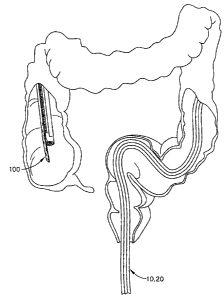
[0079] Fig. 24 is an isometric view of the distal portion of endoscope 20,
guide
apparatus 10 and another example of an intubation device, generally designated
100,
for use with guide apparatus 10. Intubation device 100, also be referred to as
a
colonic decompression tube, may be used primarily for the evacuation of fluid
such as
a gas from the colon of a patient. Intubation device 100 may include an
elongated
tube 106 defining a channel 108 therethrough. Intubation device 100 also
includes a
flexible rail 102 (also referred to as a mating part) attached to or unitarily
formed with
tube 106 along a portion or substantially the entire length of tube 106. Tube
106 and
rail 102 may be formed from an extruded polymer such as polyurethane, and have
a
similar cross-sectional profile as intubation device 24 shown in Fig. 3,
although many
CA 02582918 2007-03-29
19
other shapes are possible. Like intubation device 24, intubation device 100
may be
adapted to be slidingly engaged with carrier 22 or track 16 of guide apparatus
10.
[0080] Intubation device 100 may include a plurality of spaced-apart apertures
104
in at least the distal portion of tube 106 and in fluid communication with
channel 108.
The size and shape of apertures 104 may vary significantly, but may be
generally
large enough for the release of gas from the colon. The distal end of
intubation device
100 may be tapered as shown in Fig. 24 to facilitate atraumatic insertion into
the
colon. The proximal end of intubation device 100 (not shown) may simply be a
cut
end or may be adapted for connection to a fluid collection system (not shown).
The
length of intubation device 100 may be at least as long to extend from the
patient's
anus to the cecum of the colon, plus an additional length to extend externally
from the
patient for proper management of the released or evacuated fluid. For example,
the
length of intubation device may be approximately in the range of 100 to 200
centimeters.
[0081] Figs. 25-27 illustrate a method of placing intubation device 100 into
the
colon of a patient, using guide apparatus 10 with an endoscope, in order to
release
and/or evacuate fluid from the colon. Endoscope 20 may be provided with guide
apparatus 10 of Fig. 1 attached thereto, and may be inserted through the anus
into the
colon. As shown in Fig. 25, endoscope 20 and guide apparatus 10 may be
inserted
until the distal end of endoscope 20 extends into the desired region within
the colon,
such as in the cecum of the colon.
[0082] Intubation device 100 may be advanced along guide apparatus 10 until
the
distal end of intubation device 100 is at the desired location within the
colon, as
shown in Fig. 26. Optionally, intubation device 100 may be slidingly engaged
with
guide apparatus 10 before insertion of endoscope 20 into the colon. The distal
end of
intubation device 100 may be near the distal end of endoscope 20 prior to
insertion, or
at any location proximal to the distal end of endoscope 20.
[0083] Endoscope 20 and guide apparatus 10 maybe retracted from the colon
while
the proximal end of intubation device 100 is held stationary relative to the
patient,
thereby keeping the distal end of intubation device 100 at the desired
location within
i -1 II = CA 02582918 2007-03-29
the colon, as shown in Fig. 27. The proximal end of intubation device 100 may
be
positioned for the natural release of gas or connected to a fluid collection
system.
Although various aspects of a snaring device and associated methods have been
shown and described modifications may occur to those skilled in the art. The
present
application includes such modifications and is limited only by the scope of
the claims.