Note: Descriptions are shown in the official language in which they were submitted.
CA 02582990 2007-03-29
1
ULTRASONIC SURGICAL SYSTEM
AND METHOD
100011 Field of the Invention
100021 The present application relates to ultrasonic surgical instruments and,
more particularly, to ultrasonic surgical instruments having end effectors for
cutting and coagulating tissue. The present application also relates to
robotic
surgical systems and, more particularly, to robotic surgical systems used with
ultrasonic surgical instruments.
100031 Background of the Invention
100041 Surgeons use ultrasonic instruments in surgery to cut and coagulate
tissue. Piezoelectric elements are electrically excited at a resonant
frequency of
an ultrasonic instrument to create vibrations that are transmitted through a
resonator and amplified to produce a mechanical, standing wave vibration of
the
same frequency. An ultrasonic transmission assembly of the instrument has an
elongated, transmission waveguide that transmits this vibration to an end
effector (e.g., cutting blade) on the distal tip of the instrument. An example
of
an ultrasonic surgical instrument is the Harmonic Scalpel Laparosonic
Coagulating Shears, available from Ethicon Endo-Surgery, Inc., Cincinnati,
Ohio.
[00051 In recent years, minimally invasive robotic systems have been
developed and used for certain surgical procedures including coronary artery
bypass grafting and cholecystectomy procedures. The robotic systems provide
a number of promising benefits that generally reduce the surgical skill
required
to perform certain surgical procedures, such as by increasing dexterity and
eliminating hand tremor. The robotic systems also allow surgeons to perform
the procedures at a remote location, wherein remote may be understood as
anywhere that is "more than arm's length" from the patient. An example of a
CA 02582990 2007-03-29
2
robotic surgical system is the DAVINCI, which is available from Intuitive
Surgical Inc., Mountain View, California.
[0006] U.S. Patent No. 6,783,524 to Anderson et al. titled "Robotic Surgical
Tool with Ultrasound Cauterizing and Cutting Instrument" discloses an
ultrasonic surgical instrument mounted to a movable arm of a robotic surgical
system such as the DAVINCI. The disclosed method for enhancing robotic
surgery generally includes coupling the instrument to the robotic surgical
system, positioning an end effector of the instrument in contact with tissue
at a
surgical site and delivering ultrasound energy to the tissue. In general, the
disclosed robotic system permits a surgeon to directly control the movements
and actuation of the instrument from a remote location.
[0007] Despite such advances in surgical technologies, however, considerable
skill is still required by surgeons to perform particular steps of some
surgical
procedures. For example, in order to ultrasonically coagulate a vessel such as
the cystic duct of the gall bladder, the surgeon may press a relatively broad
surface of the ultrasonic blade against the duct, apply a light clamping force
on
the duct and sweep the ultrasonic end effector within a treatment region on
the
duct while applying an intermediate level of ultrasonic power. Then, to cut
the
duct, the surgeon may present an edge of the blade to the duct, apply a high
clamping force on the duct while holding the blade stationary and apply a high
level of ultrasonic power. The surgeon may repeat these steps several times
during a surgical procedure on a patient.
[0008] In order to reduce the time to perform certain surgical procedures and
to improve surgical outcomes, surgeons would like to employ various
techniques that are not normally practical using current surgical systems. For
example, many surgeons would like to have a way to apply a rapidly pulsed
clamping force onto tissue while applying ultrasonic energy in order to
agitate
and/or circulate fluids in the tissue to quicken fluid dessication, thereby
shortening the time to coagulate the tissue. Surgeons also would like to have
a
way to consistently apply the correct power level of ultrasonic energy for the
CA 02582990 2007-03-29
=
3
correct duration, primarily in order to avoid injury such as lateral thermal
damage to the tissue, but also to quicken the procedure while having the
assurance that the tissue seal is hemostatic. Surgeons rely greatly on their
physical senses to estimate clamping force on the tissue and the power
level/duration of ultrasonic energy that should be applied to the tissue.
Obviously, some of this sensory feedback is greatly diminished if the surgeon
is
using a robotic surgical system and separated from the patient.
[0009] Clearly, it may be very difficult, if not impossible, for a surgeon to
perform a plurality of surgical tasks in a well-coordinated manner, wherein
the
tasks may include, for example, sweeping the ultrasonic blade on tissue,
rotating
the blade, pulsing the clamping force and applying ultrasonic energy at
various
power levels.
[0010] Accordingly, there is a need for an ultrasonic surgical system and
method of ultrasonically treating tissue of a patient during a surgical
procedure,
wherein certain surgical tasks may be automatically and consistently
performed,
thereby reducing the skill level required by the surgeon to perform the tasks,
improving the surgical outcome, reducing the surgical procedure time and
further improving the ability to perform surgical procedures remotely.
[0011] Summary of the Invention
[0012] In one aspect, an ultrasonic surgical system has an ultrasonic unit
including an instrument operatively connected to an ultrasonic generator,
wherein the instrument has an end effector, which may be an ultrasonic end
effector on the distal end of a shaft. The system further includes a
positioning
unit including a movable arm adapted to hold the instrument. The positioning
unit may be adapted for releasably holding the instrument, and whereby an
operator may direct the positioning unit to position the end effector at a
surgical
site inside a body cavity of a patient for performing a plurality of surgical
tasks.
The system further includes a control unit in communication with or
operatively
connected to the ultrasonic and positioning units, wherein the control unit is
CA 02582990 2014-03-10
4
programmable with a surgical subroutine for performing the surgical tasks. The
system further includes a user interface in communication with or operatively
connected to the control unit for initiating an operative cycle of the
surgical
subroutine such that the surgical tasks are automatically performed during the
operative cycle.
[0013] More specifically, in one aspect there is provided an ultrasonic
surgical
system comprising: an ultrasonic unit including an instrument operatively
connected
to an ultrasonic generator, wherein the instrument includes an end effector
having a
clamping element and an ultrasonic blade; a positioning unit including a
movable
arm, wherein the moveable arm is adapted to hold the instrument of the
ultrasonic
unit; a control unit in communication with the ultrasonic unit and the
positioning
unit; and a user interface in communication with said control unit. The
control unit
is programmed with one or more surgical subroutines. The surgical subroutine
includes manoeuvring the end effector while powering said ultrasonic
instrument.
One or more of said surgical subroutines includes a plurality of surgical
tasks in an
operative cycle, wherein at least one surgical task of the plurality includes
reversibly
and repeatedly actuating the clamping element to apply a modulated clamping
force
to tissue clamped against the ultrasonic blade. The user interface is adapted
to
initiate an operative cycle of the surgical subroutine such that the plurality
of
surgical tasks are automatically performed during the operative cycle, such
that a
surgeon may select and initiate the one or more programmed surgical
subroutines.
[0013a] In another aspect, the system may further include a feedback system
having
at least one sensor positioned in at least one of the ultrasonic and
positioning units
and having an associated sensor circuit, wherein the feedback system is
operatively
connected to the control unit, and wherein a feedback signal associated with
an
operational performance parameter of the instrument during the operative cycle
may
be transmitted from the sensor to the control unit such that the control
system can
process the feedback signal and respond according to the surgical subroutine.
CA 02582990 2013-04-29
4a
100141 In another aspect, a method for ultrasonically treating the tissue of a
surgical
patient includes providing the ultrasonic surgical system defined in the
previous
paragraphs, programming the control unit with a surgical subroutine for
performing a
plurality of surgical tasks, positioning the end effector of the instrument at
a surgical
site inside a body cavity of the patient and initiating the surgical
subroutine of the
control unit, whereby the system automatically performs the surgical tasks
according
to the surgical subroutine.
100151 In another aspect, a method for ultrasonically treating the tissue of a
surgical
patient includes providing the ultrasonic surgical system defined in the
previous
aspects and further including a feedback system having at least one sensor
positioned
in at least one of the ultrasonic and positioning units and having an
associated
sensor circuit, wherein the feedback system is operatively connected to the
control
unit, and wherein a feedback signal associated with an operational performance
parameter of the instrument during the operative cycle may be transmitted from
the
sensor to the control unit such that the control system can process the
feedback
signal and respond according to the surgical subroutine. The method further
includes programming the control unit with a
DOCSTOR= 2690299\1
CA 02582990 2013-04-29
surgical subroutine for performing a plurality of surgical tasks, positioning
the end
effector of the instrument at a surgical site inside a body cavity of the
patient and
initiating the surgical subroutine of the control unit, whereby the system
automatically
performs the surgical tasks, processes the feedback signal and responds to the
feedback
signal according to the surgical subroutine.
[0016] Another aspect of the present invention is a use of the system
described above
for ultrasonically treating a tissue of a patient.
[0017] Another embodiment of the invention is for an energy-based medical
treatment
system including an energy-based clamp coagulator having two clamping members
adapted to coagulate patient tissue clamped between the two clamping members,
wherein at least one of the two clamping members includes, or is adapted to
function as,
a temperature sensor for measuring a temperature of the clamped patient
tissue.
[0018] Other aspects and aspects of the ultrasonic surgical system and method
of
ultrasonically cutting and coagulating tissue of a patient during a surgical
procedure are
described in the following description and shown in the accompanying drawings.
[0019] Brief Description of the Figures
[0020] FIGURE 1 is a perspective view of a robotic surgical apparatus of the
prior art;
[0021] FIGURE 2 is a perspective view of an ultrasonic surgical tool of the
prior art for
use on the robotic surgical apparatus shown in Fig. 1;
[0022] FIGURE 3 is a cut-away top view of an ultrasonic surgical instrument
according
to one aspect, wherein the instrument includes a motorized actuating unit for
operating
an end effector;
[0023] FIGURE 4 is a perspective view of the distal portion of the ultrasonic
surgical
instrument shown in Fig. 3;
CA 02582990 2007-03-29
6
[0024] FIGURE 5 is a block diagram showing an ultrasonic surgical system
according to a first aspect, which is represented by solid-line blocks and
according to a second aspect, which is represented by all the blocks;
[0025] FIGURE 6 is a side view of the distal portion of the instrument shown
in Fig. 4;
[0026] FIGURE 7 is a top view of the distal portion of the instrument shown
in Fig. 4;
[0027] FIGURE 8 is cross-sectional view, taken at line 8-8 of Fig. 7, of the
instrument shown in Fig. 4;
[0028] FIGURE 9 is a graph depicting an ultrasonic power applied to an
ultrasonic end effector versus time during an exemplary operative cycle in
accordance with one aspect of the ultrasonic surgical system;
[0029] FIGURE 10 is a graph depicting a clamping element angle of the end
effector versus time during the operative cycle in accordance with one aspect
of
the ultrasonic surgical system;
[0030] FIGURE 11 is a graph depicting a lateral position of the end effector
versus time during the operative cycle in accordance with one aspect of the
ultrasonic surgical system;
[0031] FIGURE 12 is a graph depicting a longitudinal position of the end
effector versus time during the operative cycle in accordance with one aspect
of
the ultrasonic surgical system;
[0032] FIGURE 13 is a graph depicting a rotational position of a blade of the
end effector versus time during the operative cycle in accordance with one
aspect of the ultrasonic surgical system;
[0033] FIGURE 14 is a graph depicting the ultrasonic power applied to the
end effector versus time during the operative cycle in accordance with one
CA 02582990 2013-04-29
7
aspect of the ultrasonic surgical system and is the same graph as shown in
Fig.
9;
[0034] FIGURE 15 is a graph depicting a measured clamping force feedback
of the end effector on tissue versus time during the operative cycle in
accordance with one aspect of the ultrasonic surgical system;
[0035] FIGURE 16 is a graph depicting a measured electrical impedance
feedback versus time during the operative cycle in accordance with one aspect
of the ultrasonic surgical system; and
[0036] FIGURE 17 is a graph depicting a measured acoustical impedance
feedback versus time during the operative cycle in accordance with one aspect
of the ultrasonic surgical system.
[0037] Detailed Description of the Invention
[0038] The following description pertains to an ultrasonic surgical system and
method for performing a surgical procedure according to an automated surgical
subroutine. The system and method may be particularly useful for
ultrasonically cutting and coagulating soft tissue in a patient, but the
system and
method may also be adapted to other types of surgical procedures. A surgeon
may initiate the surgical subroutine during a surgical procedure to perform a
plurality of surgical tasks simultaneously and/or in a coordinated manner,
wherein the surgical tasks may include sweeping an ultrasonic blade of the
instrument against the tissue, rotating the blade, actuating a clamping
element,
applying various power levels of ultrasonic energy and obtaining feedback
signals associated with the performance of the instrument.
[0039] The ultrasonic surgical system to be described herein may include or
be used in conjunction with a surgical robotic apparatus such as the prior art
DAVINCI system (Intuitive Surgical, Inc.) shown in Fig. 1. The robotic
apparatus, generally designated 10, is disclosed in the earlier referenced
U.S.
Patent No. 6,783,524.
CA 02582990 2013-04-29
8
Robotic apparatus 10 includes a control station 12 and a surgical
work station 20. Control station 12 includes an image display module 14 for
displaying an image of a surgical site, a support 16 on which an operator may
rest his/her forearms and a space 18 where two master control devices are
located (not shown). When using control station 12, a surgeon may sit in a
chair in front of control station 12, view the surgical site through display
module 14 and grip the master controls, one in each hand, while resting the
forearms on support 16.
[0040] Control station 12 is generally coupled to work station 20 such that
command from the master control devices may be transmitted to work station
20. Work station 20 is mobile and may be positioned adjacent to a surgical
patient. Control station 12 may be positioned a great distance from work
station
20, but typically control station 12 is positioned in the same operating room
as
work station 20.
[0041] According to various aspects, work station 20 may include at least
three robotic arm assemblies 22, 26, 27 (also referred to as movable arms),
one
of which may be configured to hold an image capture device 24 and the others
of which may be configured to hold surgical instruments 28. Coupling of work
station 20 to control station 12 generally enables display module 14 to
display
an image captured by image capture device 24 and control of movable arms 22,
26, 27.
[0042] Surgical instruments 28 typically include an elongate shaft for
endoscopic access to the surgical site inside the patient, wherein the shaft
has a
distal end effector adapted for a particular surgical task. Instruments 28 may
be
releasably mounted on wrist-like mechanisms on movable arms 22, 26, 27 such
that a surgeon may use control station 12 to move each instrument 28 in
various
directions (i.e., arms 22, 26, 27 have a plurality of degrees of freedom).
[0043] Fig. 2 is a perspective view of an alternative aspect of surgical
instrument 28 shown in Fig. 1 and is described in detail in U.S. Patent No.
CA 02582990 2007-03-29
9
6,783,524. This aspect, generally designated 30, includes an instrument base
32
having a cover 34. Instrument 30 also includes a shaft 36 extending distally
from instrument base 32 along the instrument axis 38. Shaft 36 includes an
outer sheath 40. An end effector 42 couples to the distal end of shaft 36 and
includes an ultrasonic blade 44 that cooperatively mates with a clamp 46. An
ultrasonic transducer 48 mounts to the proximal end of base 32 and a
power/control cable 50 may be operatively connected to a conventional
ultrasonic surgical generator, such as the Auto Son iXTM generator (not shown)
made by United States Surgical Corporation of Norwalk, Connecticut.
[0044] While seated at the control station, a surgeon may introduce and
position the distal portion of instrument 30 into a body cavity of a patient
and
manipulate and ultrasonically treat tissues therein. While seated at control
station 12 of robotic apparatus 10, the surgeon may steer end effector 42 to
the
surgical site inside the patient, actuate end effector 42 to clamp onto tissue
and
energize blade 44 to cut and coagulate tissue.
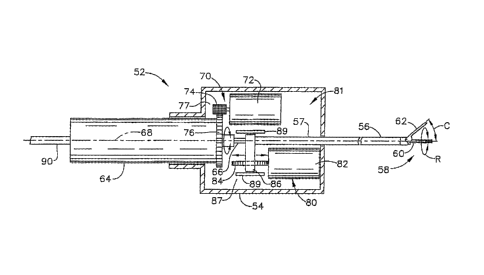
10045] Fig. 3 is a cut-away top view of an ultrasonic surgical instrument 52
that may be held by a surgeon or releasably mounted to a movable arm of a
robotic apparatus. Instrument 52 may include a base 54 and an elongate shaft
56 extending distally from the base 54. An end effector 58 on the distal end
of
shaft 56 may include a clamping element 62 (also referred to as a jaw) and an
ultrasonic blade 60. Fig. 4 is a detailed, perspective view of end effector
58.
Blade 60 is operatively connected by a waveguide 66 (Fig. 3) to an ultrasonic
transducer 64 extending from the proximal end of base 54. Transducer 64,
waveguide 66 and blade 60 may be similar in configuration and ultrasonic
operation to the surgical ultrasonic transmission assemblies disclosed in the
earlier referenced assemblies. Transducer 64 may be adapted to rotate about a
central axis 68 with respect to base 54, such that blade 60 rotates about axis
68
with respect to clamping element 62, thereby allowing clamping element 62 to
clamp against various surface portions of blade 60 for reasons to be described
herein.
CA 02582990 2007-03-29
[0046] Instrument 52 may include an actuating unit 81 having a force
transmission assembly 80 and a blade rotation assembly 70. Force transmission
assembly 80 allows electrically controlled movement of clamping element 62 in
a direction indicated by arrow "C". Blade rotation assembly 70 allows
electrically controlled rotation of blade 60 about axis 68 in either direction
as
indicated by arrow "R". These movements may be directly controlled by the
surgeon or automated as will be described herein.
[0047] In one aspect, force transmission assembly 80 may include a first
electric motor 82 operatively connected to a remote control (not shown).
Assembly 80 may include a first drive mechanism 87 that operatively engages
motor 82 to an outer sheath 57 of shaft 56, such that electrical actuation of
motor 82 moves outer sheath 57 in a distal or a proximal direction, depending
on the rotational direction of motor 82. The distal end of outer sheath 56 may
be operatively connected to clamping element 62, such that distal translation
of
sheath 57 moves clamping element 62 towards blade 60 and proximal
translation of sheath 57 moves clamping element 62 away from blade 60. Drive
mechanism 87 may include any one of a number of conventional mechanisms
for converting the rotation of motor 82 to the translation of outer sheath 57.
As
shown in Fig. 3, drive mechanism 87 may include a lead screw 84 attached to
motor 82 and operatively engaged with a follower 86 that is attached to the
proximal end of outer sheath 57 and guided between tracks 89 formed on base
54.
[0048] Still referring to the aspect shown in Fig. 3, blade rotation assembly
70
may include a second electric motor 72 operatively connected to a remote
control (not shown). Assembly 70 may include a second drive mechanism 77
operatively engaging motor 72 to ultrasonic transducer 64, such that
electrical
actuation of motor 72 causes transducer 64 to rotate in either direction about
axis 68, thereby rotating waveguide 66 and blade 60 about axis 68. A
power/control cable 90 extends proximally from transducer 64 and may be
operatively connected to a user interface (not shown) as will be described.
Second drive mechanism 77 may include any one of a number of conventional
CA 02582990 2013-04-29
11
mechanisms for converting the rotation of motor 72 to the rotation of
transducer
64. As shown in Fig. 3, drive mechanism 77 may include a pinion gear 74
attached to motor 72 and engaging a spur gear 76 attached to transducer 64.
Motor 72 may be electrically actuated such that transducer 64 rotates +1- 180
degrees about axis 68 as cable 90 flexibly twists in either direction.
100491 First and second motors 72, 82 may be selected from a large number of
commercially available DC servo-motors or other types of motors meeting
numerous system requirements, including bidirectional shaft rotation,
sufficient
torque and rotational speed modulation, microprocessor control, size and cost.
100501 As shown in Fig. 4, the distal portion of instrument 58 and the
corresponding ultrasonic transmission assembly may be similar to that
disclosed
in U.S. Patent No. 5,954,736 to Bishop et al. titled "Coagulator Apparatus
Having Indexed Rotational Positioning,".
Instrument 58 may be ultrasonically driven
by an ultrasonic generator (not shown) such as model number GEN04 available
from Ethicon Endo-Surgery, Inc., Cincinnati, OH.
100511 In another aspect of instrument 52, force transmission assembly 80 for
the remote actuation of clamping element 62 may be provided by a gripper 82
of a surgical instrument 80 as disclosed in U.S. Pat. No. 6,783,524, beginning
at
col. 15, line 29.
[0052] Surgical instrument 52 and its various aspects may be included in an
ultrasonic surgical system 100 shown in Fig. 5. System 100 may be used
according to one or more automated surgical subroutines for the coordinated
performance of a plurality of surgical tasks during a surgical procedure on a
patient.
[0053] In Fig. 5, a first aspect of system 100 is represented by solid-line
blocks and a second aspect is represented by all the blocks. A surgeon or
operator may use system 100, for example, to automatically coagulate and cut
tissue according to a predetermined surgical subroutine. All or parts of the
CA 02582990 2007-03-29
12
surgical subroutine may be programmed by the operator into system 100 prior
to the surgical procedure. Alternately, all or parts of the surgical
subroutine
may already be programmed into system 100.
[0054] System 100 generally may include a user interface 102, a
programmable control unit 104, an ultrasonic unit 106, a positioning unit 108
and an actuating unit 110. Ultrasonic unit 106 may include an ultrasonic
generator 112 and an ultrasonic surgical instrument 114. (In the following
description of system 100, references to the end effector, the blade and the
clamping element shall be applicable to end effector 58, blade 60 and clamping
element 62 shown in Fig. 4, although many other types of ultrasonic end
effectors may be adapted also for use with instrument 114.) Positioning unit
108 may include a movable arm 116 for positioning and orienting instrument
114 at a surgical site in the patient. Actuating unit 110 may include a force
transmission assembly 118 for operating clamping element 60 on instrument
114 and a blade rotation assembly 120 for rotating an ultrasonic blade on
instrument 114 about a central axis. System 100 may include or be used in
conjunction with a robotic apparatus such as robotic apparatus 10 shown in
Fig.
1. Alternatively, system 100 may be used without such a robotic apparatus,
instead requiring the surgeon to hold and position the ultrasonic surgical
instrument in a conventional manner and then initiating the automated surgical
subroutine to perform certain surgical tasks.
100551 User interface 102 may include a control box (not shown) having one
or more controls for remotely operating instrument 114, a monitor (not shown)
operatively connected to an endoscope or other type of image capture device
for
displaying an image of the surgical site and one or more controls for
initiating
and terminating an automated surgical subroutine that is programmed into
control unit 104. User interface 102 may be located remotely or near the
patient.
Alternately, all or some of user interface 102 may be positioned on instrument
114 or other components of system 100.
CA 02582990 2007-03-29
13
100561 Programmable control unit 104 may include a microprocessor
programmable with at least one surgical subroutine for performing one or more
surgical tasks simultaneously and/or in a coordinated manner. The subroutine
may automatically control one or more surgical tasks, including movements of
the blade, ultrasonic energization of the blade, actuation of the of the
clamping
element and responses to feedback signals. The duration of the subroutine is
defined herein as an operative cycle, which may range, for example, from a
fraction of a second to several seconds.
[0057] Control unit 104 may be provided with one or more surgical
subroutines pre-programmed into the microprocessor, such that the surgeon may
select and initiate a subroutine, for example, by actuating a control on user
interface 102 prior to or during the surgical procedure. For example, the
surgeon may specify that a blood vessel having a diameter of approximately
3mm will be coagulated and cut. The subroutine would then automatically
direct the instrument to provide a particular power level of ultrasonic energy
for
a particular period of time, to sweep the ultrasonic blade over the tissue and
to
actuate the clamping element according to a known effective treatment
procedure. Alternately, control unit 104 may include any one of a number of
conventional data input devices that would allow the surgeon to program the
microprocessor prior to performing a surgical procedure. Such devices would
allow inputting data using, for example, a floppy disk, a flash memory device
or
by keying in data when prompted by instructions from a programming software
and displayed on the monitor.
[0058] Ultrasonic generator 112 of ultrasonic unit 106 may be a conventional
ultrasonic generator such as the GEN04 (Ethicon Endo-Surgery, Inc.)
generator. Instrument 114 may be operatively connected to generator 112 and
releasably mounted to the movable arm 116 of positioning unit 108. Instrument
114 may be any one of the aspects of ultrasonic surgical instrument 30 shown
in
Fig. 2 and ultrasonic surgical instrument 52 shown in Figs. 3 and 4, or
equivalents thereof. A surgeon may operate user interface 102 to control
CA 02582990 2007-03-29
14
movement of arm 116 in order to position the end effector of instrument 114 at
the surgical site.
[0059] As previously noted, robotic apparatus 10 (Fig. 1) may be adapted for
positioning instrument 114 and actuating the end effector of instrument 114.
Control unit 104 may be operatively connected to apparatus 10 such that
control
unit 104 controls robotic apparatus 10 to move instrument 114 according to the
surgical subroutine. For example, the subroutine may direct the movable arm of
apparatus 10 to sweep the ultrasonic blade of instrument 114 laterally (side-
to-
side) and/or longitudinally while ultrasonic power is applied to the tissue in
order to provide a larger hemostatic margin. Or the subroutine may direct
apparatus 10 to reversibly actuate the clamping element on instrument 114 a
number of times per second while the blade is ultrasonically energized in
order
to improve the quality of the tissue weld.
[0060] Positioning unit 108 may include other types of powered mechanisms
for producing fine lateral and/or longitudinal movements of the ultrasonic
blade
and for actuating the clamping element of instrument 114 while the blade is
ultrasonically energized. For example, various types of electric motors,
electric
solenoids, pneumatic actuators, hydraulic actuators, electrically actuated
nickel/titanium shape-memory alloy mechanisms and other devices may be
incorporated into instrument 114 and operatively connected to control unit 104
for positioning and actuating instrument 114 according to the surgical
routine.
In addition, instrument 114 may be handheld and positioning unit 108, rather
than a robotic apparatus, may be a surgeon or other human operator.
[0061] Force transmission assembly 118 of actuating unit 110 actuates the
clamping element of instrument 114 to apply a modulated force on tissue
clamped against the ultrasonic blade according to the surgical subroutine.
Force
transmission assembly 118 may include force transmission components of
movable arm 116 of positioning unit 108, wherein the force transmission
components are mechanically coupled to instrument 114 for actuating the
clamping element such as described in earlier referenced U.S. Patent Numbers
CA 02582990 2013-04-29
6,783,524 and 6,352,532. Alternately, force transmission assembly 118 may
include one or more components mounted, for example, inside of instrument
114 such as described for instrument 52 in Fig. 3.
[0062] Blade rotation assembly 120 may be similar to blade rotation assembly
80 shown in Fig. 3. Blade rotation assembly 120 rotates the ultrasonic blade
about the central axis such that tissue may be clamped against various surface
portions of the blade. For example, a first surface portion may be relatively
flat
for coagulating a broad area of tissue, whereas a second surface portion may
have a relatively sharp edge for applying ultrasonic energy to a narrow region
of
tissue and cutting the tissue. The beginning of the surgical subroutine may
direct the blade to present the first surface portion towards the tissue to
create a
wide region of coagulated tissue on a blood vessel, for example, and the
ending
of the surgical subroutine may direct the blade to present the second surface
portion towards the tissue to cut through the center of the wide coagulated
region.
[0063] In addition to electric motor actuators, other types of actuators that
may be adapted for use in force transmission assembly 118 and in blade
rotation
assembly 120 include, for example, electromagnetic coil actuators, hydraulic
actuators and pneumatic actuators.
[0064] A second aspect of an ultrasonic surgical system 100, represented by
the blocks in Fig. 5, includes the elements of the first aspect and, in
addition, a
feedback system 122. Feedback system 122 may include at least one sensor 122
for measuring an operational parameter associated with the operational
performance of the ultrasonic surgical system during the operative cycle.
[0065] In one aspect, feedback system 122 may include a force sensor 126
that measures the mechanical clamping force of the clamping element. Force
sensor 126 may be any one of a number of force sensors well-known in the art,
including a strain gauge or a piezoelectric sensor mounted on the clamping
CA 02582990 2007-03-29
16
element on a force-transmitting component (not shown) of force transmission
assembly 118. Force sensor 126 may be electrically connected to control unit
104, which may process a feedback signal from force sensor 126 during the
operative cycle. Control unit 104 may then augment the ultrasonic power level,
clamping force and/or blade movements according to the surgical subroutine
and based on the force feedback. Control unit 104 may also halt the surgical
subroutine if a predetermined force is exceeded in order to prevent injury to
the
patient or damage to instrument 114.
100661 Force sensor 126 may enable control unit 104 to command actuating
unit 110 to supply any one of a plurality of clamping force-versus-time
profiles
to tissue held between the clamping element and the blade. For example, the
surgical subroutine may command the actuation of the clamping element to
provide a particular discrete force during one or more portions of the
operative
cycle. The discrete force may be pulsed or applied repetitively at a frequency
of
about 1-10 Hertz in one aspect, for example. Alternately, the surgical
subroutine may direct the actuation of the clamping element to provide an
ascending and/or descending ramped force profile or a parabolic force profile
during the operative cycle, for example. Alternately, a relatively light force
may be applied while the blade sweeps through a treatment region on the tissue
and/or rotates about the central axis during coagulation of the tissue. Then,
a
relatively high force may be applied in order to cut the tissue in the middle
of
the treatment region.
100671 In another aspect, feedback system 122 may include an electrical
impedance measuring circuit 128 for measuring the electrical impedance of the
tissue being treated by the end effector during the operation cycle. Because
electrical impedance of living tissue typically increases as the tissue is
coagulated, measurement of electrical current through the tissue as it is
being
ultrasonically treated provides an indication of the level of coagulation. The
clamping element and blade, both being electrical conductors, may be
electrically connected to an impedance measuring circuit such that current
passing through the tissue that is clamped between the clamping element and
CA 02582990 2007-03-29
17
the blade may be monitored by control unit 104. In one aspect, the surgical
subroutine may be programmed, for example, so that when electrical impedance
reaches a predetermined magnitude, the ultrasonic power is turned off and the
subroutine is halted.
[0068] In another aspect, feedback system 122 may include an acoustical
impedance measuring circuit 130, such as is well-known in the art, for
measuring acoustical impedance of the tissue being ultrasonically treated.
Acoustical impedance may be characterized as the ability of a material to
conduct a sound wave. Generally, as acoustical impedance increases, the speed
of sound through the material decreases. As tissue is coagulated, tissue
elasticity decreases and tissue density increases, thereby decreasing
acoustical
impedance. Acoustical impedance measuring circuit 130 may be physically
located inside of generator 112 of ultrasonic unit 106. Control unit 104 may
process a feedback signal from circuit 130 and modify or halt the surgical
subroutine based on the acoustical feedback.
[0069] In yet another aspect, feedback system 122 may include a temperature
sensor 132 and a temperature measuring circuit (not shown). Temperature
sensor 132 may be mounted on or near the end effector of instrument 114 in
order to measure the temperature of the blade or clamping element, the treated
tissue, an object near the treated tissue or a fluid surrounding the treated
tissue.
Temperature sensor 132 may be a thermocouple, a thermistor, an infrared
temperature sensor or any one of a number of other temperature sensors well
known in the art. Temperature feedback signals may be transmitted to control
unit 104, which may process the signal to modify or halt the surgical
subroutine.
100701 Any combination of the aforementioned aspects of feedback system
122 may be included in ultrasonic surgical system 100 to transmit feedback
signals to control unit 104, so that control unit 104 may process the signals
during the operative cycle and respond according to the surgical subroutine.
CA 02582990 2007-03-29
_
18
[0071] Figs. 6-8 illustrate examples of end effector movements that may be
automated while the blade if instrument 114 is ultrasonically energized
according to the surgical subroutine. These movements may include clamping
element angle adjustment, lateral positioning of the end effector in the x-
axis
direction, longitudinal positioning of the end effector in the z-axis
direction, and
blade rotation about the z-axis. The lateral and longitudinal positioning of
the
end effector are also referred to as "sweeping." These movements will be
defined herein as they may pertain to end effector 58 shown in Fig. 4, but
they
are also applicable to other ultrasonic end effectors having a blade and a
clamping element. For end effectors having only a blade and without a
clamping element, the movements of blade rotation and clamping element angle
adjustment are excluded.
[0072] Fig. 6 is a side view of the distal portion of instrument 52 in Fig. 4
and
shows the y-axis and the z-axis of the ordinate system positioned on an origin
142. Central axis 68 extends through shaft 56 and lies along the z-axis.
Clamping element 62 pivotally attaches to shaft 56 at a pivot 140 and may be
positioned at a clamping element angle "A" with respected to blade 60.
Clamping element angle A may be remotely adjustable, for example, to vary
approximately in the range of zero to 60 degrees by partial actuation of
clamping element 62. When clamping element 62 is actuated to move in a
closing direction towards blade 60, clamping element 62 clamps onto tissue
positioned between a serrated portion 144 and blade 60 with an average
clamping force "F" perpendicular to clamping element 62 and near the center of
serrated portion 144. When clamping element angle A is approximately zero
such as when clamping thin tissue, the average clamping force F is
approximately directed through origin 142.
[0073] Fig. 7 is a top view of the distal portion of instrument 52 shown in
Fig.
2 and shows origin 142, the z-axis and the x-axis of the ordinate system. In
certain situations, a surgeon may prefer to treat a broad area of tissue in
order,
for example, to provide safe margins of coagulated tissue for proper
hemostasis.
The surgeon may ultrasonically treat tissue in a treatment region 146 (also
CA 02582990 2007-03-29
19
referred to as a central treatment region) defined in the lateral direction of
the x-
axis by "+DX" and "-DX" and in the longitudinal direction of the z-axis by
"+DZ" and "-DZ" about origin 142.
100741 End effector 58 may be swept laterally and/or longitudinally within
treatment region 146 while blade 60 is ultrasonically energized at a low power
level and clamping element angle A is held at a desired opening. Alternately,
blade 60 may be energized at a high power level while clamping element angle
A is near zero and a high clamping force is applied to the tissue. Clamping
force may also be applied repeatedly, such as in a rapid-pulsed fashion, as
blade
60 is energized, in order to agitate and to circulate tissue fluids in the
vicinity of
the energy application. As those skilled in the art may appreciate, there are
many different combinations of clamping, energizing and sweeping that may be
used to treat various kinds of tissue in the many different kinds of surgical
situations.
10075] Fig. 8 is a cross-sectional view taken at line 8-8 in Fig. 7 of end
effector 58 when clamping element angle A is zero. Blade 60 may include a
first surface 148, a second surface 150, a third surface 152, a fourth surface
154
and a fifth surface 156, together defining a blade profile 15. Fig. 5 shows an
aspect of blade profile 160 that may be particularly useful, for example, for
transecting the cystic duct of the gall bladder during a cholecystectomy.
Blade
profile 160 may have many other geometrical shapes depending on the surgical
application and other requirements of the instrument.
[0076] Referring to Figs. 6 and 8, blade 14 is rotatable about the z-axis so
that
any one of surfaces 148, 150, 152, 154, 156 or an edge between the surfaces
may interface with clamping element 62 while tissue is held therebetween.
Blade 60 may be rotated in a positive direction "+RZ" and a negative direction
"-RZ" about the z-axis. For example, blade 60 may be rotated so that an edge
158 engages tissue when a high concentration of force on the tissue is
desired,
such as during cutting. Alternately, blade 60 may be rotated so that surface
148
engages tissue when it is desired to distribute clamping force over a wide
area
CA 02582990 2007-03-29
such as during coagulating. Or blade 60 may be rotated back and forth within
an angular range as surface 150 engages tissue, to produce a "rolling" effect
as
blade 60 is ultrasonically energized. As apparent to those skilled in the art,
many different combinations of rotating and energizing blade 60 may be
devised to rapidly and hemostatically cut and/or coagulate various types of
tissue for many different surgical situations.
[0077] Control unit 104 may be programmed to direct the fine movement of
instrument 114 along a predetermined path as blade 60 is energized and
clamping force is applied, according to the surgical subroutine, in order to
accelerate and/or enhance tissue treatment within the tissue treatment region
146 (Fig. 7). The predetermined path may include any combination of lateral
sweeping in the x-direction, longitudinal sweeping in the y-direction and
blade
rotation about the z-axis. Figs. 9 through 17 are graphs that illustrate the
operation of control system 100 according to an exemplary surgical subroutine
during an operative cycle in which a surgical procedure is performed on
tissue.
The graphs are shown without actual values and are intended to illustrate
relative parametric magnitudes and timing of the concurrent surgical tasks.
The
total time represented on the graphs may be, for example, approximately in the
range of 2 to 10 seconds. Many other surgical subroutines are possible and may
be devised according to the envisioned surgical situations and programmed into
control unit 104.
[0078] The graph shown in Fig. 9 and again in Fig. 14 illustrates an
ultrasonic
power-versus-time profile, showing ultrasonic "U/S" power (watts) versus time
"T" (seconds) for the operative cycle. Ultrasonic power is a function of
ultrasonic frequency and amplitude. The ultrasonic power transmitted to tissue
may be effectively controlled, therefore, by variation of ultrasonic frequency
and amplitude, and by selection of the clamping force (pressure) applied to
the
tissue. The ultrasonic power may be applied intermittently as shown to
coagulate tissue between each repositioning of the end effectors, and to cut
the
tissue, during which the ultrasonic power may reach a maximum value, for
example, of approximately twenty watts.
CA 02582990 2007-03-29
21
[0079] Fig. 10 illustrates a clamping element angle-versus-time profile for
the
same operative cycle as illustrated in Fig. 9. As the ultrasonic power is
applied,
the clamping element angle A may vary with time as shown, indicating how
ultrasonic power may only be applied when a clamping force is applied to the
tissue, as described in Fig. 6.
[0080] Figs. 11 and 12 show graphs that illustrate how the surgical subroutine
may concurrently direct the fine movement of the end effectors in the x-axis
direction (lateral position, DX) and the z-axis direction (longitudinal
position,
DZ), as described for Fig. 7. Fig. 11 illustrates a lateral position-versus-
time
profile and Fig. 12 illustrates a longitudinal position-versus-time profile
for the
same operative cycle shown in Fig. 9.
[0081] Fig. 13 shows a graph that illustrates a blade rotation-versus-time
profile for the same operative cycle shown in Fig. 9. Commands dictated by the
surgical subroutine direct the rotation of blade 60 about the z-axis (blade
rotation, RZ), as described for Fig. 8.
[0082] Figs. 15, 16 and 17 are graphs showing exemplary feedback signals
provided by feedback system 122 of control system 60 (Fig. 5), measured
concurrently during the operative cycle shown in Fig. 14. As noted previously,
control unit 104 may process the feedback signals and respond according to the
surgical subroutine.
[0083] According to one aspect, a method for ultrasonically treating the
tissue
of a surgical patient may include providing the ultrasonic surgical system
according to the first aspect described in Fig. 5, programming the control
unit
with a surgical subroutine for performing a plurality of surgical tasks,
positioning the end effector of the instrument at a surgical site inside a
body
cavity of the patient and initiating the surgical subroutine of the control
unit,
whereby the system automatically performs the surgical tasks according to the
surgical subroutine.
CA 02582990 2007-03-29
22
[0084] The surgical tasks of the method may be defined to include energizing
the end effector according to an ultrasonic power level profile of the
surgical
subroutine during the operative cycle and one or both of the following:
sweeping the ultrasonic end effector against the tissue in a lateral direction
within a treatment region according to a lateral sweep profile of the surgical
subroutine during the operative cycle; sweeping the ultrasonic end effector
against the tissue in a longitudinal direction within a treatment region
according
to a longitudinal sweep profile of the surgical subroutine during the
operative
cycle.
[0085] The ultrasonic surgical system provided in the method may further
include a clamping element, an ultrasonic blade and an actuating unit having a
force transmission assembly operatively connected to the clamping element and
to the control unit such that the force transmission assembly may be actuated
to
operate the clamping element to apply a variable clamping force to tissue held
between the clamping element and the blade. The surgical tasks, therefore, may
be defined to include operating the clamping element to apply a variable
clamping force to tissue held between the clamping element and the blade
according to a clamping force profile of the surgical subroutine during the
operative cycle.
[0086] The ultrasonic surgical system provided in the method may further
include the end effector having an ultrasonic blade and an actuating unit
having
a blade rotation assembly operatively connected to the blade and to the
control
unit such that the blade rotation assembly may be actuated to rotate the blade
about a central axis. The surgical tasks, therefore, may be defined to include
rotating the blade about the central axis according to a blade rotation
profile of
the surgical subroutine during the operative cycle.
[0087] In another aspect, a method for ultrasonically treating the tissue of a
surgical patient may include providing the ultrasonic surgical system
according
to the second aspect described in Fig. 5, wherein the system further includes
a
feedback system having at least one sensor positioned in at least one of the
CA 02582990 2007-03-29
23
ultrasonic and positioning units and having an associated sensor circuit,
wherein the feedback system is operatively connected to the control unit, and
wherein a feedback signal associated with an operational performance
parameter of the instrument during the operative cycle may be transmitted from
the sensor to the control unit such that the control system can process the
feedback signal and respond according to the surgical subroutine. The method
may further include programming the control unit with a surgical subroutine
for
performing a plurality of surgical tasks, positioning the end effector of the
instrument at a surgical site inside a body cavity of the patient and
initiating the
surgical subroutine of the control unit, whereby the system automatically
performs the surgical tasks, processes the feedback signal and responds to the
feedback signal according to the surgical subroutine.
[0088] Although various aspects of an ultrasonic surgical system and method
have been shown and described, it should be understood that modifications may
occur to those skilled in the art. The present application contemplates and
includes such modifications and is limited only by the scope of the claims.