Note: Descriptions are shown in the official language in which they were submitted.
CA 02583095 2013-08-02
WO 2006/043052
PCT/GB2005/004022
DVT Detection Devices and Methods
The present invention relates to the detection of a
range of clinical conditions including Deep Vein Thrombosis
(DVT) and diabetic peripheral neuropathy, critical limb
ischaemia, autonomic neural function and arterial and
venous disease by the assessment of the vasomotor activity
in the micro-circulation at individual sites on a body, and
in particular, the detection of Deep Vein Thrombosis (DVT)
and diabetic peripheral neuropathy.
Deep vein thrombosis (DVT) in the legs is a condition
whereby a blood clot, develops in a vein causing partial or
complete blockage of the vessel. The cause of the clot can
be due to vessel damage, either from surgical procedures or
trauma, or from a period of haemostasis due to prolonged
periods of inactivity (e.g. long haul flight, disability).
The perceivable consequences of a DVT can range from mild
pain and swelling to a fatal pulmonary embolism.
Known tests used in clinical practices for the
detection of DVT include imaging tests such as venography
and duplex ultrasonography. Venography requires the
injection of a radio opaque imaging medium and X-ray
imaging requiring expert interpretation and is hazardous
and uncomfortable to the patient, time consuming, expensive
and not suitable for primary care or a General Practitioner
(GP). Similarly, Duplex ultrasonography is a time consuming
and expensive process not suitable for primary care or for
GPs requiring highly skilled practitioners.
Plethysmography is a known test which is low cost,
relatively quick, and is used in trained primary care or by
a trained GP. However plethysmography requires the patient
to exercise during the test which is not suitable for all
patients and the test requires an expert operator and is
not always reliable. There is also D-dimer assay, test that
CA 02583095 2007-04-02
WO 2006/043052 PCT/GB2005/004022
2
measures the clotting agents in blood and is recommended to
be used in conjunction with other tests. The
plethysmography and D-dimer tests are used as a front line
screening means to remove as many patients as possible
without a DVT from progressing to the more onerous imaging
tests of duplex ultrasonography or venography.
The invention seeks to make improvements.
Accordingly, the present invention provides a device
comprising a light transmission and detection system to
assess vasomotor activity in the micro-circulation at
individual sites on a body for the monitoring and
assessment of a range of clinical conditions including
suspected DVT, diabetic peripheral neuropathy, critical
limb ischaemia, autonomic neural function and arterial and
venous disease.
Vasomotor activity in the micro-circulation is the
continuous process of contraction and dilatation of the
micro-vessels and serves several important functions
including blood pressure regulation,
temperature
regulation, tissue oxygenation and nutrition.-The control
of this process is both local and systemic. Local control
is activated by chemical signalling from the adjacent
tissues while the systemic control originates from the
autonomic sympathetic nervous system, principally for the
regulation of core temperature and systemic blood pressure.
The resulting local blood volume variation provides
information on many of the biological processes both
locally and systemically.
In a preferred embodiment, the invention comprises a
light transmission and detection system including wave
transducers, the wave transducers placed at one or more
sites on a body, control means to measure the light
absorbed and/or reflected at the or more sites and provide
signals relating to the absolute value at the or more sites
CA 02583095 2007-04-02
WO 2006/043052 PCT/GB2005/004022
3
and/or the differential value between the sites.
Preferably, the transducers are infra red wave transducers.
The present invention uses the transducers to monitor
the micro-circulation blood. volume variation beneath the
transducer continuously. The light absorption is
proportional to the volume of blood or, conversely, light
reflection is inversely proportional to blood volume. For a
resting patient in a stable environment, either seated or
supine, the major changes of blood volume are
manifestations of systemic control. Further, in the limbs,
the systemic vasomotor control is symmetrical. Therefore,
by placing a transducer on the sole of each foot of a
healthy subject, the signal from each transducer will be
similar if not identical. The presence of a unilateral DVT
can be detected by measuring the dissimilarity between the
two transducer signals as the distal volume of the affected
leg is increased due to increased outflow resistance. This
imposes altered frequency and phase characteristics in the
vasomotor variation of the affected leg and therefore
affects the bilateral symmetry.
In another aspect of the invention the signals
received from the transducers are used in the assessment of
autonomic systemic and peripheral neuropathy. Conventional
systemic, autonomic function testing, analyses heart rate
variability, usually derived from the ECG waveform.
However, cardiac pulsation can be seen in the signal
collected at most points on the skin around the body using
the transducer. Therefore, heart rate variability can be
derived from this signal. Analysis of the variation in the
heart rate component can then be compared to the low
frequency variation of the signal from the transducer,
allowing a direct comparison of peripheral and systemic
autonomic function. In the healthy subject both sources of
variation should be similar, whereas in the patient
CA 02583095 2007-04-02
WO 2006/043052 PCT/GB2005/004022
4
suffering with peripheral neuropathy alone there will be a
dissimilarity.
The advantages of using vasomotor activity in the feet
to assess DVT, vascular disease and neurological function
include the ability to use a passive test requiring no
movement on the part of the patient. Preferably, the
neurological function test is augmented by stress testing
such as valsalva manoeuvre or mild graduation of exhalation
impedance. The sites to be used on the patient's body are
easily accessible, requiring low cost instruments, lower
level of skill than existing tests and providing reliable
results.
To date, there is little work published on the use of
vasomotor activity for the
assessment , of clinical
conditions such as those of the present invention due to
the poor understanding of vasomotor activity and related
biological processes. We have found that the vasomotor
signal provides valuable information concerning the many
biological processes occurring simultaneously within
healthy and unhealthy bodies.
The invention will now be described by way of example
only, with reference to the following drawings, of which:
Figure 1 shows the light transmission and detection
system according to the invention;
Figure 2 shows a block diagram of the transducers in
Figure 1;
Figures 3a, b, c are schematic views of a preferred
embodiment of the invention in Figure 1 applied to
different sites on a patient;
Figure 4 is a signal output from the embodiment as
applied in Figure 3a;
Figure 5 shows another preferred embodiment of the
invention;
CA 02583095 2007-04-02
WO 2006/043052 PCT/GB2005/004022
Figure 6 shows the output from the embodiment as shown
in Figure 5 from the various sites of the legs of a
patient; and
Figure 7 shows the signal response to increased
5 breathing impedance and hand grip.
Figure 8 shows the vasomotor signal and extraction of
the heart rate variation.
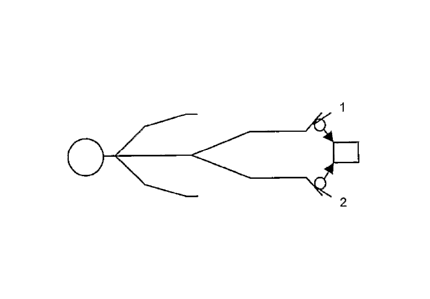
Referring to Figures 1 and 2, the invention comprises
a light transmission and detection system including
transducers 1, 2 comprising an LED and photo-detector with
suitable amplifiers 3, 4 as shown in Figure 2. Once the
transducers 1, 2 are attached to the skin the central
control unit 5 calibrates them by driving the LED 1 with a
voltage appropriate to detect a mid-scale voltage from the
photo-detector 2. The
photo-detector 2 signals are
digitised by A/D1 and A/D2.
The drive voltages for the
LEDS are produced from the output of D/A1 and D/A2. Once
the calibration process is complete the central control
unit 5 collects data from the photo-detector 4 (Figure 2)
at a sampling rate appropriate for the application. For
DVT detection a sample rate of 6 Hz is used. A user input
device 6 such as a keypad and a display for output, for
example an LCD screen or LED indicators or similar is used.
There is also provided an input/output port for PC
connection, printer or other form of data logging device.
Figures 3a to c show a preferred embodiment of the
invention using a two channel system using two transducers
1, 2 for differential signal analysis. For the purpose of
DVT detection, the transducers 1, 2 are positioned on the
soles of the feet of a patient as shown in Figure 3a. The
configuration of 3b can give an indication of the
approximate location of DVT. If the vasomotor signals are
similar the DVT will be located in the thigh whereas if the
vasomotor signals are dissimilar the DVT will be located in
CA 02583095 2007-04-02
WO 2006/043052 PCT/GB2005/004022
6
the calf. The arrangement in Figure 3c indicates the pulse
transit time between the upper and lower extremities and
thus an indication of arterial stiffness. Figure 4 shows
the signal derived from the soles of the feet of a healthy
subject using a two channel system. The signal from each
transducer is similar if not identical. The presence of a
unilateral DVT is detected by measuring any dissimilarity
between the two signals.
The output presented to the user can take the form of
a detailed display of vasomotor signals collected from the
transducers 1, 2 as shown in Figure 4 to a simple
indication of a condition being present or absent. The
display can be configured to the application.
The sampling rate of the transducer 1, 2 signals is
such that the heart rate component can be resolved to
within +/- 1 ms or better if the heart rate is of interest
in the assessment being performed, for example in autonomic
function testing. Otherwise sampling frequencies that meet
the Nyquist requirements are adequate.
The signals acquired from each transducer 1, 2 are
subject to appropriate analytical algorithms. The signals
are subject to amongst others complex demodulation a
mathematical technique used for investigating the vasomotor
activity centred at specific frequencies with a bandwidth
chosen in accordance with the application, for example DVT
detection.
The output of the complex demodulation
algorithm consists of an amplitude signal and a phase
signal which when combined, produce a time varying signal
modulated by both amplitude and phase with limited
bandwidth, all centred on the demodulating frequency.
As well as the arrangements shown in Figures 3a to c,
another preferred embodiment has two further transducers 7,
8 applied behind the knees for a four channel system as
shown in Figure 5.
The signals are passed through the
CA 02583095 2007-04-02
WO 2006/043052 PCT/GB2005/004022
7
stages of signal pre-processing including filtering and DC
removal followed by complex demodulation at a set of chosen
frequencies, for example 8 to 30 cycles per minute. The
mean absolute phase differences (MAPD) from the right foot
(RF) and the left foot (LF) are calculated for each
frequency to produce a spectrum RFLF(MAPD) and the
RFLF(MAPD) is then used by a pattern classifier such as a
pre-trained artificial neural network to provide an output
on a screen that there is either "DVT PRESENT" or "DVT NOT
PRESENT".
For a four channel system as shown in Figure 5, there
will be six MAPDs as shown in Figure 6:-
Right Foot Left Foot : RFLF = mean ( abs ( RF ($) - LF () ) ) ,
Right Knee Left Knee : RKLK = mean( abs( RK W - LK () ) ) ,
Right Foot Right Knee : RFRK = mean( abs( RF() - RK() ) ) ,
Left Foot Left Knee : LFLK = mean( abs( LF () - LK () ) ),
Right Foot Left Knee : RFLK = mean( abs( RF() - LK () ) ) ,
Right Knee Left Foot : RKLF = mean( abs( RK (1o) - LF() ) ) ,
giving six times the diagnostic information of the two
channel system, described above.
In addition to detecting DVT, the present invention
can monitor and assess a range of clinical conditions
including diabetic peripheral neuropathy, critical limb
ischaemia, autonomic neural function and arterial and
venous disease.
In each of these conditions the vasomotor activity of
the micro circulation possesses a unique signature which is
extracted and assessed using the appropriate signal
processing algorithms. These algorithms are tuned to the
appropriate frequency bands determined by the clinical
condition of interest. The algorithms exploit the property
of vasomotor symmetry between the left and right feet and
also use the similarity between the low frequency
components of the vasomotor activity and the low frequency
components of heart rate variation. As shown in Figure 8,
the device according to the invention, extracts from the
CA 02583095 2007-04-02
WO 2006/043052 PCT/GB2005/004022
8
vasomotor signal the heart rate variation and direct
comparison of the simultaneous low frequency heart rate
variation and the low frequency vasomotor variation
provides information relating to diabetic sympathetic
neuropathy, any dissimilarity between the two components
indicating diabetic sympathetic neuropathy.
Figure 7 shows the changes in vasomotor activity
related to increased breathing resistance and the hand grip
test of a healthy person. These tests affect systemic blood
pressure and cardiac output which in turn cause
neurologically mediated responses in heart rate and
peripheral vasomotor activity as observed with the
transducers on the soles of the feet. Any changes from the
signals in Figure 7 between the resting phase and the
increased breathing resistance and the hand grip test will
indicate diabetic sympathetic neuropathy since the
pathology of the sympathetic nerve fibres which innovate
the micro-blood vessels within the feet will cause
significant change in vasomotor behaviour.