Note: Descriptions are shown in the official language in which they were submitted.
DEMANDE OU BREVET VOLUMINEUX
LA PRESENTE PARTIE DE CETTE DEMANDE OU CE BREVET COMPREND
PLUS D'UN TOME.
CECI EST LE TOME 1 DE 2
CONTENANT LES PAGES 1 A 72
NOTE : Pour les tomes additionels, veuillez contacter le Bureau canadien des
brevets
JUMBO APPLICATIONS/PATENTS
THIS SECTION OF THE APPLICATION/PATENT CONTAINS MORE THAN ONE
VOLUME
THIS IS VOLUME 1 OF 2
CONTAINING PAGES 1 TO 72
NOTE: For additional volumes, please contact the Canadian Patent Office
NOM DU FICHIER / FILE NAME:
NOTE POUR LE TOME / VOLUME NOTE:
CA 02583208 2013-02-01
ANTI-CD70 ANTIBODY AND ITS USE FOR THE TREATMENT AND
PREVENTION OF CANCER AND IMMUNE DISORDERS
CROSS REFERENCES TO RELATED APPLICATIONS
[0001] <deleted>
BACKGROUND OF THE INVENTION
[0002] CD70 is a member of the tumor necrosis factor (TNF) family of cell
membrane-bound
and secreted molecules that are expressed by a variety of normal and malignant
cell types. The
primary amino acid (AA) sequence of CD70 predicts a transmembrane type II
protein with its
carboxyl terminus exposed to the outside of cells and its amino terminus found
in the cytosolic
side of the plasma membrane (Bowman et al., 1994,1 Immunol. 152:1756-61;
Goodwin etal.,
1993, Cell 73:447-56). Human CD70 is composed of a 20 AA cytoplasmic domain,
an 18 AA
transmembrane domain, and a 155 AA extracytoplasmic domain with two potential
N-linked
glycosylation sites (Bowman etal., supra; Goodwin etal., supra). Specific
immunoprecipitation
of radioisotope-labeled CD70-expressing cells by anti-CD70 antibodies yields
polypeptides of 29
and 50 kDa (Goodwin etal., supra; Hintzen etal., 1994,1 Immunol. 152:1762-73).
Based on its
homology to TNF-alpha and TNF-beta, especially in structural strands C, D, H
and I, a trimeric
structure is predicted for CD70 (Petsch etal., 1995, Mol. Immunol. 32:761-72).
[0003] Original immunohistological studies revealed that CD70 is expressed on
germinal center
B cells and rare T cells in tonsils, skin, and gut (Hintzen et al., 1994, Mt.
Immunol. 6:477-80).
Subsequently, CD70 was reported to be expressed on the cell surface of
recently antigen-activated
T and B lymphocytes, and its expression wanes after the removal of antigenic
stimulation (Lens et
al., 1996, Eur. I Immunol. 26:2964-71; Lens et al., 1997, Immunology 90:38-
45). Within the
lymphoid system, activated natural killer cells (Orengo etal., 1997, Clin.
Exp. Immunol. 107:608-
13) and mouse mature peripheral dendritic cells (Akiba et al., 2000,1 Exp.
Med. 191:375-80)
also express CD70. In non-lymphoid lineages, CD70 has been detected on thymic
medullar
epithelial cells (Hintzen etal., 1994, supra; Hishima etal., 2000, Am. 1 Surg.
Pathol. 24:742-46).
- I -
CA 02583208 2007-04-03
WO 2006/044643 PCT/US2005/036994
[00041 In addition to expression on normal cells, CD70 expression has been
reported in
different types of cancers including lymphomas, carcinomas, and tumors of
neural origin. In
malignant B cells, 71% of diffuse large B-cell lymphomas, 33% of follicle
center
lymphomas, 25% of mantle lymphomas, and 50% of B-CLL have been reported to
express
CD70 (Lens et at., 1999, Br. J. Haenzatol. 106:491-503). CD70 is frequently
expressed
together with other lymphoid activation markers on the malignant Hodgkin and
Reed-
Sternberg cells of Hodgkin's disease (Gruss and Kadin, 1996, Bailieres Clin.
Haematol.
9:417-46). One report demonstrates CD70 expression on 88% (7 of 8 cases) of
thymic
carcinomas and 20% (1 of 5 cases) of atypical thymomas (Hishima et al., 2000,
supra). The
second type of carcinoma on which CD70 has been detected is nasopharyngeal
carcinoma.
One study reports the presence of CD70 on 80% (16 of 20 cases) of snap-frozen
tumor
biopsies obtained from undifferentiated nasopharyngeal carcinomas
(Agathanggelou et at.,
1995, Am J Path 147:1152-60). CD70 has also been detected on brain tumor
cells, especially
glioma cell lines, solid human gliomas, and meningiomas (Held-Feindt and
Mentlein, 2002,
Int. J. Cancer 98:352-56; Wischlusen et al., 2002, Can. Res. 62:2592-99).
[0005] The receptor for CD70 is CD27, a glycosylated type I transmembrane
protein of
about 551(Da (Goodwin et al., 1993, Cell 73:447-56; Hintzen et al., 1994,
supra). CD70 is
sometimes referred to as CD27L. CD27, which exists as a homodimer on the cell
surface
(Gravestein et al., 1993, Eur. J. Immunol. 23:943-50), is a member of the TNF
receptor
superfamily as defined by cysteine-rich repeats of about 40 amino acids in the
extracellular
domain (Smith et at., 1990, Science 248:1019-23; Locksley et al., 2001, Cell
104:487-501).
CD27 is expressed by thymocytes, NK, T, and B cells (Hintzen et al., 1994,
Immunol Today
15:307-11; Lens et al., 1998, Semin. Immunol. 10:491-99). On resting T cells,
CD27 is
constitutively expressed, yet antigenic triggering further upregulates CD27
expression (de
Jong et at., 1991, J. Immunol. 146:2488-94; Hintzen et at., 1993, J. Immunol.
151:2426-35).
Further, triggering of T cells via their T cell antigen receptor complex alone
or in
combination with the accessory molecule CD28 releases soluble CD27 from
activated T cells
(Hintzen et al., 1991, J. Immunol. 147:29-35). Naive B cells do not express
CD27, but its
expression is induced and, in contrast to CD70, sustained after antigenic
triggering of B cells
(Jacquot et al., 1997, J. Immunol. 159:2652-57; Kobata et al., 1995, Proc.
Natl. Acad. Sci.
USA 92:11249-53).
2
CA 02583208 2007-04-03
WO 2006/044643 PCT/US2005/036994
[0006] In marked contrast to the restricted expression of CD27 and CD70 in
normal B
lineage cells, both CD27 and CD70 are frequently co-expressed in many B cell
non-
Hodgkin's lymphomas and leukemias. This could potentially lead to functional
CD27-CD70
interactions on these cells in the form of an autocrine loop, resulting in
CD27 signaling and in
CD70-induced proliferation, thereby providing a growth advantage to malignant
cells (Lens
et al., 1999, supra).
[0007] The role of CD7O-CD27 co-stimulation in cell-mediated autoimmune
diseases has
been investigated in a model of experimental autoimmune encephalomyelitis
(EAE)
(Nakajima et al., 2000, J. Neuroimmunol. 109:188-96). In vivo administration
of the anti-
mouse CD70 mAb (clone FR-70) suppressed the onset of EAE by inhibiting antigen-
induced
TNF-alpha production without affecting B and T cell number, T cell priming, Ig
production
or TH1/TH2 cell balance. However, such treatment had little efficacy in
established disease.
[0008] Graft versus host disease (GVHD) is aTH1-mediated immune response that
is a
major and often lethal consequence of allogeneic bone marrow transplantation
(BMT)
therapy that occurs when histocompatibility antigen differences between the BM
donor and
the recipient of the transplant are present (den Haan et al., 1995, Science
268:1476). GVHD
is an immune reaction against host tissues mounted by mature T cells present
in the
transplanted donor marrow (Giralt and Champlin, 1994, Blood 84:3603). It is
noteworthy
that CD70 has been detected in vivo on CD4+ cells in conditions characterized
by allogeneic
reaction, as in cases of maternal T cell engraftment in severe combined immune
deficiency
patients (Brugnoni et al., 1997 Immunol. Lett. 55:99-104). Prophylaxis of GVHD
is achieved
by pan-T cell immunosuppressive agents such as cyclosporine, corticosteroids,
or
methotrexate. Howver, these agents are not specific and cause significant
adverse side
effects.
[0009] As indicated supra, CD70 is not expressed on normal non-hematopoietic
cells.
CD70 expression is mostly restricted to recently antigen-activated T and B
cells under
physiological conditions, and its expression is down-regulated when antigenic
stimulation
ceases. Evidence from animal models suggests that CD70 may contribute to
immunological
disorders such as, e.g., rheumatoid arthritis (Brugnoni et al., 1997, Immunol.
Lett. 55:99-104),
psoriatic arthritis (Brugnoni et al., 1997, Immunol. Lett. 55:99-104), and
lupus (Oelke et al.,
2004, Arthritis Rheum. 50:1850-60). In addition to its potential role in
inflammatory
responses, CD70 is also expressed on a variety of transformed cells including
lymphoma B
3
CA 02583208 2007-04104
pEAtus 31 AU G .20t1Ø
p 11,5 OF 3 1E, 5.43
Rc:1413.11114,76L1,0. is-cr age 4)
cells, Hodgkin and Reed-Sternberg cells, malignant cells of neural origin, and
a number of
carcinomas.
[0010] Accordingly, there is a need for anti-CD70 antibodies and other CD70
binding
agents that can exert a clinically useful cytotoxic, cytostatic, or
irnmunosuppressive effect on
CD70-expressing cells, particularly without exerting undesirable effects on
non-CD70-
expressing cells. Such binding agent would be useful against cancers that
express CD70 or
immune disorders that are mediated by CD70-expressing cells.
BRIEF SUMMARY OF THE INVENTION
[00111 The present invention provides CD70 antibodies and other CD70 binding
agents and
methods relating to the use of such binding agents for the prophylaxis or
treatment of CD70-
7Th
expressing cancers and immunological disorders where CD70-expressing cells are
present.
The antibody or other binding agent binds to CD70 and exhibits a cytotoxic,
cytostatic,
and/or immunosuppressive effect on CD70-expressing cells in the absence of
conjugation to a
therapeutic agent.
[00121 In one aspect, a method of treating a CD70-expressing cancer in a
subject is
provided. The method generally includes administering to the subject an
effective amount of
a binding agent having an antigen-binding region that binds to CD70, and at
least one effector
domain mediating at least an ADCC, ADCP or CDC response in the subject,
wherein the
binding agent exerts a cytostatic or cytotoxic effect in the absence of
conjugation to a
therapeutic agent. The CD70-binding agent can be, for example, an antibody,
such as a
chimeric, humanized, or fiffly human antibody. The antibody can include, for
example, an
effector domain of a human IgM or IgG antibody. The IgG antibody can be, for
example, a
.human Igal or IgG3 subtype. In some embodiments, the antibody includes a
human constant
region.
[00131 In some embodiments, the antibody competes Tor binding to CD70 with
monoclonal
antibody I F6 or 2F2. In other embodiments, the antibody is a humanized 1F6 or
2F2 or a
chimeric 1F6 or 2F2 antibody. The antibody can be, for example, monovalent,
divalent or
.multivalent.
[00141 The CD70-expressing cancer can be, for example, a kidney tumor, a B
cell
lymphoma, a colon carcinoma, Hodgkin's Disease, multiple myeloma, non-
Hodgkin's
AMENDED SHEET
==
CA 02583208 2007-04-04
p. C: /111 S.11 , ' :"44 ct:41
ip7,1,itilillInlusa in [pl.
trage 5)
= ipEmjs 31 AUG '2,C7DIt9
lymphoma, chronic lymphocitic leukemia, acute lymphatic leukemia, a
nasopharyngeal
carcinoma, brain tumor or a thymic carcinoma, The kidney tumor can be, for
example, a
renal cell carcinoma. The brain tumor can be, for example, a glioma, a
glioblastoma, or a
meningioma. The subject can be, for example, a mammal, such as a human being.
[0015] In another aspect, a method for treating an immunological disorder is
provided. The
method includes administering to a subject an effective amount of a binding
agent having an
antigen-binding region that binds to CD70, and at least one effector domain
mediating at least
an ADCC, ADCP or CDC response in the subject, wherein the binding agent exerts
a
cytostatic, cytotoxic, or immunosuppressive effect in the absence of
conjugation to a
therapeutic agent. The CD70 binding agent can be, for example, an antibody,
such as a
chimeric, humanized, or fully human antibody. The antibody can include,for
example, an
(
j
\---;>' effector domain of a human IgM or IgG antibody. The IgG antibody
can be, for example, a
human IgGlor IgG3 subtype. In some embodiments, the antibody includes a human
constant
region.
[00161 In some embodiments, the antibody competes for binding to CD70 with
monoclonal
antibody 1F6 or 2F2. In other embodiments, the antibody is a humanized 1F6 or
2F2 or a
chimeric 1F6 or 2F2 antibody. The antibody can be, for example, monovalent,
divalent or
multivalent.
[0017] The immunological disorder can be, for example, a T cell-mediated
immunological
disorder. In some embodiments, the T cell mediated immunogical disorder
comprises
activated T cells expressing CDT/ In some embodiments, resting T cells are not
substantially depleted by administration of the binding agent. The T cell-
mediated
immunological disorder also can be, for example, rheumatoid arthritis,
systemic lupus E
(SLE), Type I diabetes, asthma, atopic dermitus, allergic rhinitis,
thrombocytopenic purpura,
multiple sclerosis, psoriasis, Sjorgren's syndrome, Hashimoto's thyroiditis,
Grave's disease,
primary biliary cirrhosis, Wegener's granulomatosis, tuberculosis, or graft
versus host
disease. In other embodiments, the immunological disorder is an activated B-
lymphocyte
disorder. The subject can be, for example, a mammal, such as a human being.
[00181 In another aspect, an antibody includes an antigen-binding region that
binds to
CD70 is provided. The antibody includes at least one effector domain mediating
at least an
ADCC, ADCP or CDC response in a subject, and exerts a cytostatic* or cytotoxic
effect
AMENDED SHEET
.=
CA 02583208 2014-03-04
on a CD70 expressing cancer, which cytostatic or cytotoxic effect is achieved
in the absence of
conjugation to a cytostatic or cytotoxic agent, and wherein the antibody is
not monoclonal
antibody 1F6 or 2F2. The antibody can compete for binding to CD70 with
monoclonal antibody
1F6 and 2F2.
[0019] In another aspect, the antibody includes an antigen-binding region that
binds to CD70,
and at least one effector domain mediating at least an ADC, ADCC, ADCP or CDC
response in a
subject, and exerts an immunosuppressive effect on a CD70 expressing
immunological disorder,
which immunosuppressive effect is achieved in the absence of conjugation to a
cytostatic or
cytotoxic agent, and wherein the antibody is not monoclonal antibody 1F6 or
2F2. The antibody
can compete for binding to CD70 with monoclonal antibody 1F6 and 2F2.
[0020] In a related aspect, also provided is a pharmaceutical composition for
the treatment of a
CD70-expressing cancer or an immunological disorder. The composition includes
a CD70-
binding antibody and at least one pharmaceutically compatible ingredient.
Further provided is a
pharmaceutical kit including a container including a CD70-binding antibody,
wherein the
antibody is lyophilized, and a second container comprising a pharmaceutically
acceptable
diluent.
[0020a] Various embodiments of the present invention relate to use of a
humanized or
chimeric antibody for the treatment of a CD70-expressing cancer in a subject,
wherein the
antibody comprises an antigen-binding region that binds to CD70 and at least
one effector
domain mediating at least an ADCC, ADCP or CDC response in the subject,
wherein the
antigen-binding region comprises a heavy chain variable region comprising a
first
complementarity determining region (CDR) having the amino acid sequence of
residues 45-54 of
SEQ ID NO:2, a second CDR having the amino acid sequence of SEQ ID NO:8, and a
third
CDR having the amino acid sequence of SEQ ID NO:10, and a light chain variable
region
comprising a first CDR having the amino acid sequence of SEQ ID NO:16, a
second CDR
having the amino acid sequence of SEQ ID NO:18, and a third CDR having the
amino acid
sequence of SEQ ID NO:20, and wherein the effector domain is the effector
domain of a human
6
CA 02583208 2014-03-04
IgM or IgG antibody, and the antibody exerts a cytotoxic effect and is not
conjugated to a
therapeutic agent.
[0020b] Various embodiments of the present invention relate to use of a
humanized or
chimeric antibody in the manufacture of a medicament for the treatment of a
CD70-expressing
cancer in a subject, wherein the antibody comprises an antigen-binding region
that binds to
CD70 and at least one effector domain mediating at least an ADCC, ADCP, or CDC
response in
the subject, wherein the antigen-binding region comprises a heavy chain
variable region
comprising a first CDR having the amino acid sequence of residues 45-54 of SEQ
ID NO:2, a
second CDR having the amino acid sequence of SEQ ID NO:8, and a third CDR
having the
amino acid sequence of SEQ ID NO:10, and a light chain variable region
comprising a first CDR
having the amino acid sequence of SEQ ID NO:16, a second CDR having the amino
acid
sequence of SEQ ID NO:18, and a third CDR having the amino acid sequence of
SEQ ID
NO:20, and wherein the effector domain is the effector domain of a human IgM
or IgG antibody,
and the antibody exerts a cytotoxic effect and is not conjugated to a
therapeutic agent.
[0020c] Various embodiments of the present invention relate to use of a
humanized or
chimeric antibody for the treatment of an immunological disorder characterized
by expression of
CD70 on immune cells in a subject, wherein the antibody comprises: an antigen-
binding region
that binds to CD70, and at least one effector domain mediating at least an
ADCC, ADCP or CDC
response in the subject, wherein the antigen-binding region comprises a heavy
chain variable
region comprising a first CDR having the amino acid sequence of residues 45-54
of SEQ ID
NO:2, a second CDR having the amino acid sequence of SEQ ID NO:8, and a third
CDR having
the amino acid sequence of SEQ ID NO:10, and a light chain variable region
comprising a first
CDR having the amino acid sequence of SEQ ID NO:16, a second CDR having the
amino acid
sequence of SEQ ID NO:18, and a third CDR having the amino acid sequence of
SEQ ID
NO:20, and wherein the effector domain is the effector domain of a human IgM
or IgG antibody,
and the antibody exerts a cytotoxic effect and is not conjugated to a
therapeutic agent.
[0020d] Various embodiments of the present invention relate to use of a
humanized or
chimeric antibody in the manufacture of a medicament for the treatment of an
immunological
6a
CA 02583208 2014-03-04
disorder characterized by expression of CD70 on immune cells in a subject,
wherein the antibody
comprises an antigen-binding region that binds to CD70 and at least one
effector domain
mediating at least an ADCC, ADCP, or CDC response in the subject, wherein the
antigen-
binding region comprises a heavy chain variable region comprising a first CDR
having the amino
acid sequence of residues 45-54 of SEQ ID NO:2, a second CDR having the amino
acid
sequence of SEQ ID NO:8, and a third CDR having the amino acid sequence of SEQ
ID NO:10,
and a light chain variable region comprising a first CDR having the amino acid
sequence of SEQ
ID NO:16, a second CDR having the amino acid sequence of SEQ ID NO:18, and a
third CDR
having the amino acid sequence of SEQ ID NO:20, and wherein the effector
domain is the
effector domain of a human IgM or IgG antibody, and the antibody exerts a
cytotoxic effect and
is not conjugated to a therapeutic agent.
[0020e] Various embodiments of the present invention provide a humanized or
chimeric
antibody comprising an antigen-binding region that binds to CD70, and at least
one effector
domain mediating at least an ADCC, ADCP or CDC response in a subject, wherein
the antigen-
binding region comprises a heavy chain variable region comprising a first CDR
having the amino
acid sequence of residues 45-54 of SEQ ID NO:2, a second CDR having the amino
acid
sequence of SEQ ID NO:8, and a third CDR having the amino acid sequence of SEQ
ID NO:10,
and a light chain variable region comprising a first CDR having the amino acid
sequence of SEQ
ID NO:16, a second CDR having the amino acid sequence of SEQ ID NO:18, and a
third CDR
having the amino acid sequence of SEQ ID NO:20, and wherein the effector
domain is the
effector domain of a human IgM or IgG antibody, and the antibody exerts a
cytotoxic effect on a
CD70 expressing cancer, which cytotoxic effect is achieved in the absence of
conjugation to a
cytotoxic agent.
[002011 Various embodiments of the present invention provide a humanized or
chimeric
antibody comprising an antigen-binding region that binds to CD70, and at least
one effector
domain mediating at least an ADCC, ADCP or CDC response in a subject, wherein
the antigen-
binding region comprises a heavy chain variable region comprising a first CDR
having the amino
acid sequence of residues 45-54 of SEQ ID NO:2, a second CDR having the amino
acid
sequence of SEQ ID NO:8, and a third CDR having the amino acid sequence of SEQ
ID NO:10,
6b
CA 02583208 2014-03-04
=
and a light chain variable region comprising a first CDR having the amino acid
sequence of SEQ
ID NO:16, a second CDR having the amino acid sequence of SEQ ID NO:18, and a
third CDR
having the amino acid sequence of SEQ ID NO:20, and wherein the effector
domain is the
effector domain of a human IgM or IgG antibody, and the antibody exerts a
cytotoxic effect on a
CD70 expressing immune cell, which cytotoxic effect is achieved in the absence
of conjugation
to a cytotoxic agent.
[0020g] Various embodiments of the present invention provide a pharmaceutical
composition
for the treatment of a CD70-expressing cancer or an immunological disorder
characterized by
CD70-expressing immune cells, the composition containing an antibody as
defined above and at
least one pharmaceutically compatible ingredient.
[0020h] Various embodiments of the present invention provide a pharmaceutical
kit
comprising: a container comprising an antibody as defined above, wherein the
antibody is
lyophilized, and a second container comprising a pharmaceutically acceptable
diluent.
[0021] The present invention may be more fully understood by reference to the
following
detailed description of the invention, non-limiting examples of specific
embodiments of the
invention and the appended figures.
BRIEF DESCRIPTION OF THE DRAWINGS
[0022] Figure 1. The 1F6 VL and VH cDNA and amino acid sequences. The coding
and amino
acid sequences for the light (VL, upper 2 panels; SEQ ID NOs:11 and 12) and
heavy chain (VH,
lower 2 panels; SEQ ID NOs:1 and 2) variable regions of 1F6 were determined.
The
complementarity determining regions (CDRs) for the VL and VH were identified
according to
criteria described in Kabat et al. (1991, Sequences of Proteins of Immunogical
Interest,
Washington, DC, US Department of Health and Public Services; Chothia and Lesk,
1987,1 Mol.
Biol. 196: 901-17). Amino acid residues corresponding to the CDRs are
underlined. The signal
peptide for the VL and VH are identified to be amino residues ¨20 to -1 and
¨19 to -1,
respectively.
6c
CA 02583208 2007-04-04
jpEA/us 3 1 AUG ;2.00:41=2
R3 ri; / 741 G 9 Yff:-4! ;71;_ipt Gage 7)
[00231 Figure 2. The 2F2 VL and VII cDNA and amino acid sequences. The coding
and
amino acid sequences for the light (VL, upper 2 panels; SEQ ID NOs:31 and 32)
and heavy
chain (VH, lower 2 panels; SEQ JD NOs:21 and 22) variable regions of 2F2 were
determined. The complementarity determining regions (CDRs) for the VL and VH
were
identified according to criteria described in Kabat et al., supra; Chothia and
Lesk, supra.
Amino acid residues corresponding to the CDRs are underlined. The signal
peptides for the
VL and VH were identified to be amino residues -20 to -1 and -19 to -1,
respectively.
[00241 Figure 3. Amino acid sequence comparisons between the 1F6 and 2F2 CDRs
(SEQ
NOs:16, 36, 18, 38, 20, 40, 6, 26, 8, 28, 10 and 30, respectively). The amino
acid
sequences of 1F6 and 2F2 CDRs are aligned. Underlined residues represent
conservative
substitutions and boxed and italic residues represent divergent substitutions.
rt-
[00251 Figure 4. Chimeric 1F6 Expression Vector pDEF14-1F6. The structure of
an
expression vector for expression of antibodies is shown.
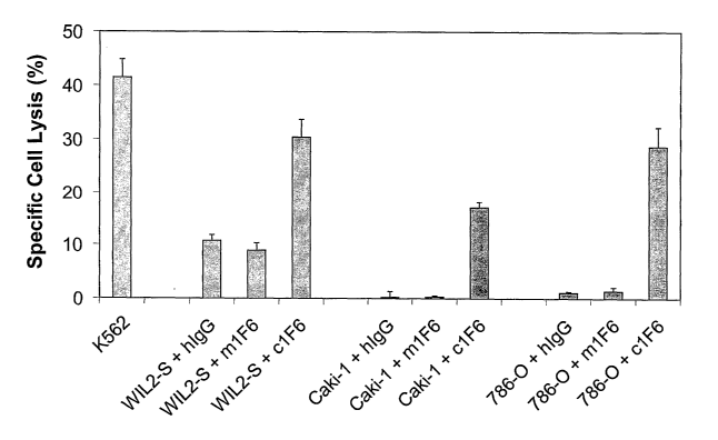
100261 Figure 5. Chimeric 1F6 anti-CD70 antibody mediates antibody-dependent
cellular
cytotoxicity (ADCC). Na251Cr04-labeled target cells (WIL2-S B lymphoblastoid
cells, Caki-
1. renal cell carcinoma cells, and 786-0 renal cell carcinoma cells) were
coated with chimeric
1F6 (c1F6), murine 1.F6 (m1F6), or human IgG (hIgG) and mixed with peripheral
blood
mononuclear cells (PBMC) at an effector to target ratio of 30 CD16+ cells to 1
target cell.
After 4 hours, the supernatants from lysed cells were measured on a
scintillation counter.
The percent specific lysis was calculated as West sample cpm ¨ spontaneous
cpm) (total
k
cpm ¨ spontaneous cpm)) X 100. Points represent the mean standard deviation
of triplicate
samples.
[00271 Figure 6. Chimeric 1F6-coated target cells recognized by PBMC from
multiple -
donors. Na251Cr04-labeled Caki-1 renal cell carcinoma cells were coated with
varying
concentrationssof chimeric-1F6 or non-binding control human IgG (hIgG) and
mixed with -
PBMC from t*o normal donors (2051661 and ND016) at an effector to target cell
ratio of 17
CD16+ cells to 1. target cell. peci.fic lysis was assessed by measuring
chromium-51 activity
in culture supernatants four hours later as described in Figure 5.
[00281 Figure 7. Chimeric 1F6 mediates ADCC against lymphoid cell lines. CD70+
B
lymphoblastoid cells (WIL2-S) and cutaneous T cell lymphoma cells (FM) were
labeled with
Na251Cr04 then mixed with chimeric 1F6 or human Ig (hIgG) at various
concentrations as
7
.=.
.
*
col A
CA 02583208 2007-04-03
WO 2006/044643 PCT/US2005/036994
indicated. PBMC-containing CD16+ cells were added to the target cells at a
ratio of 18:1
(CD16+ cells:target) and percent lysis determined after a four-hour incubation
as described in
Figure 5.
[0029] Figure 8. Chimeric 1F6 mediates ADCC against CD70+ multiple myeloma
cell
lines. (A) Expression of CD70 by multiple myeloma cell lines. L-363, JJN-3, LP-
1 and U-
266 cells were stained with a murine anti-CD70 antibody (open histograms) or a
non-binding
murine IgG control antibody (solid histograms). Antibody binding was detected
with FITC-
conjugated anti-mouse IgG and the cells analyzed by flow cytometry. (B) ADCC
activity of
c1F6. CD38+/CD138+/CD70+ multiple myeloma cell lines were labeled with
Na25'Cr04 and
then mixed with chimeric 1F6 (solid squares) or human Ig (solid triangle) at
various
concentrations as indicated. CD16+ cells enriched from PBMC were added to the
target cells
at a ratio of 15:1 (CD16+ cells:target) and the percent lysis determined after
a four-hour
incubation as described in Figure 5. ADCC activity was blocked by pre-
incubating the
CD16+ effector cells with antibody to FcyRIII (CD16, open squares). Numbers
within each
graph indicate the number of CD70 molecules expressed by each cell lines
estimated using
the QIFIKIT (DakoCytomation, Carpinteria, CA)
[0030] Figure 9. Chimeric 1F6 mediates ADCC against Hodgkin's disease (HD)
cell lines.
CD70+ HD cell lines Hs445 and L428 were labeled with Na251Cr04 then mixed with
chimeric
1F6 or human Ig (hIgG) at concentrations indicated. PBMC-containing CD16+
cells were
added to the target cells at a ratio of 18:1 (CD16+ cells:target) and percent
lysis determined
after a four-hour incubation as described in Figure 5. Numbers within each
graph indicate the
number of CD70 molecules expressed by each cell lines estimated using the
QIFIKIT
(DakoCytomation, Carpinteria, CA)
[0031] Figure 10. CD70 induced during antigen-specific T cell expansion. PBMCs
from a
normal HLA-A0201 donor were stimulated with the M1 peptide derived from the
influenza
virus matrix protein. (A) and B show a representative example of specific CD70
induction on
the expanding CD8+NI317+ after stimulation with the M1 peptide for five days.
Binding of
the control IgG (open curves) and anti-CD70 mAb (closed curves) on the
CD8+Nr317- or
CD8+NI317+ cells are shown.
[0032] Figure 11. Dose response comparison of c1F6 on depletion of antigen-
specific
CD8+/Vf317+ cells. PBMCs from a normal HLA-A0201 donor were stimulated with
the M1
8
CA 02583208 2007-04-03
WO 2006/044643 PCT/US2005/036994
peptide as described in Figure 10. Peptide-stimulated cultures were untreated
or initiated
with concurrent addition of irrelevant control mAb, murine anti-CD70 antibody
(m1F6) or
graded doses of chimeric anti-CD70 antibody (c1F6), as indicated. The percent
CD8W017+
cells after 9 days was determined by flow cytometry.
[0033] Figure 12. Chimeric 1F6 mediates complement-dependent cytotoxicity in
CD70+B
cells. CD70+ lymphoblastic NHL line (MHH-PREB-1), EBV- Burkitt's lymphoma line
(MC116), lymphoblastoid B cell line (WIL2-S), and multiple myeloma cell line
(LP-1) were
incubated with graded doses of the indicated antibodies in the presence of 10%
normal
human serum. For MHH-PREB-1, MC116 and WIL2-S antibodies were used at 50, 5,
0.5,
and 0.05 lAg/mL, while for LP-1 antibodies were used at 50, 10, 2, and
0.4m/mL. Human
IgG (hIgG) was used as a non-binding negative control antibody. Cell lysis was
assayed by
cell permeability to the DNA dye, propidium iodide detected by flow cytometry.
Background cell lysis in medium only was subtracted to give specific cell
lysis.
[0034] Figure 13. Chimeric 1F6 mediates complement-dependent cytotoxicity in
CD70+ T
cells. c1F6-mediated CDC of the CD70+ cutaneous T cell lymphoma line HH and a
CD70+
activated normal T cell line (C9D) was evaluated as described in Figure 12.
[0035] Figure 14. Chimeric 1F6 mediates antibody-dependent cellular
phagocytosis
(ADCP) against CD70+ cells. CD70+ lymphoblastoid cells (W1L2-S) were labeled
with a
green fluorescent cell membrane dye (PKH67), treated with graded doses of c1F6
then mixed
with monocyte-derived macrophages. After two hours, the mixture was incubated
with a PE-
conjugated anti-CD1lb antibody to label the macrophage surface. Uptake of
antibody-coated
target cells by macrophages was determined by flow cytometric analysis of
green and red
double fluorescent cells. For fluorescent microscopy, CD1113+ cells were
additionally stained
with Alexa FluorTm568 goat anti-mouse IgG to enhance the red signal. (A) WIL2-
S cells
were treated with control antibody (hIgG1) or c1F6 and mixed with macrophages.
The
percent phagocytic cells (of total macrophages) that ingested antibody-coated
target cells are
indicated in the upper right quadrant. (B) W1L2-S cells were treated with
graded doses of
c1F6 (triangles) or nonbinding control Ig (hIgGl, circle) and the percent of
phagocytic cells
that ingested target cells was determined by flow cytometry.
[0036] Figure 15. Chimeric 1F6 mediates ADCP against multiple CD70+ cell
targets. The
indicated CD70+ lymphoma, multiple myeloma, and renal cell carcinoma cell
lines were used
9
CA 02583208 2007-04-03
WO 2006/044643 PCT/US2005/036994
as targets for chimeric 1F6-mediated ADCP assays as described in Figure 15.
Percentage
specific ADCP activity at saturating concentrations of chimeric 1F6 is
tabulated.
[0037] Figure 16. In vivo antitumor activity of c1F6 in CD70+ xenograft
lymphoma
models. SC1D mice (n=10/group) were inoculated intravenously with 1 x 10⁶
Ramos cells or
IM-9 cells one day prior to drug treatment. A single dose of chimeric 1F6 was
administered
at 1 or 4 mg/kg and a single dose of the non-binding control antibody (IgG)
was administered
at 4 mg/kg. Survival was monitor and difference between treatment groups was
compared
using the log-rank test as indicated by the P values.
DETAILED DESCRIPTION OF THE INVENTION
[0038] The present invention provides CD70 binding agent and methods for using
such
binding agents for the the prophylaxis or treatment of CD70-expressing cancers
and
immunological disorders. The CD70 binding agent includes domain that binds to
CD70 (e.g.,
the extracellular domain) and an effector domain. The present inventors have
discovered that
a CD70 binding agent containing an effector domain can induces a cytotoxic,
cytostatic, or
immunosuppressive effect on CD70-expressing cells in the absence of
conjugation to a
therapeutic agent. The cytotoxic, cytostatic, or immunosuppressive effect can
be induced, for
example, by recruiting and activating cytotoxic white blood cells, e.g.,
natural killer (NK)
cells, phagocytotic cells (e.g., macrophages) and/or serum complement
components.
[0039] In one aspect, the methods and compositions relate to antibodies and
antibody
derivatives that bind to CD70. In an exemplary embodiment, the antibodies or
derivatives
thereof compete with monoclonal antibody 1F6 or 2F2 for binding to CD70. A
cytotoxic,
cytostatic, and/or immunosuppressive effect is mediated by the CD70 antibody
or derivative
and effector cells or complement components that interact with an effector
domain (e.g., an
Fc region) of the antibody. The cytotoxic, cytostatic, and/or
immunosuppressive effect
depletes or inhibits the proliferation of CD70-expressing cells. CD70
antibodies can be
monoclonal, chimeric, humanized, and human antibodies. In some embodiments,
the
antibody constant regions are of the IgG subtype. In some embodiments, the
antibody is not
a mouse monoclonal antibody.
[0040] In another aspect, the methods and compositions relate to other CD70-
binding
agents that bind to CD70. The CD70-binding agent binds to an extracellular
domain of
CA 02583208 2007-04-03
WO 2006/044643 PCT/US2005/036994
CD70. A cytotoxic, cytostatic, and/or immunosuppressive effect is mediated by
the CD70-
binding agent and effector cells or complement components that interact with
an effector
domain (e.g., an Fc region). The cytotoxic, cytostatic, and/or
immunosuppressive effect
depletes or inhibits the proliferation of CD70-expressing cells. CD70-binding
agents can be,
for example, CD27 and derivatives thereof.
I. Definitions and Abbreviations
[0041] Unless defined otherwise, all technical and scientific terms used
herein have the same
meaning as commonly understood by one of ordinary skill in the art pertinent
to the methods
and compositions described. As used herein, the following terms and phrases
have the
meanings ascribed to them unless specified otherwise.
[0042] The terms "inhibit" or "inhibition of" as used herein means to reduce
by a measurable
amount, or to prevent entirely.
[0043] The term "CD 70 binding agent" as used herein means an anti-CD70
antibody, a
derivative of an anti-CD70 antibody, or other agent that binds to CD70, such
as an
extracellular domain or a portion thereof.
[0044] A "therapeutic agent" is an agent that exerts a cytotoxic, cytostatic,
or
immunosuppressive effect on cancer cells or activated immune cells.
[0045] A "cytotoxic effect" refers to the depletion, elimination and/or the
killing of a target
cell. A "cytotoxic agent" refers to an agent that has a cytotoxic effect on a
cell.
[0046] A "cytostatic effect" refers to the inhibition of cell proliferation. A
"cytostatic
agent" refers to an agent that has a cytostatic effect on a cell (or a
specific subset of cells),
thereby inhibiting the growth and/or expansion of the cell (or specific subset
of cells).
[0047] The term "deplete," in the context of the effect of a CD70-binding
agent on CD70-
expressing cells, refers to a reduction or elimination of the CD70-expressing
cells.
[0048] The term "immunosuppressive agent" as used herein refers to an agent
that inhibits
the development or maintenance of an immunologic response. Such inhibition can
be
effected by, for example, elimination of immune cells (e.g., T or B
lymphocytes); induction
or generation of immune cells that can modulate (e.g., down-regulate) the
functional capacity
11
CA 02583208 2007-04-03
WO 2006/044643 PCT/US2005/036994
of other cells; induction of an unresponsive state in immune cells (e.g.,
anergy); or increasing,
decreasing or changing the activity or function of immune cells, including,
for example,
altering the pattern of proteins expressed by these cells (e.g., altered
production and/or
secretion of certain classes of molecules such as cytokines, chemokines,
growth factors,
transcription factors, kinases, costimulatory molecules or other cell surface
receptors, and the
like). In typical embodiments, an immunosuppressive agent has a cytotoxic or
cytostatic
effect on an immune cell that promotes an immune response.
[0049] "Immune cell" as used herein refers to a cell of hematopoietic lineage
involved in
regulating an immune response. In typical embodiments, an immune cell is a T
lymphocyte,
a B lymphocyte, an NK cell, a monocyte/macrophage, or a dendritic cell.
[0050] The term "polypeptide" refers to a polymer of amino acids and its
equivalent and does
not refer to a specific length of a product; thus, "peptides" and "proteins"
are included within
the definition of a polypeptide. Also included within the definition of
polypeptides are
"antibodies" as defined herein. A "polypeptide region" refers to a segment of
a polypeptide,
which segment may contain, for example, one or more domains or motifs (e.g., a
polypeptide
region of an antibody can contain, for example, one or more complementarity
determining
regions (CDRs)). The term "fragment" refers to a portion of a polypeptide
typically having at
least 20 contiguous or at least 50 contiguous amino acids of the polypeptide.
A "derivative"
is a polypeptide or fragment thereof having one or more non-conservative or
conservative
amino acid substitutions relative to a second polypeptide; or a polypeptide or
fragment
thereof that is modified by covalent attachment of a second molecule such as,
e.g., by
attachment of a heterologous polypeptide, or by glycosylation, acetylation,
phosphorylation,
and the like. Further included within the definition of "derivative" are, for
example, a
polypeptides containing one or more analogs of an amino acid (e.g., unnatural
amino acids
and the like), polypeptides with unsubstituted linkages, as well as other
modifications known
in the art, both naturally and non-naturally occurring.
[0051] The term "antibody" as used herein refers to (a) immunoglobulin
polypeptides and
immunologically active portions of immunoglobulin polypeptides (i.e.,
polypeptides of the
immunoglobulin family, or fragments thereof, that contain an antigen binding
site that
immunospecifically binds to a specific antigen (e.g., CD70)), or (b)
conservatively
substituted derivatives of such immunoglobulin polypeptides or fragments that
12
CA 02583208 2007-04-03
WO 2006/044643 PCT/US2005/036994
immunospecifically bind to the antigen (e.g., CD70). Antibodies are generally
described in,
for example, Harlow and Lane, Antibodies: A Laboratory Manual (Cold Spring
Harbor
Laboratory Press, 1988).
[0052] In the context of immunoglobulin polypeptides or fragments thereof as
defined above,
"conservative substitution" means one or more amino acid substiutions that do
not
substantially reduce specific binding (e.g., as measured by the KD) of the
immunoglobulin
polypeptide or fragment thereof to an antigen (i.e., substitutions that
increase binding, that do
not significantly alter binding, or that reduce binding by no more than about
40%, typically
no more than about 30%, more typically no more than about 20%, even more
typically no
more than about 10%, or most typically no more than about 5%, as determined by
standard
binding assays such as, e.g., ELISA).
[0053] An "antibody derivative" as used herein refers to an antibody, as
defined above, that
is modified by covalent attachment of a heterologous molecule such as, e.g.,
by attachment of
a heterologous polypeptide, or by glycosylation, acetylation or
phosphorylation not normally
associated with the antibody, and the like. In some embodiments, the
heterologous molecule
is not a therapeutic agent. In some embodiments, the heterologous molecule
does not exhibit
a cytostatic or cytotoxic effect by itself.
[0054] The term "monoclonal antibody" refers to an antibody that is derived
from a single
cell clone, including any eukaryotic or prokaryotic cell clone, or a phage
clone, and not the
method by which it is produced. Thus, the term "monoclonal antibody" as used
herein is not
limited to antibodies produced through hybridoma technology.
[0055] The term "heterologous," in the context of a polypeptide, means from a
different
source (e.g., a cell, tissue, organism, or species) as compared with another
polypeptide, so
that the two polypeptides are different. Typically, a heterologous polypeptide
is from
different species.
[0056] As used herein, the term "functional," in the context of an CD70
binding agent
indicates that the binding agent is (1) capable of binding to CD70 and (2)
depletes or inhibits
the proliferation of CD70-expressing cells without conjugation to a cytotoxic
or cytostatic
agent, or has an immunsuppressive effect on an immune cell without conjugation
to an
immunosuppressive agent.
13
CA 02583208 2007-04-04
Pr C T :4" '12; ,0" 315.ept '1:3 114ir 14iiiraminunei, ge
14)
PEA/us 31 AUG .2#01*
[0057] The term "antibody effector function(s)," or AEF, as used herein refers
to a function
contributed by an Fe effector domain(s) of an Ig (e.g., the Fe region of an
immunoglobulin).
Such function can be effected by, for example, binding of an Fe effector
domain(s) to an Fc
receptor on an immune cell with phagocytic or lytic activity or by binding of
an Fe effector
domain(s) to components of the complement system. Typically, the effect(s)
mediated by the
Fe-binding cells or complement components result in inhibition and/or
depletion of the CD70
targeted cell.
[0058]
The
term "antibody-dependent cellular cytotoxicity", or ADCC, is a mechanism
for inducing cell death that depends upon the interaction of antibody-coated
target cells (i.e.,
cells with bound antibody) with immune cells possessing lytic activity (also
referred to as
effector cells). Such effector cells include natural killer cells,
monocytesimacrophages and
neutrophils. ADCC is triggered by interactions between the Fe region of an
antibody bound
to a tumor cell and Fey receptors, particularly FcyRI and FcyR111, on immune
effector cells
such as neutrophils, macrophages and natural killer cells. The tumor cell is
eliminated by
phagocytosis or lysis, depending upon the type of mediating effector cell.
Death of the
antibody-coated target cell occurs as a result of effector cell activity.
[0059] The term "antibody-dependent cellular phagocytosis", or ADCP, refers to
the
process by which antibody-coated cells are internalized, either in whole or in
part, by
õ
phagocytic immune cells (e.g., macrophages, neutrophils and dendritic cells)
that bind to an
,
\
immunogiobulin Fc region.
[0060] The term "complement-dependent cytotoxicity" or CDC refers to a
mechanism for
inducing cell death in which an Fe effector domain(s) of a target-bound
antibody activates a
series of enzymatic reactions culminating in the formation of holes in the
target cell
membrane. Typically, antigen-antibody complexes such as those on antibody-
coated target
cells bind and activate complement component Clq which in turn activates the
complement
cascade leading to target cell death. Activation of complement may also result
in deposition
of complement components on the target cell surface that facilitate ADCC by
binding
complement receptors (e.g., CR3) on leukocytes.
[0061] The terms "identical" or "percent identity," in the context of two or
more nucleic
=
acids or polypeptide sequences, refer to two or more sequences or subsequences
that are the
14
t SHE.
CA 02583208 2007-04-03
WO 2006/044643 PCT/US2005/036994
same or have a specified percentage of nucleotides or amino acid residues that
are the same,
when compared and aligned for maximum correspondence. To determine the percent
identity, the sequences are aligned for optimal comparison purposes (e.g.,
gaps can be
introduced in the sequence of a first amino acid or nucleic acid sequence for
optimal
alignment with a second amino or nucleic acid sequence). The amino acid
residues or
nucleotides at corresponding amino acid positions or nucleotide positions are
then compared.
When a position in the first sequence is occupied by the same amino acid
residue or
nucleotide as the corresponding position in the second sequence, then the
molecules are
identical at that position. The percent identity between the two sequences is
a function of the
number of identical positions shared by the sequences (i.e., % identity = # of
identical
positions/total # of positions (e.g., overlapping positions) x 100). In some
embodiments, the
two sequences are the same length.
[0062] The term "substantially identical," in the context of two nucleic acids
or polypeptides,
refers to two or more sequences or subsequences that have at least 50%, at
least 55%, at least
60%, or at least 65% identity; typically at least 70% or at least 75%
identity; more typically at
least 80% or at least 85% identity; and even more typically at least 90%, at
least 95%, or at
least 98% identity (e.g., as determined using one of the methods set forth
infra).
[0063] The terms "similarity" or "percent similarity" in the context of two or
more
polypeptide sequences, refer to two or more sequences or subsequences that
have a specified
percentage of amino acid residues that are the same or conservatively
substituted when
compared and aligned for maximum correspondence, as measured using one of the
methods
set forth infra. By way of example, a first amino acid sequence can be
considered similar to a
second amino acid sequence when the first amino acid sequence is at least 50%,
60%, 70%,
75%, 80%, 90%, or 95% identical, or conservatively substituted, to the second
amino acid
sequence when compared to an equal number of amino acids as the number
contained in the
first sequence, or when compared to an alignment of polypeptides that has been
aligned by a,
e.g., one of the methods set forth infra.
[0064] The terms "substantial similarity" or "substantially similar," in the
context of
polypeptide sequences, indicates that a polypeptide region has a sequence with
at least 70%,
typically at least 80%, more typically at least 85%, or at least 90% or at
least 95% sequence
CA 02583208 2007-04-03
WO 2006/044643 PCT/US2005/036994
similarity to a reference sequence. For example, a polypeptide is
substantially similar to a
second polypeptide, when the two peptides differ by one or more conservative
substitutions.
[0065] In the context of anti-CD70 antibodies or derivatives thereof, a
protein that has one or
more polypeptide regions substantially identical or substantially similar to
one or more
antigen-binding regions (e.g., a heavy or light chain variable region, or a
heavy or light chain
CDR) of an anti-CD70 antibody retains specific binding to an epitope of CD70
recognized by
the anti-CD70 antibody, as determined using any of various standard
immunoassays known
in the art or as referred to herein.
[0066] The determination of percent identity or percent similarity between two
sequences can
be accomplished using a mathematical algorithm. A preferred, non-limiting
example of a
mathematical algorithm utilized for the comparison of two sequences is the
algorithm of
Karlin and Altschul, 1990, Proc. Natl. Acad. Sci. USA 87:2264-2268, modified
as in Kahn
and Altschul, 1993, Proc. Natl. Acad. Sci. USA 90:5873-5877. Such an algorithm
is
incorporated into the NBLAST and )(BLAST programs of Altschul et al., 1990, J.
Mol. Biol.
215:403-410. BLAST nucleotide searches can be performed with the NBLAST
program,
score = 100, wordlength = 12, to obtain nucleotide sequences homologous to a
nucleic acid
encoding a protein of interest. BLAST protein searches can be performed with
the XBLAST
program, score = 50, wordlength = 3, to obtain amino acid sequences homologous
to protein
of interest. To obtain gapped alignments for comparison purposes, Gapped BLAST
can be
utilized as described in Altschul et al., 1997, Nucleic Acids Res. 25:3389-
3402.
Alternatively, PSI-Blast can be used to perform an iterated search which
detects distant
relationships between molecules (Id.). When utilizing BLAST, Gapped BLAST, and
PSI-
Blast programs, the default parameters of the respective programs (e.g.,
)(BLAST and
NBLAST) can be used Another preferred, non-limiting example of a mathematical
algorithm
utilized for the comparison of sequences is the algorithm of Myers and Miller,
CABIOS
(1989). Such an algorithm is incorporated into the ALIGN program (version 2.0)
which is
part of the GCG sequence alignment software package. When utilizing the ALIGN
program
for comparing amino acid sequences, a PAM120 weight residue table, a gap
length penalty of
12, and a gap penalty of 4 can be used. Additional algorithms for sequence
analysis are
known in the at and include ADVANCE and ADAM as described in Torellis and
Robotti,
1994, Comput. Appl. Biosci. 10:3-5; and FASTA described in Pearson and Lipman,
1988,
Proc. Natl. Acad. Sci.USA 85:2444-8. Within FASTA, ktup is a control option
that sets the
16
CA 02583208 2007-04-03
WO 2006/044643 PCT/US2005/036994
sensitivity and speed of the search. If ktup=2, similar regions in the two
sequences being
compared are found by looking at pairs of aligned residues; if ktup=1, single
aligned amino
acids are examined. ktup can be set to 2 or 1 for protein sequences, or from 1
to 6 for DNA
sequences. The default if ktup is not specified is 2 for proteins and 6 for
DNA.
[0067] Alternatively, protein sequence alignment may be carried out using the
CLUSTAL W
algorithm, as described by Higgins et al., 1996, Methods Enzynzol. 266:383-
402.
[0068] As used herein, the terms "prevention" or "prevent" refer to
administration of an anti-
CD70 antibody or derivative or other binding agent to a subject before the
onset of a clinical
or diagnostic symptom of a CD70-expressing cancer or immunological disorder
(e.g.,
administration to an individual with a predisposition or at a high risk of
acquiring the CD70-
expressing cancer or immunological disorder) to (a) block the occurrence or
onset of the
CD70-expressing cancer or immunological disorder, or one or more of clinical
or diagnostic
symptoms thereof, (b) inhibit the severity of onset of the CD70-expressing
cancer or
immunological disorder, or (c) to lessen the likelihood of the onset of the
CD70-expressing
cancer or immunological disorder.
[0069] As used herein, the terms "treatment" or "treat" refer to slowing,
stopping, and/or
reversing the progression of a CD70-expressing cancer or immunological
disorder in a
subject, as evidenced by a decrease or elimination of a clinical or diagnostic
symptom of the
disease, by administration of an anti-CD70 antibody or derivative thereof or
other binding
agent to the subject after the onset of the clinical or diagnostic symptom of
the CD70-
expressing cancer or immunological disorder at any clinical stage. Treatment
can include, for
example, a decrease in the severity of a symptom, the number of symptoms, or
frequency of
relapse.
[0070] The term "pharmaceutically acceptable" as used herein means approved by
a
regulatory agency of the Federal or a state government or listed in the U.S.
Pharmacopeia or
other generally recognized pharmacopeia for use in animals, and more
particularly in
humans. The term "pharmaceutically compatible ingredient" refers to a
pharmaceutically
acceptable diluent, adjuvant, excipient, or vehicle with which an anti-CD70-
binding agent is
administered.
17
CA 02583208 2007-04-04
PEA/US 31 AUG Aitio
plc: "T i "õ:"-¶11. 115 gçjfr.6
õ1:;:l! i. 8)
[00711 The term "effective amount" refers to the amount of the antibody or
derivative or
other binding agent that is sufficient to inhibit the occurrence or ameliorate
one or more
clinical or diagnostic symptoms of a CD70-expressing cancer or immunological
disorder in a
subject. An effective amount of an agent is administered according to the
methods described
herein in an "effective regime." The term "effective regime" refers to a
combination of
amount of the agent and dosage frequency adequate to accomplish treatment or
prevention of
a CD70-expressing cancer or immunological disorder.
II.Anti-CD70 Antibodies and Derivatives Thereof
[00721 The methods and compositions described herein encompass the use of a
CD70
binding agent that specifically binds to CD70 and exerts a cytotoxic,
cytostatic or
imrnunosuppressive effect on CD70-expressing cancer cells or activated immune
cells. The
CD70 binding agent can be, for example, an anti-CD70 antibody, an antigen-
binding
fragment of an anti-CD70 antibody, a derivative thereof, or other CD70-binding
agent. The
CD70-binding agent includes an antibody effector domain fimction that mediates
or
stimulates ADCC, ADCP and/or CDC responses against a CD70-expressing target
cell. The
effector domain(s) can be, for example, an Fe region of an Ig molecule. The
CD70-binding
agent exerts a cytotoxic or cytostatic effect on CD70-expressing cancer cells,
or exerts a
cytotoxic, cytostatic, or immunosuppressive effect on activated lymphocytes or
dendritic
cells, for the treatment of a CD70-expressing cancer or an immunological
disorder,
respectively. Typically, the CD70-binding agent recruits and/or activates
cytotoxic white
blood cells (e.g., natural killer (1\TIC) cells, phagocytotic cells (e.g.,
macrophages), and/or
serum complement components). In some embodiments, the CD70 binding agent is
monoclonal antibody (mAb) 1F6 or 2F2 or a derivative thereof. In other
embodiments, the
anti-CD70 antibody or derivative thereof competes with monoclonal antibody 1.
F6 or 2F2 for
binding to CD70. In some embodiments, the CD70 binding agent does not induce
an
agonistic or antagonistic signal when binding to CD70.
[0073] An anti-CD70 antibody typically is or is derived from a monoclonal
antibody and can
include, for example, a chimeric (e.g., having a human constant region and
mouse variable
region), a humanized, or a fully human antibody; a single, chain antibody; a
maxibody, a
minibody, an antigen binding region, or the like. The antibody molecule
includes at least one
effector domain that can functionally interact with and activate cytotoxic
white blood cells
18
AMENDED
CA 02583208 2007-04-03
WO 2006/044643 PCT/US2005/036994
and/or serum complement components. In some embodiments, a CD70 antigen
binding
region can be joined to an effector domain or domains such as, for example,
hinge-CH2-CH3
domains of an immunoglobulin, or a portion or fragment of an effector
domain(s) having
effector function. Antigen-binding antibody fragments, including single-chain
antibodies,
can comprise for example the variable region(s) in combination with the
entirety or a portion
of an effector domain (e.g., a CH2 and/or CH3 domain alone or in combination
with a CH1,
hinge and/or CL domain). Also, antigen-binding fragments can comprise any
combination of
effector domains. In some embodiments, the anti-CD70 antibody can be a single
chain
antibody comprising a CD70-binding variable region joined to hinge-C2-CH3
domains.
[0074] Typically, the antibodies are of human, or non-human origin (e.g.,
rodent (e.g.,
mouse or rat)), donkey, sheep, rabbit, goat, guinea pig, camelid, horse, or
chicken) of specific
Ig isotypes that can mediate effector function. As used herein, "human"
antibodies include
antibodies having the amino acid sequence of a human immunoglobulin and
include
antibodies isolated from human immunoglobulin libraries, from human B cells,
or from
animals transgenic for one or more human immunoglobulin, as described infra
and, for
example in U.S. Patent Nos. 5,939,598 and 6,111,166.
[0075] The effector domain of an antibody can be from any suitable vertebrate
animal
species and isotypes. The isotypes from different animal species differ in the
abilities to
mediate effector functions. For example, the ability of human immunoglobulin
to mediate
CDC and ADCC/ADCP is generally in the order of IgMzIgG1zIgG3>IgG2>IgG4 and
IgG14gG3>IgG2/IgM/IgG4, respectively. Murine immunoglobulins mediate CDC and
ADCC/ADCP generally in the order of murine IgM4gG3>>IgG2b>IgG-2a>>IgG1 and
IgG2b>IgGa>IgGl>>IgG3, respectively. In another example, murine IgG2a mediates
ADCC while both murine IgG2a and IgM mediate CDC. In some embodiments, the
CD70
binding agent consists of antibody variable and effector domains. In other
embodiments, the
CD70 binding agent consists essentially of antibody variable and effector
domains, and can
further include an additional compound(s) that is not a therapeutic agent (s).
A CD70-binding
polypeptide also can be expressed as a recombinant fusion protein comprising
of the
appropriate constant domains to yield the desired effector function(s).
[0076] Upon binding to target cells, the antibodies or derivatives can trigger
in vitro and in
vivo target cell destruction through effector domain (e.g., Fc-) mediated
effector functions.
19
CA 02583208 2007-04-03
WO 2006/044643 PCT/US2005/036994
Without intending to be bound by any particular theory, Fc regions of
antibodies can recruit
Fc receptor (FcR)-expressing cells and juxtapose them with antibody-coated
target cells.
Cells expressing surface FcR for IgGs including FcyRIII (CD16), FcyRII (CD32)
and FcyRIII
(CD64) can act as effector cells for the destruction of IgG-coated cells. Such
effector cells
include monocytes, macrophages, natural killer (Nk) cells, neutrophils and
eosinophils.
Engagement of FcyR by IgG activates antibody-dependent cellular cytotoxicity
(ADCC) or
antibody-dependent cellular phagocytosis (ADCP). ADCC is mediated by CD16+
effector
cells through the secretion of membrane pore-forming proteins and proteases,
while
phagocytosis is mediated by CD32+ and CD64+ effector cells (see Fundamental
Immunology,
4th ed., Paul ed., Lippincott-Raven, N.Y., 1997, Chapters 3, 17 and 30; Uchida
et al., 2004, J.
Exp. Med. 199:1659-69; Akewanlop et al., 2001, Cancer Res. 61:4061-65;
Watanabe et al.,
1999, Breast Cancer Res. Treat. 53:199-207). In addition to ADCC and ADCP, Fc
regions
of cell-bound antibodies can also activate the complement classical pathway to
elicit
complement-dependent cytotoxicity (CDC). C lq of the complement system binds
to the Fc
regions of antibodies when they are complexed with antigens. Binding of Clq to
cell-bound
antibodies can initiate a cascade of events involving the proteolytic
activation of C4 and C2
to generate the C3 convertase. Cleavage of C3 to C3b by C3 convertase enables
the
activation of terminal complement components including C5b, C6, C7, C8 and C9.
Collectively, these proteins form membrane-attack complex pores on the
antibody-coated
cells. These pores disrupt the cell membrane integrity, killing the target
cell (see
Immunobiology, 6th ed., Janeway et al., Garland Science, N. Y., 2005, Chapter
2).
[0077] The antibodies can be monospecific, bispecific, trispecific, or of
greater
multispecificity. Multispecific antibodies may be specific for different
epitopes of CD70
and/or may be specific for both CD70 as well as for a heterologous protein.
(See, e.g., PCT
Publications WO 93/17715, WO 92/08802, WO 91/00360, and WO 92/05793; Tutt et
al.,
1991, J. Immunol. 147:60-69; U.S. Patent Nos. 4,474,893; 4,714,681; 4,925,648;
5,573,920;
and 5,601,819; Kostelny et al., 1992, J. Immunol. 148:1547-1553.)
Multispecific antibodies,
including bispecific and trispecific antibodies, useful for practicing the
methods described
herein are antibodies that immunospecifically bind to both CD70 (including but
not limited to
antibodies that have the CDRs of the monoclonal antibodies 2F2 and 1F6) and a
second cell
surface receptor or receptor complex that mediates ADCC, phagocytosis, and/or
CDC, such
as CD16/FcgRIII, CD64/FcgRI, killer inhibitory or activating receptors, or the
complement
CA 02583208 2007-04-03
WO 2006/044643 PCT/US2005/036994
control protein CD59. In a typical embodiment, the binding of the portion of
the
multispecific antibody to the second cell surface molecule or receptor complex
enhances the
effector functions of the anti-CD70 antibody or other CD70 binding agent.
[0078] In one aspect, an anti-CD70 antibody comprises one or more
complementarity
determining regions (CDRs) substantially identical or substantially similar to
one or more
CDR(s) of monoclonal antibody 1F6 (see Table 1). For example, the antibody can
include a
heavy chain CDR and/or a light chain CDR that is substantially identical or
substantially
similar to a corresponding heavy chain CDR (H1, H2, or H3 regions) or
corresponding light
chain CDR (L1, L2, or L3 regions) of mAb 1F6 (SEQ ID NO:6; SEQ ID NO:8; SEQ ID
NO:10; SEQ ID NO:16; SEQ ID NO:18; or SEQ ID NO:20, respectively). In typical
embodiments, the anti-CD70 antibody has two or three heavy chain CDRs and/or
two or
three light chain CDRs that are substantially identical or substantially
similar to
corresponding heavy and/or light chain CDRs of mAb 1F6. In specific
embodiments, a CDR
substantially identical or substantially similar to a heavy or light chain CDR
of 1F6 has the
amino acid sequence set forth in SEQ lD NO:6, SEQ ID NO:8, SEQ ID NO:10, SEQ
ID
NO:16, SEQ ID NO:18, or SEQ ID NO:20.
[0079] For example, in some embodiments, where an anti-CD70 antibody has at
least one
heavy chain CDR substantially identical or substantially similar to a heavy
chain CDR of
mAb 1F6, the antibody or derivative thereof further includes at least one
light chain CDR that
is substantially identical or substantially similar to a light chain CDR of
mAb 1F6.
[0080] In some embodiments, an anti-CD70 antibody includes a heavy or light
chain variable
domain, the variable domain having (a) a set of three CDRs substantially
identical or
substantially similar to corresponding CDRs of mAb 1F6, and (b) a set of four
framework
regions. For example, an anti-CD70 antibody can include a heavy or light chain
variable
domain, the variable domain having (a) a set of three CDRs, in which the set
of CDRs are
from monoclonal antibody 1F6, and (b) a set of four framework regions of the
IgG type.
[0081] In some embodiments, the anti-CD70 antibody is a chimeric antibody. A
chimeric
antibody is a molecule in which different portions of the antibody are derived
from different
animal species, such as for example antibodies having a variable region
derived from a
murine monoclonal antibody and a human IgG immunoglobulin constant region.
Methods
for producing chimeric antibodies are known in the art. (See e.g., Morrison,
Science, 1985,
21
CA 02583208 2007-04-03
WO 2006/044643
PCT/US2005/036994
229:1202; Oi et al., 1986, BioTechniques 4:214; Gillies etal., 1989, J.
hninunol. Methods
125:191-202; U.S. Patent Nos. 5,807,715; 4,816,567; and 4,816,397.)
[0082] In an exemplary embodiment, the anti-CD70 antibody is a chimeric
antibody that
includes a heavy chain variable region that is substantially identical or
substantially similar to
the heavy chain variable region of mAb 1F6 (i.e., substantially identical or
substantially
similar to the amino acid sequences set forth in SEQ ID NO:2, see Table 1)
and/or a light
chain variable region that is substantially identical or substantially similar
to the light chain
variable regions of mAb 1F6 (i.e., substantially identical or substantially
similar to the amino
acid sequences set forth in SEQ ID NO:12, see Table 1). For example, the
antibody can
include a heavy chain variable region having the amino acid sequence set forth
in SEQ ID
NO:2 and, optionally, can further include a light chain variable region having
the amino acid
sequence set forth in SEQ ID NO:12. The heavy and light chain antibody
constant regions
are of the IgG type. In an exemplary embodiment, the anti-CD70 antibody is a
chimeric IgG
mAb 1F6.
[0083] In some embodiments, an anti-CD70 antibody is a chimeric antibody that
includes
one or more CDRs substantially identical or substantially similar to one or
more CDR(s) of
monoclonal antibody 2F2 (see Table 1). For example, the antibody can include a
heavy chain
CDR and/or a light chain CDR that is substantially identical or substantially
similar to a
corresponding heavy chain CDR (H1, H2, or H3 regions) or corresponding light
chain CDR
(L1, L2, or L3 regions) of mAb 2F2 (SEQ ID NO:26, SEQ ID NO:28; SEQ ID NO:30;
SEQ
ID NO:36, SEQ ID NO:38 or SEQ ID NO:40). In typical embodiments, the anti-CD70
antibody has two or three heavy chain CDRs and/or two or three light chain
CDRs that are
substantially identical or substantially similar to corresponding heavy and/or
light chain
CDRs of mAb 2F2. In specific embodiments, a CDR substantially identical or
substantially
similar to a heavy or light chain CDR of 2F2 has the amino acid sequence set
forth in SEQ ID
NO:26, SEQ ID NO:28, SEQ ID NO:30; SEQ ID NO:36, SEQ ID NO:38, or SEQ ID
NO:40.
[0084] For example, in some embodiments, where an anti-CD70 antibody has at
least one
heavy chain CDR substantially identical or substantially similar to a heavy
chain CDR of
mAb 2F2, the antibody or derivative thereof further includes at least one
light chain CDR that
is substantially identical or substantially similar to a light chain CDR of
mAb 2F2.
22
CA 02583208 2007-04-03
WO 2006/044643
PCT/US2005/036994
[0085] In some embodiments, an anti-CD70 antibody includes a heavy or light
chain variable
domain, the variable domain having (a) a set of three CDRs substantially
identical or
substantially similar to corresponding CDRs of mAb 2F2, and (b) a set of four
framework
regions. For example, an anti-CD70 antibody can include a heavy or light chain
variable
domain, the variable domain having (a) a set of three CDRs, in which the set
of CDRs are
from monoclonal antibody 2F2, and (b) a set of four framework regions, in
which the set of
framework regions are of the IgG type. In an exemplary embodiment, the anti-
CD70
antibody is a chimeric IgG mAb 2F2.
[0086] In an embodiment, the anti-CD70 antibody includes a heavy chain
variable region that
is substantially identical or substantially similar to the heavy chain
variable region of mAb
2F2 (i.e., substantially identical or substantially similar to the amino acid
sequences set forth
in SEQ ID NO:22, see Table 1) and/or a light chain variable region that is
substantially
identical or substantially similar to the light chain variable regions of mAb
2F2 (i.e.,
substantially identical or substantially similar to the amino acid sequences
set forth in SEQ
ID NO:32, see Table 1). For example, the antibody can include a heavy chain
variable region
having the amino acid sequence set forth in SEQ ID NO:22 and, optionally, can
further
include a light chain variable region having the amino acid sequence set forth
in SEQ ID
NO:32. In one exemplary embodiment, the anti-CD70 antibody is mAb 2F2.
[0087] In some embodiments, the antibody comprises a 1F6 VH and a 2F2 VL or a
1F6 VH
and a 2F2 VL.
[0088] The following table indicates the regions of 1F6 or 2F2 to which each
SEQ ID NO.
corresponds.
Table 1
NUCLEOTIDE OR AMINO
MOLECULE ACID SEQ ID NO
1F6 Heavy Chain Variable Region Nucleotide 1
1F6 Heavy Chain Variable Region Amino Acid
23
CA 02583208 2007-04-03
WO 2006/044643
PCT/US2005/036994
Table 1
NUCLEOTIDE OR AMINO
MOLECULE ACID SEQ ID NO
1F6 Heavy Chain Signal Peptide Nucleotide 3
1F6 Heavy Chain Signal Peptide Amino Acid 4
1F6 Heavy Chain-CDR1(H1) Nucleotide 5
1F6 Heavy Chain-CDR1 (H1 ) Amino Acid 6
1F6 Heavy Chain-CDR2(H2) Nucleotide 7
1F6 Heavy Chain-CDR2(H2) Amino Acid 8
1F6 Heavy Chain-CDR3(H3) Nucleotide 9
1F6 Heavy Chain-CDR3(H3) Amino Acid 10
1F6 Light Chain Variable Region Nucleotide 11
1F6 Light Chain Variable Region Amino Acid 12
1F6 Light Chain Signal Peptide Nucleotide 13
1F6 Light Chain Signal Peptide Amino Acid 14
1F6 Light Chain-CDR1(L1) Nucleotide 15
1F6 Light Chain-CDR1 (L1) Amino Acid 16
1F6 Light Chain-CDR2(L2) Nucleotide 17
1F6 Light Chain-CDR2(L2) Amino Acid 18
1F6 Light Chain-CDR3(L3) Nucleotide 19
24
CA 02583208 2007-04-03
WO 2006/044643
PCT/US2005/036994
Table 1
NUCLEOTIDE OR AMINO
MOLECULE ACID SEQ ID
NO
1F6 Light Chain-CDR3(L3) Amino Acid 20
2F2 Heavy Chain Variable Region Nucleotide 21
2F2 Heavy Chain Variable Region Amino Acid 22
2F2 Heavy Chain Signal Peptide Nucleotide 23
2F2 Heavy Chain Signal Peptide Amino Acid 24
2F2 Heavy Chain-CDR1(H1) Nucleotide 25
2F2 Heavy Chain-CDR1(H1) Amino Acid 26
2F2 Heavy Chain-CDR2(H2) Nucleotide 27
2F2 Heavy Chain-CDR2(H2) Amino Acid 28
2F2 Heavy Chain-CDR3(H3) Nucleotide 29
2F2 Heavy Chain-CDR3(H3) Amino Acid 30
2F2 Light Chain Variable Region Nucleotide 31
2F2 Light Chain Variable Region Amino Acid 32
2F2 Light Chain Signal Peptide Nucleotide 33
2F2 Light Chain Signal Peptide Amino Acid 34
2F2 Light Chain-CDR1(L1) Nucleotide 35
2F2 Light Chain-CDR1(L1) Amino Acid 36
CA 02583208 2007-04-03
WO 2006/044643 PCT/US2005/036994
Table 1
NUCLEOTIDE OR AMINO
MOLECULE ACID SEQ ID NO
2F2 Light Chain-CDR2(L2) Nucleotide 37
2F2 Light Chain-CDR2(L2) Amino Acid 38
2F2 Light Chain-CDR3(L3) Nucleotide 39
2F2 Light Chain-CDR3(L3) Amino Acid 40
[0089] Anti-CD70 antibodies and derivatives thereof and other binding agents
may also be
described or specified in terms of their binding affinity to CD70. Typical
binding affinities
include those with a dissociation constant or Kd less than 5 X 10⁻²M, 10-2M, 5
X i0 M, 10-3
M, 5 X HO M, i0 M, 5 X 1.0-5 M, lir M, 5 X le M, 10-6M, 5 X i0 M, i0 M, 5 X 10-
8
M, 10-8 M, 5 X le NI, i0 M, 5 X 10⁻¹ M, 1040 M, 5 X 10-11M, 10-11M, 5 X 10-
12M, 10-12
M, 5 X -13 M,10-13 M, 5 X 10-14M, 10-14M, 5 X10-15 M, or l0' M.
[0090] The antibodies can be generated by methods known in the art. For
example,
monoclonal antibodies can be prepared using a wide variety of techniques
including, e.g., the
use of hybridoma, recombinant, and phage display technologies, or a
combination thereof.
Hybridoma techniques are generally discussed in, for example, Harlow et al.,
Antibodies: A
Laboratory Manual (Cold Spring Harbor Laboratory Press, 2nd ed., 1988); and
Hammerling,
et al., In Monoclonal Antibodies and T-Cell Hybridomas, pp. 563-681 (Elsevier,
N.Y., 1981).
Examples of phage display methods that can be used to make the anti-CD70
antibodies
include, e.g., those disclosed in (Hoogenboom and Winter, 1991, J. Mol. Biol.
227:381;
Marks et al., 1991, J. Mol. Biol. 222:581; Quan and Carter, 2002, The rise of
monoclonal
antibodies as therapeutics in Anti-IgE and Allergic Disease, Jardieu and Fick
Jr., eds., Marcel
Dekker, New York, NY, Chapter 20, pp. 427-469; Brinkman et al., 1995, J.
Immunol.
Methods 182:41-50; Ames et al., 1995, J. Immunol. Methods 184:177-186;
Kettleborough et
al., 1994, Eur. J. Immunol. 24:952-958; Persic et al., 1997, Gene 187:9-18;
Burton et al.,
1994, Advances in Immunology 57:191-280; PCT Application No. PCT/GB91/01134;
PCT
Publications WO 90/02809, WO 91/10737, WO 92/01047, WO 92/18619, WO 93/11236,
26
CA 02583208 2013-02-01
WO 95/15982, WO 95/20401, and U.S. Patent Nos. 5,698,426; 5,223,409;
5,403,484; 5,580,717;
5,427,908; 5,750,753; 5,821,047; 5,571,698; 5,427,908; 5,516,637; 5,780,225;
5,658,727;
5,733,743 and 5,969,108.
[0091] Examples of techniques that can be used to produce single-chain
antibodies include those
described in U.S. Patents 4,946,778 and 5,258,498; Huston etal., 1991, Methods
in Enzymology
203:46-88; Shu etal., 1993, Proc. Natl. Acad. Sci. USA 90:7995-7999; and
Skerra etal., 1988,
Science 240:1038-1040.
[0092] Methods for making bispecific antibodies are known in the art.
Traditional production
of full-length bispecific antibodies is based on the coexpression of two
immunoglobulin heavy
chain-light chain pairs, where the two chains have different specificities
(see, e.g., Milstein etal.,
1983, Nature 305:537-39). Because of the random assortment of immunoglobulin
heavy and light
chains, these hybridomas (quadromas) produce a potential mixture of 10
different antibody
molecules, of which only one has the correct bispecific structure. Similar
procedures are
disclosed in International Publication No. WO 93/08829, and in Traunecker
etal., 1991, EMBO J.
10:3655-59.
[0093] According to a different approach, antibody variable domains with the
desired binding
specificities (antibody-antigen combining sites) are fused to immunoglobulin
constant domain
sequences. The fusion preferably is with an immunoglobulin heavy chain
constant domain,
comprising at least part of the hinge, CH2, and CH3 regions. In some
embodiments, the fusion
includes a first heavy-chain constant region (CHI) containing the site
necessary for light chain
binding, present in at least one of the fusions. Nucleic acids with sequences
encoding the
immunoglobulin heavy chain fusions and, if desired, the immunoglobulin light
chain, are inserted
into separate expression vectors, and are co-transfected into a suitable host
organism. This
provides for great flexibility in adjusting the mutual proportions of the
three polypeptide
fragments in embodiments when unequal ratios of the three polypeptide chains
used in the
construction provide the optimum yields. It is, however, possible to insert
the coding sequences
for two or all three polypeptide chains in one expression vector when the
expression of at least
two polypeptide chains in equal ratios results in high yields or when the
ratios are of no particular
significance.
- 27 -
CA 02583208 2013-02-01
=
[0094] In an embodiment of this approach, the bispecific antibodies have a
hybrid
immunoglobulin heavy chain with a first binding specificity in one arm, and a
hybrid
immunoglobulin heavy chain-light chain pair (providing a second binding
specificity) in the other
arm. This asymmetric structure facilitates the separation of the desired
bispecific compound from
unwanted immunoglobulin chain combinations, as the presence of an
immunoglobulin light chain
in only one half of the bispecific molecule provides for a facile way of
separation (see, e.g,
International Publication No. WO 94/04690.
[0095] For further discussion of bispecific antibodies see, for example,
Suresh et al., 1986,
Methods in Enzymology 121:210; Rodrigues et al., 1993, J. Immunology 151:6954-
61; Carter et
al., 1992, Bio/Technology 10:163-67; Carter et al., 1995,1 Hematotherapy 4:463-
70; Merchant et
al., 1998, Nature Biotechnology 16:677-81. Using such techniques, bispecific
antibodies can be
prepared for use in the treatment or prevention of disease as defined herein.
[0096] Bifunctional antibodies are also described in European Patent
Publication No. EPA 0
105 360. As disclosed in this reference, hybrid or bifunctional antibodies can
be derived either
biologically, i.e., by cell fusion techniques, or chemically, especially with
cross-linking agents or
disulfide-bridge forming reagents, and may comprise whole antibodies or
fragments thereof.
Methods for obtaining such hybrid antibodies are disclosed for example, in
International
Publication WO 83/03679 and European Patent Publication No. EPA 0 217 577.
[0097] An anti-CD70 antibody can also be a humanized antibody. Humanized
antibodies are
antibody molecules that bind the desired antigen and have one or more CDRs
from a non-human
species, and framework and constant regions from a human immunoglobulin
molecule. Often,
framework residues in the human framework regions will be substituted with the
corresponding
residue from the CDR donor antibody to alter, preferably improve, antigen
binding. These
framework substitutions are identified by methods well known in the art, e.g.,
by modeling of the
interactions of the CDR and framework residues to identify framework residues
important for
antigen binding and sequence comparison to identify unusual framework residues
at particular
positions. (See, e.g., Queen et al., U.S. Patent No. 5,585,089; Riechmann et
al., 1988, Nature
332:323.) Antibodies can be humanized using a variety of techniques known in
the art including,
for example, CDR-grafting (see, e.g., EP 0 239 400; PCT publication WO
91/09967; U.S. Patent
Nos. 5,225,539; 5,530,101; and 5,585,089), veneering or resurfacing (see,
e.g., EP 0 592 106; EP
- 28 -
CA 02583208 2013-02-01
=
0 519 596; Padlan, Molecular Immunology, 1991, 28(4/5):489-498; Studnicka et
al., 1994,
Protein Engineering 7(6):805-814; Roguska et al., 1994, Proc. Natl. Acad. Sci.
USA 91:969-973),
and chain shuffling (see, e.g., U.S. Patent No. 5,565,332).
[0098] Humanized monoclonal antibodies can be produced by recombinant DNA
techniques
known in the art, for example using methods described in International
Publication No. WO
87/02671; European Patent Publication No. 0 184 187; European Patent
Publication No. 0 171
496; European Patent Publication No. 0 173 494; International Publication No.
WO 86/01533;
U.S. Patent No. 4,816,567; European Patent Publication No. 0 012 023; Berter
etal., 1988,
Science 240:1041-43; Liu et al., 1987, Proc. Natl. Acad. Sci. USA 84:3439-43;
Liu etal., 1987, J.
Immunol. 139:3521-26; Sun etal., 1987, Proc. Natl. Acad. Sci. USA 84:214-18;
Nishimura etal.,
1987, Cancer. Res. 47:999-1005; Wood etal., 1985, Nature 314:446-449; and Shaw
etal., 1988,
J. Natl. Cancer Inst. 80:1553-59; Morrison, 1985, Science 229:1202-07; Oi
etal., 1986,
BioTechniques 4:214; U.S. Patent 5,225,539; Jones etal., 1986, Nature 321:552-
25; Verhoeyan et
al., 1988, Science 239:1534; and Beidler etal., 1988,J. Immunol. 141:4053-60.
[0099] In some embodiments, the anti-CD70 antibody is a human IgG antibody.
Human
antibodies can be made by a variety of methods known in the art including,
e.g., phage display
methods (see supra) using antibody libraries derived from human immunoglobulin
sequences.
See also, e.g., U.S. Patent Nos. 4,444,887 and 4,716,111; and PCT Publications
WO 98/46645,
WO 98/50433, WO 98/24893, WO 98/16654, WO 96/34096, WO 96/33735, and WO
91/10741.
= In addition, a human antibody recognizing a selected epitope can be
generated using a technique
referred to as "guided selection," in which a selected non-human monoclonal
antibody, e.g., a
mouse antibody, is used to guide the selection of a completely human antibody
recognizing the
same epitope (see, e.g., Jespers etal., 1994, Bio/technology 12:899-903).
Human antibodies can
also be produced using transgenic mice that express human immunoglobulin
genes. Monoclonal
antibodies directed against the antigen can be obtained from the immunized,
transgenic mice
using conventional hybridoma technology. For an overview of this technology
for producing
human antibodies, see Lonberg and Huszar, 1995, Int. Rev. Immunol. 13:65-93.
For a detailed
discussion of this technology for producing human antibodies and human
monoclonal antibodies
and protocols for producing such antibodies, see, e.g., PCT Publications WO
98/24893, WO
92/01047, WO 96/34096, and WO 96/33735; European Patent No. 0 598 877; U.S.
Patent Nos.
- 29 -
CA 02583208 2013-02-01
=
5,413,923; 5,625,126; 5,633,425; 5,569,825; 5,661,016; 5,545,806; 5,814,318;
5,885,793;
5,916,771; and 5,939,598.
[0100] In addition, companies such as Abgenix, Inc. (Fremont, CA), Genpharm
(San Jose, CA),
and Medarex (Princeton, NJ) can be engaged to provide human antibodies
directed against a
selected antigen using technology similar to that described above. Completely
human antibodies
can be produced using transgenic mice that are incapable of expressing
endogenous
immunoglobulin heavy and light chains genes, but which can express human heavy
and light
chain genes. The transgenic mice are immunized in the normal fashion with a
selected antigen,
e.g., all or a portion of a polypeptide of the invention. Monoclonal
antibodies directed against the
antigen can be obtained using conventional hybridoma technology. The human
immunoglobulin
transgenes harbored by the transgenic mice rearrange during B cell
differentiation, and
subsequently undergo class switching and somatic mutation. Thus, using such a
technique, it is
possible to produce therapeutically useful IgG, IgA, IgM and IgE antibodies.
For an overview of
this technology for producing human antibodies, see Lonberg and Huszar (1995,
Int. Rev.
Irnmunol. 13:65-93). For a detailed discussion of this technology for
producing human antibodies
and human monoclonal antibodies and protocols for producing such antibodies.
(See, e.g., U.S.
Patent Nos. 5,625,126; 5,633,425; 5,569,825; 5,661,016; 5,545,806.
[0101] As set forth supra, a CD70 binding agent can be a derivative of an anti-
CD70 antibody.
Generally, an anti-CD70 antibody derivative comprises an anti-CD70 antibody
(including e.g., an
antigen-binding fragment or conservatively substituted polypeptides) and at
least one polypeptide
region or other moiety heterologous to the anti-CD70 antibody. For example, an
anti-CD70
antibody can be modified, e.g., by the covalent attachment of any type of
molecule, such that
covalent attachment does not prevent the antibody derivative from specifically
binding to CD70
via the antigen-binding region or region derived therefrom, or the effector
domains(s) from
specifically binding Fc receptor. Typical modifications include,
- 30 -
CA 02583208 2007-04-03
WO 2006/044643 PCT/US2005/036994
e.g., glycosylation, acetylation, pegylation, phosphorylation, amidation,
derivatization by
known protecting/blocking groups, proteolytic cleavage, linkage to a cellular
ligand or other
protein, and the like. Any of numerous chemical modifications may be carried
out by known
techniques, including, but not limited to specific chemical cleavage,
acetylation, formylation,
metabolic synthesis of tunicamycin, etc.
[0102] In some embodiments, the antibody derivative is a multimer, such as,
for example, a
dimer, comprising one or more monomers, where each monomer includes (i) an
antigen-
binding region of an anti-CD70 antibody, or a polypeptide region derived
therefrom (such as,
e.g., by conservative substitution of one or more amino acids), and (ii) a
multimerizing (e.g.,
dimerizing) polypeptide region, such that the antibody derivative forms
multimers (e.g.,
homodimers) that specifically bind to CD70. In typical embodiments, an antigen
binding
region of an anti-CD70 antibody, or a polypeptide region derived therefrom, is
recombinantly
or chemically fused with a heterologous protein, wherein the heterologous
protein comprises
a dimerization or multimerization domain. Prior to administration of the
antibody derivative
to a subject for the purpose of treating or preventing immunological disorders
or CD70-
expressing cancers, the derivative is subjected to conditions that allow
formation of a
homodimer or heterodimer. A heterodimer, as used herein, may comprise
identical
dimerization domains but different CD70 antigen-binding regions, identical
CD70 antigen-
binding regions but different dimerization domains, or different CD70 antigen-
binding
regions and dimerization domains.
[0103] Typical dimerization domains are those that originate from
transcription factors. In
one embodiment, the dimerization domain is that of a basic region leucine
zipper ("bZlF'")
(see Vinson et al., 1989, Science 246:911-916). Useful leucine zipper domains
include, for
example, those of the yeast transcription factor GCN4, the mammalian
transcription factor
CCAAT/enhancer-binding protein C/EBP, and the nuclear transform in oncogene
products,
Fos and Jun. (See Landschultz et al., 1988, Science 240:1759-64; Baxevanis and
Vinson,
1993, Curr. Op. Gen. Devel. 3:278-285; O'Shea et al., 1989, Science 243:538-
542.) In
another embodiment, the dimerization domain is that of a basic-region helix-
loop-helix
("bHLH") protein. (See Murre et al., 1989, Cell 56:777-783. See also Davis et
al., 1990,
Cell 60:733-746; Voronova and Baltimore, 1990, Proc. Natl. Acad. Sci. USA
87:4722-26.)
Particularly useful hHLH proteins are myc, max, and mac.
31
CA 02583208 2007-04-03
WO 2006/044643
PCT/US2005/036994
[0104] In yet other embodiments, the dimerization domain is an immunoglobulin
constant
region such as, for example, a heavy chain constant region or a domain thereof
(e.g., a CH1
domain, a CH2 domain, and/or a CH3 domain). (See, e.g., U.S. Patent Nos.
5,155,027;
5,336,603; 5,359,046; and 5,349,053; EP 0 367 166; WO 96/04388.)
[0105] Heterodimers are known to form between Fos and Jun (Bohmann et al.,
1987, Science
238:1386-1392), among members of the ATIVCREB family (Hai et at., 1989, Genes
Dev.
3:2083-2090), among members of the C/EBP family (Cao et at., 1991, Genes Dev.
5:1538-
52; Williams et at., 1991, Genes Dev. 5:1553-67; Roman et at., 1990, Genes
Dev. 4:1404-
15), and between members of the ATF/CREB and Fos/Jun families (Hai and Curran,
1991,
Proc. Natl. Acad. Sci. USA 88:3720-24). Therefore, when a CD70-binding protein
is
administered to a subject as a heterodimer comprising different dimerization
domains, any
combination of the foregoing may be used.
[0106] In other embodiments, an anti-CD70 antibody derivative is an anti-CD70
antibody
conjugated to a second antibody (an "antibody heteroconjugate") (see U.S.
Patent No.
4,676,980). Heteroconjugates useful for practicing the present methods
comprise an antibody
that binds to CD70 (e.g., an antibody that has the CDRs and/or heavy chains of
the
monoclonal antibodies 2F2 or 1F6) and an antibody that binds to a surface
receptor or
receptor complex, such as CD16/FcgRIII, CD64/FcgRI, killer cell activating or
inhibitory
receptors, or the complement control protein CD59. In a typical embodiment,
the binding of
the portion of the multispecific antibody to the second cell surface molecule
or receptor
complex enhances the effector functions of an anti-CD70 antibody.
[0107] In some embodiments, the anti-CD70 antibody or derivative thereof
competitively
inhibits binding of mAb 1F6 or 2F2 to CD70, as determined by any method known
in the art
for determining competitive binding (such as e.g., the immunoassays described
herein). In
typical embodiments, the antibody competitively inhibits binding of 1F6 or 2F2
to CD70 by
at least 50%, at least 60%, at least 70%, or at least 75%. In other
embodiments, the antibody
competitively inhibits binding of 1F6 or 2F2 to CD70 by at least 80%, at least
85%, at least
90%, or at least 95%.
[0108] Antibodies can be assayed for specific binding to CD70 by any of
various known
methods. Immunoassays which can be used include, for example, competitive and
non-
competitive assay systems using techniques such as Western blots,
radioiminunoassays,
32
CA 02583208 2007-04-03
WO 2006/044643
PCT/US2005/036994
ELISA (enzyme linked immunosorbent assay), "sandwich" immunoassays,
immunoprecipitation assays, precipitin reactions, gel diffusion precipitin
reactions,
immunodiffusion assays, agglutination assays, complement-fixation assays,
immunoradiometric assays, fluorescent immunoassays, and protein A
immunoassays. Such
assays are routine and well-known in the art. (See, e.g., Ausubel et at.,
eds., Short Protocols
in Molecular Biology (John Wiley and Sons, Inc., New York, 4th ed. 1999);
Harlow and
Lane, Using Antibodies: A Laboratory Manual (Cold Spring Harbor Laboratory
Press, Cold
Spring Harbor, N.Y., 1999.)
[0109] Further, the binding affinity of an antibody to CD70 and the off-rate
of an antibody
CD70 interaction can be determined by competitive binding assays. One example
of a
competitive binding assay is a radioimmunoassay comprising the incubation of
labeled CD70
(e.g., 3H or 1251) with the antibody of interest in the presence of increasing
amounts of
unlabeled CD70, and the detection of the antibody bound to the labeled CD70.
The affinity
of the antibody for CD70 and the binding off-rates can then be determined from
the data by
Scatchard plot analysis. Competition with a second antibody (such as e.g., mAb
1F6 or 2F2)
can also be determined using radioimmunoassays. For example, CD70 is incubated
with the
antibody of interest conjugated to a labeled compound (e.g., 3H or 1251) in
the presence of
increasing amounts of an unlabeled second antibody. Alternatively, the binding
affinity of an
antibody to CD70 and the on- and off-rates of an antibody-CD70 interaction can
b
determined by surface plasmon resonance. In some embodiments, the anti-CD70
antibodies
or derivatives thereof can be targeted to and accumulate on the membrane of a
CD70-
expressing cell.
[0110] The anti-CD70 antibodies and derivatives thereof that are useful in the
present
methods can be produced by methods known in the art for the synthesis of
proteins, typically,
e.g., by recombinant expression techniques. Recombinant expression of an
antibody or
derivative thereof that binds to CD70 and depletes or inhibits the
proliferation of CD70-
expressing cells requires construction of an expression vector containing a
nucleic acid that
encodes the antibody or derivative thereof. A vector for the production of the
protein
molecule may be produced by recombinant DNA technology using techniques known
in the
art. Standard techniques such as, for example, those described in Sambrook and
Russell,
Molecular Cloning: A Laboratory Manual (Cold Spring Harbor Laboratory Press,
Cold
Spring Harbor, N.Y., 3rd ed., 2001); Sambrook et al., Molecular Cloning: A
Laboratory
33
CA 02583208 2007-04-03
WO 2006/044643
PCT/US2005/036994
Manual (Cold Spring Harbor Laboratory Press, Cold Spring Harbor, N.Y., 2nd
ed., 1989);
Short Protocols in Molecular Biology (Ausubel et al., John Wiley and Sons, New
York, 4th
ed., 1999); and Glick and Pasternak, Molecular Biotechnology: Principles and
Applications
of Recombinant DNA (ASM Press, Washington, D.C., 2nd ed., 1998) can be used
for
recombinant nucleic acid methods, nucleic acid synthesis, cell culture,
transgene
incorporation, and recombinant protein expression.
[0111] For example, for recombinant expression of an anti-CD70 antibody, an
expression
vector may encode a heavy or light chain thereof, or a heavy or light chain
variable domain,
operably linked to a promoter. An expression vector may include, for example,
the
nucleotide sequence encoding the constant region of the antibody molecule
(see, e.g., PCT
Publication WO 86/05807; PCT Publication WO 89/01036; and U.S. Patent No.
5,122,464),
and the variable domain of the antibody may be cloned into such a vector for
expression of
the entire heavy or light chain. The expression vector is transferred to a
host cell by
conventional techniques, and the transfected cells are then cultured by
conventional
techniques to produce the anti-CD70 antibody. In typical embodiments for the
expression of
double-chain antibodies, vectors encoding both the heavy and light chains can
be co-
expressed in the host cell for expression of the entire immunoglobulin
molecule.
[0112] A variety of prokaryotic and eukaryotic host-expression vector systems
can be
utilized to express an anti-CD70 antibody or derivative thereof. Typically,
eukaryotic cells,
particularly for whole recombinant anti-CD70 antibody molecules, are used for
the
expression of the recombinant protein. For example, mammalian cells such as
Chinese
hamster ovary cells (CHO), in conjunction with a vector such as the major
intermediate early
gene promoter element from human cytomegalovirus, is an effective expression
system for
the production of anti-CD70 antibodies and derivatives thereof (see, e.g.,
Foeckin_g et al.,
1986, Gene 45:101; Cockett et al., 1990, Bio/Technology 8:2).
[0113] Other host-expression systems include, for example, plasmid-based
expression
systems in bacterial cells (see, e.g., Ruther et al., 1983, EMBO 1,2:1791;
Inouye and Inouye,
1985, Nucleic Acids Res. 13:3101-3109; Van Heeke and Schuster, 1989, J. Biol.
Chem.
24:5503-5509); insect systems such as, e.g., the use of Autographa californica
nuclear
polyhedrosis virus (AcNPV) expression vector in Spodoptera frugiperda cells;
and viral-
based expression systems in mammalian cells, such as, e.g., adenoviral-based
systems (see,
34
CA 02583208 2007-04-03
WO 2006/044643
PCT/US2005/036994
e.g., Logan and Shenk, 1984, Proc. Natl. Acad. Sci. USA 81:355-359; Bittner et
al., 1987,
Methods in Enzymol. 153:51-544).
[0114] In addition, a host cell strain can be chosen that modulates the
expression of the
inserted sequences, or modifies and processes the gene product in the specific
fashion
desired. Appropriate cell lines or host systems can be chosen to ensure the
correct
modification and processing (e.g., glycosylation, phosphorylation, and
cleavage) of the
protein expressed. To this end, eukaryotic host cells which possess the
cellular machinery for
proper processing of the primary transcript and gene product can be used. Such
mammalian
host cells include, for example, CHO, VERO, BHK, HeLa, COS, MDCK, 293, 3T3,
and
W138.
[0115] A stable expression system is typically used for long-term, high-yield
production of
recombinant anti-CD70 antibody or derivative thereof. For example, cell lines
that stably
express the anti-CD70 antibody or derivative thereof can be engineered by
transformation of
host cells with DNA controlled by appropriate expression control elements
(e.g., promoter,
enhancer, sequences, transcription terminators, polyadenylation sites) and a
selectable
marker, followed by growth of the transformed cells in a selective media. The
selectable
marker confers resistance to the selection and allows cells to stably
integrate the DNA into
their chromosomes and grow to form foci which in turn can be cloned and
expanded into cell
lines. A number of selection systems can be used, including, for example, the
herpes simplex
virus thymidine kinase, hypoxanthineguanine phosphoribosyltransferase, and
adenine
phosphoribosyltransferase genes, which can be employed in tk-, hgprt- or aprf
cells,
respectively. Also, antimetabolite resistance can be used as the basis of
selection for the
following genes: dhfr, which confers resistance to methotrexate; gpt, which
confers resistance
to mycophenolic acid; neo, which confers resistance to the aminoglycoside G-
418; and hygro,
which confers resistance to hygromycin. Methods commonly known in the art of
recombinant DNA technology can be routinely applied to select the desired
recombinant
clone, and such methods are described, for example, in Current Protocols in
Molecular
Biology (Ausubel et al. eds., John Wiley and Sons, N.Y., 1993); Kriegler, Gene
Transfer and
Expression, A Laboratory Manual (Stockton Press, N.Y., 1990); Current
Protocols in Human
Genetics (Dracopoli et al. eds., John Wiley and Sons, N.Y., 1994, Chapters 12
and 13); and
Colberre-Garapin et al., 1981, J. Mol. Biol. 150:1.
CA 02583208 2007-04-03
WO 2006/044643 PCT/US2005/036994
[0116] The expression levels of an antibody or derivative can be increased by
vector
amplification. (See generally, e.g., Bebbington and Hentschel, The Use of
Vectors Based on
Gene Amplification for the Expression of Cloned Genes in Mammalian Cells in
DNA
Cloning, Vol.3 (Academic Press, New York, 1987).) When a marker in the vector
system
expressing an anti-CD70 antibody or derivative thereof is amplifiable, an
increase in the level
of inhibitor present in host cell culture media will select host cells that
have increased copy
number of a marker gene conferring resistance to the inhibitor. The copy
number of an
associated antibody gene will also be increased, thereby increasing expression
of the antibody
or derivative thereof (see Crouse et al., 1983, Mol. Cell. Biol. 3:257).
[0117] Where the anti-CD70 antibody comprises both a heavy and a light chain
or
derivatives thereof, the host cell may be co-transfected with two expression
vectors, the first
vector encoding the heavy chain protein and the second vector encoding the
light chain
protein. The two vectors may contain identical selectable markers which enable
equal
expression of heavy and light chain proteins. Alternatively, a single vector
may be used
which encodes, and is capable of expressing, both heavy and light chain
proteins. In such
situations, the light chain is typically placed before the heavy chain to
avoid an excess of
toxic free heavy chain (see Proudfoot, 1986, Nature 322:52; Kohler, 1980,
Proc. Natl. Acad.
Sci. USA 77:2 1 97). The coding sequences for the heavy and light chains may
comprise
cDNA or genomic DNA.
[0118] Once an anti-CD70 antibody or derivative thereof has has been produced
(e.g., by an
animal, chemical synthesis, or recombinant expression), it can be purified by
any suitable
method for purification of proteins, including, for example, by chromatography
(e.g., ion
exchange or affinity chromatography (such as, for example, Protein A
chromatography for
purification of antibodies having an intact Fc region)), centrifugation,
differential solubility,
or by any other standard technique for the purification of proteins. An anti-
CD70 antibody or
derivative thereof can, for example, be fused to a marker sequence, such as a
peptide, to
facilitate purification by affinity chromatography. Suitable marker amino acid
sequences
include, e.g., a hexa-histidine peptide, such as the tag provided in a pQE
vector (QIAGEN,
Inc., Chatsworth, CA, 91311), and the "HA" tag, which corresponds to an
epitope derived
from the influenza hemagglutinin protein (Wilson et al., 1984, Cell 37:767),
and the "flag"
tag.
36
CA 02583208 2007-04-03
WO 2006/044643
PCT/US2005/036994
[0119] Once an anti-CD70 antibody or derivative thereof is produced, its
ability to exert a
cytostatic or cytotoxic effect on CD70-expressing cancer cells or an
immunosuppressive
effect on a CD70-expressing immune cell is determined by the methods described
infra or as
known in the art.
[0120] To minimize activity of the anti-CD70 antibody outside the activated
immune cells or
CD70-expressing cancer cells, an antibody that specifically binds to cell
membrane-bound
CD70, but not to soluble CD70, can be used, so that the anti-CD70 antibody is
concentrated
at the cell surface of the activated immune cell or CD70-expressing cancer
cell.
[0121] Typically, the anti-CD70 antibody or derivative is substantially
purified (e.g.,
substantially free from substances that limit its effect or produce undesired
side-effects). In
some embodiments, the anti-CD70 antibody or derivative is at least about 40%
pure, at least
about 50% pure, or at least about 60% pure. In some embodiments, the anti-CD70
antibody
or derivative is at least about 60-65%, 65-70%, 70-75%, 75-80%, 80-85%, 85-
90%, 90-95%,
or 95-98% pure. In some embodiments, the anti-CD70 antibody or derivative is
approximately 99% pure.
HI. Other CD7O-Binding Agent
[0122] Further CD70-binding agents include fusion proteins (i.e., proteins
that are
recombinantly fused or chemically conjugated, including both covalent and non-
covalent
conjugation) to heterologous proteins (of typically at least 10, 20, 30, 40,
50, 60, 70, 80, 90 or
at least 100 amino acids). Such CD70-binding agents include a portion that
binds to CD70
and an immunoglobulin effector domain or a functional equivalent thereof. As
used herein, a
functional equivalent of immunoglobulin effector domain binds to an Fc
receptor on an
immune cell with phagocytic or lytic activity or by binding of an Fc effector
domain(s) to
components of the complement system. The fusion protein does not necessarily
need to be
direct, but may occur through linker sequences.
[0123] For example, CD70-binding agent can be produced recombinantly by fusing
the
coding region of one or more of the CDRs of an anti-CD70 antibody in frame
with a
sequence coding for a heterologous protein. The heterologous protein includes
an effector
domain or a functional equivalent thereof and may provide one or more of the
following
37
CA 02583208 2007-04-03
WO 2006/044643 PCT/US2005/036994
characteristics: promote stable expression; provide a means of facilitating
high yield
recombinant expression; and/or provide a multimerization domain.
[0124] In some embodiments, the CD70-binding agent can include one or more
CDRs from
an antibody that binds to CD70 and depletes or inhibits the proliferation of
CD70-expressing
cells alone, without conjugation to a cytotoxic agent.
[0125] In an aspect, a CD70-binding agent can include CD27 and variants or
fragments
thereof that bind to CD70. CD70-binding agent can further include peptides,
ligands and
other molecules that specifically bind to CD70.
[0126] CD70-binding agent can be identified using any method suitable for
screening for
protein-protein interactions. Typically, proteins are initially identified by
their ability to
specifically bind to CD70. The ability of such a binding protein to exert a
cytostatic or
cytotoxic effect on activated lymphocytes or CD70-expressing cancer cells by
recruiting and
activating cytotoxic white blood cells, e.g., natural killer (NK) cells,
phagocytotic cells, e.g.,
macrophages, and serum complement components, without conjugation to a
cytotoxic or
cytostatic agent, or an immunosuppressive effect on an immune cell by
themselves, without
conjugation to an immunosuppressive agent, can be determined. Among the
traditional
methods which can be employed are "interaction cloning" techniques which
entail probing
expression libraries with labeled CD70 in a manner similar to the technique of
antibody
probing of 4111 libraries. By way of example and not limitation, this can be
achieved as
follows: a cDNA clone encoding CD70 (or a 1F6 or 2F2 binding domain thereof)
can be
modified at the C-terminus by inserting the phosphorylation site for the heart
muscle kinase
(HMK) (see, e.g., Blanar and Rutter, 1992, Science 256:1014-18). The
recombinant protein
is expressed in E. coli and purified on a GDP-affinity column to homogeneity
(Edery et al.,
1988, Gene 74:517-25) and labeled using 732P-ATP and bovine heart muscle
kinase (Sigma)
to a specific activity of 1x10⁸ cpm/pg, and used to screen a human placenta
Agt11 cDNA
library in a "far-Western assay" (Blanar and Rutter, 1992, Science 256:1014-
18). Plaques
which interact with the CD70 probe are isolated. The cDNA inserts of positive
A, plaques are
released and subcloned into a vector suitable for sequencing, such as
pBluescript KS
(Stratagene, La Jolla, CA).
38
CA 02583208 2007-04-03
WO 2006/044643 PCT/US2005/036994
[0127] One method which detects protein interactions in vivo is the two-hybrid
system. One
version of this system has been described (Chien et al., 1991, Proc. Natl.
Acad. Sci. USA
88:9578-82) and is commercially available from Clontech (Palo Alto, CA).
[0128] Once a CD70-binding protein is identified, its ability (alone or when
multimerized or
fused to a dimerization or multimerization domain) to exert a cytostatic or
cytotoxic effect on
CD70-expressing cancer cells or an immunosuppressive effect on a
CD70¨expressing
immune cell can be determined by the methods described infra.
IV. Methods to improve effector functions of anti-CD70-targeting agents.
[0129] In some embodiments, the effector function of a CD70-binding agent can
be
augmented by improving its effector functions using one or more antibody
engineering
approaches known in the art. Illustrative, non-limiting examples for such
approaches are
provided below.
[0130] ADCC and ADCP are mediated through the interaction of cell-bound
antibodies
with Fey receptors (FcyR) expressed on effector cells. Both the glycosylation
status and
primary amino acid sequence of the IgG Fc region have functional effects on
the Fcy-FcyR
interaction. A stronger Fcy-FcyR interaction is associated with better target
cell killing by
effector cells.
[0131] Oligosaccharides covalently attached to the conserved Asn297 are
required for the
Fc region of an IgG to bind FcyR (Lund et al., 1996, J. Inimunol. 157:4963-69;
Wright and
Morrison, 1997, Trends Biotechnol. 15:26-31). Engineering of this glycoform on
IgG can
significantly improve IgG-mediated ADCC. Addition of bisecting N-
acetylglucosamine
modifications (Umana et al., 1999, Nat. BiotechnoL 17:176-180; Davies et al.,
2001, Biotech.
Bioeng. 74:288-94) to this glycoform or removal of fucose (Shields et al.,
2002, J. Biol.
Chem. 277:26733-40; Shinkawa et al., 2003,1 Biol. Chem. 278:6591-604-; Niwa et
al., 2004,
Cancer Res. 64:2127-33) from this glycoform are two examples of IgG Fc
engineering that
improves the binding between IgG Fc and FcyR, thereby enhancing Ig-mediated
ADCC
activity.
[0132] A systemic substitution of solvent-exposed amino acids of human IgG1 Fc
region
has generated IgG variants with altered FcyR binding affinities (Shields et
al., 2001, J. Biol.
39
CA 02583208 2007-04-03
WO 2006/044643 PCT/US2005/036994
Chem. 276:6591-604). When compared to parental IgGl, a subset of these
variants involving
substitutions at Thr256/Ser298, Ser298/G1u333, Ser298/Lys334, or
Ser298/G1u333/Lys334 to
Ala demonstrate increased in both binding affinity toward FcyR and ADCC
activity (Shields
et al., 2001, J. Biol. Chem. 276:6591-604; Okazaki et al., 2004, J. Mol. Biol.
336:1239-49).
[0133] Antibody-mediated CDC begins with the binding of Clq to cell bound IgG
molecules. Specific amino acid residues on human IgG1 responsible for Clq
binding and
species-specific differences of Clq binding have been reported (Idusogie et
at., 2000, J.
Immunol. 164:4178-4184). Complement fixation activity of antibodies have been
improved
by substitutions at Lys326 and G1u333; for e.g., such substitutions can
improve both Clq
binding and CDC activity of the human IgG1 antibody rituximab (Idusogie et
at., 2001, J.
Immunol. 166:2571-2575). The same substitutions on a human IgG2 backbone can
convert
an antibody isotype that binds poorly to Clq and is severely deficient in
complement
activation activity to one that can both bind Clq and mediate CDC (Idusogie et
at., 2001, J.
Immunol. 166:2571-75). Several other methods have also been applied to improve
complement fixation activity of antibodies. For example, the grafting of an 18-
amino acid
carboxyl-terminal tail piece of IgM to the carboxyl-termini of IgG greatly
enhances their
CDC activity. This is observed even with IgG4, which normally has no
detectable CDC
activity (Smith et at., 1995, J. Immunol. 154:2226-36). Also, substituting
Ser444 located
close to the carboxy-terminal of IgG1 heavy chain with Cys induced tail-to-
tail dimerization
of IgG1 with a 200-fold increase of CDC activity over monomeric IgG1 (Shopes
et at., 1992,
J. Immunol. 148:2918-22). In addition, a bispecific diabody construct with
specificity for
Clq also confers CDC activity (Kontermann et at., 1997, Nat. Biotech. 15:629-
31).
[0134] The in vivo half-life of an antibody can also impact on its effector
functions. In
some embodiments, it is desirable to increase or decrease the half-life of an
antibody to
modify its therapeutic activities. FcRn is a receptor that is structurally
similar to MHC Class
I antigen that non-covalently associates with 132-microglobulin. FcRn
regulates the
catabolism of IgGs and their transcytosis across tissues (Ghetie and Ward,
2000, Annu. Rev.
Immunol. 18:739-766; Ghetie and Ward, 2002, Imnzunol. Res. 25:97-113). The IgG-
FcRn
interaction takes place at pH 6.0 (pH of intracellular vesicles) but not at pH
7.4 (pH of
blood); this interaction enables IgGs to be recycled back to the circulation
(Ghetie and Ward,
2000, Ann. Rev. Immunol. 18:739-766; Ghetie and Ward, 2002, Iinnatnol. Res.
25:97-113).
The region on human IgG1 involved in FcRn binding has been mapped (Shields et
at., 2001,
CA 02583208 2007-04-03
WO 2006/044643 PCT/US2005/036994
J. Biol. Chem. 276:6591-604). Alanine substitutions at positions Pro238,
Thr256, Thr307,
Gln311, Asp312, G1u380, G1u382, or Asn434 of human IgG1 enhance FcRn binding
(Shields
et al., 2001, J. Biol. Chem. 276:6591-604). IgG1 molecules harboring these
substitutions are
expected to have longer serum half-lives. Consequently, these modified IgG1
molecules may
be able to carry out their effector functions, and hence exert their
therapeutic efficacies, over
a longer period of time compared to unmodified IgGl.
V. Assays for Cytotoxic, Cytostatic, and Immunosuppressive Activities
[0135] Methods of determining whether an antibody mediates effector function
against a
target cell are known. Illustrative examples of such methods are described
infra.
[0136] For determining whether an anti-CD70 antibody or derivative mediates
antibody-
dependent cellular cytotoxicity against activated immune cells or CD70-
expressing cancer
cells, an assay that measures target cell death in the presence of antibody
and effector
immune cells may be used. An assay used to measure this type of cytotoxicity
can be based
on determination of 51Cr release from metabolically-labeled targets cells
after incubation in
the presence of effector cells and target-specific antibody (see, e.g.,
Perussia and Loza, 2000,
Methods in Molecular Biology 121:179-92 and "51Cr Release Assay of Antibody-
Dependent
Cell-Mediated Cytotoxicity (ADCC)," in Current Potocols in linmunology,
Coligan et al.
eds., Wileyand Sons, 1993). For example, activated immune cells (e.g.,
activated
lymphocytes) or CD70-expressing cancer cells labeled with Na_251Cr04 and
plated at a density
of 5,000 cells per well of a 96-well plate can be treated with varying
concentrations of anti-
CD70 antibody for 30 minutes and then mixed with normal human peripheral blood
mononuclear cells (PBMC) for 4 hours. The membrane disruption that accompanies
target
cell death releases 51Cr into the culture supernatant which may be collected
and assessed for
radioactivity as a measure of cytotoxic activity. Other assays to measure ADCC
may involve
nonradioactive labels or be based on induced release of specific enzymes. For
example, a
non-radioactive assay based on time-resolved fluorometry is commercially
available
(Delphia, Perkin Elmer). This assay is based on loading target cells with an
acetoxymethyl
ester of fluorescence enhancing ligand (BATDA) that penetrates the cell
membrane then
hydrolyses to form a membrane impermeable hydrophilic ligand (TDA). When mixed
with
target specific antibody and PBMC effector cells, TDA is released from lysed
cells and is
41
CA 02583208 2007-04-03
WO 2006/044643 PCT/US2005/036994
available to form a highly fluorescent chelate when mixed with Europium. The
signal,
measured with a time-resolved fluorometer, correlates with the amount of cell
lysis.
[0137] To determine whether an anti-CD70 antibody or derivative mediates
antibody-
dependent cellular phagocytosis against activated immune cells or CD70-
expressing cancer
cells, an assay that measures target cell internalization by effector immune
cells (e.g., fresh
cultured macrophages or established macrophage-like cell line) may be used
(see, e.g., Munn
and Cheung, 1990, J. Exp. Med. 172:231-37; Keler et al., 2000, J. Immunol.
164:5746-52;
Akewanlop et al., 2001, Cancer Res. 61:4061-65). For example, target cells may
be labeled
with a lipophilic membrane dye such as PKH67 (Sigma), coated with target-
specific
antibody, and mixed with effector immune cells for 4-24 hours_ The effector
cells may then
be identified by counterstaining with a fluorochrome-labeled antibody specific
for a
phagocytic cell surface marker (e.g., CD14) and the cells analyzed by two-
color flow
cytometry or fluoresence microscopy. Dual-positive cells represent effector
cells that have
internalized target cells. For these assays, effector cells may be monocytes
derived from
PBMC that have been differentiated into macrophages by culture for 5-10 days
with M-CSF
or GM-CSF (see, e.g., Munn and Cheung, supra). Human macrophage-like cell
lines U937
(Larrick et al., 1980, J. Immunology 125:6-12) or THP-1 (Tsuchiya et al.,
1980, Int. J.
Cancer 26:171-76) which are available from ATCC may be used as an alternative
phagocytic
cell source.
[0138] Methods of determining whether an antibody mediates complement-
dependent
cytotoxicity upon binding to target cells are also known. The same methods can
be applied to
determine whether a CD70-binding agent mediates CDC activated immune cells or
CD70-
expressing cancer cells. Illustrative examples of such methods are described
infra.
[0139] The source of active complements can either be normal human serum or
purified from
laboratory animal including rabbits. In a standard assay, a CD70-binding agent
is incubated
with CD70-expressing activated immune cells (e.g., activated lymphocytes) or
CD70-
expressing cancer cells in the presence of complements. The ability of such
CD70-binding
agent to mediate cell lysis can be determined by several readouts. In one
example, a
Na51Cr04 release assay is used. In this assay, target cells are labeled with
Na51Cra4=
Unincorporated Na51Cr04 is washed off and cells are plated at a suitable
density, typically
between 5,000 to 50,000 cells/well, in a 96-well plate. Incubation with the
CD70-binding
42
CA 02583208 2007-04-03
WO 2006/044643
PCT/US2005/036994
agent in the presence of normal serum or purified complements typically last
for 2-6 hours at
37 C in a 5% CO2 atmosphere. Released radioactivity, indicating cell lysis, is
determined in
an aliquot of the culture supernatant by gamma ray counting. Maximum cell
lysis is
determined by releasing incorporated Na51Cr04 by detergent (0.5-1% NP-40 or
Triton X-100)
treatment. Spontaneous background cell lysis is determined in wells where only
complements are present without any CD70-binding agents. Percentage cell lysis
is
calculated as (CD70-binding agent-induced lysis ¨ spontaneous lysis)/maximum
cell lysis.
The second readout is a reduction of metabolic dyes, e.g., Alamar Blue, by
viable cells. In
this assay, target cells are incubated with CD70-binding agent with
complements and
incubated as described above. At the end of incubation, 1/10 volume of Alamar
Blue
(Biosource International, Camarillo, CA) is added. Incubation is continued for
up to 16 hours
at 37 C in a 5% CO2 atmosphere. Reduction of Alamar Blue as an indication of
metabolically active viable cells is determined by fluorometric analysis with
excitation at 530
nm and emission at 590 nm. The third readout is cellular membrane permeability
to
propidium iodide (PI). Formation of pores in the plasma membrane as a result
of
complement activation facilitates entry of PI into cells where it will diffuse
into the nuclei
and bind DNA. Upon binding to DNA, PI fluorescence in the 600 nm significantly
increases.
Treatment of target cells with CD70-binding agent and complements is carried
out as
described above. At end of incubation, PI is added to a final concentration of
5 Rg/ml. The
cell suspension is then examined by flow cytometry using a 488 nm argon laser
for
excitation. Lysed cells are detected by fluorescence emission at 600 nm.
VI. Animal Models of Immunological Disorders or CD 70-Expressing Cancers
[0140] The anti-CD70 antibodies or derivatives can be tested or validated in
animal models
of immunological disorders or CD70-expressing cancers. A number of established
animal
models of immunological disorders or CD70-expressing cancers are known to the
skilled
artisan, any of which can be used to assay the efficacy of the anti-CD70
antibody or
derivative. Non-limiting examples of such models are described infra.
[0141] Examples for animal models of systemic and organ-specific autoimmune
diseases
including diabetes, lupus, systemic sclerosis, Sjogren's Syndrome,
experimental autoimmune
encephalomyelitis (multiple sclerosis), thyroiditis, myasthenia gravis,
arthritis, uveitis,
inflammatory bowel disease have been described by Bigazzi, "Animal Models of
43
CA 02583208 2007-04-03
WO 2006/044643
PCT/US2005/036994
Autoimmunity: Spontaneous and Induced," in The Autoimmune Diseases (Rose and
Mackay
eds., Academic Press, 1998); in "Animal Models for Autoimmune and Inflammatory
Disease," in Current Protocols in Immunology (Coligan et at. eds., Wiley and
Sons, 1997);
and Peng, "Experimental Use of Murine Lupus Models," in Methods in Molecular
Medicine,
Vol. 102. Autoimmunity: Methods and Protocols (Pen l ed., Humana Press Inc.).
[0142] Allergic conditions, e.g., asthma and dermatitis, can also be modeled
in rodents.
Airway hypersensitivity can be induced in mice by ovalbumin (Tomkinson et al.,
2001, J.
Immunol. 166:5792-800) or Schistosoma nzansoni egg antigen (Tesciuba et al.,
2001, J.
Immunol. 167:1996-2003). The Nc/Nga strain of mice show marked increase in
serum IgE
and spontaneously develop atopic dermatitis-like leisons (Vestergaard et al.,
2000, Mol. Med.
Today 6:209-10; Watanabe et al., 1997, Int. Immunol. 9:461-66; Saskawa et al.,
2001, Int.
Arch. Allergy Immunol. 126:239-47).
[0143] Injection of immuno-competent donor lymphocytes into a lethally
irradiated histo-
incompatible host is a classical approach to induce GVHD in mice.
Alternatively, the parent
B6D2F1 murine model provides a system to induce both acute and chronic GVHD.
In this
model the B6D2F1 mice are Fl progeny from a cross between the parental strains
of
C57BL/6 and DBA/2 mice. Transfer of DBA/2 lymphoid cells into non-irradiated
B6D2F1
mice causes chronic GVHD, whereas transfer of C57BL/6, C57BL/10 or BlO.D2
lymphoid
cells causes acute GVHD (Slayback et at., 2000, Bone Marrow Transpl. 26:931-
938; Kataoka
et al., 2001, Immunology 103:310-318).
[0144] Additionally, both human hematopoietic stem cells and mature peripheral
blood
lymphoid cells can be engrafted into SCID mice, and these human lympho-
hematopoietic
cells remain functional in the SCID mice (McCune et al., 1988, Science
241:1632-1639;
Kamel-Reid and Dick, 1988, Science 242:1706-1709; Mosier et at., 1988, Nature
335:256-
259). This has provided a small animal model system for the direct testing of
potential
therapeutic agents on human lymphoid cells. (See, e.g., Tournoy et at., 2001,
J. Immunol.
166:6982-6991.)
[0145] Moreover, small animal models to examine the in vivo efficacies of the
anti-CD70
antibodies or derivatives can be created by implanting CD70-expressing human
tumor cell
lines into appropriate immunodeficient rodent strains, e.g., athymic nude mice
or SCID mice.
Examples of CD70-expressing human lymphoma cell lines include, for example,
Daudi
44
CA 02583208 2007-04-03
WO 2006/044643
PCT/US2005/036994
(Ghetie et at., 1994, Blood 83:1329-36; Ghetie et al., 1990, Int. J. Cancer
15:481-85; de
Mont et at., 2001, Cancer Res. 61:7654-59), Ramos (Ma et at., 2002, Leukemia
16:60-6;
Press et al., 2001, Blood 98:2535-43), HS-Sultan (Cattan and Maung, 1996,
Cancer
Chernother. Phannacol. 38:548-52; Cattan and Douglas, 1994, Leuk. Res. 18:513-
22), Raji
(Ochakovskaya et at., 2001, Clin. Cancer Res. 7:1505-10; Breisto et at., 1999,
Cancer Res.
59:2944-49), and CA46 (Kreitman et at., 1999, Int. J. Cancer 81:148-55). Non-
limiting
example of a CD70-expressing Hodgkin's lymphoma line is L540cy (Barth et at.,
2000,
Blood 95:3909-14; Wahl et at., 2002, Cancer Res. 62:3736-42). Non-limiting
examples of
CD70 expressing human renal cell carcinoma cell lines include 786-0 (Ananth et
at., 1999,
Cancer Res. 59:2210-16; Datta et at., 2001, Cancer Res. 61:1768-75), ACHN
(Hara et at.,
2001, J. Urol. 166:2491-94; Miyake et at., 2002, J. Urol. 167:2203-08), Caki-1
(Prewett et
at., 1998, Clin. Cancer Res. 4:2957-66; Shi and Siemann, 2002, Br. J. Cancer
87:119-26),
and Caki-2 (Zellweger et at., 2001, Neoplasia 3:360-67). Non-limiting examples
of CD70-
expressing nasopharyngeal carcinoma cell lines include C15 and C17 (Busson et
at., 1988,
Int. J. Cancer 42:599-606; Bernheim et at., 1993, Cancer Genet. Cytogenet.
66:11-5). Non-
limiting examples of CD70-expressing human glioma cell lines include U373
(Palma et at.,
2000, Br. J. Cancer 82:480-7) and U87MG (Johns et at., 2002, Int. J. Cancer
98:398-408).
These tumor cell lines can be established in immunodeficient rodent hosts
either as solid
tumor by subcutaneous injections or as disseminated tumors by intravenous
injections. Once
established within a host, these tumor models can be applied to evaluate the
therapeutic
efficacies of the anti-CD70 antibody or derivatives as described herein on
modulating in vivo
tumor growth.
VII. Immune Disorders and CD 70-expressing Cancers
[01461 The anti-CD70 antibodies or derivatives as described herein are useful
for treating or
preventing a CD70-expressing cancer or an immunological disorder characterized
by
expression of CD70 by inappropriate activation of immune cells (e.g.,
lymphocytes or
dendritic cells). Such expression of CD70 can be due to, for example,
increased CD70
protein levels on the cells surface and/or altered antigenicity of the
expressed CD70.
Treatment or prevention of the immunological disorder, according to the
methods described
herein, is achieved by administering to a subject in need of such treatment or
prevention an
effective amount of the anti-CD70 antibody or derivative, whereby the antibody
or derivative
(i) binds to activated immune cells that express CD70 and that are associated
with the disease
CA 02583208 2007-04-03
WO 2006/044643 PCT/US2005/036994
state and (ii) exerts a cytotoxic, cytostatic, or immunosuppressive effect on
the activated
immune cells without conjugation to a cytotoxic, cytostatic, or
immunosuppressive agent.
[0147] Immunological diseases that are characterized by inappropriate
activation of immune
cells and that can be treated or prevented by the methods described herein can
be classified,
for example, by the type(s) of hypersensitivity reaction(s) that underlie the
disorder. These
reactions are typically classified into four types: anaphylactic reactions,
cytotoxic (cytolytic)
reactions, immune complex reactions, or cell-mediated immunity (CMI) reactions
(also
referred to as delayed-type hypersensitivity (DTH) reactions). (See, e.g.,
Fundamental
Immunology (William E. Paul ed., Raven Press, N.Y., 3rd ed. 1993).)
[0148] Specific examples of such immunological diseases include the following:
rheumatoid
arthritis, autoimmune demyelinative diseases (e.g., multiple sclerosis,
allergic
encephalomyelitis), endocrine ophthalmopathy, uveoretinitis, systemic lupus
erythematosus,
myasthenia gravis, Grave's disease, glomerulonephritis, autoimmune
hepatological disorder,
inflammatory bowel disease (e.g., Crohn's disease), anaphylaxis, allergic
reaction, Sjogren's
syndrome, type I diabetes mellitus, primary biliary cirrhosis, Wegener's
granulomatosis,
fibromyalgia, polymyositis, dermatomyositis, multiple endocrine failure,
Schmidt's
syndrome, autoimmune uveitis, Addison's disease, adrenalitis, thyroiditis,
Hashimoto's
thyroiditis, autoimmune thyroid disease, pernicious anemia, gastric atrophy,
chronic hepatitis,
lupoid hepatitis, atherosclerosis, subacute cutaneous lupus erythematosus,
hypoparathyroidism, Dressler's syndrome, autoimmune thrombocytopenia,
idiopathic
thrombocytopenic purpura, hemolytic anemia, pemphigus vulgaris, pemphigus,
dermatitis
herpetiformis, alopecia arcata, pemphigoid, scleroderma, progressive systemic
sclerosis,
CREST syndrome (calcinosis, Raynaud's phenomenon, esophageal dysmotility,
sclerodactyly, and telangiectasia), male and female autoimmune infertility,
ankylosing
spondolytis, ulcerative colitis, mixed connective tissue disease,
polyarteritis nedosa, systemic
necrotizing vasculitis, atopic dermatitis, atopic rhinitis, Goodpasture's
syndrome, Chagas'
disease, sarcoidosis, rheumatic fever, asthma, recurrent abortion, anti-
phospholipid
syndrome, farmer's lung, erythema multiforme, post cardiotomy syndrome,
Cushing's
syndrome, autoimmune chronic active hepatitis, bird-fancier's lung, toxic
epidermal
necrolysis, Alport's syndrome, alveolitis, allergic alveolitis, fibrosing
alveolitis, interstitial
lung disease, erythema nodosum, pyoderma gangrenosum, transfusion reaction,
Takayasu's
arteritis, polymyalgia rheumatica, temporal arteritis, schistosomiasis, giant
cell arteritis,
46
CA 02583208 2007-04-03
WO 2006/044643 PCT/US2005/036994
ascariasis, aspergillosis, Sampter's syndrome, eczema, lymphomatoid
granulomatosis,
Behcet's disease, Caplan's syndrome, Kawasaki's disease, dengue,
encephalomyelitis,
endocarditis, endomyocardial fibrosis, endophthalmitis, erythema elevatum et
diutinum,
psoriasis, erythroblastosis fetalis, eosinophilic faciitis, Shulman's
syndrome, Felty's
syndrome, filariasis, cyclitis, chronic cyclitis, heterochronic cyclitis,
Fuch's cyclitis, IgA
nephropathy, Henoch-Schonlein purpura, graft versus host disease,
transplantation rejection,
cardiomyopathy, Eaton-Lambert syndrome, relapsing polychondritis,
cryoglobulinemia,
Waldenstrom's macroglobulemia, Evan's syndrome, and autoimmune gonadal
failure.
[0149] Accordingly, the methods described herein encompass treatment of
disorders of B
lymphocytes (e.g., systemic lupus erythematosus, Goodpasture's syndrome,
rheumatoid
arthritis, and type I diabetes), Thrlymphocytes (e.g., rheumatoid arthritis,
multiple sclerosis,
psoriasis, Sjorgren's syndrome, Hashimoto's thyroiditis, Grave's disease,
primary biliary
cirrhosis, Wegener's granulomatosis, tuberculosis, or graft versus host
disease), or Th2-
lymphocytes (e.g., atopic dermatitis, systemic lupus erythematosus, atopic
asthma,
rhinoconjunctivitis, allergic rhinitis, Omenn's syndrome, systemic sclerosis,
or chronic graft
versus host disease). Generally, disorders involving dendritic cells involve
disorders of Thi-
lymphocytes or Th2-lymphocytes.
[0150] In some embodiments, the immunological disorder is a T cell-mediated
immunological disorder, such as a T cell disorder in which activated T cells
associated with
the disorder express CD70. Anti-CD70 antibodies or derivatives can be
administered to
deplete such CD70-expressing activated T cells. In a specific embodiment,
administration of
anti-CD70 antibodies or derivatives can deplete CD70-expressing activated T
cells, while
resting T cells are not substantially depleted by the anti-CD70 or derivative.
In this context,
"not substantially depleted" means that less than about 60%, or less than
about 70% or less
than about 80% of resting T cells are not depleted.
[0151] The anti-CD70 antibodies and derivatives as described herein are also
useful for
treating or preventing a CD70-expressing cancer. Treatment or prevention of a
CD70-
expressing cancer, according to the methods described herein, is achieved by
administering to
a subject in need of such treatment or prevention an effective amount of the
anti-CD70
antibody or derivative, whereby the antibody or derivative (i) binds to CD70-
expressing
cancer cells and (ii) exerts a cytotoxic or cytostatic effect to deplete or
inhibit the
47
CA 02583208 2007-04-03
WO 2006/044643 PCT/US2005/036994
proliferation of the CD70-expressing cancer cells alone (i.e., without
conjugation to a
cytotoxic, cytostatic, or immunosuppressive agent).
[0152] CD70-expressing cancers that can be treated or prevented by the methods
described
herein include, for example, different subtypes of indolent Non-Hodgkin's
Lymphoma
(indolent NHLs) (e.g., follicular NHLs, small lyniphocytic lymphomas,
lymphoplasmacytic
NHLs, or marginal zone NHLs); Non-Hodgkin's Lymphoma, cancers of the B-cell
lineage,
including, e.g., Burkitt's lymphoma and chronic lymphocytic leukemia; multiple
myeloma,
renal cell carcinomas; nasopharyngeal carcinomas; thymic carcinomas; gliomas;
glioblastoma, Waldenstroms macroglobulinemia; meningiomas; and colon, stomach,
and
rectal carcinomas.
VIA Pharmaceutical Compositions Comprising Anti-CD70 Antibodies and
Derivatives and
Administration Thereof
[0153] A composition containing a CD70 binding agent (e.g., an anti-CD70
antibody or
derivative) can be administered to a subject having or at risk of having an
immunological
disorder or a CD70-expressing cancer. The invention further provides for the
use of a CD70
binding agent (e.g., an anti-CD70 antibody or derivative) in the manufacture
of a medicament
for prevention or treatment of a CD70 expressing cancer or immunological
disorder. The
term "subject" as used herein means any mammalian patient to which a CD70-
binding agent
can be administered, including, e.g., humans and non-human mammals, such as
primates,
rodents, and dogs. Subjects specifically intended for treatment using the
methods described
herein include humans. The antibodies or derivatives can be administered
either alone or in
combination with other compositions in the prevention or treatment of the
immunological
disorder or CD70-expressing cancer.
[0154] Various delivery systems are known and can be used to administer the
CD70 binding
agent. Methods of introduction include but are not limited to intradermal,
intramuscular,
intraperitoneal, intravenous, subcutaneous, intranasal, epidural, and oral
routes. The CD70
binding agent can be administered, for example by infusion or bolus injection,
by absorption
through epithelial or mucocutaneous linings (e.g., oral mucosa, rectal and
intestinal mucosa,
and the like) and can be administered together with other biologically active
agents such as
chemotherapeutic agents. Administration can be systemic or local.
48
CA 02583208 2007-04-03
WO 2006/044643 PCT/US2005/036994
[0155] In specific embodiments, the CD70 binding agent composition is
administered by
injection, by means of a catheter, by means of a suppository, or by means of
an implant, the
implant being of a porous, non-porous, or gelatinous material, including a
membrane, such as
a sialastic membrane, or a fiber. Typically, when administering the
composition, materials to
which the anti-CD70 antibody or derivative does not absorb are used.
[0156] In other embodiments, the anti-CD70 antibody or derivative is delivered
in a
controlled release system. In one embodiment, a pump may be used (see Langer,
1990,
Science 249:1527-1533; Sefton, 1989, CRC Crit. Ref Biomed. Eng. 14:201;
Buchwald et al.,
1980, Surgery 88:507; Saudek et al., 1989, N. Engl. J. Med. 321:574). In
another
embodiment, polymeric materials can be used. (See Medical Applications of
Controlled
Release (Langer and Wise eds., CRC Press, Boca Raton, Florida, 1974);
Controlled Drug
Bioavailability, Drug Product Design and Peifonnance (Smolen and Ball eds.,
Wiley, New
York, 1984); Ranger and Peppas, 1983, Macromol Sci. Rev. Macromol. Chem.
23:61. See
also Levy et al., 1985, Science 228:190; During et al., 1989, Ann. Neurol.
25:351; Howard et
al., 1989, J. Neurosurg. 71:105.) Other controlled release systems are
discussed, for
example, in Langer, supra.
[0157] A CD70 binding agent (e.g., an anti-CD70 antibody or derivative) can be
administered as pharmaceutical compositions comprising a therapeutically
effective amcount
of the binding agent and one or more pharmaceutically compatible ingredients.
For example,
the pharmaceutical composition typically includes one or more pharmaceutical
carriers (e.g.,
sterile liquids, such as water and oils, including those of petroleum, animal,
vegetable or
synthetic origin, such as peanut oil, soybean oil, mineral oil, sesame oil and
the like). VVater
is a more typical carrier when the pharmaceutical composition is administered
intravenously.
Saline solutions and aqueous dextrose and glycerol solutions can also be
employed as liquid
carriers, particularly for injectable solutions. Suitable pharmaceutical
excipients includ, for
example, starch, glucose, lactose, sucrose, gelatin, malt, rice, flour, chalk,
silica gel, sodium
stearate, glycerol monostearate, talc, sodium chloride, dried skim milk,
glycerol, propylene
glycol, water, ethanol, and the like. The composition, if desired, can also
contain minor
amounts of wetting or emulsifying agents, or pH buffering agents. These
compositions can
take the form of solutions, suspensions, emulsion, tablets, pills, capsules,
powders, sustained-
release formulations and the like. The composition can be formulated as a
suppository, -with
traditional binders and carriers such as triglycerides. Oral formulations can
include standard
49
CA 02583208 2007-04-03
WO 2006/044643 PCT/US2005/036994
carriers such as pharmaceutical grades of mannitol, lactose, starch, magnesium
stearate,
sodium saccharine, cellulose, magnesium carbonate, etc. Examples of suitable
pharmaceutical carriers are described in "Remington's Pharmaceutical Sciences"
by E.W.
Martin. Such compositions will contain a therapeutically effective amount of
the nucleic acid
or protein, typically in purified form, together with a suitable amount of
carrier so as to
provide the form for proper administration to the patient. The formulations
correspond to the
mode of administration.
[0158] In typical embodiments, the pharmaceutical composition is formulated in
accordance
with routine procedures as a pharmaceutical composition adapted for
intravenous
administration to human beings. Typically, compositions for intravenous
administration are
solutions in sterile isotonic aqueous buffer. Where necessary, the
pharmaceutical can also
include a solubilizing agent and a local anesthetic such as lignocaine to ease
pain at the site of
the injection. Generally, the ingredients are supplied either separately or
mixed together in
unit dosage font', for example, as a dry lyophilized powder or water free
concentrate in a
hermetically sealed container such as an ampoule or sachette indicating the
quantity of active
agent. Where the pharmaceutical is to be administered by infusion, it can be
dispensed with
an infusion bottle containing sterile pharmaceutical grade water or saline.
Where the
pharmaceutical is administered by injection, an ampoule of sterile water for
injection or
saline can be provided so that the ingredients can be mixed prior to
administration.
[0159] Further, the pharmaceutical composition can be provided as a
pharmaceutical kit
comprising (a) a container containing a CD70 binding agent (e.g., an anti-CD70
antibody or
derivative) in lyophilized form and (b) a second container containing a
pharmaceutically
acceptable diluent (e.g., sterile water) for injection. The pharmaceutically
acceptable diluent
can be used for reconstitution or dilution of the lyophilized anti-CD70
antibody or derivative.
Optionally associated with such container(s) can be a notice in the form
prescribed by a
governmental agency regulating the manufacture, use or sale of pharmaceuticals
or biological
products, which notice reflects approval by the agency of manufacture, use or
sale for human
administration.
[0160] The amount of the CD70 binding agent (e.g., anti-CD70 antibody or
derivative) that is
effective in the treatment or prevention of an immunological disorder or CD70-
expressing
cancer can be determined by standard clinical techniques. In addition, in
vitro assays may
CA 02583208 2007-04-03
WO 2006/044643 PCT/US2005/036994
optionally be employed to help identify optimal dosage ranges. The precise
dose to be
employed in the formulation will also depend on the route of administration,
and the stage of
immunological disorder or CD70-expressing cancer, and should be decided
according to the
judgment of the practitioner and each patient's circumstances. Effective doses
may be
extrapolated from dose-response curves derived from in vitro or animal model
test systems.
[0161] For example, toxicity and therapeutic efficacy of the anti-CD70
antibody or derivative
can be determined in cell cultures or experimental animals by standard
pharmaceutical
procedures for determining the LD50 (the dose lethal to 50% of the population)
and the ED50
(the dose therapeutically effective in 50% of the population). The dose ratio
between toxic
and therapeutic effects is the therapeutic index and it can be expressed as
the ratio LD50/ED50=
A CD70-binding agent (e.g., an anti-CD70 antibody or derivative) that exhibits
a large
therapeutic index is preferred. Where a CD70-binding agent exhibits toxic side
effects, a
delivery system that targets the CD70-binding agent to the site of affected
tissue can be used
to minimize potential damage non-CD70-expressing cells and, thereby, reduce
side effects_
[0162] The data obtained from the cell culture assays and animal studies can
be used in
formulating a range of dosage for use in humans. The dosage of the CD70
binding agent
typically lies within a range of circulating concentrations that include the
ED50 with little or
no toxicity. The dosage may vary within this range depending upon the dosage
form
employed and the route of administration utilized. For any CD70 binding agent
used in the
method, the therapeutically effective dose can be estimated initially from
cell culture assays.
A dose can be formulated in animal models to achieve a circulating plasma
concentration
range that includes the IC50 (i.e., the concentration of the test compound
that achieves a half-
maximal inhibition of symptoms) as determined in cell culture. Such
information can be
used to more accurately determine useful doses in humans. Levels in plasma can
be
measured, for example, by high performance liquid chromatography.
[0163] Generally, the dosage of an anti-CD70 antibody or derivative
administered to a
patient with an immunological disorder or CD70-expressing cancer is typically
0.1 mg/kg to
100 mg/kg of the subject's body weight. The dosage administered to a subject
is 0.1 mg/kg
to 50 mg/kg of the subject's body weight, 1 mg/kg to 30 mg/kg, 1 mg/kg to 20
mg/kg, 1
mg/kg to 15 mg/kg, or 1 mg/kg to 10 mg/kg of the subject's body weight.
Generally, human
antibodies have a longer half-life within the human body than antibodies from
other species
51
CA 02583208 2007-04-03
WO 2006/044643
PCT/US2005/036994
due to the immune response to the foreign proteins. Thus, lower dosages of
anti-CD70
antibody or derivative comprising humanized, chimeric or human antibodies and
less
frequent administration is often possible.
[0164] In some embodiments, the pharmaceutical compositions comprising the
CD70
binding agent can farther comprise a therapeutic agent (i.e., a non-conjugated
cytotoxic or
immunosuppressive agent such as, for example, any of those described herein).
The anti-
CD70 binding agent also can be co-administered in combination with one or more
therapeutic
agents for the treatment or prevention of immunological disorders or CD70-
expressing
cancers. For example, combination therapy can include a therapeutic agent
(e.g., a cytc=static,
cytotoxic, or immunosuppressive agent, such as an unconjugated cytostatic,
cytotoxic, or
immunosuppressive agent such as those conventionally used for the treatment of
cancers or
immunological disorders). Combination therapy can also include, e.g.,
administration c=f an
agent that targets a receptor or receptor complex other than CD70 on the
surface of activated
lymphocytes, dendritic cells or CD70-expressing cancer cells. An example of
such an agent
includes a second, non-CD70 antibody that binds to a molecule at the surface
of an activated
lymphocyte, dendritic cell or CD70-expressing cancer cell. Another example
includes a
ligand that targets such a receptor or receptor complex. Typically, such an
antibody or ligand
binds to a cell surface receptor on activated lymphocytes, dendritic cell or
CD70-expressing
cancer cell and enhances the cytotoxic or cytostatic effect of the anti-CD70
antibody by
delivering a cytostatic or cytotoxic signal to the activated lymphocyte,
dendritic cell or
CD70-expressing cancer cell. Such combinatorial administration can have an
additive or
synergistic effect on disease parameters (e.g., severity of a symptom, the
number of
symptoms, or frequency of relapse).
[0165] With respect to therapeutic regimens for combinatorial administration,
in a specific
embodiment, an anti-CD70 binding agent is administered concurrently with a
therapeutic
agent. In another specific embodiment, the therapeutic agent is administered
prior or
subsequent to administration of the anti-CD70 antibody or derivative, by at
least an hour and
up to several months, for example at least an hour, five hours, 12 hours, a
day, a week, a
month, or three months, prior or subsequent to administration of the anti-CD70
antibody or
derivative. In some embodiments, the subject is monitored following
administration of the
anti-CD70 binding agent, and optionally the therapeutic agent.
52
CA 02583208 2007-04-03
WO 2006/044643
PCT/US2005/036994
[0166] The therapeutic agent can be, for example, any agent that exerts a
therapeutic effect
on cancer cells or activated immune cells. Typically, the therapeutic agent is
a cytotoxic or
immunosuppressive agent.
[0167] Useful classes of cytotoxic or immunosuppressive agents include, for
example,
antitubulin agents, auristatins, DNA minor groove binders, DNA replication
inhibitors,
alkylating agents (e.g., platinum complexes such as cis-platin,
mono(platinum), bis(platinum)
and tri-nuclear platinum complexes and carboplatin), anthracyclines,
antibiotics, antifolates,
antimetabolites, chemotherapy sensitizers, duocarmycins, etoposides,
fluorinated
pyrimidines, ionophores, lexitropsins, nitrosoureas, platinols, pre-for-ming
compounds, purine
antimetabolites, puromycins, radiation sensitizers, steroids, taxanes,
topoisomerase inhibitors,
vinca alkaloids, or the like.
[0168] Individual cytotoxic or immunosuppressive agents include, for example,
an androgen,
anthramycin (AMC), asparaginase, 5-azacytidine, azathioprine, bleomycin,
busulfan,
buthionine sulfoximine, camptothecin, carboplatin, carmustine (BSNU), CC-1065,
chlorambucil, cisplatin, colchicine, cyclophosphamide, cytarabine, cytidine
arabinosicle,
cytochalasin B, dacarbazine, dactinomycin (formerly actinomycin),
daunorubicin,
decarbazine, docetaxel, doxorubicin, an estrogen, 5-fluordeoxyuridine, 5-
fluorouracil,
gramicidin D, hydroxyurea, idarubicin, ifosfamide, irinotecan, lomustine
(CCNU),
mechlorethamine, melphalan, 6-mercaptopurine, methotrexate, mithramycin,
mitomycin C,
mitoxantrone, nitroimidazole, paclitaxel, plicamycin, procarbizine,
streptozotocin,
tenoposide, 6-thioguanine, thioTEPA, topotecan, vinblastine, vincristine,
vinorelbine, VP-16
and VM-26.
[0169] In some typical embodiments, the therapeutic agent is a cytotoxic
agent. Suitable
cytotoxic agents include, for example, dolastatins (e.g., auristatin E, AFP,
MMAF, MN4AE),
DNA minor groove binders (e.g., enediynes and lexitropsins), duocarmycins,
taxanes (e.g.,
paclitaxel and docetaxel), puromycins, vinca alkaloids, CC-1065, SN-38,
topotecan,
morpholino-doxorubicin, rhizoxin, cyanomorpholino-doxorubicin, echinomycin,
combretastatin, netropsin, epothilone A and B, estramustine, cryptophysins,
cemadotin.,
maytansinoids, discodermolide, eleutherobin, and mitoxantrone.
[0170] In some embodiments, the cytotoxic agent is a conventional
chemotherapeutic such
as, for example, doxorubicin, paclitaxel, melphalan, vinca alkaloids,
methotrexate, mitomycin
53
CA 02583208 2007-04-03
WO 2006/044643
PCT/US2005/036994
C or etoposide. In addition, potent agents such as CC-1065 analogues,
calicheamicia,
maytansine, analogues of dolastatin 10, rhizoxin, and palytoxin can be linked
to the anti-
CD70 antibodies or derivatives thereof.
[0171] In specific embodiments, the cytotoxic or cytostatic agent is
auristatin E (also known
in the art as dolastatin-10) or a derivative thereof. Typically, the
auristatin E derivative is,
e.g., an ester formed between auristatin E and a keto acid. For example,
auristatin E can be
reacted with paraacetyl benzoic acid or benzoylvaleric acid to produce AEB and
AEVB,
respectively. Other typical auristatin derivatives include AFP, MMAF, and
MMAE. The
synthesis and structure of auristatin E and its derivatives are described in
U.S. Patent
Application Nos. 09/845,786 (U.S. Patent Application Publication No.
20030083263) and
10/001,191; International Patent Application No. PCT/US03/24209, International
Patent
Application No. PCT/US02/13435, and U.S. Patent Nos. 6,323,315; 6,239,104;
6,034,065;
5,780,588; 5,665,860; 5,663,149; 5,635,483; 5,599,902; 5,554,725; 5,530,097;
5,521,284;
5,504,191; 5,410,024; 5,138,036; 5,076,973; 4,986,988; 4,978,744; 4,879,278;
4,816,444;
and 4,486,414.
[0172] In specific embodiments, the cytotoxic agent is a DNA minor groove
binding agent.
(See, e.g., U.S. Patent No. 6,130,237.) For example, in some embodiments, the
minor groove
binding agent is a CBI compound. In other embodiments, the minor groove
binding agent is
an enediyne (e.g., calicheamicin).
[0173] Examples of anti-tubulin agents include, but are not limited to,
taxanes (e.g., Taxol
(paclitaxel), Taxotere (docetaxel)), T67 (Tularik), vinca alkyloids (e.g.,
vincristine,
vinblastine, vindesine, and vinorelbine), and dolastatins (e.g., auristatin E,
AFP, MMAF,
MMAE, AEB, AEVB). Other antitubulin agents include, for example, baccatin
derivatives,
taxane analogs (e.g., epothilone A and B), nocodazole, colchicine and
colcimid, estrarmstine,
cryptophysins, cemadotin, maytansinoids, combretastatins, discodermolide, and
eleuth_erobin.
[0174] In some embodiments, the cytotoxic agent is a maytansinoid, another
group of anti-
tubulin agents. For example, in specific embodiments, the maytansinoid is
maytansine or
DM-1 (ImmunoGen, Inc.; see also Chari et al., 1992, Cancer Res. 52:127-131).
[0175] In some embodiments, the therapeutic agent is not a radioisotope.
54
CA 02583208 2007-04-03
WO 2006/044643
PCT/US2005/036994
[0176] In some embodiments, the cytotoxic or immunosuppressive agent is an
antimtabolite.
The antimetabolite can be, for example, a purine antagonist (e.g. azothioprine
or
mycophenolate mofetil), a dihydrofolate reductase inhibitor (e.g.,
methotrexate), acyclovir,
gangcyclovir, zidovudine, vidarabine, ribavarin, azidothymidine, cytidine
arabinoside,
amantadine, dideoxyuridine, iododeoxyuridine, poscarnet, or trifluridine.
[0177] In other embodiments, the cytotoxic or immunosuppressive agent is
tacrolimus,
cyclosporine or rapamycin. In further embodiments, the cytoxic agent is
aidesleukin,
alemtuzumab, alitretinoin, allopurinol, altretamine, amifostine, anastrozole,
arsenic trioxide,
bexarotene, bexarotene, calusterone, capecitabine, celecoxib, cladribine,
Darbepoetin alfa,
Denileukin diftitox, dexrazoxane, dromostanolone propionate, epirubicin,
Epoetin alfa,
estramustine, exemestane, Filgrastim, floxuridine, fludarabine, fulvestrant,
gemcitabine,
gemtuzumab ozogamicin, goserelin, idarubicin, ifosfamide, imatinib mesylate,
Interferon
alfa-2a, irinotecan, letrozole, leucovorin, levamisole, meclorethamine or
nitrogen mustard,
megestrol, mesna, methotrexate, methoxsalen, mitomycin C, mitotane, nandrolone
phenpropionate, oprelvekin, oxaliplatin, pamidronate, pegademase,
pegaspargase,
pegfilgrastim, pentostatin, pipobroman, plicamycin, porfimer sodium,
procarbazine,
quinacrine, rasburicase, Sargramostim, streptozocin, tamoxifen, temozolomide,
teniposide,
testolactone, thioguanine, toremifene, Tositumomab, Trastuzumab, tretinoin,
uracil mustard,
valrubicin, vinblastine, vincristine, vinorelbine and zoledronate.
[0178] In additional embodiments, the therapeutic agent is an antibody, such
as a
humanized anti HER2 monoclonal antibody, RITUXAN (rituximab; Genentech; a
chirneric
anti CD20 monoclonal antibody); OVAREX (AltaRex Corporation, MA); PANOREK
(Glaxo
Wellcome, NC; a murine IgG2a antibody); Cetuximab Erbitux (Imclone Systems
Inc., NY;
an anti-EGFR IgG chimeric antibody); Vitaxin (MedImmune, Inc., MD; Campath I/H
(Leukosite, MA; a humanized IgG1 antibody); Smart MI95 (Protein Design Labs,
Inc_ , CA; a
humanized anti-CD33 IgG antibody); LymphoCide (Immunomedics, Inc., NJ; a
humanized
anti-CD22 IgG antibody); Smart lD10 (Protein Design Labs, Inc., CA; a
humanized anti-
HLA-DR antibody); Oncolym (Techniclone, Inc., CA; a radiolabeled murine anti-
HLA-Dr10
antibody); Allomune (BioTransplant, CA; a humanized anti-CD2 mAb); Avastin
(Gen_entech,
Inc., CA; an anti-VEGF humanized antibody); Epratuzamab (Immunomedics, Inc.,
NJ and
Amgen, CA; an anti-CD22 antibody); and CEAcide (Immunomedics, NJ; a humanized
anti-
CEA antibody).
CA 02583208 2007-04-03
WO 2006/044643
PCT/US2005/036994
[0179] Other suitable antibodies include, but are not limited to, antibodies
against the
following antigens: CA1 25, CA15-3, CA19-9, L6, Lewis Y, Lewis X, alpha
fetoprotein, CA
242, placental alkaline phosphatase, prostate specific membrane antigen,
prostatic acid
phosphatase, epidermal growth factor, MAGE-1, MAGE-2, MAGE-3, MAGE -4, anti-
transferrin receptor, p97, MUC1-KLH, CEA, gp100, MART 1, Prostate Specific
Antigen, IL-
2 receptor, CD20, CD52, CD33, CD22, human chorionic gonadotropin, CD38, CD40,
mucin,
P21, MPG, and Neu oncogene product.
[0180] In some embodiments, the therapeutic agent is an immunosuppressive
agent. The
immunosuppressive agent can be, for example, gancyclovir, etanercept,
tacrolimus,
cyclosporine, rapamycin, cyclophosphamide, azathioprine, mycophenolate mofetil
or
methotrexate. Alternatively, the immunosuppressive agent can be, for example,
a
glucocorticoid (e.g., cord sol or aldosterone) or a glucocorticoid analogue
(e.g., prednisone or
dexamethasone).
[0181] In some typical embodiments, the immunosuppressive agent is an anti-
inflammatory
agent, such as arylcarboxylic derivatives, pyrazole-containing derivatives,
oxicam derivatives
and nicotinic acid derivatives. Classes of anti-inflammatory agents include,
for example,
cyclooxygenase inhibitors, 5-lipoxygenase inhibitors, and leukotriene receptor
antagonists.
[0182] Suitable cyclooxygenase inhibitors include meclofenamic acid, mefenamic
acid,
carprofen, diclofenac, diflunisal, fenbufen, fenoprofen, ibuprofen,
indomethacin, ketoprofen,
nabumetone, naproxen, sulindac, tenoxicam, tolmetin, and acetylsalicylic acid.
[0183] Suitable lipoxygenase inhibitors include redox inhibitors (e.g.,
catechol butane
derivatives, nordihydroguaiaretic acid (NDGA), masoprocol, phenidone,
Ianopalen,
indazolinones, naphazatrom, benzofuranol, alkylhydroxylamine), and non-redox
inhibitors
(e.g., hydroxythiazoles, inethoxyalkylthiazoles, benzopyrans and derivatives
thereof,
methoxytetrahydropyran, boswellic acids and acetylated derivatives of
boswellic acids, and
quinolinemethoxyphenyla.cetic acids substituted with cycloalkyl radicals), and
precursors of
redox inhibitors.
[0184] Other suitable lipoxygenase inhibitors include antioxidants (e.g.,
phenols, propyl
gallate, flavonoids and/or naturally occurring substrates containing
flavonoids, hydroxylated
derivatives of the flavones, flavonol, dihydroquercetin, luteolin, galangin,
orobol, derivatives
56
CA 02583208 2007-04-03
WO 2006/044643
PCT/US2005/036994
of chalcone, 4,2',4'-trihydroxychalcone, ortho-aminophenols, N-hydroxyureas,
benzofuranols, ebselen and species that increase the activity of the reducing
selenoenzymes),
iron chelating agents (e.g., hydroxamic acids and derivatives thereof, N-
hydroxyureas, 2-
benzyl-1-naphthol, catechols, hydroxylamines, carnosol trolox C, catechol,
naphthol,
sulfasalazine, zyleuton, 5-hydroxyanthranilic acid and 4-(omega-
arylalkyl)phenylalkanoic
acids), imidazole-containing compounds (e.g., ketoconazole and itraconazole),
phenothiazines, and benzopyran derivatives.
[0185] Yet other suitable lipoxygenase inhibitors include inhibitors of
eicosanoids (e.g.,
octadecatetraenoic, eicosatetraenoic, docosapentaenoic, eicosahexaenoic and
docosahexaenoic acids and esters thereof, PGE1 (prostaglandin El), PGA2
(prostaglandin
A2), viprostol, 15-monohydroxyeicosatetraenoic, 15-monohydroxy-eicosatrienoic
and 15-
monohydroxyeicosapentaenoic acids, and leukotrienes B5, C5 and D5), compounds
interfering with calcium flows, phenothiazines, diphenylbutylamines,
verapamil, fuscoside,
curcumin, chlorogenic acid, caffeic acid, 5,8,11,14-eicosatetrayenoic acid
(ETYA),
hydroxyphenylretinamide, Ionapalen, esculin, diethylcarbamazine,
phenantroline, baicalein,
proxicromil, thioethers, diallyl sulfide and di-(1-propenyl) sulfide.
[0186] Leukotriene receptor antagonists include calcitriol, ontazolast, Bayer
Bay-x-1005,
Ciba-Geigy CGS-25019C, ebselen, Leo Denmark ETH-615, Lilly LY-293111, Ono ONO-
4057, Terumo TMK-688, Boehrin.ger Ingleheim BI-RM-270, Lilly LY 213024, Lilly
LY
264086, Lilly LY 292728, Ono ONO LB457, Pfizer 105696, Perdue Frederick PF
10042,
Rhone-Poulenc Rorer RP 66153, SmithKline Beecham SB-201146, SmithKline Beecham
SB-201993, SmithKline Beecham SB-209247, Searle SC-53228, Sumitamo SM 15178,
American Home Products Way 121006, Bayer Bay-o-8276, Warner-Lambert CI-987,
Warner-Lambert CI-987BPC-15I-Y 223982, Lilly LY 233569, Lilly LY-255283,
MacroNex
MNX-160, Merck and Co. MK-591, Merck and Co. MK-886, Ono ONO-LB-448, Purdue
Frederick PF-5901, Rhone-Poulenc Rorer RG 14893, Rhone-Poulenc Rorer RP 66364,
Rhone-Poulenc Rorer RP 69698, Shionoogi S-2474, Searle SC-41930, Searle SC-
50505,
Searle SC-51146, Searle SC-52798, SmithKline Beecham SKandF-104493, Leo
Denmark
SR-2566, Tanabe T-757 and Teijin '1E1-1338.
[0187] Examples
57
CA 02583208 2007-04-03
WO 2006/044643
PCT/US2005/036994
[0188] The invention is further described in the following examples, which are
in not
intended to limit the scope of the invention. Cell lines described in the
following examples
were maintained in culture according to the conditions specified by the
American Type
Culture Collection (ATCC) or Deutsche Sammlung von Mikroorganismen und
Zellkulturen
GmbH, Braunschweig, Germany (DMSZ). Cell culture reagents were obtained from
Invitrogen Corp., Carlsbad, CA.
[0189] Example 1. Construction of Chimeric AntiCD70 Antibody.
[0190] To determine the cDNA sequences encoding the light (VL) and heavy (VH)
chain
variable regions of 1F6 and 2F2 mAb, total RNA was isolated from the 1F6 and
2F2
hybridomas using TRIzol Reagent (Invitrogen, Carlsbad, CA) according to the
manufacturer's instructions. Gene-specific primers mIgcKl: 5'-CTT CCA C'TT GAC
ATT
GAT GTC TTT G-3' (SEQ ID NO:41) and mIgGl: 5'-CAG GTC ACT GTC ACT GGC
TCA G-3' (SEQ ID NO:42) were applied to reverse transcribe the light chain
variable (VL)
and heavy chain variable (VH) first strand cDNAs from both RNA preparations,
respectively.
First strand cDNA reactions were run using the SuperScriptTM First Strand
Synthesis System
for RT-PCR from Invitrogen. The VL and VH cDNAs were then poly-G tailed using
terminal
deoxynucleotidyl transferase (TdT) and the supplied TdT buffer, according to
conditions
specified by the manufacturer (Invitrogen). Poly-G tailed VL and VH first
strand cDNAs
were then subjected to PCR amplification. The forward primer for both the VL
and VH PCRs
was ANCTA1L: 5' GTC GAT GAG CTC TAG AAT TCG TGC CCC CCC CCC CCC C-3'
(SEQ ID NO:43). The reverse primer for amplifying the VL was HBS-mck: 5'-CGT
CAT
GTC GAC GGA TCC AAG CTT CAA GAA GCA CAC GAC TGA GGC AC-3' (SEQ ID
NO:44). The reverse primer for amplifying the VH was HBS-mG1: 5'-CGT CAT GTC
GAC
GGA TCC AAG CTT GTC ACC ATG GAG TTA GTT TGG GC-3' (SEQ ID NO:45).
PCRs were run with Ex Taq and the supplied reaction buffer in conditions
specified by the
manufacturer (Fisher Scientific, Pittsburgh, PA). The VL and VH PCR products
were then cut
by HindIII and EcoRI and cloned into Hindiff/EcoRI-cut pUC19. Recombinant
plasmid
clones were identified, and the nucleotide sequences for the 1F6 and 2F2
hybridomas were
determined.
[0191] Complementarity determining regions (CDRs) in the heavy and light
chains of 1F6
and 2F2 mAbs were determined according to the criteria described in Kabat et
al., 1991,
58
CA 02583208 2007-04-03
WO 2006/044643 PCT/US2005/036994
Sequences of Proteins of Inzmunological Interest, Washington DC, US Department
of Health
and Public Services; Chothia and Lesk, 1987, J. Mol. Biol. 196:901-17 (Figures
1 and 2).
Sequence alignments at both the cDNA and amino acid levels revealed that
closely related
light chain genes were probably utilized in both hybridomas. There is a 92%
sequence
identity between 1F6 VL and 2F2 VL at the amino acid level. Sequence
comparison of the
CDRs shows that 1F6 CDR-L1 is identical to 2F2 CDR-L1, only one divergent
substitution is
present between 1F6 CDR-L2 and 2F2 CDR-L2, and only 2 conservative
substitutions are
present between 1F6 CDR-L3 and 2F2 CDR-L3 (Figure 3). On the other hand, a
higher
degree of sequence diversity is present between 1F6 VH and 2F2 VH; about 66 of
137 amino
acid residues are different between the 2 VHS. Sequence comparison of the CDRs
shows that
of 10 residues are different between 1F6 CDR-H1 and 2F2 CDR-H1 (3 of the 5
substitutions are divergent), 12 of 17 residues are different between 1F6 CDR-
I-12 and 2F2
CDR-H2 (9 of the 12 substitutions are divergent), and 5 of 9 residues are
different between
1F6 CDR-H3 and 2F2 CDR-H3 (4 of the 5 substitutions are divergent) (Figure 3).
[0192] An expression vector containing both the chimeric 1F6 heavy and light
chains was
constructed, as described infra. In this vector, each of the polypeptide
chains is under the
control of a copy of the CHEF1 promoter, along with an associated CHEF1 intron
sequence
and an immunoglobulin polyA region. For construction of this large vector, it
was necessary
to assemble the chimerized heavy and light chain sequences in separate
"chimerization"
plasmids, each encoding a section of the final construct. The final expression
construct was
then assembled via three-way ligation with a third vector encoding the rest of
the necessary
control regions. DG44 CHO cells were transformed with this plasmid and a
clonal line with
good production was isolated.
[0193] Construction of the heavy chain chimerization vector
[0194] A chimerization vector, pSG850, had been previously constructed by
cloning a 6 kb
NotI-XhoI fragment of a CHEF1 vector into a Bluescript vector. This fragment
contains part
of the CHEF1 5' intron, a chimeric IgGi antibody heavy chain, and a portion of
a genomic
sequence from the region just downstream of the human 'gat constant region
containing a
polyA signal. Replacement of the heavy chain variable region with that from
1F6 resulted in
a plasmid carrying the chimeric 1F6 sequence. To prepare this construct, the
entirelF6 heavy
chain variable region sequence was amplified via PCR from the sequencing
vector. The
59
CA 02583208 2007-04-03
WO 2006/044643 PCT/US2005/036994
forward oligonucleotide primer 5'-ATA AAT AAG CTT ACC GCC ACC ATG GCT TGG
GTG TGG ACC TTG-3' (SEQ ID NO:46) encoding a HindIII restriction site, a
consensus
Kozac sequence 5' to the coding sequence for the heavy chain leader, and
sequence
homologous to the leader sequence was used. The reverse primer 5'-ATA AAG GCT
AGC
TGA GGA GAC GGT GAC TGA GGT-3' (SEQ ID NO:47) encoded sequence homologous
to the 3' end of the variable region and a NheI restriction site. The PCR
product was digested
with Hindi:II and NheI. The pSG850 vector was digested with the same enzymes
to remove
the existing variable region and the larger vector fragment was isolated.
Ligation of the 1F6
VH and the pSG850 vector fragments resulted in the construction of pJC140
containing the
chimeric 1F6 heavy chain.
[0195] Construction of the light chain chimerization vector
[0196] A chimerization vector, pSG855, had been previously constructed by
cloning a 4 kb
XhoI-XbaI fragment of a CHEF1 vector into a Bluescript vector. This fragment
contains a
portion of the human IgG4 downstream region, the CHEF1 promoter region, the
CHEF1
5'intron, a chimeric antibody kappa light chain, and the human kappa
downstream region
containing a polyA signal. Replacement of the existing light chain variable
region with that
from 1F6 resulted in a plasmid carrying the chimeric 1F6 kappa sequence. To
prepare this
construct, the entire 1F6 light chain variable region sequence was amplified
via PCR from the
sequencing vector. The forward oligonucleotide primer 5'-ATA AAG AAG CTT ACC
GCC
ACC ATG GAG ACA GAC ACA CTC CTG-3' (SEQ ID NO:48) encoding a HindIII
restriction site, a consensus Kozac sequence 5' to the coding sequence for the
light chain
leader, and sequence homologous to the leader sequence was used. The reverse
primer 5'-
ATA AAG GAA GAC AGA TGG TGC AGC CAC AGT CCG TIT GAT rpm CAG CTT
GGT GCC-3' (SEQ ID NO:49) encoded sequence complementary to the last 24 base
pairs of
the light chain variable region and the first 24 base pairs of the kappa
constant region,
including a BbsI restriction site. The PCR product was digested with HindIlif
and BbsI. The
pSG855 vector was digested with the same enzymes to cut out the existing
variable region
and the larger vector fragment was isolated. Ligation of the 1F6 VL and the
pSG855 vector
fragments resulted in the construction of pJC160 containing the chimeric 1F6
light chain.
[0197] Assembly of the c1F6 expression vector
CA 02583208 2007-04-03
WO 2006/044643 PCT/US2005/036994
[0198] The expression vector carrying both chains of the chimeric 1F6 antibody
was
assembled via a three-way ligation. The CHEF1 expression vector pDEF14 was
digested
with NotI and XbaL and the 19.7 kb vector fragment was isolated. pJC140 was
digested with
NotI and XhoI and the 6 kilobase fragment was isolated. pJC160 was digested
with XhoI and
XbaI and the 4 kilobase fragment was isolated. These three fragments were
mixed in a 1:1:1
molar ratio in a ligation reaction, and the ligation product was used to
transform XL10-Gold
cells. Clones were screened by restriction mapping, and a correct clone was
confirmed by
sequencing of the heavy and light chain coding regions. Figure 4 shows the
plasmid map of
the final product.
[0199] Transfection of DG44 cells with pDEF14-1F6
[0200] 200 lig of pDEF14-1F6 plasmid DNA was linearized with PvuLI overnight
at 37 C,
and then ethanol precipitated after the addition of 100 pg sonicated salmon
sperm DNA
(Specialty Media cat# S-005-G Lavallette, NJ). The DNA was resuspended in 350
pl sterile
dH20 and 450 l 2X HeBS (40 mM HEPES-NaOH pH 7.0, 274 mM NaCl, 10 mM KC1, 1.4
mM Na2HPO4, and 12 mM dextrose).
[0201] DG44 Chinese Hamster Ovary cells were from a bank of cells which had
been
previously adapted to serum-free suspension. The DG-111 were cultured in shake
flasks to a
density of approximately 1 x 10⁶ cells/ml in nonselective media, serum-free
Exce11325 (JRH
Biosciences, Inc., Lenexa, KS) supplemented with recombinant human insulin, L-
glutamine,
hypoxanthine and thymidine. 2 x 10⁷ cells were harvested by centrifugation in
sterile 15 ml
tubes, washed with CMF-PBS and repelleted. The washed DG44 cells were
resuspended in
the DNA solution and pulsed once in a BioRad GenePulser II electroporator (960
pF, 290
volts, time constant 9 - 11 msec) (Bio-Rad, Hercules, CA). The cells were
allowed to recover
at room temperature for 8 - 10 minutes. Cells were then added to 10 ml of the
non-selective,
serum-free medium above, transferred to stationary T75 flasks and allowed to
recover for two
days at 37 C and 5% CO2. The transfection pool was then centrifuged at low
speed and
resuspended in selective medium (same as the medium described above except
without
hypoxanthine and thymidine, designated Exce11325SEL) and passaged until the
culture
reached a viability of >90%. A fed-batch culture of this transfection pool
yielded cAb with
specificity comparable to the murine 1F6 Ab.
61
CA 02583208 2007-04-03
WO 2006/044643
PCT/US2005/036994
[0202] Subcloning of c1F6 transfection pool
[0203] Using a feeder-cell method, the c1F6 transfection pool cells were mixed
with DG44
cells at a ratio of 1:2000 in Exce11325SEL (without hypoxanthine or thymidine)
and plated at
a density of 1000 cells/well into four 96-well plates. This gave an effective
density of 0.5
c1F6 cells/well along with 1000 DG44 feeder cells/well. The DG44 cells died
off after a few
days due to their requirement for HT. DG44 cells plated at 10,000 cells/well
under the same
conditions were shown to exhibit no survival. The two plates yielded 34 wells
with
outgrowth of single colonies. The 34 clonal wells were scaled up to 24¨well
plates and
extinct-culture supernatants from the wells were screened for cAb titer -via a
standard anti-
chimeric antibody ELISA. Six subclones were chosen based on titer and growth
for further
screening via shake flask fed-batch culture. One of these clones demonstrated
a cell-specific
productivity of approximately 20 picograms per cell per day, with a final fed-
batch titer of
1.1 g/l.
[0204] Example 2: Chimeric 1F6 mediates ADCC against CD70+ tumor cell lines.
[0205] The ability of c1F6 to mediate ADCC against the CD70+ cell lines W1L2-
S, Caki-1
and 786-0 was measured using a standard 51Cr release assay. Tumor cells were
labeled for
one hour with 100 [t.Ci Na251Cr04, washed thoroughly to remove unincorporated
radioisotope, then plated in a 96-well plate at a concentration of 5,000 cells
per well.
Antibodies elF6, m1F6 or human Ig were added to appropriate wells at a final
concentration
of 1 pg/m1 for 0.5 hours prior to the addition of effector PBMC. PBMC were
adjusted to
reflect an effector cell to target cell ratio of 30 CD16+ cells:1 target cell.
After 4 hours
incubation, the 51Cr released from lysed cells was measured and the percent
specific lysis
calculated as [(test sample cpm ¨ spontaneous cpm) (total cpm ¨ spontaneous
cpm)1 X
100. Spontaneous release of isotope was determined from the supernatant of
target cells
incubated in medium alone. Total counts were determined from target cells
lysed with 2%
triton-X. As shown in Figure 5, c1F6 effectively induced the lysis of each
tumor target
whereas tumor cells treated with CD70-binding murine 1F6 (m1F6) or non-binding
control
human Ig (hIg) were minimally affected. Lysis of the NK sensitive target K562,
in the
absence of antibody, confirmed the presence of effector cells with cytolytic
potential within
PBMC.
62
CA 02583208 2007-04-03
WO 2006/044643
PCT/US2005/036994
[0206] Example 3: Chimeric 1F6-coated target cells recognized by PBMC fi-om
multiple
donors.
[0207] Caki-1 renal cell carcinoma cells were labeled with Na051Cr04, treated
with graded
doses of c1F6, and then incubated with effector PBMC from two normal donors.
Specific
lysis was assessed after 4 hours incubation as described in Example 2. Donor
2051661 and
ND016 efficiently lysed Caki-1 target cells treated with 1 or 0.1 pg/m1 c1F6
(Figure 6).
Specific lysis decreased in an antibody dose-dependent manner thereafter, and
was negligible
when target cells were treated with 0.001 pg/m1 c1F6. Target cells mixed with
non-binding
control Ig (hIg) were not lysed by PBMC from either donor.
[0208] Example 4: Chimeric 1F6-coated lymphoid cell lines are susceptible to
lysis by
PBMC.
[0209] To determine if transformed cells of B and T cell lineage were also
susceptible to
c1F6-mediated ADCC activity, CD70+ B lymphoblastoid cells (WIL2-S) and
cutaneous T
cell lymphoma cells (HH) were labeled with Na251Cr04 and then mixed with c1F6
or human
Ig at various concentrations, as indicated. PBMC were added to the target
cells at a ratio of
18:1 (CD16+ cells:target) and percent lysis determined after a four-hour
incubation as
described in Example 2. Figure 7 shows that both WIL2-S and HH are recognized
and lysed
by PBMC effector cells in the presence of c1F6. Whereas treatment of target
cells with non-
binding control antibody resulted in minimal cell lysis, treatment of WIL2-S
and HH cells
with c1F6 at 1 p,g/m1 enabled 43.5% and 37.5% target cell death, respectively.
[0210] The multiple myeloma cell lines L-363, JJN-3, LP-1 and U-266 were
tested for
expression of CD70 by flow cytometry. As shown in Figure 8A, CD70 was readily
detected
on each. The susceptibility of these multiple myeloma cell lines to chimeric
1F6-mediated
ADCC was deteunined as described in Example 2. Target cells treated with 1 or
0.1 pg/m1
c1F6 were efficiently lysed (Figure 8B). Specific lysis decreased in an
antibody dose-
dependent manner thereafter, and was negligible when target cells were treated
with 0.001
pg/m1 c1F6. Target cells mixed with non-binding control Ig (hIg) were not
lysed. Blockade
of FcyRIII by pre-incubating effector cells with anti-CD16 antibody abolished
ADCC
activity, confirming the dependence of lytic activity on interaction of
antibody with FcR-
63
CA 02583208 2007-04-03
WO 2006/044643
PCT/US2005/036994
bearing effector cells. Similar dose-dependent chimeric 1F6-mediated ADCC was
also
observed using CD70+ Hodgkin's disease cell lines as targets (Figure 9).
[0211] Example 5: CD70 Expression on Activated T Cells During an Antigen-
Specific In
Vitro Immune Response.
[0212] A 9-amino acid peptide (GILGFVFTL, M1 peptide, (SEQ ID NO:50)) derived
from
the influenza virus matrix protein binds to the peptide-binding groove of the
HLA-A0201
molecule. Presentation of the M1 peptide by HLA-A0201 expressing antigen
presenting cells
to autologous T cells specifically stimulates the activation and expansion of
CD8+ cytotoxic T
cells expressing the T cell receptor v317 chain (Lehner et al., 1995, J. Exp.
Med. 181: 79-
91), constituting a convenient in vitro experimental system to track the
activation and
expansion of antigen-specific T cells to their cognate antigen.
[0213] To examine CD70 expression on activated antigen-specific T cells, PBMCs
from a
normal donor expressing HLA-A0201 were stimulated with the M1 peptide. PBMCs
were
seeded at 2 x 10⁶ cells/ml with 5 Ag/m1 of M1 peptide in AIIVIV medium
supplemented with
5% human AB serum. IL-2 (Proleukin, Chiron, Emeryville, CA) and IL-15 (R and D
Systems, Minneapolis, MN) were added to a final concentration of 20 IU/m1 and
5 ng/ml,
respectively, once every two days beginning on day 2 after culture initiation.
The expansion
of CD8+/VP17+ T cells and induction of CD70 on the CD8+NP17+ were followed by
three-
color flow cytometry. VI317+ T cells were identified by the anti-TCRVP17 mAb
clone
E17.5F3 (Beckman Coulter, Miami, FL). Results showed that only 0.9% of the
cells within
the lymphocyte population was CD8+NP17+ two days after culture initiation. T
cell
expansion was only evident within the CD8+NP17+ population. The percentage of
CD8+7VI317+ progressively increased to 1.9% on day 5, to 14% on day 7, and to
23% on day
11. CD70 expression became detectable 3 days after antigen stimulation and
increased to
approximately 60% of the expanding CD8+/V017+ cells on day 7 (Figure 10A). The
highest
level of CD70 expression as indicated by the mean fluorescence intensity (MEI)
was also
detected on day 7 (Figure 10B). The percentage of CD70+/CD8+/V-017+ cells and
the MFI of
CD70 expression on the CD8+NP17+ started to decline thereafter. Whereas CD70
was
clearly expressed on the CD8+/VP17+ cells, no CD70 could be detected on the
CD8+NI317-
64
CA 02583208 2007-04-03
WO 2006/044643
PCT/US2005/036994
cells. These results confirmed that CD70 was induced on activated T cells
responding to the
antigenic stimulation but not the bystander, antigen non-specific T cells.
[0214] Example 6: In Vitro Deletion of CD70+ Antigen-Specific T Cells by c1F6.
[0215] To test the ability of c1F6 to deplete antigen-specific activated T
cells, PBMC from
a normal donor expressing HLA-A0201 were stimulated with the M1 peptide in the
presence
or absence of anti-CD70 antibody. PBMC were seeded in a 24-well plate at a
concentration
of 0.5 x 10⁶ cells/ml with 5 ms/m1 M1 peptide in 2 ml medium supplemented with
IL-2 and
IL-15, as described supra. Nonbinding immunoglobulin, murine 1F6 (m1F6) or
chimeric
1F6 (c1F6) antibody was added at a concentration of 1 g/ml at the start of
the culture and on
days 2 and 5 post culture initiation. On day 5, half of the spent culture
supernatant was
replaced with fresh cytokine-containing medium. On day 9, the percentage of
antigen-
reactive cells (CD8+N1317+ population) was determined by flow cytometric
analysis of cells
stained with FITC-conjugated anti-V13 17- and PE-Cy5-conjugated anti-CD8
antibodies.
Some of the cell cultures were additionally supplemented on days 0 and 5 with
0.25 x 10⁶
CD16+ cell-enriched PBMC. To enrich for CD16+ cells, T cells, B cells and
monocytes were
depleted by labeling PBMC with anti-CD8, anti-CD4, anti-CD20, and anti-CD14
antibodies
followed by inununomagnetic bead selection. Over the course of the study, the
antigen-
specific CD8+/VI317+ cells expanded to comprise 47.2% of all viable cells
within the culture
in the absence of antibody. Similarly, CD8+N317+ cells represented 48.3% and
38.5% of all
cells present in cultures treated with the nonbinding antibody and murine anti-
CD70
antibody, respectively. In contrast, substantial inhibition of CD8+NP17+
expansion was
observed in cultures treated with chimeric anti-CD70 antibody (12.5%). The
reduction in
antigen-reactive cells was even more pronounced when CD16+ effector cells were
added to
the culture. CD8+Ni317+ cells made up only 1.5% of all cells in c1F6-treated
cultures
compared to 32.6% in untreated cultures and 22.4% or 22.9% of cells treated
with irrelevant
and m1F6 antibodies, respectively. These results demonstrate that c1F6
selectively targets
and prevents the expansion of antigen-activated T cells.
[0216] Example 7: Dose response comparison of anti-CD 70 on depletion of
antigen-
specific CD8-1-114317+ cells.
CA 02583208 2007-04-03
WO 2006/044643
PCT/US2005/036994
[0217] To confirm the ability of c1F6 to prevent the expansion of antigen-
activated T cells
and to further evaluate the antibody-dependent nature of this response, a
second study was
performed in which Ml-peptide stimulated PBMCs were initiated in the presence
of graded
doses of c1F6 (Figure 11). Antigen-specific CD8+/V1317+ cells recovered on day
9 from
untreated cultures represented 56% of all viable cells. In contrast, addition
of c1F6 to the
cultures on day 0 significantly limited expansion of the antigen-reactive
population in a dose
dependent manner. CD8W1317+ cells comprised 7.8, 5.8, and 16.9% of all cells
in cultures
treated with 1, 0.1 and 0.01 jig/m1 c1F6, respectively. c1F6 antibody added at
a
concentration of 0.001 i.tg/m1 did not prevent CD8WP17+ cell expansion.
[0218] Example 8. Chimeric 1F6 mediates complement-dependent cytotoxicity in
CD70+ B
and T cells.
[0219] The ability of c1F6 to mediate complement-dependent cytotoxicity was
examined
using CD70+ B and T cells. In these experiments, normal human serum that was
not heat-
inactivated was used as the source of complement. Target cells were treated
with graded
doses of c1F6 or a non-binding human IgG control in the presence of normal
human serum.
After incubation at 37 C for 2 hours, propidium iodide was added to a final
concentration of
Ag/ml. Cell preparations were then examined by flow cytometry. Cells stained
by
propidium iodide were considered to have lost plasma membrane integrity (cell
lysis) as a
result of antibody-mediated complement activation and formation of the
membrane attach
complex. Spontaneous background lysis was subtracted from antibody-mediated
cell lysis to
yield specific cell lysis. Using this assay, c1F6 mediated dose-dependent
lysis of several
CD70+ B cell targets (Figure 12). These targets included the lymphoblastic non-
Hodgkin's
lymphoma line (MHB-PREB-1), the EBV- Burkitt's lymphoma line (MC116), and the
EBV+
lymphoblastoid B cell line (WIL2-S). For both MHH-PREB-1 and WIL2-S, maximum
specific lysis was >50%. Specific lysis of MC116 was around 15% at the highest
concentration of c1F6 used.
[0220] The sensitivity of two CD70+ T cell targets was also examined. HH is a
cutaneous
T cell lymphoma cell line that constitutively expresses CD70. C9D is a normal
T cell line
that is maintained and propagated in culture. Stimulation of resting C9D with
phytohemagglutinin (PHA, 2 [tg/m1), EL-2 (100 Ill/ml Proleukin (aldesleukin)
Chiron,
Emeryville, CA), and irradiated CESS cells (ATCC, Manassas, VA) at a 1:1
CESS:T cell
66
CA 02583208 2007-04-03
WO 2006/044643
PCT/US2005/036994
ratio initiates a cell activation and expansion cycle which is accompanied by
inducible
surface expression of CD70. HH and CD70+ C9D cells were incubated with c1F6 or
control
IgG as described above in the presence of normal human serum. Cell lysis was
assessed by
propidium iodide permeability and flow cytometry (Figure 13). Cell lysis was
detected in
both targets in a dose-dependent manner, demonstrating that c1F6 also mediates
CDC on
CD70+ T cell targets.
67
CA 02583208 2007-04-03
WO 2006/044643 PCT/US2005/036994
[0221] Example 9: Chimeric 1F6 mediates ADCP against CD70+ tumor cell lines.
[0222] The ability of c1F6 to mediate antibody-dependent cellular phagocytosis
was
examined using CD70+ WlL2-S cells and monocyte-derived macrophages as a source
of
phagocytic cells. To generate macrophages, monocytes from PBMC were adhered to
tissue
culture flasks for approximately 1 hour in medium containing 1% human serum.
Nonadherent cells were decanted and the remaining adherent cells cultured in
serum-free X-
VIVO medium (BioWhittaker, Walkersville, MD) supplemented with 500 units/mL
rhGM-
CSF for 11-14 days. Macrophages were 75% viable, were CD3-/CD14+/CD1 lb, and
expressed FcyRI, II and III. To detect c1F6-mediated ADCP, 8x10⁵ WlL2-S target
cells were
stained with PKH67 (Sigma-Aldrich Corp., St. Louis, MO), a green fluorescent
cell
membrane dye, according to the manufacturer's protocol. Th cells were then pre-
incubated
with 2ug/mL of c1F6 in PBS for 30 minutes on ice and washed once with PBS to
remove
excess antibody. The target cells were combined with 2x10⁵ macrophages in a 96-
well U-
bottom microtiter plate at a final ratio of 1 macrophage cell to 4 target
cells in RPMI 1640
medium supplemented with 10% Ultra Low IgG PBS (Invitrogen Corp.). After a two-
hour
incubation at 37 C in a 5% CO2 humidified incubator, the cell mixture was
labeled with PE-
conjugated mouse anti-CD 1 lb antibody to surface label the macrophages. The
cells were
washed once with PBS, fixed in 1% paraformaldehyde in PBS, and analyzed by
flow
cytometry to detect double fluorescence as a measure of phagocytic activity.
For
fluorescence microscopy, CD11b+ cells were further labeled with Alexa FluorTM
568 goat
anti-mouse IgG (Molecular Probes, Inc., Eugene, OR) to enhance the red
fluorescent signal.
As shown in Figure 14A, 79% of macrophages phagocytosed WIL2-S target cells
when the
targets were coated with c1F6. In contrast, limited phagocytosis was observed
by
macrophages mixed with WIL2-S cells treated with non-binding Ig control
antibody (12.8%).
The double fluorescence staining indicative of phagocytosis was a result of
target cell
ingestion and was not due to conjugate formation, as judged by fluorescent
microscopy.
Green WIL2-S cellular material was clearly shown to be localized within
macrophages whose
membranes were stained red. WlL2-S cells treated with non-binding Ig were seen
to be
separate and apart from red-stained macrophages. Phagocytic activity was
dependent upon
antibody in a dose specific manner (Figure 14B). Further examination revealed
that CD70+
transformed cell lines derived from different cancer types were all sensitive
to chimeric 1F6-
mediated ADCP (Figure 15).
68
CA 02583208 2007-04-03
WO 2006/044643
PCT/US2005/036994
[0223] Example 10: In vivo antitumor activity of c1F6.
[0224] CD70 positive Burkitt's lymphoma line Raji and EBV-transformed
lymphoblastoid
cell line I[VI-9 were obtained from the ATCC (Manassas, VA). Cells were grown
in RPMI
(Life Technologies Inc., Gaithersburg, MD) and supplemented with 10% Fetal
Bovine
Serum. To establish disseminated disease, 1 x 10⁶ Raji or IM-9 cells washed
and
resuspended in 0.2 ml PBS were injected into the lateral tail vein of C.B.-17
SCID mice
(Harlan, Indianapolis, IN). After injection, all of the mice were pooled and
then placed
randomly into the various treatment groups. A single dose of 1 or 4 mg/kg of
c1F6, or 4
mg/kg of control IgG was given one day after the cell implant by intravenous
injection into
the lateral tail vein. Mice were weighed and evaluated for signs of disease at
least once per
week. Mice were removed from the experiment and sacrificed when they exhibited
signs
indicative of disease onset characterized by one or more of the following:
weight loss of 15-
20% from day 0 body weight, hunched posture, lethargy, cranial swelling, or
dehydration. In
the Raji model median survival for the untreated and control IgG-treated
groups was 21 and
24 days, respectively. Chimeric 1F6 prolonged survival in a dose-dependent
manner, with a
median survival of 31 and 72 days in the groups treated at 1 and 4 mg/kg of
chimeric 1F6,
respectively (Figure 16). A similar increase in survival was also observed in
the IM-9 model.
In the absence of treatment or treatment with control IgG, the median survival
was 35 and 28
days, respectively. Treatment with 1 or 4 mg/kg of c1F6 increased the median
survival time
to 53 and > 100 days, respectively (Figure 16). In both models the increase in
survival was
found to be statistically significant based on the log-rank test as indicated
in Figure 16.
[0225] Example 11: Treatment of Experimental Allergic Encephalomyelitis by
Administration of Anti-CD 70 Antibodies
[0226] Studies indicate a role for CD70/CD27-mediated T cell-T cell
interactions in
enhancing the Thi-mediated immune responses in cell-mediated autoimmune
diseases,
including, for example, autoimmune demyelinating diseases. In this example,
experimental
allergic encephalomyelitis (EAE), an animal model of the demylelinating
disease multiple
sclerosis (MS), is treated with a chimeric or humanized anti-CD70 antibody
that recognizes
an epitope of murine CD70 correspoding the 1F6 epitope of human CD70.
69
CA 02583208 2007-04-03
WO 2006/044643 PCT/US2005/036994
[0227] Induction and clinical assessment of Experimental Allergic
Encephalomyelitis
(EAE): R-EAE (relapsing EAE) is induced in six- to seven-week-old female SJL
mice by
subcutaneous immunization with 100 I of complete Freund's adjuvant (CFA)
emulsion
containing 200 pz of Mycobacterium tuberculosis H37Ra and 40 jig of the
immunodominant
epitope of proteolipid protein, PLP139-151. The signs of EAE are scored on a 0
to 5 scale as
follows: (0) normal; (1) limp tail or hind limb weakness; (2) limp tail and
hind-limb
weakness (waddling gait); (3) partial hind-limb paralysis; (4) complete hind-
limb paralysis;
and (5) moribund. A relapse is defined as a sustained increase (more than 2
days) in at least
one full grade in clinical score after the animal had improved previously at
least a full clinical
score and had stabilized for at least 2 days. The data are plotted as the mean
clinical score for
all animals in a particular treatment group or as the relapse rate (total
number of relapses in a
group divided by the total number of mice in that group).
[0228] Anti-CD 70 Administration Regimens: Anti-CD70 antibody (0.1-3 mg/kg
body
weight) is administered intraperitoneally in a total volume of 100 RI. Mice
are treated 3 times
per week for 3 consecutive weeks (9 total treatments). Treatment is initiated
before disease
onset (day 7) or at the peak of acute disease (day 14). As a control, one
group of EAE-
induced mice are left untreated.
[0229] Inhibition of TNF- a and IFN-y induction: Demonstration of the presence
of TNF-a
and IFN-7 in the brains of mice with EAE shows an inflammatory disease process
indicative
of EAE disease progression. Inhibition of the induction of these cytokines in
the brains of
SJL mice treated with anti-CD70 antibody indicates the value of anti-CD70
antibody therapy
in preventing or treating EAE. Brains are obtained from from at least three
animals treated
preclinically (at day 13, after three treatments, and day 26, after nine
treatments) and at peak
of acute disease (at day 20, after three treatments, and day 33, after nine
treatments). Brains
are fixed (10% buffered formalin), and tissues are embedded in paraffin and
sectioned.
Sections are then independently stained for TNF-a or IFN-7 by incubation with
a primary
antibody specific for the respective cytokine, followed by incubation with a
secondary
antibody conjugated to FITC. Tissue sections are then mounted in mounting
media and
analyzed by immunofluorescence microscopy. Decreased TNF-a or INF-7 staining
in anti-
CD70 antibody-treated mice versus the untreated EAE-induced mice shows
inhibition of
inflammatory cytokine induction using anti-CD70 antibody therapy.
CA 02583208 2007-04-03
WO 2006/044643
PCT/US2005/036994
[0230] Inhibition of Disease Symptoms or Relapse Rates: EAE-induced SJL mice
in the anti-
CD70 antibody treatment group are compared with untreated EAE-induced mice to
assess the
efficacy of anti-CD70 antibody therapy in either preventing disease onset or
treating
established disease. For mice treated preclinically, a decrease in the mean
score for EAE
disease, as compared to the untreated control group, demonstrates the efficacy
of anti-CD70
antibody therapy in preventing disease. For mice treated at the peak of acute
disease, either
(a) a decrease in the relapse rate or (b) a decrease in the post-treatment
mean score for EAE,
as compared to the untreated control group, demonstrates the efficacy of anti-
CD70 antibody
therapy in treating established disease.
[0231] Example 12: Treatment of Graft versus Host Disease by Administration of
Anti-
CD70 Antibodies
[0232] The hu-SCID model has proven to be an effective system for
investigating human
immunological diseases. In this model of graft versus host disease, the
effects of anti-CD70
antibodies and antibody drug conjugates on human PBLs and/or PBMCs are
studied.
[0233] Establishment of Human Immune Cells in Immunodeficient Mice: Prior to
injection
of human PBL or PBMC, the following effector cells are depleted in the mice
with the
indicated reagents: for Natural Killer (NK) cells, e.g., anti-asialo-GM1 or
TMB-1 antibody;
for macrophages/monocytes, e.g., chlodronate encapsulated liposomes; for
neutrophils, e.g.,
anti-Gr-1 antibody; for complement, e.g.., cobra venom factor. Human PBL or
PBMC (1-
30x107) are transplanted into SCM mice (female CB.17-SCID, SCID-NOD, or CB.17-
SCM/beige mice, 8-12 weeks old) to establish a stable long-term reconstitution
of a
functional human immune system in the mice.
[0234] Human cell engraftment in SC1D mice is assessed by analyzing sera for
the
presence human immunoglobulin during the experiment. Engraftment efficiency is
also
measured by human cell counts in blood, peritoneal exudates, and spleens of
anesthetized or
euthanized animals throughout the study. Upon successful establishment of
human cell
engraftment, the mice are treated with anti-CD70 antibody or anti-CD70
antibody drug
conjugate (1-10 mg/kg body weight, given intraperitoneally or intravenously,
four-seven
doses every four to seven days). The effects of antibody or antibody conjugate
treatment on
human cells is investigated by examining the numbers of the human cells in
mouse blood
and/or spleen taken at different days (1, 4, 7, 14, and 28) after treatment.
71
CA 02583208 2013-02-01
= =
[0235] Collection of tissues: At specific time points post injection and at
the end of the
experiment, the following tissues are collected and analyzed for disease
progression and
cellular infiltration from euthanasized mice: spleen, lymph nodes, thymus,
liver, bone
marrow, lung, brain, intestine, colon, skin, pancreas, peritoneal exudate, and
blood.
[0236] Example 13: Treatment of Astma by Administration of Anti-CD70
Antibodies.
[0237] The efficacy of anti-CD70 antibody was investigated in a murine model
of asthma.
Balb/c mice were treated with 10 mg OVA/Alum for sensitization at days 0, 7
and 14. An
anti-murine CD70 antibody (clone 3B9) at a dose of 10 mg/kg body weight, was
administered
intraperitoneally Q7dx4 starting at day 0. The mice were then challenged with
5%
aerosolized ovalbumin at days 21, 22, and 23. On day 26, the mice were
sacrificed and the
bronchioalveolar lavage fluid, blood, draining lymph nodes, spleen, and lung
were collected.
The results obtained indicated a milder cellular infiltration to the lung in
the 3B9 treated
group in comparison to the control group.
[0238] The present invention is not to be limited in scope by the specific
embodiments
described herein. Indeed, various modifications of the invention in addition
to those
described herein will become apparent to those skilled in the art from the
foregoing
description and accompanying figures.
- 72 -
DEMANDE OU BREVET VOLUMINEUX
LA PRESENTE PARTIE DE CETTE DEMANDE OU CE BREVET COMPREND
PLUS D'UN TOME.
CECI EST LE TOME 1 DE 2
CONTENANT LES PAGES 1 A 72
NOTE : Pour les tomes additionels, veuillez contacter le Bureau canadien des
brevets
JUMBO APPLICATIONS/PATENTS
THIS SECTION OF THE APPLICATION/PATENT CONTAINS MORE THAN ONE
VOLUME
THIS IS VOLUME 1 OF 2
CONTAINING PAGES 1 TO 72
NOTE: For additional volumes, please contact the Canadian Patent Office
NOM DU FICHIER / FILE NAME:
NOTE POUR LE TOME / VOLUME NOTE: