Note: Descriptions are shown in the official language in which they were submitted.
CA 02583584 2007-04-12
WO 2006/022699 PCT/US2004/025780
1
SOFT TISSUE BIOPSY INSTRUMENT
Background of the Invention
I. Cross Reference to Related Application:
This application is a continuation-in-part of U. U.S. Patent Application
Serial No. 10, 391,737, filed March 19, 2003, and entitled "Soft Tissue Biopsy
Instrument".
U. Field of the Invention: This invention relates generally to a
biopsy instrument, and more particularly to an improved apparatus for
performing soft tissue biopsy.
III. Discussion of the Prior Art: In the "Background of the
Invention" section of U.S. Patent 5,036,860, there is set forth a discussion
of
prior art soft tissue biopsy devices and the shortcomings of those devices.
The
contents of U.S. Patent 5,036,860 are hereby incorporated by reference as if
set
forth in full herein.
The preferred embodiment of the invention described in the'860 patent
comprises an elongated housing having somewhat the shape of a ballpoint pin
and with a small opening at one end. A first and hollow cannula is positioned
within the housing and is reciprocally moveable. One end of the first cannula
extends through the opening in the elongate housing and has a sharpened tip
for
insertion into tissue from which a biopsy specimen is to be taken. A needle-
like
stylet is positioned within the first cannula and is reciprocally moveable
within
the lumen of the first cannula. The needle has a sharpened tip for
facilitating
insertion into tissue and proximate the sharpened tip is a notch or recess
into
which the tissue specimen projects when the needle is inserted into soft
tissue.
The needle stylet is mounted in a slide, allowing it to move independently
of the first, outer cannula. A spring and latch mechanism is provided that
allows
the needle and cannula combination to be placed in a cocked position. Once the
device is cocked, it is inserted into the soft tissue from which a specimen is
to be
withdrawn and the device is "fired". In a two-step sequence, the needle stylet
is
CA 02583584 2007-04-12
WO 2006/022699 PCT/US2004/025780
2
first returned to its uncocked position and then the outer cannula also is
advanced to slide over and sever the biopsy sample from surrounding tissue and
to capture the specimen contained in the stylet's notch as the needle and
first
cannula are simultaneously withdrawn from the target tissue.
During a soft tissue biopsy procedure, it is often desirable to collect
multiple samples proximate a suspected tumor or the like. In the prior art
devices described in the '860 patent, only a single sample can be taken for
any
one penetration of tissue by the outer cannula. This is because the outer
cannula
and the stylet housed therein must be removed from the patient before a first
sample can be released for microscopic examination. Thus, it would be
advantageous to have a soft tissue biopsy device that would allow multiple
samples to be extracted from the patient without having to create multiple
puncture wounds, thereby reducing patient trauma.
PCT International Publication WO 96/04851 describes a biopsy
instrument that is designed to be fired twice to collect two biopsy specimens
in a
notched stylet without removing the instrument from the patient's body.
However, it has no provision for adjusting the length of the multiple
specimens.
They are each necessarily of the same length.
While prior art biopsy devices of the type described have permitted
adjustment of the sample size to be excised, none, so far as is known, has
allowed multiple samples of different sizes to be extracted without having to
make multiple punctures with the cannula.
In the prior art arrangement described in the'860 patent, the release of a
spring force for driving the sampling stylet results in the triggering of the
outer
cannula as the stylet reaches its end of travel point. It would be
advantageous in
an instrument of the type described if the outer cannula movement can be made
independent of stylet firing if so desired in a fully automated device.
Then, too, it is important that the soft tissue biopsy instrument provide
for one-handed operation and that it be safe to use, having suitable
interlocks for
preventing premature, unintended firing of the stylet and/or outer cannula.
CA 02583584 2007-04-12
WO 2006/022699 PCT/US2004/025780
3
SUMMARY OF THE INVENTION
The foregoing objects and advantages are achieved by providing a soft
tissue biopsy instrument that comprises a housing member having a generally
hollow handle that is partitioned into first and second compartments. The
housing member has closed distal and proximal ends but with a small aperture
formed through the distal end. A tubular cannula of a predetermined inside
diameter has a tubular hub affixed to its proximal end. The distal end of the
tubular cannula is beveled to a sharp, tissue piercing point and the outside
diameter of the cannula allows it to freely pass through the aperture in the
distal
end of the housing.
The instrument further comprises a stylet that is adapted to be slidably
inserted into and removed from the lumen of the cannula. The stylet has a
slide
member affixed to a proximal thereof and a sharpened distal end. Formed a
predetermined distance proximal of the distal end of the stylet is a notch of
a
predetermined length and depth in which multiple tissue specimens are to be
collected. The slide member on the distal end of the stylet is reciprocally
moveable in a guideway formed in the housing member. First and second
compression springs are individually disposed in the first and second
compartments formed in the housing. The first spring is operatively disposed
between the housing and the slide member on the proximal end of the stylet and
the second spring is operatively disposed between the housing and the tubular
hub on the proximal end of the cannula. In order to compress and store energy
in the springs, a cocking assembly is slidably mouiited on the housing and is
operatively coupled to the first and second springs for compressing both
springs,
while simultaneously retracting the cannula and the stylet in a proximal
direction
in discrete steps where the number of steps establishes the length of the
specimen(s). The cocking assembly further supports a release button which,
when depressed, sequentially releases energy stored in the first and second
springs to first drive the stylet in the distal direction and then drive the
cannula in
a distal direction whereby a single tissue sample is cut free of surrounding
tissue
CA 02583584 2007-04-12
WO 2006/022699 PCT/US2004/025780
4
and retained in the stylet's notch. The device can be recocked to collect a
further
specimen or the stylet can be withdrawn from the lumen of the cannula without
a
need to also remove the cannula from its position within the body of the
patient.
As such, the specimen(s) collected in the stylet's notch can be removed and
the
stylet replaced within the cannula without having to reinsert the cannula.
DESCRIPTION OF THE DRAWINGS
The foregoing features, objects and advantages of the invention will
become apparent to those skilled in the art from the following detailed
description of a preferred embodiment, especially when considered in
conjunction
with the accompanying drawings in which like numerals in the several views
refer
to corresponding parts.
Figure 1 is a perspective view of the biopsy instrument;
Figure 2 is a perspective drawing of the stylet used in the biopsy device of
Figure 1;
Figure 3 is a perspective drawing of a piece part comprising the
instrument's handle;
Figure 4 is a perspective view of the stylet spring retention sleeve;
Figure 5 is a perspective view of the cannula spring retention sleeve;
Figure 6 is a perspective view of the handle's rear cover member;
Figure 7 is a bottom perspective view of the device of Figure 1 with the
cocking and trigger assembly removed;
Figure 8 is a perspective view of the handle's front cover;
Figure 9 is a perspective view of the sequence actuating shutter forming
part of the assembly of Figure 1; and
Figure 10 is an exploded view of the biopsy instrument of Figure 1
showing the internal components in their appropriate orientation.
DESCRIPTION OF THE PREFERRED EMBODIMENT
The preferred embodiment of the invention illustrated in the drawings
constitutes a new and improved automated tissue biopsy device that permits
unique operating features and ease of operation, not available in other
CA 02583584 2007-04-12
WO 2006/022699 PCT/US2004/025780
commercially available automated needle biopsy devices. Included as features
of
the invention and described in detail hereinbelow are:
= Following insertion of the needle into soft tissue and the firing of the
device, a stylet containing the tissue sample can be removed from the
5 device without extracting the biopsy needle from the patient;
= The stylet can be replaced in the device and the device can be cocked
and refired while it remains in the patient;
= Multiple samples of the same or differing length can be collected by
multiple cocking and firing sequences without removing the stylet from
the cutting cannula.
= A single button/slide assembly on the device is used to control all of the
functions of the device, namely, the cocking, setting of tissue sample
size, sequential or closely simultaneous firing of the stylet and cannula
and allows the removal of the stylet from the cannula; a unique firing
mechanism built into spring retention sleeves permits automatic firing of
both the needle and the cannula either individually or sequentially;
= A unique force divider substantially reduces the cocking force, thereby
permitting simultaneous cocking of the stylet and cannula drive springs
and selective adjustment of the tissue sample size to be extracted.
Reduction of the cocking force tends to insure that the cannula will not
be moved during the cocking operation;
= A unique mechanism prevents the device from being fired before
cocking is completed;
= A unique sequencing actuator controls the firing sequence whereby the
stylet is fired first and the cannula second and also controls the latching
sequence whereby needle orientation is properly managed.
The way in which the foregoing features are realized will now be explained.
Certain terminology will be used in the following description for
convenience in reference only and will not be limiting. The words "upwardly",
CA 02583584 2007-04-12
WO 2006/022699 PCT/US2004/025780
6
"downwardly", "rightwardly" and "leftwardly" will refer to directions in the
drawings to which reference is made. The words "inwardly" and "outwardly"
will refer to directions toward and away from, respectively, the geometric
center
of the device and associated parts thereof. Said terminology will include the
words above specifically mentioned, derivatives thereof and words of similar
import.
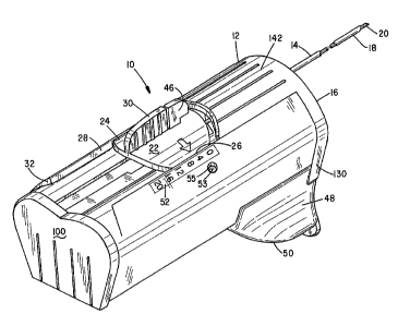
Referring to Figure 1, there is indicated generally by numeral 10 a
somewhat enlarged perspective view of a soft tissue biopsy device. It is seen
to
comprise a molded plastic handle member 12 having an outer needle or cannula
14 projecting through an opening formed in a front face 16 of the device. The
needle or cannula 14 is preferably formed from stainless steel and may
comprise
hypodermic stock of a predetermined length and a diameter in a range of from
about 14 ga. to about 20 ga. Its distal end 18 is beveled to a sharp point to
facilitate its ability to pass through soft tissue.
The cannula 14 has a lumen for receiving a tissue sampling stylet 20
therethrough. The stylet 20 is affixed to a molded plastic grip member 22
having
dove-tail side edges 24 and 26 that ride in a slot 28 provided in the handle
12.
The grip member includes an outwardly projecting ear 30 having serrated side
surfaces to facilitate its being gripped between a thumb and forefinger to
facilitate its being pulled rearward beyond the end 32 of the guideway 28 so
that
the stylet 20 can be fully extracted from the confines of the outer tubular
cannula
14.
Referring momentarily to Figure 2, it will be seen that the stylet 20
includes an elongated notch 34 in which one or multiple sample(s) can be
captured and retained following the firing of the device, all as will be
further
explained. The distal end of the stylet 20 is beveled to a sharp cutting edge
36.
Its proximal end 38 extends through a tubular bore 40 formed in a downwardly
projecting rib 42 that is integrally formed with the grip member 22. The grip
member 22 also includes a transversely extending slot 44 for receiving a latch
member 46 (Figure 1) therein. When the latch member is in the position
CA 02583584 2007-04-12
WO 2006/022699 PCT/US2004/025780
7
illustrated in Figure 1, the grip member 22 is effectively connected to an
internal
spring-driven ferrule which will be further described when the exploded view
of
Figure 4 is explained.
Also slidable mounted to the handle member 12 is a combination cocking
slide 48 and firing trigger 50. As the cocking slide 48 is pulled rearward by
the
user's finger, springs associated with the cannula 14 and stylet 20 are
simultaneously compressed to store energy. Also, sliding the cocking lever 48
rearward displaces the grip member 22 rearward to establish how much of the
notch 34 will become exposed out the end of the tubular needle 14 during a
first
phase of the firing sequence of the device. This establishes the size of the
specimen that will be collected. To aid the user, a numeric scale 52 is
mounted
alongside the guideway 28 and a fiducial mark (arrow) on the grip member 22
points to the scale to indicate the size of the sample to be extracted. A
further
indicator 53 is visible through a hole 55 in the handle 12. When the device
has
been cocked and is ready to fire, the indicator 53 shows red. Once the trigger
50
has been depressed to fire, both the stylet 20 and the cannula 14, the
indicator 53
shows green.
A molded piece part comprising the housing 12 is illustrated in
perspective in Figure 3. Molded from a suitable medical grade plastic in an
injection molding operation, the housing 12 includes a generally flat base 54
having a slot 56 formed longitudinally through it. Supported on the base are
first
and second generally tubular portions 58 and 60 with a common wall 62
extending between them. Formed through the thickness dimension of the tubular
member 60 is a longitudinally extending slot 64 leading away from a generally
rectangular aperture 66. Likewise, the tubular member 58 also includes a
longitudinally extending slot 68. The wall 62 where the tubular members 58 and
60 merge with one another define a V-shaped groove 70. Integrally formed with
and projecting upwardly from the top surface of the tubular members 58 and 60
are wedge-shaped wings 72 and 74 whose vertical walls 76 and 78 define a
guideway for the stylet gripper member 22. That is, the stylet gripper member
22
CA 02583584 2007-04-12
WO 2006/022699 PCT/US2004/025780
8
is dimensioned to fit between the vertical walls 76 and 78 and with the fin 42
resting in the V-shaped groove 70.
Referring next to Figure 4, there is shown a stylet spring retention sleeve
47 which is generally cylindrical and which has the latch member 46 integrally
molded therewith. The sleeve 80 has a generally open rearward end 82 and a
closed forward end 84. The closed end 84 includes a rectangular aperture 86.
The outside diameter of the sleeve 82 is sized so as to allow it to freely
slip into
the second tubular portion 60 of the handle member 12 with the latch member 46
projecting upward through the slot 64.
Figure 5 is a perspective view of the cannula spring retention sleeve,
which is indicated generally by numeral 88. It, too, is cylindrical and hollow
with
an open rearward end 90 and a closed forward end 92. A rectangular aperture
94 extends through the otherwise closed end 92. Extending radially outward
from the exterior surface of the cannula spring retention sleeve 88 is a
protuberance 96 that has a bore 98 formed through it for receiving a proximal
end portion of the cannula 14 therein. When the sleeve 88 is inserted into the
housing member 12, the protuberance 96 extends out through the longitudinal
slot 68 when the sleeve 88 is contained within the first tubular portion 58 of
the
handle 12. Also one or the other of colored areas 99 or 101 will be visible
through the aperture 55 depending on whether the device is cocked or not.
Turning next to Figure 6, it shows a perspective view of a rear cover 100
for the housing 12. Projecting outward from the inner face of the cover plate
100 are barb latches 102 and 104 that are adapted to mate with rectangular
apertures 106 and 108 formed through the wall of the housing member 12. The
barb members 102 and 104 are sufficiently resilient to allow them to deflect
as
the cover plate 100 is pushed against the rear edge of the housing. Upon
reaching the apertures 106 and 108, the barbs spring through those openings to
latch the cover in place.
Also projecting perpendicularly from the rear face of the cover plate 100
are longitudinally extending posts 110 and 112 each having a plurality of
ratchet
CA 02583584 2007-04-12
WO 2006/022699 PCT/US2004/025780
9
teeth 114 and 116 formed thereon. As can be seen in the exploded view of
Figure 10, helically wound compression springs 118 and 120 surround the posts
110 and 112 and fit into the sleeves 80 and 88 that are held within the
housing 12
when assembled. The end portions 122 and 124 of the posts 110 and 112 extend
through the rectangular openings 86 and 94 formed in the closed ends of the
sleeves 80 and 88. The spacing between adjacent ratchet teeth establishes the
resolution in specimen size that can be collected.
Referring again to the cover member 100, a further cylindrical post 126
projects perpendicularly from the rear face of the cover 100 and a further
compression return spring 128 (Figure 7) is disposed on the post 126 for a
purpose that will be further explained hereinbelow.
The front cover for the housing 12 is shown in Figure 8 and is indicated
generally by numeral 130. A stepped rib 134 having a first portion 136 of a
predetermined height dimension and a second portion 138 of approximately
twice the height of the portion of the rib 136, thereby defining a stop or
shoulder
140 is centrally disposed on the inner surface of the front cover 130 and acts
as a
glide for a shutter 144.
Integrally molded with the front cover is a top member 142 that fits
between the vertical edges 76 and 78 of the wedge-shaped wings 72 and 74 of
the handle 12.
Figure 9 is a perspective view of a sequence actuating shutter 144 which
is adapted to cooperate with the stepped rib 134 that is formed on the inner
face
of the front cover 130. Formed inwardly from the side edge 146 is a notch 150
having a first reference surface 152 at a first predetermined distance from a
reference end 154 of the shutter member. In a like way, a notch 156 having a
reference surface 158 extends inward from the side edge 148 of the shutter.
The
reference surface 158 is at a slightly greater displacement from the reference
edge
154 than is the reference surface 152.
The shutter 144 further includes the central groove 160 formed partially
through the thickness dimension of the shutter 144 and leading to a slot 162
that
CA 02583584 2007-04-12
WO 2006/022699 PCT/US2004/025780
extends completely through the thickness dimension of the shutter. The shutter
148 is juxtaposed to the rear face of the front cover 130 so that the portion
136
of the rib 134 fits within the groove 160 of the shutter while the portion 138
of
double thickness extends into the slot 162. The shutter is dimensioned and the
5 groove 160 is sized to allow the shutter 144 to slide relative to the inside
surface
of the front cover until a point is reached where the shoulder 140 engages the
bottom 164 of the slot 162 thereby providing a stop mechanism preventing the
posts 110 and 112 from becoming hyper extended. The trigger 50 is likewise
protected from over extension.
10 Turning now to the exploded assembly drawing of Figure 10, with the spring
retaining sleeves 80 and 88 inserted into the respective first and second
tubular
portions 58 and 60 (Figure 3) of the handle 12 and the rear cover plate 100
also
affixed to the handle, the inner ends of the springs 118 and 120 abut the
closed
ends of the sleeves 80 and 88 while the ends 122 and 124 of the posts 110 and
112 extend through the rectangular apertures 86 and 94 of the sleeves. Front
cushions 166 and 168 are adhesively affixed to the closed ends of the sleeves
80
and 88 and these cushions or pads have rectangular openings that align with
the
rectangular openings in the ends of the sleeves 80 and 88.
When the front cover 16 is assembled onto the handle 12, the ends 122
and 124 (Figure 6) of the posts 110 and 112 fit into the slots 150 and 156 of
the
sequence actuating shutter 144.
A slide member 170 (Figure 10) is dimensioned to fit in sliding relation to
the housing 12. More particularly, the slide member 170 includes a pair of
flanges 172 and 174 adapted to ride in channels 176 and 178 of the housing 12.
The cocking lever 48 has a pair of lateral edge channels 171 and 173 designed
to
fit into guideways 177 and 179 of housing 12 (Fig. 3). A gear rack 175 is
molded into the base of the cocking lever 48. Formed through the raised center
portion of the slide plate 170 is a rectangular opening 182 and fitted into
that
opening is a pinion gear 184 that is journaled for rotation on a pin 186 that
passes through a transverse bore 188 formed in the raised center portion 180.
CA 02583584 2007-04-12
WO 2006/022699 PCT/US2004/025780
11
With reference to Figure 7, it can be seen that a similar gear rack 190 is
formed
along the length of the handle 12 in alignment with the pinion gear 184. The
slide 170 further includes an outwardly projecting rib 192 at a front edge
thereof
that is adapted to cooperate with the closed front ends of the sleeves 80 and
88.
In operation, as the cocking lever 48 is pulled rearward by the user's
index finger, the projection 192 on slide 70 is in engagement with the spring
retaining sleeves 80 and 88 and pulls those sleeves rearward, compressing the
springs 118 and 120 as they move. An edge of the rectangular openings in the
spring retention sleeves 80 and 88 engage the teeth 114 and 116 on the posts
110
and 112 to hold the sleeves 80 and 88 in place when finger pressure is
removed.
In that the cannula 14 is attached to the protuberance 96 on the sleeve 88
and the stylet moves with the sleeve 80 by virtue of the engagement of the
latch
member 46 with the transverse slot 44 in the grip member 22, it moves rearward
with the displacement of the spring retaining sleeve 80. Once the cocking
slide
has been drawn rearward a desired measured amount as reflected by the arrow
on the stylet grip 22 and the numerical indicia 52, the soft tissue biopsy
device is
ready for use in collecting a first specimen of a pre-set length.
Using appropriate imaging, the physician advances the cannula 14 and the
stylet 20 projecting from the front end 16 of the handle into the area of the
body
where a tissue sample is to be taken. As the trigger button 50 is depressed,
the
front edge 51 thereof is brought into engagement with the bottom edge of the
sequence actuating shutter 144, displacing it along the guide 136 of the front
cover 130 and first elevating the post supporting the spring 120. When that
post
is elevated to the point where its teeth no longer engage the mating edge of
the
rectangular aperture 86 of the spring retaining sleeve 80, the spring drives
the
sleeve 80 forward until its cushioned front end hits the closed end of housing
12.
In that the latch member 46 is engaged with the notch 44 of the stylet grip
22,
the stylet will be driven into the tissue where the sample is to be taken. The
tissue fills the portion of the notch 34 extending beyond the end of the
cannula
14. When the release button 50 is further depressed, it elevates the shutter
CA 02583584 2007-04-12
WO 2006/022699 PCT/US2004/025780
12
member 144 to the point where the teeth 116 on the post 112 supporting the
spring 118 no longer engages the edge of the rectangular opening on the front
end of the sleeve 88, thus allowing the spring 118 to drive the sleeve 88
forward
against the closed end of the housing 12. This drives the cannula affixed to
the
protuberance 96 forward to slice the tissue sample contained within the notch
of
the stylet free of surrounding tissue.
At this point, the cocking lever 48 can again be pulled proximally a
desired measured distance while the cannula 18 remains in place within the
tissue
to again cock the biopsy device in preparation of collecting a further
specimen.
The distal end of the cannula can be advanced, if desired, and when the
trigger 50
is again depressed and the stylet driven distally, the friction between the
earlier
collected specimen and the inner wall of the cannula will cause the specimen
to
move to the proximal end of the notch 34, making room for the further specimen
to prolapse into the notch before the cannula again is driven distally to
sever the
specimen from surrounding tissue. This process can be repeated until the
notch.
34 in the stylet becomes filled.
Now, with the device of the present invention, the tissue sample(s) can be
removed from the device without displacing the cannula from its current
position
within the body. This is done by rotating the latch member 46 out from the
notch
44 in the stylet grip 22 and then pulling back on the ear 30 on the grip
member to
slide the stylet out from the lumen of the cannula 14. Once the tissue
sample(s)
is removed from the notch in the stylet, the stylet can be replaced by sliding
its
distal end into the proximal end of the cannula and guiding the grip member 22
to
its frontmost position, at which point the latch member 46 can again be
rotated
into the groove 44, latching the stylet and its grip to the spring retaining
sleeve
80. With the instrument still in its position within the body of the patient,
it can
be recocked by again drawing back on the cocking slide member 48 preparatory
to again firing the instrument.
By providing a gear rack on the undersurface of the cocking slide 48 as
well as the undersurface of the housing 12, and by providing the pinion gear
184,
CA 02583584 2007-04-12
WO 2006/022699 PCT/US2004/025780
13
a mechanical advantage is achieved lessening the finger force required to
compress the springs 118 and 120. The arrangement of the pinion gear 184 with
the racks reduces the distance traveled by the slide member 170 by a 2:1
ratio,
allowing a shorter return spring 128 to be used.
By depressing the firing trigger 50 down firmly in a single stroke, the
stylet and the cannula will be advanced in rapid succession determined by the
offset in the height of surfaces 158 and 152 relative to the reference surface
154
of the sequence actuating shutter 144. When desired, by slowly depressing the
firing trigger 50, the stylet can be advanced without automatically releasing
the
cannula. By further depressing the firing trigger 50 at a later time, the
cannula
will be advanced. The surface 151 on shutter 144 (Fig. 9) is arranged to
cooperate with the end portion 125 of the post 112 (Fig. 6) to prevent the
stylet
sleeve 80 from latching until after the cannula sleeve 88 (Fig. 5) becomes
latched,
thereby synchronizing the latching sequence.
It should be noted that the biopsy device 10 cannot be fired while the
cocking action is taking place. Until the cocking lever 48 has been returned
to its
forwardmost position by the return spring 128, the edge 51 of the trigger
button
50 cannot engage the edge 154 of the shutter 144 to lift the posts 110 and 112
so
that their teeth no longer engage the bottom edge of the rectangular openings
in
the two spring retention sleeves.
This invention has been described herein in considerable detail in order to
comply with the patent statutes and to provide those skilled in the art with
the
information needed to apply the novel principles and to construct and use such
specialized components as are required. However, it is to be understood that
the
invention can be carried out by specifically different equipment and devices,
and
that various modifications, both as to the equipment and operating procedures,
can be accomplished without departing from the scope of the invention itself.
What is claimed is: