Note: Descriptions are shown in the official language in which they were submitted.
CA 02585890 2007-04-23
ACCOMMODATIVE INTRAOCULAR LENS SYSTEM
Background of the Invention
This invention relates generally to the field of intraocular lenses (IOL) and,
more
particularly, to accommodative IOLs.
The human eye in its simplest terms functions to provide vision by
transmitting light
through a clear outer portion called the cornea, and focusing the image by way
of a crystalline
lens onto a retina. The quality of the focused image depends on many factors
including the
io size and shape of the eye, and the transparency of the cornea and the lens.
When age or disease causes the lens to become less transparent, vision
deteriorates
because of the diminished light which can be transmitted to the retina. This
deficiency in the
lens of the eye is medically known as a cataract. An accepted treatment for
this condition is
surgical removal of the lens and replacement of the lens function by an
artificial intraocular
lens (IOL).
In the United States, the majority of cataractous lenses are removed by a
surgical
technique called phacoemulsification. During this procedure, an opening is
made in the
anterior capsule and a thin phacoemulsification cutting tip is inserted into
the diseased lens
and vibrated ultrasonically_ The vibrating cutting tip liquifies or emulsifies
the lens so that
the lens may be aspirated out of the eye. The diseased lens, once removed, is
replaced by an
artificial lens.
In the natural lens, bifocality of distance and near vision is provided by a
mechanism
known as accommodation. The natural lens, early in life, is soft and contained
within the
capsular bag. The bag is suspended from the ciliary muscle by the zonules.
Relaxation of the
ciliary muscle tightens the zonules, and stretches the capsular bag. As a
result, the natural
lens tends to flatten. Tightening of the ciliary muscle relaxes the tension on
the zonules,
allowing the capsular bag and the natural lens to assume a more rounded shape.
In the way,
the natural lens can be focus alternatively on near and far objects.
As the lens ages, it becomes harder and is less able to change shape in
reaction to the
tightening of the ciliary muscle. This makes it harder for the lens to focus
on near objects, a
medical condition known as presbyopia. Presbyopia affects nearly all adults
over the age of
45 or 50.
Prior to the present invention, when a cataract or other disease required the
removal of
the natural lens and replacement with an artificial IOL, the IOL was a
monofocal lens,
requiring that the patient use a pair of spectacles or contact lenses for near
vision. Advanced
Medical Optics has been selling a bifocal IOL, the Array lens, for several
years, but due to
quality of issues, this lens has not been widely accepted.
CA 02585890 2007-04-23
2
Several designs for accommodative IOLs are being studied. For example, several
designs manufactured by C&C Vision are currently undergoing clinical trials.
See U.S.
Patent Nos. 6,197,059, 5,674,282, 5,496,366 and 5,476,514 (Cununing), the
entire contents of
which being incorporated herein by reference. The lens described in these
patents is a single
s optic lens having flexible haptics that allows the optic to move forward and
backward in
reaction to movement of the ciliary muscle. A similar designs are described in
U.S. Patent
No. 6,302,911 B 1(Hanna), 6,261,321 B 1 and 6,241,777 B 1(both to Kellan), the
entire
contents of which being incorporated herein by reference. The amount of
movement of the
optic in these single-lens systems, however, may be insufficient to allow for
a useful range of
accommodation. In addition, as described in U.S. Patent Nos. 6,197,059,
5,674,282,
5,496,366 and 5,476,514, the eye must be paralyzed for one to two weeks in
order for
capsular fibrosis to entrap the lens that thereby provide for a rigid
association between the
lens and the capsular bag. In addition, the commercial models of these lenses
are made from
a hydrogel or silicone material. Such materials are not inherently resistive
to the formation of
is posterior capsule opacification ("PCO"). The only treatment for PCO is a
capsulotomy using
a Nd:YAG laser that vaporizes a portion of the posterior capsule. Such
destruction of the
posterior capsule may destroy the mechanism of accommodation of these lenses.
There have been some attempts to make a two-optic accommodative lens system.
For
example, U.S. Patent No. 5,275,623 (Sarfarazi), WIPO Publication No. 00/66037
(Glick, et
al.) and WO 01/34067 A1 (Bandhauer, et al), the entire contents of which being
incorporated
herein by reference, all disclose a two-optic lens system with one optic
having a positive
power and the other optic having a negative power. The optics are connected by
a hinge
mechanism that reacts to movement of the ciliary muscle to move the optics
closer together or
further apart, thereby providing accommodation. In order to provide this "zoom
lens" effect,
movement of the ciliary muscle must be adequately transmitted to the lens
system through the
capsular bag, and none of these references disclose a mechanism for ensuring
that there is a
tight connection between the capsular bag and the lens system. In addition,
none of these
lenses designs have addressed the problem with PCO noted above.
Therefore, a need continues to exist for a safe and stable accommodative
intraocular
lens system that provides accommodation over a broad and useful range.
Brief Summary of the Invention
The present invention improves upon the prior art by providing a two-optic
accommodative lens system. The first lens has a negative power and is located
posteriorly
within the capsular bag and lying against the posterior capsule. The periphery
of the first lens
is attached to a ring-like structure having a side wall. The second lens is
located anteriorly to
CA 02585890 2007-04-23
3
the first lens within of the capsular bag and is of a positive power. The
peripheral edge of the
second lens contains a plurality of haptics that are arranged in a spiral
pattern and project
posteriorly from the second lens and toward the first lens. The haptics are
relatively firm, yet
still flexible and ride within the side wall of the ring-like structure, so
that flattening or
steepening of the capsule in reaction to movement of the ciliary muscle and
causes the second
lens to move along the optical axis of the lens system.
Accordingly, one objective of the present invention is to provide a safe and
biocompatible intraocular lens.
Another objective of the present invention is to provide a safe and
biocompatible
io intraocular lens that is easily implanted in the posterior chamber.
Still another objective of the present invention is to provide a safe and
biocompatible
intraocular lens that is stable in the posterior chamber.
Still another objective of the present invention is to provide a safe and
biocompatible
accommodative lens system.
is These and other advantages and objectives of the present invention will
become
apparent from the detailed description and claims that follow.
Brief Description of the Drawing
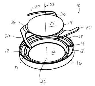
20 FIG. 1 is an enlarged, exploded perspective view of the lens system of the
present
invention.
FIG. 2 is an enlarged perspective view of the first lens of the lens system of
the
present invention.
FIG. 3 is an enlarged cross-sectional view of the first lens of the lens
system of the
25 present invention.
FIG. 4 is an enlarged top plan view of the second lens of the lens system of
the present
invention.
FIG. 5 is an enlarged elevational view of the second lens of the lens system
of the
present invention.
Detailed Description of the Invention
As best seen in the figures, lens system 10 of the present invention generally
consists
of posterior lens 12, anterior lens 14 and circumferential ring 16. Lens 12 is
preferably
integrally formed with ring 16. Lens 12 preferably is made from a soft,
foldable material that
is inherently resistive to the formation of PCO, such as a soft acrylic. Lens
14 preferable is
made from a soft, foldable material such as a hydrogel, silicone or soft
acrylic. Lens 12 may
CA 02585890 2007-04-23
4
be any suitable power, but preferably has a negative power. Lens 14 may also
be any suitable
power but preferably has a positive power. The relative powers of lenses 12
and 14 should be
such that the axial movement of lens 14 toward or away from lens 12 should be
sufficient to
adjust the overall power of lens system 10 at least one diopter and
preferably, at least three to
four diopters, calculation of such powers of lenses 12 and 14 being within the
capabilities of
one skilled in the art of designing ophthalmic lenses by, for example, using
the following
equations:
P= PI + PZ - T/n * PIP2 (1)
SP = -ST/n * PiP2 (2)
Lens 12 is generally symmetrical about optical axis 22. Peripheral band 18
connects
lens 12 with ring 16 is relatively stiff, so as to allow some, but not
excessive, flexing in
response to ciliary muscle contraction and relaxation. Peripheral band 18 may
contain a
plurality of holes 19 for allowing the release or aspiration of any
viscoelastic material used
during surgery from behind optic 12 and/or band 18. Ring 16 has generally
upright sidewall
28 that project anteriorly. Lens 14 contains a plurality of haptics 20 that
project outward from
optic 24 of lens 14 and away from optic 24 of lens 14 along optical axis 22.
Second flexible
haptics 20 are connected to lens 14 by regions 26 that are relatively stiff
and allow second
haptics 20 to exhibit resistive, spring-like movement. When compressed, second
haptics 20
store energy, releasing that energy when uncompressed. Regions 26 also help
create a space
between the anterior capsule (not shown) and optic 24 that allow fluid to flow
between the
posterior and anterior sides of optic 24.
In use, lens 12 is implanted into the capsular bag prior to the implantation
of lens 14.
Lens 12 is held within the capsular bag by ring 16. Lens 14 is implanted so
that second
haptics 20 ride within sidewal128 of ring 16. Lenses 12 and 14 are free-
floating and not
connected to each other. Upon implantation of lens 14, haptics 20 will flex in
response to
flattening and steepening of the lens capsule resulting from contraction and
relaxation of the
ciliary muscles. Flattening of the capsule caused by relaxation of the ciliary
muscles will
compress haptics 20 axially and allow second optic 24 to move posteriorly
along optical axis
22 toward lens 12. Reduction of zonular tension caused by contraction of the
ciliary muscles
will allow energy stored in compressed haptics 20 to release, allowing optic
24 of lens 14 to
move anteriorly along optical axis 22 and away from lens 12 because of the
vaulted and spiral
arrangement of haptics 20.
CA 02585890 2007-04-23
This description is given for purposes of illustration and explanation. It
will be apparent to
those skilled in the relevant art that changes and modifications may be made
to the invention
described above without departing from its scope or spirit.
