Note: Descriptions are shown in the official language in which they were submitted.
CA 02586147 2007-05-01
WO 2006/055251 PCT/US2005/039691
Attorney Docket No. 22884.04108
METHOD AND APPARATUS FOR DETERMINING CORRELATION BETWEEN
SPATIAL COORDINATES IN BREAST
CROSS REFERENCE TO RELATED APPLICATION
[0001] The present application claims the benefit of U.S. Provisional
Application No.
60/624,349 filed November 2, 2004, the disclosure of which is incorporated
herein by
reference in its entirety.
STATEMENT REGARDING FEDERALLY SPONSORED RESEARCH OR
DEVELOPMENT
[0002] This work was supported, at least in part, by a Department of Defense
Congressionally Directed Medical Research Program Concept Award Grant (Grant
No.
BC032942). The U.S. Govermnent may have certain rights in this invention.
FIELD
[0003] Applicants' inventive concept relates generally to medical imaging and,
more
specifically, to methods and devices for predicting the spatial coordinates of
an anatomic
structure or point of interest in or on a breast to be found on one method of
evaluation based
on information on the location of the item of interest determined from another
method of
evaluation.
BACKGROUND
[0004] Physical examination and breast imaging are important to breast health.
In addition to
breast self-examination (BSE) and clinical breast examination (CBE)--
inspection and manual
palpation of the breast which is performed by physicians and other medical
caregivers--many
imaging modalities are used to identify and evaluate breast lumps and tissues
such as tumors,
CA 02586147 2007-05-01
WO 2006/055251 PCT/US2005/039691
cysts, and other abnormalities ("lesions") and to help differentiate benign
(noncancerous) and
malignant (cancerous) breast disease.
[0005] CBE is challenging to learn, and then accurately perform, since on
palpation the
breast is normally non-uniform ("lumpy-bumpy") in texture. Furthermore,
abnormalities,
when present, may be subtle and difficult for the examiner's fingers to
distinguish from
adjacent normal tissue. Hence, imaging modalities have become vital to patient
care and
wellbeing. Conversely, definite pathological abnormalities detected on
physical examination
may be difficult to see or appreciate with one or more imaging modalities.
Furthermore, even
if an abnormality is seen on one image of one or more of the modalities, it
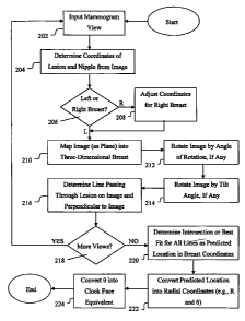
may be difficult to
detect or appreciate on another image of the same or a different imaging
modality.
[0006] Mammography, ultrasound and magnetic resonance imaging (MRI) are
imaging
modalities commonly used to search for and evaluate breast tissue
abnormalities.
Mammography and ultrasound are the imaging modalities most commonly employed
to non-
invasively evaluate the breast. MRI is generally used for further
investigation, if warranted.
Mammography may be deemed "Diagnostic" when it is used to evaluate a patient
who has
symptoms, signs or a history of breast disease or "Screening" when the
technique is applied
as a cancer surveillance examination for the general population of women who
are
asymptomatic. Ultrasound is rarely used for screening and generally reserved
for further
evaluation of breast abnormalities detected on mammography and physical
examination.
[0007] If an abnormality is identified on physical examination and/or using
one of these
imaging modalities, it is common to examine the patient with another of the
imaging
modalities to clarify any ambiguous finding and achieve greater accuracy and
completeness
in diagnosis.
[0008] The breast is very pliable and its geometry, as a whole, responds to
the effects of
gravity and other external forces imparted during examination.
[0009] SBE and CBE are performed with the patient both upright and supine,
with the
examining fingers trapping breast tissue between skin and chest wall as lumps
are sought. As
a result, the breast tissue is displaced toward and against the chest wall and
the skin at the site
of examination is oriented generally parallel to the chest wall.
2
CA 02586147 2007-05-01
WO 2006/055251 PCT/US2005/039691
[0010] Conversely, during a mammogram, the majority of the skin and underlying
breast is
displaced away from the chest wall and flattened into a plane which is
perpendicular to the
chest wall.
[0011] Mammography is a specific type of imaging that uses a low-dose X-ray
system for the
examination of breasts. As currently clinically practiced, during a
mammography exam, the
patient is typically upright and the technique entails pulling one breast at a
time away from
the body and resting it on the surface of a plate with another plate pressed
firmly against
opposite side of the breast to hold and flatten out the breast tissue. Breast
compression
during mammography spreads out the tissue which minimizes and evens out the
thickness.
This compression is important because it improves overall visualization of the
tissue and
lessens the chance that abnormalities are obscured by overlying breast tissue.
Compression
also holds the breast still to eliminate blurring of the image caused by
motion and reduces X-
ray scatter to increase sharpness of the image. X-rays passing through the
breast tissue are
detected and processed into an image for display on film or a monitor (an
electronic viewing
device such as, for example, a cathode ray tube (CRT), liquid crystal display
(LCD) or
plasma monitor). The resultant image or "view" is a two dimensional
representation of the
complex three dimensional structure of the breast.
[0012] The Cranio-Caudal (CC) view and a Mediolateral Oblique (MLO) view are
two views
that are commonly used in mammography. Other views used in mammography include
a
Latero-Medial (LM) view, a Mediolateral (ML) view, etc.
[0013] The CC view, or head-to-toe view, images the breast from above. A CC
view of a
right breast is illustrated in FIG. IA and a CC view of a left breast is
illustrated in FIG. 1B.
The MLO view images the breast from a side-to-side perspective at an oblique
angle. An
MLO view of the right breast is illustrated in FIG. 1C and an MLO view of the
left breast is
illustrated in FIG. 1 D.
[0014] On mammography, the location of a lesion or site of interest can be
described in many
ways including a rough, intuitive estimation of clock-face, the quadrant, and
approximate
depth (expressed as anterior, middle or posterior breast). It is not uncommon
for examiners
to have difficulty describing the exact location of the mammographic
abnormality. In
addition to having to integrate information from two separate views,
estimation of lesion
location is challenging since the CC views and the MLO views are not at 90
degrees with
3
CA 02586147 2007-05-01
WO 2006/055251 PCT/US2005/039691
respect to each other and the angle at which the MLO views are done is quite
variable
(generally between about 30 and 60 degrees; the technologist tries to conform
to the lateral
edge of the pectoralis muscles). Thus, an estimate of the clock face position,
or even the
quadrant in which the lesion resides, will also be affected by MLO angle,
particularly when
the lesion is closer to the periphery of the breast near the 12:00-6:00 axis
or the 3:00-9:00
axis. A lesion seen on one view of the breast may be occult on another view.
The best
clinical description, derived from experience and intuition, is an estimate of
clock-face
position and depth (anterior, middle or posterior breast).
[0015] In addition to the difficulties in predicting where a mammographic
lesion will be
found on ultrasound or physical examination, similar difficulties arise when
trying to predict
where a lesion seen on ultrasound or on physical exam will be on the
mammogram.
[0016] Breast ultrasound is typically done with the patient supine (laying on
her back) using
a hand-held probe (ultrasound transducer) which is in contact with the skin
surface and
oriented in a fashion to be perpendicular or roughly perpendicular to the
chest wall. Optimal
sonographic technique requires compression to be applied; but, as with
physical examination,
the pressure is directed between skin and chest wall wherein the site of
interest is trapped as it
is acoustically examined. The typical ultrasound display is thus a two
dimensional image
directed toward the chest wall. At least two orthogonal images are done of the
site of
interest: longitudinal and transverse ("north/south" and "side to side",
respectively) with
respect to the long axis of the body or radial and anti-radial with respect to
the nipple.
Various descriptions (annotations) of the location of the site being imaged
have been deemed
clinically acceptable, for example, describing the lesion according to the
quadrant it is in, the
clock-face position, or a combination of clock-face position and distance from
the nipple.
The most informative description is clock-face position and distance from the
nipple, but
there is variation clinically in how these measurements are done (e.g., as
patients may be
positioned supine or in a variation of supine).
[0017] An MRI of a breast is generally performed with the patient in a prone
position and the
breast oriented and hanging dependently within the well of a breast coil. MRI
uses
radiofrequency waves and a strong magnetic field rather than X-rays to provide
detailed
images of internal organs and tissues. The technique has proven very valuable
for the
diagnosis of a broad range of pathologic conditions in all parts of the body
including cancer,
4
CA 02586147 2007-05-01
WO 2006/055251 PCT/US2005/039691
heart and vascular disease, stroke, and joint and musculoskeletal disorders.
MRI requires
specialized equipment and expertise and allows evaluation of some body
structures that may
not be as visible with other imaging methods. MRI of the breast is becoming
important for
many clinical indications including characterization of indeterminate lesions,
the extent of
disease, search of occult disease in patients with malignant adenopathy,
surveillance of
patients at high risk, etc. The MRI data can be used to produce volume and
planar images,
the latter in any orientation to the body. As with the other imaging
modalities, the location of
a lesion can be described in various ways.
[0018] Assignment of lesion location is dependent on the training, skill and
experience of the
examiner(s).
[0019] Close correlation of location of a lesion found on physical examination
and ultrasound
is possible if the lesion can be definitely and unequivocally identified and
the patient is
similarly positioned for the examinations and careful measurement of clock-
face position and
distance from the nipple are done.
[0020] However, equating lesion location estimates between the physical
examination and
mammography and ultrasound and mammography is, in general, difficult to
perform and
prone to error because of the considerable differences in patient positioning,
direction of
compression and the individual exam techniques noted above. Accurate location
estimates
that can be equated to ultrasound and physical examination are difficult to
achieve and are
even more dependent on the experience and expertise of the radiology physician
reading the
examination.
[0021] From a practical standpoint, it can be difficult to intuitively predict
the location where
a lesion detected on mammography or ultrasound will be found on a subsequent
MRI.
Conversely, it is not uncommon to have one or more unexpected findings on an
MRI, which
then require reappraisal of the patient's mammograms and/or a return to the
ultrasound suite
to attempt to determine the significance of the unexpected MRI findings.
[0022] Excellence in patient care requires that there be complete concordance
between any
and all of the modalities used to examine the patient, to ensure that it is
truly the same
abnormality that is being identified and evaluated on the studies.
CA 02586147 2007-05-01
WO 2006/055251 PCT/US2005/039691
[0023] In general day to day clinical practice, when mammography is used, it
is often
difficult to predict the location where a discovered abnormality will be found
using another
modality (e.g., ultrasound or physical examination), or, conversely, where it
must be found
on mammography because of the type of views which are commonly used in
mammography.
[0024] Thus, a need exists for readily predicting the location of an item of
interest (e.g., a
lesion) for one modality, including physical examination, based upon where the
item of
interest is noted by another means of evaluation.
SUMMARY
[0025] Accordingly, it is one aspect to provide a method and system for
predicting the
location of an object of interest in or on a breast applicable to one method
of evaluation from
data on the location of the object of interest determined using another method
of evaluation.
[0026] It is another aspect to provide a method and apparatus for predicting
the location of an
object of interest in or on a breast on an ultrasound and/or physical
examination from data on
the location of the object of interest determined by mammography of the
breast.
[0027] It is still another aspect to provide a method and apparatus for
predicting the location
of an object of interest in or on a breast on a mammogram from data on the
location of the
object of interest determined by an ultrasound and/or physical examination of
the breast.
[0028] It is yet another aspect to provide a method and apparatus for
predicting the location
of an object of interest in or on a breast on a mammogram, ultrasound and/or
physical
examination from data on the location of the object of interest determined by
an MRI of the
breast.
[0029] It is an aspect to define a standard position for a patient to assume
when examiners
perform ultrasound (or when SBE or CBE are done with the patient lying down)
in order to
reduce lateral displacement of the breast during examination so as to
facilitate examination
and minimize variation in technique and measurement.
[0030] It is another aspect to provide a method and apparatus for predicting
the location of an
object of interest in or on a breast on a mammogram view from data on the
location of the
object of interest determined from another mammogram view. Accordingly, if two
views of
6
CA 02586147 2007-05-01
WO 2006/055251 PCT/US2005/039691
a breast are imaged by mammography and a lesion only appears in one of the
views, a region
containing the location of the lesion on the other view can be estimated.
[0031] It is still another aspect to designate visually, as with a graphical
overlay, an estimated
location, for example, on a mammogram, on an image of the breast in the
standard position,
etc.
[0032] It is another aspect to correct spatial registration anomalies between
prior and
subsequent congruent mammogram views.
[0033] It is an aspect to provide a tool for teaching and improving image
interpretation and
physical examination skills.
DESCRIPTION OF THE DRAWINGS
[0034] The above and additional aspects, features and advantages will become
more apparent
by describing in detail exemplary embodiments with reference to the attached
drawings, in
which:
[0035] FIG. lA is a drawing illustrating a CC view of a right breast;
[0036] FIG. 1B is a drawing illustrating a CC view of a left breast;
[0037] FIG. 1 C is a drawing illustrating an MLO view of the right breast;
[0038] FIG. 1D is a drawing illustrating an MLO view of the left breast;
[0039] FIG. 2 is a flowchart illustrating a method for predicting a location
to assist in
performing an ultrasound from known mammogram data, according to an exemplary
embodiment;
[0040] FIG. 3A is a drawing illustrating a CC view of a left breast having a
lesion therein;
[0041] FIG. 3B is a drawing illustrating an MLO view of the left breast having
the lesion
therein;
7
CA 02586147 2007-05-01
WO 2006/055251 PCT/US2005/039691
[0042] FIG. 4 is a photograph illustrating an exemplary mammography machine
capable of
rotating an image plane about two independent axes;
[0043] FIG. 5A is a photograph showing a perspective view of a patient in a
supine position;
[0044] FIG. 5B is a photograph showing a perspective view of the patient in a
standard
position;
[0045] FIG. 6A is a drawing illustrating a top view of a left breast of a
patient in the standard
position with a predicted location marked thereon;
[0046] FIG. 6B is a drawing illustrating a side view of the left breast of the
patient in the
standard position, as viewed from the patient's feet looking toward the
patient's head, with a
predicted depth shown thereon;
[0047] FIG. 7 is a drawing illustrating another top view of the left breast of
the patient in the
standard position with the predicted location marked thereon;
[0048] FIG. 8A is a drawing illustrating a top view of a left breast of a
patient in the standard
position having a lesion therein;
[0049] FIG. 8B is a drawing illustrating a side view of a left breast of a
patient in the
standard position, as viewed from the patient's feet looking toward the
patient's head, having
a lesion therein;
[0050] FIG. 9 is a drawing illustrating a CC view of the left breast of the
patient having the
predicted location of the lesion marked thereon;
[0051] FIG. 10 is a graph illustrating the correlation between predicted and
measured clock-
face position of a lesion for a sample set of patients, according to an
exemplary embodiment;
and
[0052] FIG. 11 is a graph illustrating the correlation between predicted and
measured
distance from the nipple to the lesion for the sample set of patients,
according to an
exemplary embodiment.
8
CA 02586147 2007-05-01
WO 2006/055251 PCT/US2005/039691
DESCRIPTION OF EXEMPLARY EMBODIMENTS
[0053] Hereinafter, exemplary embodiments will be described in detail with
reference to the
accompanying drawings. The exemplary methods and systems described herein
allow for the
estimation of the location of an item of interest for use in performing
mammography,
ultrasound, MRI or physical examination of a breast, given a known position of
the item from
performing at least one of the other modalities. The estimated location can
be, for example, a
point, a line, a curve, a surface, a region, etc.
[0054] By way of example and not by way of limitation, an exemplary embodiment
directed
to a method for predicting the location of an item an interest (e.g., a
lesion) of a breast for use
in performing an ultrasound, given known location information of the lesion
from two
mammograms will be described herein with reference to FIG. 2.
[0055] Preferably, but not necessarily, at least two views of a breast being
examined are
provided (steps 202, 206 and 218), with the lesion being visible on each of
the mammograms.
For example, as shown in FIG. 3A, a mammogram 300 representing a CC view of a
left
breast 304 and nipple 302 illustrates a lesion 306 within the breast. In FIG.
3B, the lesion
306 appears in a mammogram 320 representing an MLO view of the left breast
304, nipple
302 and chest wall 308.
[0056] Mammograms 300 and 320 can be produced, for example, with screen-film
cassettes
that are exposed to X-rays during mammography. In such a case, the data on the
mammograms 300 and 320 is preferably, but not necessarily, digitized. In this
manner, the
data derived from the mammograms 300 and 320, which represents the images from
the
respective films, can be readily stored, transmitted and processed.
[0057] Such digitization is unnecessary if the mammograms 300 and 320 are
produced by
digital mammography, wherein the X-rays passing through the compressed breast
are
recorded by means of an electronic digital detector instead of the film. In
digital
mammography, the resulting electronic image can be, for example, displayed on
a monitor
and/or printed onto film.
[0058] For mammogram 300 (i.e., the CC view of the breast), an angle of
rotation of the
developed image with respect to the left breast 304 is known, as is a tilt
angle of the left
9
CA 02586147 2007-05-01
WO 2006/055251 PCT/US2005/039691
breast 304 in the developed image. For example, the angle of rotation and tilt
angle are
known based on the positions of an axis of rotation and a tilt axis,
respectively, of the
mammography machine at the time the mammogram 300 is taken.
[0059] An illustrative mammography machine 400 having an X-ray emitter 405, as
shown in
'FIG. 4, includes an axis of rotation 410 and a tilt axis 420. As some
mammography machines
lack a tilt axis, a mammogram taken by such a machine will not have a tilt
component. In an
exemplary embodiment, a mammogram view from a machine lacking a tilt axis is
treated as
having a tilt value of 0. Other information may be known from the state of the
mammography machine 400 at the time a mammogram is taken. For example, the
amount of
compression is known from the distance between a first plate 430 and second
plate 432
between which the breast being examined is compressed.
[0060] Additionally, the physical size (i.e., the width and height) of the
scanned CC image
(e.g., in millimeters), as well as the pixel dimensions (e.g., 3600 x 4800
pixels) of the
scanned CC image are known. Furthermore, a location of the nipple 302 and a
location of the
lesion 306 are determined from the mammogram 300, e.g., from data derived from
the
mammogram 300 (step 204).
[0061] For example, x and y coordinates of the nipple 302 (e.g., in image
pixels) are
determined, as are x and y coordinates of the lesion 306 (e.g., in image
pixels). The
coordinates of the nipple 302 and the lesion 306 may be determined, for
example, manually
by a technician (e.g., a radiologist or clinician). Alternatively, image
processing techniques
may be able to identify the nipple 302 and/or lesion 306 in a mammogram so as
to determine
the corresponding coordinates. In an exemplary embodiment, an image processing
technique
for identifying the nipple 302 and/or lesion 306 in a mammogram uses
information on
contour of the breast 304.
[0062] Additional devices may be used to aid in identifying the nipple 302
and/or the lesion
306 in the mammogram 300. For example, a metal ball may be placed near the
center of the
nipple 302 prior to performing mammography on the breast. Furthermore, a hook-
wire may
be used to skewer the lesion 306 to highlight the lesion on the mammogram and
to assist in
confirming the identification of the lesion among different views. Other
techniques which
may be useful include needle localization and skin mapping.
CA 02586147 2007-05-01
WO 2006/055251 PCT/US2005/039691
[0063] Similar information is known and/or determined for mammogram 320 (i.e.,
the MLO
view of the breast). Based on the known position information of the lesion 306
in the
mammograms 300 and 320, a predicted location of the lesion 306 for an
ultrasound
examination is determined.
[0064] In an ultrasound examination, a probe containing one or more acoustic
transducers
sends pulses of sound into the breast. In general, whenever a sound wave
encounters a
material with a different acoustical impedance, part of the sound wave is
reflected, which the
probe detects as an echo. The time it takes for the echo to travel back to the
probe is
measured and used to calculate the depth of the tissue causing the echo.
[0065] It has been a common practice for both clinical practitioners
conducting physical
examination and imagers performing ultrasound of the breast to study the
recumbent patient
supine, i.e., flat on the back, with the ipsilateral arm in a variable amount
of extension (see,
e.g., FIG. 5A). Additionally, in an attempt to increase the sensitivity of the
exam by
"thinning" the part of the breast being examined the patient may be turned a
variable amount
to the ipsilateral or contralateral side by displacing the bulk of the
remainder of the breast.
Although aspects of this approach have merit, the inherent variability of
positioning and
overall technique combined with the mobility and plasticity of the breast
makes it difficult or
impossible to either assign or predict the location of lesion with consistency
and accuracy.
Simply put, this shifting of the breast makes predicting the location where
the sonographer
should search for a lesion 306, known to be present mammographically or with
MRI, with the
ultrasound transducer more difficult. Conversely, the variability of
positioning and the
displacements of the breast that occur this method of breast examination deter
assignment of
unique spatial coordinates that can be related to lesion location on
mammography and MRI.
[0066] Accordingly, a standard position is defined herein for positioning the
patient during
both clinical and ultrasound examination.
[0067] The standard position (see, e.g., FIG. 5B) is a variation of supine in
which the patient
is turned to the contralateral side (hips and shoulders uniformly) a
sufficient amount to flatten
the breast evenly against the chest wall in the ML direction (side to side).
Furthermore, the
patient's arm is abducted a sufficient amount to flatten the tail of the
breast against the chest
wall as well. In most patients, with these maneuvers, the superior and
inferior portions of the
breast will also be evenly displaced on the chest wall. However, occasionally,
patients have a
11
CA 02586147 2007-05-01
WO 2006/055251 PCT/US2005/039691
"bell-shaped" chest (i.e., the caudal aspect of the thorax slopes more
anteriorly than usual)
which results in a tendency for the bulk of breast to shift asymmetrically
superiorly whenever
the patient is recumbent. In this circumstance, to fully achieve the standard
position, the head
of the examining table may need to be elevated (to some degree of Fowler's
position) or the
entire table itself placed in reverse Trandelenburg's position a sufficient
amount to make the
anterior chest wall more parallel to the floor so gravity can shift the breast
away from the
head and towards the feet a sufficient amount so that the superior and
inferior portions of the
breast are also distributed evenly on the chest wall. To facilitate stability
of positioning and
patient comfort, support may be placed under the arm and ipsilateral aspect of
the chest,
abdomen and hip.
[0068] When the patient is in the standard position the nipple areolar complex
is centered
with the bulk of the remainder of the breast evenly distributed about it, in
the medial, lateral,
superior, and inferior directions. Achieving the patient standard position is
easy, since the
examiner merely needs to maneuver the patient until the nipple-areolar complex
is centered
on the bulk of the breast.
[0069] An intent of the standard position is a symmetrical and reproducible
displacement of
the breast when the patient is recumbent, so that it is evenly distributed
upon the chest wall
for either physical or ultrasound examination. The result of the standard
position is that
measurement of lesion location (e.g., clock-face position and distance from
the nipple) can be
done and reported in the same fashion with CBE and ultrasound, and the results
of one of
these exams can easily be correlated with the other.
[0070] As shown in FIG. 5, the breast of a patient in the supine position 500
has shifted
laterally. Conversely, the breast of the patient in the standard position 520
is substantially
flat again the chest all, with minimal lateral shifting. Accordingly, with the
patient in this
standard position, a sonographer can easily and accurately ascertain the
position on the breast
for the ultrasound to be performed, based on the predicted location.
[0071] The information on the location of the lesion 306 to be predicted for
use in the
ultrasound examination includes R, which is the radius (distance) of the
lesion 306 from the
nipple 302 (in centimeters), 0, which is the angular location of the lesion
306 (e.g., in
degrees), and D, which is the depth of the lesion 306 from the skin surface
(in millimeters).
For example, in FIGS. 6A and 6B, the predicted location on a left breast 304
of a patient in
12
CA 02586147 2007-05-01
WO 2006/055251 PCT/US2005/039691
the standard position to search for the lesion 306 via ultrasound is marked by
an "X" 602,
corresponding to a radius R and an angle 0 of approximately 30 degrees (i.e.,
approximately
the 2:00 clock face position), and an "X" 604 corresponding to a depth D.
[0072] The R and 0 values alone may be sufficient to identify a predicted
location of the
lesion (i.e., a point (x,y)) for use in performing an ultrasound. As polar
coordinates (R, 0), R
is the radial distance from the origin (e.g., the nipple) to the point (x,y)
and 0 is the polar
angle measured as the angle counterclockwise from the positive .x-axis (i.e.,
the 3:00 clock
face position) to the line from the origin to the point (x,y).
[0073] With respect to a location on the breast, however, it is common for
clinicians to use a
clock face position instead of B. A clock face position is an angular
measurement, measured
in hours, in the clockwise direction, from the 12:00 position (i.e., the
positive y-axis). For
example, a clock face position of 7:00 corresponds to an angle of 240 degrees.
As a practical
matter, a 0 value may be readily converted to a clock face position and vice
versa.
[0074] In addition to the R and 0 values, the ultrasound itself may then
determine the D
value. Accordingly, in this exemplary embodiment, only the R and 0 values are
predicted
based on the information known from mammograms 300 and 320, as follows. In
other
exemplary embodiments, the D value could be predicted as well.
[0075] For each of mammograms 300 and 320 (i.e., the CC and MLO views of the
left breast
304), the position of the lesion 306 with respect to the lower left corner of
the image (e.g.,
based on the data derived from the mammograms 300 and 320) is computed (step
204).
Additionally, the position of the nipple 302 with respect to the lower left
corner of the image
is computed (step 204). Such positions are referred to as "film coordinates"
or "image
coordinates."
[0076] A line corresponding to the chest wall 308 is estimated. In this
exemplary
embodiment, the left edge of the image (i.e., the left edge of mammograms 300
and 320) is
estimated to be the line (i.e., the chest wall 308). In other exemplary
embodiments, the line
could be estimated by image processing or based on information provided by,
for example,
the radiologist. In a two-dimensional coordinate system where the nipple 302
represents the
origin, unit vectors perpendicular to the line representing the chest wall 308
head to the right
13
CA 02586147 2007-05-01
WO 2006/055251 PCT/US2005/039691
side of the image and unit vectors parallel to the line representing the chest
wall 308 head to
the top of the image, the location of the lesion 306 with respect to the
nipple 302 is computed.
[0077] In a three-dimensional coordinate system where the nipple 302
represents the origin,
the axes are perpendicular to the standard body cross-sectional (i.e.,
axial/transverse, sagittal
and coronal) directions. Coordinates in this coordinate system are referred to
as "body
coordinates" or "breast coordinates."
[0078] The image (e.g., based on the data derived from the respective
mammogram 300, 320)
as a plane is located in three-dimensional space as an axial slice, with the
nipple 302 in the
image corresponding to the nipple in body coordinates, to form an initial
location of the
image plane (step 210). Then, the image plane is rotated about an axis
perpendicular to a
coronal slice by the angle of rotation known for the respective mammogram 300,
320 (step
212).
[0079] Thereafter, the image plane is rotated about an axis perpendicular to a
sagittal slice by
the tilt angle, if any, known for the respective mammogram 300, 320 (step
214). The lesion
location on the image plane after this rotation is now in breast coordinates.
Accordingly, the
angle of rotation and the tilt angle known from the mammograms 300 and 320 are
accounted
for by mathematically transforming the spatial coordinates representing the
image plane.
[0080] In this exemplary embodiment, angle of rotation is addressed before
tilt angle,
because of the manner in which the mammograms 300, 320 were produced (e.g., by
mammography machine 400). In other exemplary embodiments, it may be necessary
to
address the tilt angle before the angle of rotation.
[0081] Then, the line passing through the lesion location on the image plane
and
perpendicular to the image plane is computed, in breast coordinates (step
216). This line
represents a backprojection of the locations within the three-dimensional left
breast 304 at
which the lesion 306 could have been located to result in the marking at the
known location
in the two-dimensional image (i.e., mammograms 300, 320).
[0082] With the line passing through the lesion location on the image plane
and
perpendicular to the image plane computed for each of the mammogram views, as
described
above, the intersection of the lines is then computed in a lesion localization
process (step
14
CA 02586147 2007-05-01
WO 2006/055251 PCT/US2005/039691
220). In an exemplary embodiment, the lesion localization process includes
backprojecting
multiple lines (corresponding to multiple views) to aid in predicting the
location of the lesion,
for example, for an ultrasound modality. Other exemplary embodiments may
incorporate
mathematical breast compression models in the lesion localization process.
[0083] In practice, the lines will often not intersect: In such a case, the
best "fit" for the
intersection is computed (step 220). According to an exemplary embodiment, a
least squares
fit for the intersection point of the lines is performed. The intersection
point of the lines or
the best "fit" for the intersection of the lines represents the predicted
location of the lesion in
breast coordinates. In another exemplary embodiment, the best "fit" is
achieved through
iterative calculations that are used to minimize an error measure.
[0084] In other exemplary embodiments, the set of points which may have given
rise to the
location identified on the mammogram image can be curves or regions instead of
lines. The
shape of the curves or regions can be deduced, for example, from physical
characteristics of
the breast (e.g., breast density) and the amount of compression used when
acquiring the
mammogram.
[0085] Thereafter, the predicted location of the lesion in breast coordinates
is projected into
coordinates useful for performing an ultrasound evaluation. In this exemplary
embodiment,
ultrasound coordinates R and 0 are defined by polar coordinates in the coronal
plane (step
222). The polar angle defined by 0 can be converted into a clock face reading
to be used for
the ultrasound (step 224).
[0086] By usirtg the known pixel dimensions and physical size of the image
(i.e.,
mammograms 300, 320), it is possible to convert from pixel units to physical
(e.g., millimeter
and/or centimeter) units. Accordingly, the predicted lesion location can be
presented in
physical ultrasound coordinates.
[0087] In other exemplary embodiments, the D coordinate may be defined in the
direction
orthogonal to the R/9 plane. In still other exemplary embodiments, physical
characteristics of
the breast (e.g., size, tissue density, location of the chest wall, etc.) may
be taken into account
when projecting the breast coordinates into the ultrasound coordinates.
CA 02586147 2007-05-01
WO 2006/055251 PCT/US2005/039691
[0088] According to this exemplary embodiment, the method is applicable to the
right breast
as well, with minor modifications (step 208). For example, the right breast is
handled by
transforming to the geometry of the left breast and then using the left breast
geometry. In an
exemplary embodiment, this is accomplished by measuring image coordinates from
the upper
right corner of the image (as opposed to the lower left corner of the image),
and by reversing
the sign of the angle of rotation.
[0089] In another exemplary embodiment, the predicted location is used to aid
in performing
a physical examination of the breast. Preferably, but not necessarily, the
physical
examination is performed while the patient is in the standard position. For
the physical
examination, the lesion is expected to be found, if it is palpable, at the
same coordinates
known from an ultrasound, or predicted from the mammogram data or the MRI
data.
[0090] As shown in FIG. 7, the left breast 304 of a patient in the standard
position is
substantially flat against the patient's chest wall 308. Based on the values
of R and 0
determined from mammograms 300, 320, the examiner knows the predicted location
602 on
the left breast 304 at which to initially focus the examination. If an
estimation of D was
determined from the mammograms 300, 320 as well, the examiner will also know
the
predicted depth 604 of the lesion. Use of the standard position, as described
above, is
particularly advantageous in this instance since compression of the breast in
the standard
position tends to preserve the 0 value.
[0091] In a similar fashion, according to another exemplary embodiment, a
known ultrasound
location of an item of interest can be used to predict a location of the item
on a mammogram.
Preferably, but not necessarily, the ultrasound is administered with the
patient in the standard
position.
[0092] For example, from an ultrasound (e.g., a transverse or longitudinal
view) of a left
breast 304 having a lesion 306 therein, information on the location of the
lesion 306 (e.g., the
R and 0 values) can be determined. For example, as shown in FIGS. 8A and 8B,
an
ultrasound of the left breast 304 having the lesion 306 is determined to have
an R value, a 0
value (corresponding approximately to a clock face position of 2:00) and a D
value. The
ultrasound may reveal additional information, such as a T value, which is the
breast thickness
(in millimeters) at the site of the lesion 306.
16
CA 02586147 2007-05-01
WO 2006/055251 PCT/US2005/039691
[0093] In another exemplary embodiment, if the lesion 306 is palpable, the
values of R, 0 and
D may be approximated based on a physical examination of the left breast 304,
instead of
ultrasound. Preferably, but not necessarily, the patient is in the standard
position for the
physical examination.
[0094] From this known position information, whether from an ultrasound or
physical
examination, a location 902 of the lesion 306 on a mammogram (e.g., a CC view)
900, as
shown in FIG. 9, can be predicted for a specified angle of rotation and tilt
angle.
[0095] In addition to the angle of rotation and tilt angle for the mammogram,
the physical
size (i.e., the width and height) and the pixel dimensions (e.g., 3600 x 4800
pixels) of the
desired mammogram must be provided, for example, by operator input.
[0096] As noted above, the ultrasound reveals information such as the polar
coordinates (R,
0) indicating the position of the lesion 306 with respect to the nipple 302.
Preferably, but not
necessarily, the lesion position is measured with respect to the nipple 302
with the breast 304
in the standard position.
[0097] Other information known from the ultrasound data includes, for example,
D, which is
the depth of the lesion 306 from the skin surface and D1, which is the depth
of the chest wall.
Other exemplary embodiments may use additional known information, such as an
amount of
compression of the breast 304, the size of the breast 304, the tissue density,
etc. in predicting
the location of the lesion 306 on the mammogram.
[0098] The nipple 302, the lesion 306 and the chest wall 308 are located in
body/breast
coordinates (i.e., a three-dimensional Cartesian coordinate system), with the
nipple 302
considered to be the origin of the breast 304. In an exemplary embodiment, a
breast to chest
wall direction is the direction perpendicular to the coronal plane, and the
chest wall is a
coronal plane at a depth given by the nipple 302 to chest wall 308 distance.
[0099] The location of the lesion 306 (in a coronal plane passing through the
lesion) is
indicated by polar coordinates (R, 0) with respect to the nipple 302. The
lesion depth D (with
respect to the nipple 302) indicates the appropriate coronal slice.
17
CA 02586147 2007-05-01
WO 2006/055251 PCT/US2005/039691
[00100] Using the specified angle of rotation and tilt angle, the direction
perpendicular to the
image plane is computed. With the nipple 302 as the origin, the three-
dimensional location
of the lesion 306 is projected onto the image plane, as the plane of the
mammogram to be
estimated. In other exemplary embodiments, alternate methods of incorporating
the
geometry of the breast could be used to project (e.g., the lesion 306) onto
the image plane.
[00101] The chest wall 308 is located on the plane of the mammogram as a line,
which is the
intersection (in three dimensions) of the chest wall plane with the mammogram
plane. The
angle of this line is determined for the coordinate system, and the distance
of this line to the
nipple 302 (on the mammogram plane) is computed. Then, the mammogram plane is
rotated
such that the vertical direction is parallel to the chest wall 308. As noted
above, in one
exemplary embodiment, a breast to chest wall direction is the direction
perpendicular to the
coronal plane, and the chest wall is a coronal plane at a depth given by the
nipple 302 to chest
wa11308 distance.
[00102] The coordinates of the lesion with respect to this rotated mammogram
plane (e.g.,
with all distances currently in centimeters) are determined. Optionally, the
origin of the
rotated coordinate system is shifted.
[00103] In an exemplary embodiment, the origin of the coordinate system may be
shifted
horizontally (with respect to the nipple 302) by the nipple-chest wall
distance and vertically
by half the height of the desired mammogram. The left edge of the mammogram
plane is
now coincident with the line on the mammogram plane which represents the chest
wall 308,
and the vertical position of the nipple 302 is halfway between the top and
bottom of the
mammogram to be estimated. The origin is now located at the lower left corner
of the
mammogram to be estimated. The location of the nipple and the lesion are
computed in this
shifted coordinate system.
[00104] From the known physical size and pixel resolution of the mammogram to
be
estimated, all distances are converted into pixel units. Accordingly, the
predicted nipple 302
and lesion 306 locations are now given in image coordinates. Thereafter, the
estimated
location of the nipple 302 and/or lesion 306 can be presented to a user (e.g.,
textually,
graphically, etc.).
18
CA 02586147 2007-05-01
WO 2006/055251 PCT/US2005/039691
[00105] According to another exemplary embodiment, a method (and apparatus for
practicing
the method) are provided for predicting the location of an object of interest
(e.g., a lesion) of
a breast for a mammogram, ultrasound and/or physical examination from data on
the location
of the lesion 306 determined by an MRI on the breast.
[00106] As noted above, an MRI is typically performed on a breast with a
patient lying down
on her stomach (e.g., on a table within the MRI device) such that her breasts
hang down due
to gravity. The MRI system can identify the locations of the nipple 302 and
the lesion 306 in
a three-dimensional (breast) coordinate system. Additionally, the MRI system
can provide
information on other items as well, for example, the chest wall 308, the skin
outline, etc.
[00107] The steps described above for transforming breast coordinates (e.g.,
of the nipple 302
and the lesion 306) to coordinates useful for performing an ultrasound or
physical
examination may be applied to the data from the MRI.
[00108] In an exemplary embodiment, this transformation is a projection along
lines
perpendicular to the coronal plane. A more general model of the transformation
of the breast
geometry from the MRI position to the standard position could be used. For
example, since
the breast tissue typically falls directed toward the chest wall 308, a
projection modeling this
transformation could be used.
[00109] In another exemplary embodiment, the locations of the nipple 302 and
the lesion 306,
as determined in breast coordinates from the MRI data, can be converted into
mammogram
coordinates, in a manner similar to that described above for predicting a
location on a
mammogram from ultrasound data.
[00110] In yet another exemplary embodiment, if an item of interest (e.g., a
lesion) is visible
on a first mammogram view but not visible on a second mammogram view, the
region in
which the item could be expected to be found on the second mammogram view is
predicted
based on the first mammogram view.
[00111] For example, in a first mammogram (e.g., a CC view of the left breast
304), the lesion
306 is detected, but in a second mammogram (e.g., an MLO view of the left
breast 304), the
lesion 306 is not detected.
19
CA 02586147 2007-05-01
WO 2006/055251 PCT/US2005/039691
[00112] A line, curve or region of possible source locations (in breast
coordinates) is
determined for the detected lesion 306 in the first mammogram. Using the angle
of rotation
and tilt angle, known from the first mammogram, the location of the chest wall
308 is
transformed into a plane, surface or region in three-dimensional space (i.e.,
in breast
coordinates). Accordingly, the region in space which could have projected onto
the chest
wall 308 is located. In an exemplary embodiment, the left edge of the image is
backprojected
into a plane in breast coordinates. Thus, the lesion 306 is located as a line
in breast
coordinates and the chest wall 308 is located as a plane in breast
coordinates.
[00113] Using the specified angle of rotation and tilt angle for the second
mammogram, the
direction perpendicular to the image plane is computed.
[00114] According to an exemplary embodiment, the chest wall 308 is located on
the plane of
the second mammogram as a line, which is the intersection (in three
dimensions) of the chest
wall plane with the plane of the second mammogram. The angle of this line is
determined for
the coordinate system on the plane of the second mammogram, and the distance
of this line to
the nipple 302 (on the mammogram plane) is computed. Then, the plane of the
second
mammogram is rotated such that the vertical direction is parallel to the chest
wall plane.
[00115] The coordinates of the lesion with respect to this rotated mammogram
plane (e.g.,
with all distances currently in centimeters) are determined. Then, the origin
of the rotated
coordinate system is shifted horizontally (with respect to the nipple 302) by
the nipple-chest
wall distance, and vertically by half the height of the desired mammogram. The
left edge of
the mammogram plane is now coincident with the line on the mammogram plane
which
represents the chest wall 308, and the vertical position of the nipple 302 is
halfway between
the top and bottom of the mammogram to be estimated. The origin is now located
at the
lower left corner of the mammogram to be estimated. The location of the nipple
is computed
in this shifted coordinate system.
[00116] The line (or curve) of possible source locations (in breast
coordinates), as determined
above, is projected onto the image plane of the second mammogram. In an
exemplary
embodiment, each point on the line (or curve) is projected as a point (in film
coordinates) on
the image plane of the second mammogram, thereby yielding a one-parameter
family of
points for the second mammogram. The estimated locations (region) can be
present to the
user (e.g., textually, graphically, etc.). For example, the predicted location
of the item may
CA 02586147 2007-05-01
WO 2006/055251 PCT/US2005/039691
be displayed as a graphical overlay on the second mammogram view as an aid in
interpreting
the second mammogram.
[00117] In another exemplary embodiment, an apparatus for predicting the
location of an item
of interest (e.g., a lesion) of a breast for use in performing an ultrasound,
given known
location information of the lesion from mammograms (e.g., data derived from a
mammogram) is provided. The apparatus may be a device for performing the
exemplary
methods described above and variations thereof.
[00118] As one example, the apparatus includes a computer (e.g., a general
purpose computer)
for executing a predefined algorithm (computer program) to predict the
location of the lesion
306. The computer receives data representing different views (e.g., CC and MLO
views) of a
breast 304 with the lesion 306 indicated thereon. If digital mammography was
not used, the
data can be obtained by digitizing films of the two different views.
[00119] The location of the lesion 306 for each view is manually input by an
operator.
Optionally, the computer may be able to process the data to identify the
location of the lesion
in each of the views. For example, the operator could identify the lesion 306
for a view
displayed on the computer (e.g., by using a mouse to click on the lesion).
Thereafter, the
computer could determine the image coordinates of the lesion 306 based on the
location that
the operator clicked and the known image and/or pixel dimensions.
[00120] As another example, the computer could employ image processing to
process the
mammographic data in order to identify the lesion 306 for each view. This
image processing
could use information on the contour of the breast 304.
[00121] Thereafter, according to an exemplary embodiment, the aforementioned
lesion
localization process is used by the computer to locate the identified lesion
locations onto
regions in three-dimensional space, wherein the regions represent the points
in the breast
through which the probing X-rays have passed.
[00122] By computing the intersection, or the likely region for the
intersection, the computer
determines the likely location of the lesion in three-dimensional space. The
computer then
transforms the three-dimensional region to the geometry of the breast in the
standard position
21
CA 02586147 2007-05-01
WO 2006/055251 PCT/US2005/039691
for ultrasound imaging. For example, a value of R and 0 can be determined from
the
transformed three-dimensional point and the known location of the nipple.
[00123] Similarly, the predicted location (e.g., R and 0 values) may be used
to aid in
performing a physical examination of the breast. Preferably, but not
necessarily, the physical
examination is performed while the patient is in the standard position.
[00124] In another exemplary embodiment, the computer includes an interface at
which a user
may use an input device (e.g., keyboard, mouse, pointing device, etc.) to
indicate the lesion of
interest on one or more mammogram views, wherein the computer then outputs the
expected
location of the lesion for an ultrasound examination of physical examination
(e.g., for a
patient in the standard position). The expected location may be output as
numerical
coordinates, for example, displayed on the mammogram display, a computer
monitor or some
other display device.
[00125] In still another exemplary embodiment, the location of a lesion
determined from one
or more mammographic views could be displayed as a graphical overly on an
image of the
breast in the standard position as an aid to the sonographer. Similarly, a
graphical overlay of
the position or range of positions of where a lesion may be expected to be
palpated on
physical examination with the patient in the Standard Position could be
supplied to the
clinician on paper or via electronic means to facilitate the physical
examination. For
example, a projector could be utilized to project the graphical overlay (e.g.,
an "X" symbol)
directly onto the breast of the patient in the standard position at the
predicted location.
[00126] In still another exemplary embodiment, if the item of interest is
determined by
physical examination and the clinician examines the patient in the standard
position and
measures the coordinates of the lesion in the radial coordinates (e.g., R and
0), a graphical
overlay is displayed on the site of the area of concern on the mammograms
(e.g., displayed
on a mammography workstation), or on a representation of the breast on the
sonographer's
console.
[00127] According to an exemplary embodiment, a method for correcting spatial
registration
anomalies between prior and subsequent congruent mammogram views, due to
patient
positioning and other variables, so that computer aided detection (CAD)
devices can more
readily compare similar mammograms, including current and prior images, to
look for
22
CA 02586147 2007-05-01
WO 2006/055251 PCT/US2005/039691
changes that may represent cancer to increase the sensitivity and specificity
of CAD devices
is provided.
[00128] For example, the CAD device could use the aforementioned lesion
location process to
predict a location of an item of interest in a subsequent mammogram view from
the data
known from the prior mammogram view. Additionally, the CAD device could
spatially
transform the image plane of the subsequent mammogram to account for
differences in
patient positioning and other variables.
[00129] According to an exemplary embodiment, a method for determining the
probability
that a possible lesion (marked as suspicious by CAD) seen on one mammographic
view is the
same structure as a possible lesion marked on another mammographic view of the
same
breast. Furthermore, the possible lesion indicated by the CAD could further be
used to
predict an expected location on ultrasound or MRI from the mammographic view
or views.
[00130] According to another exemplary embodiment, a system using three-
dimensional
modeling and a virtual reality (VR) display teaches image interpretation and
physical
examination skills. As one example, a student wearing VR equipment is
presented with a
virtual mammogram view (e.g., a CC view of a right breast) and a corresponding
virtual,
three-dimensional image of the right breast. In this example, when the student
selects a
location on the virtual mammogram, the corresponding location on the virtual
breast is
predicted and an indication (e.g., an "X" symbol) is displayed on the virtual
breast at the
predicted location. Through repeated examples, the student learns to
appreciate the spatial
correlations between a two-dimensional mammogram and a three-dimensional
breast.
[00131] To evaluate and establish the veracity of Applicants' general
inventive concept and, in
particular, the exemplary embodiments relating to the estimation of a location
of an item an
interest of a breast for use in performing an ultrasound, given known location
information of
the item from at least one and preferably two or more mammograms, the
following steps
were taken.
[00132] A data set of mammograms from a plurality of patients with focal
mammographic
abnormalities was collected. For the data set, the amount of compression used,
the angle at
which the image was acquired and patient data particulars (e.g., contour of
the breast, breast
size, etc.) were tracked. For each patient, at least one mammogram view was
taken, and
23
CA 02586147 2007-05-01
WO 2006/055251 PCT/US2005/039691
preferably two views were taken (e.g., typically the CC and MLO views). The
mammogram
data set for each patient was digitized. For each patient's data, the location
of the lesion from
the one or more mammogram views was determined in relation to the nipple in
two-
dimensional coordinates. The outer edge of the nipple was generally used as a
reference
point. If two views were taken, then the lesion location could be determined
in three
dimensional coordinates.
[00133] A data set of ultrasounds from the same set of patients, which were
examined in the
standard position, was also collected. From the ultrasound data set, the
location of the lesion
for each patient was determined in radial coordinates (R, 0, D and T), wherein
R is the radius
of the lesion from the nipple in centimeters; 0 is the angular location of the
lesion in degrees
counterclockwise from the positive x-axis, which was then converted into a
clock face
position measured clockwise from the 12:00 position; D is the depth of the
lesion from the
skin surface in millimeters, and T is breast thickness in millimeters at the
site of the lesion.
[00134] From the mammogram and ultrasound data sets, a representative sample
of patients
was selected for evaluating the algorithm. In particular, every patient having
a lesion which
was clearly identified in each of the CC and MLO views and in the ultrasound
was selected.
Accordingly, the algorithm was run on data for approximately 105 patients,
which was the
population size for which such complete data was available.
[00135] Applicants' general inventive concept encompasses the use of
techniques (e.g.,
artificial intelligence, evolutionary algorithms, etc.) for parsing the
mammogram and
ultrasound data sets to identify relationships and parameters for use in the
lesion localization
process or similar algorithm.
[00136] By comparing the actual ultrasound location with the location
predicted based on the
known mammogram locations, it was possible to observe the effectiveness of the
algorithm
(e.g., the lesion localization process). For example, as shown in FIG. 10,
with no equipment
correction methods or compression model techniques employed, the ultrasound
coordinates
were predicted with an absolute angular error of 27.7 degrees (i.e., less than
one hour on the
clock face). In FIG. 10, the straight line in the graph depicts where the
predicted and the
measured clock-face position are the same.
24
CA 02586147 2007-05-01
WO 2006/055251 PCT/US2005/039691
[00137] In FIG. 11, the correlation between the predicted and the measured
distance from the
nipple to the lesion is shown. Here, the mean radial error was 2.4 cm. In FIG.
11, the
straight line in the graph depicts where the predicted and the measured
distance are the same.
Additionally, the absolute x-coordinate error was determined to 1.4 cm and the
absolute y-
coordinate error was determined to be 3.3 cm.
[00138] Exemplary embodiments have been provided herein for purposes of
illustration and
are not intended to in any way be limiting. Indeed, additional advantages and
modifications
will readily appear to those skilled in the art, without departing from the
spirit and the scope
of Applicants' general inventive concept. For example, while various
embodiments have
been described herein as using two mammogram views, Applicants' general
inventive
concept includes use of a single mammogram view, as well as three or more
mammogram
views, for predicting a location of an object of interest for another
modality. As another
example, while various embodiments described herein have identified locations
as lines,
curves, surfaces or points, the locations are not so limited and Applicants'
general inventive
concept includes the identification of these locations as three-dimensional
regions.