Note: Descriptions are shown in the official language in which they were submitted.
CA 02586504 2007-04-27
= BIOPSY CANNULA ADJUSTABLE DEPTH STOP
FIELD OF THE INVENTION
poi] The present invention relates in general to biopsy devices, and more
particularly to
biopsy devices having a cutter for severing tissue, and even more particularly
to a
localization and guidance fixture that guides insertion of a probe, or a
sleeve that
subsequently receives the probe of a biopsy device.
BACKGROUND OF THE INVENTION
100021 When a suspicious tissue mass is discovered in a patient's breast
through
examination, ultrasound, MRI, X-ray imaging or the like, it is often necessary
to perform
a biopsy procedure to remove one or more samples of that tissue in order to
determine
whether the mass contains cancerous cells. A biopsy may be performed using an
open or
percutaneous method.
100031 An open biopsy is performed by making a large incision in the breast
and removing
either the entire mass, called an excisional biopsy, or a substantial portion
of it, known as
an incisional biopsy. An open biopsy is a surgical procedure that is usually
done as an
outpatient procedure in a hospital or a surgical center, involving both high
cost and a high
level of trauma to the patient. Open biopsy carries a relatively higher risk
of infection and
bleeding than does percutaneous biopsy, and the disfigurement that sometimes
results
from an open biopsy may make it difficult to read future mammograms. Further,
the
aesthetic considerations of the patient make open biopsy even less appealing
due to the
risk of disfigurement. Given that a high percentage of biopsies show that the
suspicious
tissue mass is not cancerous, the downsides of the open biopsy procedure
render this
method inappropriate in many cases.
100041 Percutaneous biopsy, to the contrary, is much less invasive than open
biopsy.
Percutaneous biopsy may be performed using fine needle aspiration (FNA) or
core needle
biopsy. In FNA, a very thin needle is used to withdraw fluid and cells from
the suspicious
tissue mass. This method has an advantage in that it is very low-pain, so low-
pain that
local anesthetic is not always used because the application of it may be more
painful than
the FNA itself. However, a shortcoming of FNA is that only a small number of
cells are
1
CA 02586504 2014-07-03
. obtained through the procedure, rendering it relatively less
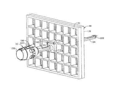
useful in analyzing the
suspicious tissue and making an assessment of the progression of the cancer
less simple if
the sample is found to be malignant.
[0005] During a core needle biopsy, a small tissue sample is removed allowing
for a
pathological assessment of the tissue, including an assessment of the
progression of any
cancerous cells that are found. The following patent documents disclose
various core
biopsy devices: US 6,273,862 issued Aug. 14, 2001; US 6,231,522 issued May 15,
2001;
US 6,228,055 issued May 8, 2001; US 6,120,462 issued September 19, 2000; US
6,086,544 issued July 11, 2000; US 6,077,230 issued June 20, 2000; US
6,017,316 issued
Jan. 25, 2000; US 6,007,497 issued Dec. 28, 1999; US 5,980,469 issued Nov. 9,
1999; US
5,964,716 issued Oct. 12, 1999; US 5,928,164 issued July 27, 1999; US
5,775,333 issued
July 7, 1998; US 5,769,086 issued June 23, 1998; US 5,649,547 issued July 22,
1997; US
5,526,822 issued June 18, 1996; and US Patent Application 2003/0199753
published Oct.
23, 2003 to Hibner et al.
100061 In U.S. Pat. Appin. Publ. No. 2005/0283069A1, "MRI biopsy device
localization
fixture" to Hughes et al., a localization mechanism, or fixture, is described
that is used in
conjunction with a breast coil for breast compression and for guiding a core
biopsy
instrument during prone biopsy procedures in both open and closed Magnetic
Resonance
Imaging (MRI) machines. The localization fixture includes a three-dimensional
Cartesian
positionable guide for supporting and orienting an MRI-compatible biopsy
instrument,
and, in particular, a sleeve to a biopsy site of suspicious tissues or
lesions.
100071 A z-stop enhances accurate insertion, and prevents over-insertion or
inadvertent
retraction of the sleeve. In particular, the Z-stop is engaged to the
localization fixture at a
distance from the patient set to abut a handle of the biopsy device as an
attached biopsy
probe reaches the desired depth. Similarly, another biopsy cannula may be a
sleeve with a
hub corresponding to a handle that contacts the z-stop.
100081 While such a localization fixture with a depth stop feature provides
clinical
advantages, some surgeons may prefer other types of methods of positioning a
biopsy
probe or similar biopsy cannula. For instance, some clinicians may prefer a
manually
guided biopsy probe, such as when being directed by on-going diagnostic
imaging (e.g.,
2
CA 02586504 2007-04-27
ultrasonic). It would thus be desirable to incorporate preventing over-
insertion of a biopsy
probe when not employing a three-axis insertion guidance apparatus.
SUMMARY OF THE INVENTION
[0009] The present invention addresses these and other problems of the prior
art by providing
an apparatus and method for use of a depth stop device longitudinally
positioned on a
biopsy cannula prior to insertion into tissue. The depth stop device
advantageously has an
unlocked condition that allows positioning followed by a locking condition
such that
inadvertent over-insertion is affirmatively blocked. Thereby, even manual
insertion of a
biopsy device or trocar/sleeve has the benefits of guided procedures to
prevent
overshooting with a piercing tip of the biopsy cannula.
fool oi In one aspect of the invention, a device serves as the depth stop by
presenting a
guiding portion that substantially circumferentially encompasses a shaft of a
biopsy
cannula. A locking portion moves into binding engagement with the biopsy
cannula when
at a desired longitudinal position thereon. A transverse portion of the device
precludes
over insertion by coming into abutment with the skin of the patient or some
proximate
structure that localizes the body portion being biopsied.
(00111 In another aspect of the invention, a biopsy cannula has measurement
indicia that aids
in longitudinal positioning of a depth stop device, the measurement indicia
being
representative of depth of penetration achieved thereby.
100121 These and other objects and advantages of the present invention shall
be made
apparent from the accompanying drawings and the description thereof.
3
CA 02 5 8 65 0 4 2 0 07-0 4-2 7
BRIEF DESCRIPTION OF THE DRAWINGS
=
[0013] While the specification concludes with claims particularly pointing out
and distinctly
claiming the present invention, it is believed the same will be better
understood by
reference to the following description, taken in conjunction with the
accompanying
drawings in which:
=
[0014] FIGURE 1 is an isometric view of a biopsy system including a control
module
remotely coupled to a biopsy device, and including a localization fixture with
a lateral
grid plate used in conjunction with a rotatable cube to position a
trocar/obturator or a
probe of the biopsy device to a desired insertion depth as set by a ring stop.
100151 FIGURE 2 is an isometric view of the breast coil receiving the
localization fixture of
FIG. 1.
[0016] FIGURE 3 is an isometric view of the biopsy device inserted through the
rotatable
cube within the cube plate of the localization fixture attached to a breast
coil of FIG. 1.
[00171 FIGURE 4 is an isometric view of a two-axis rotatable guide cube of the
biopsy
system of FIG. 1.
100181 FIGURE 5 is a diagram of nine guide positions achievable by the two-
axis rotatable
guide cube of FIG. 5.
=
100191 FIGURE 6 is an isometric view of a two-axis rotatable guide cube
inserted into a
lateral grid with the backing of the localization fixture of FIG. 1.
100201 FIGURE 7 is an isometric view of the trocar and sleeve of the biopsy
system of FIG.
1.
100211 FIGURE 8 is an isometric exploded view of the trocar and sleeve of FIG.
7.
[0022] FIGURE 9 is an isometric view of a trocar and sleeve of FIG. 7 with a
depth stop
device of FIG. 1 inserted through the guide cube and grid plate of FIG. 6.
[0023] FIGURE 10 is an alternative guide cube for the biopsy system of FIG. 1
with two-
axes of rotation and self-grounding features.
[0024] FIGURE 11 is an isometric view of the trocar and sleeve of FIG. 7
inserted into one
of two guide cubes of FIG. 10 inserted into the grid plate of FIG. 1.
4
CA 02586504 2007-04-27
= [0025] FIGURE 12 is an aft isometric view of a further alternative guide
cube with four
angled, parallel guide holes for the biopsy system of FIG. 1.
[0026] FIGURE 13 is a front isometric view of the guide cube of FIG. 12.
100271 FIGURE 14 is a right side view of the guide cube of FIG. 12 with the
angled, parallel
guide holes depicted in phantom.
[0028] FIGURE 15 is an aft view in elevation of yet another alternative guide
cube for the
biopsy system of FIG. 1 with a pair of converging guide holes and a pair of
diverging
guide holes.
[0029] FIGURE 16 is a left side view of the guide cube of FIG. 15 taken in
cross section
along lines 16-16 through the pair of converging guide holes.
[0030] FIGURE 17 is a left side view of the guide cube of FIG. 15 taken in
cross section
along lines 17-17 through the pair of diverging guide holes.
loom FIGURE 18 is an isometric view of a two hole guide cube for the biopsy
system of
FIG. 1.
100321 FIGURE 19 is an isometric view of a one-hole guide cube for the biopsy
system of
FIG. 1.
[0033] FIGURE 20 is a rotating guide for guiding the trocar and sleeve of FIG.
7 into either
of the two-hole guide cube of FIG. 18 or the one-hole guide cube of FIG. 19.
100341 FIGURE 21 is an aft isometric view of the trocar and sleeve of FIG. 7
inserted
through the rotating guide of FIG. 20 into the two-hole guide cube of FIG. 18.
100351 FIGURE 22 is an isometric locking 0-ring for the biopsy system of FIG.
1.
100361 FIGURE 23 is an aft view of the locking 0-ring of FIG. 22 with a cross
section of a
biopsy instrument cannula shown in both an unlocked orientation and rotated a
quarter
turn into a locked orientation depicted in phanton.
100371 FIGURE 24 is an isometric view of a cylindrical rotating guide formed
of elastomeric
material with an oval through hole for the biopsy system of FIG. 1.
CA 02586504 2014-07-03
[0038] FIGURE 25 is an aft view of the cylindrical rotating guide of FIG. 24
with a cross
sectional view of an unlocked oval-shaped biopsy instrument cannula inserted
in the oval
through hole.
[0039] FIGURE 26 is an aft view of the cylindrical rotating guide and biopsy
instrument
cannula of FIG. 25 with the cylindrical rotating guide rotated a quarter turn
relative to the
cannula to elastomerically lock thereon.
[0040] FIGURE 27 is an isometric view of a flattened oval rotating guide for
the biopsy
system of FIG. 1.
[0041] FIGURE 28 is an isometric view of a triangular clip depth stop for the
biopsy system
of FIG. 1.
[0042] FIGURE 29 is an isometric view of a scissor-like depth stop clip for
the biopsy
system of FIG. 1.
100431 FIGURE 30 is an aft isometric view of a shutter depth stop with an
inserted biopsy
instrument cannula for the biopsy system of FIG. 1.
[0044] FIGURE 31 is an aft view of the shutter depth stop of FIG. 30 prior to
use.
[0045] FIGURE 32 is a front isometric view of the shutter depth stop and
inserted biopsy
instrument cannula of FIG. 30.
[0046] FIGURE 33 is an aft view of the shutter depth stop and biopsy
instrument cannula of
FIG. 30 with the shutter depth stop vertically compressed into an unlocked
state.
DETAILED DESCRIPTION OF THE INVENTION
[0047] Turning
to the Drawings, wherein like numerals denote like components throughout
the several views, in FIGS. 1-3, a Magnetic Resonance Imaging (MR1) compatible
biopsy
system 10 has a control module 12 that typically is placed outside of a
shielded room
containing an MRI machine (not shown) or at least spaced away to mitigate
detrimental
interaction with its strong magnetic field and/or sensitive radio frequency
(RF) signal
detection antennas. As described in U.S. Pat. No. 6,752,768, a range of
preprogrammed
functionality is incorporated into the control module 12 to assist in taking
these tissue
samples. The control module 12
6
1 I I I AI ..ir
CA 02586504 2007-04-27
controls and powers an MRI biopsy device 14 that is positioned and guided by a
localization fixture 16 attached to a breast coil 18 that is placed upon a
gantry (not shown)
of the MRI machine.
100481 The control module 12 is mechanically, electrically, and
pneumatically coupled to
the MM biopsy device 14 so that components may be segregated that need to be
spaced
away from the strong magnetic field and the sensitive RF receiving components
of the
MM machine. A cable management spool 20 is placed upon a cable management
attachment saddle 22 that projects from a side of the control module 12. Wound
upon the
cable management spool 20 is a paired electrical cable 24 and mechanical cable
26 for
communicating control signals and cutter rotation/advancement motions
respectively. In
particular, electrical and mechanical cables 24, 26 each have one end
connected to
respective electrical and mechanical ports 28, 30 in the control module 12 and
another
end connected to a reusable holster portion 32 of the MRI biopsy device 14. An
MRI
docking cup 34, which may hold the holster portion 32 when not in use, is
hooked to the
control module 12 by a docking station mounting bracket 36.
100491 An interface lock box 38 mounted to a wall provides a tether 40 to a
lockout port 42
on the control module 12. The tether 40 is advantageously uniquely terminated
and of
short length to preclude inadvertent positioning of the control module 12 too
close to the
MRI machine. An in-line enclosure 44 may advantageously register the tether
40,
electrical cable 24 and mechanical cable 26 to their respective ports 42, 28,
30 on the
control module 12.
[0050] Vacuum assist is provided by a first vacuum line 46 that connects
between the
control module 12 and an outlet port 48 of a vacuum canister 50 that catches
liquid and
solid debris. A tubing kit 52 completes the pneumatic communication between
the control
module 12 and the MRI biopsy device 14. In particular, a second vacuum line 54
is
connected to an inlet port 56 of the vacuum canister 50. The second vacuum
line 54
divides into two vacuum lines 58, 60 that are attached to the MM biopsy device
14. With
the MRI biopsy device 14 installed in the holster portion 32, the control
module 12
performs a functional check. Saline is manually injected into biopsy device 14
to serve as
a lubricant and to assist in achieving a vacuum seal. The control module 12
actuates a
cutter mechanism (not shown) in the MR' biopsy device 14, monitoring full
travel.
Binding in the mechanical cable 26 or within the biopsy device 14 is monitored
with
7
CA 02586504 2007-04-27
reference to motor force exerted to turn the mechanical cable 26 and/or an
amount of
twist in the mechanical cable 26 sensed in comparing rotary speed or position
at each end
of the mechanical cable 26.
100511 A remote keypad 62, which is detachable from the reusable holster
portion 32,
communicates via the electrical cable 24 to the control panel 12 to enhance
clinician
control of the MRI biopsy device 14, especially when controls that would
otherwise be on
the MRI biopsy device 14 itself are not readily accessible after insertion
into the
localization fixture 16 and/or placement of the control module 12 is
inconveniently
remote (e.g., 30 feet away). An aft end thumbwheel 63 on the reusable holster
portion 32
is also readily accessible after insertion to rotate the side from which a
tissue sample is to
be taken.
[0052] Left and right parallel upper guides 64, 66 of a localization
framework 68 are
laterally adjustably received respectively within left and right parallel
upper tracks 70, 72
attached to an under side 74 and to each side of a selected breast aperture 76
formed in a
patient support platform 78 of the breast coil 18. A base 80 of the breast
coil 18 is
connected by centerline pillars 82 that are attached to the patient support
platform 78
between the breast apertures 76. Also, a pair of outer vertical support
pillars 84, 86 on
each side spaced about a respective breast aperture 76 respectively define a
lateral recess
88 within which the localization fixture 16 resides.
00531 It should be appreciated that the patient's breasts hang pendulously
respectively into
the breast apertures 76 within the lateral recesses 88. For convenience,
herein a
convention is used for locating a suspicious lesion by Cartesian coordinates
within breast
tissue referenced to the localization fixture 16 and to thereafter selectively
position an
instrument, such as a probe 90 (FIG. 1) of a disposable probe assembly 91 that
is engaged
to the reusable holster portion 32 to form the MRI biopsy device 14. To
enhance hands
off use of the biopsy system 10, especially for repeated reimaging within the
narrow
confines of a closed bore MM machine, the MRI compatible biopsy system 10 may
also
guide a trocar ("introducer") 92 encompassed by a sleeve 94. Depth of
insertion is
controlled by a depth stop device 95 longitudinally positioned on either the
probe 90 or
the sleeve 94.
8
CA 02586504 2007-04-27
100541 This guidance is specifically provided by a lateral fence, depicted
as a grid plate 96,
which is received within a laterally adjustable outer three sided plate
bracket 98 attached
below the left and right parallel upper guides 64, 66. Similarly, a medial
fence with
respect to a medial plane of the chest of the patient, depicted as a medial
plate 100, is
received within an inner three-sided plate bracket 102 attached below the left
and right
parallel upper guides 64, 66 close to the centerline pillars 82 when installed
in the breast
coil 18. To further refine the insertion point of the instrument (e.g., probe
90,
trocar/sleeve 92, 94), a guide cube 104 is inserted into the backside of the
grid plate 96.
Noss] The selected breast is compressed along an inner (medial) side by the
medial plate
100 and on an outside (lateral) side of the breast by the grid plate 96, the
latter defining an
X-Y plane. The X-axis is vertical (sagittal) with respect to a standing
patient and
corresponds to a left to right axis as viewed by a clinician facing the
externally exposed
portion of the localization fixture 16. Perpendicular to this X-Y plane
extending toward
the medial side of the breast is the Z-axis, which typically corresponds to
the orientation
and depth of insertion of the probe 90 of the MRI biopsy device 14 or the
trocar/sleeve
92, 94. For clarity, the term Z-axis may be used interchangeably with "axis of
penetration", although the latter may or may not be orthogonal to the spatial
coordinates
used to locate an insertion point on the patient. Versions of the localization
fixture 16
described herein allow a nonorthogonal axis of penetration to the X-Y axis to
a lesion at a
convenient or clinically beneficial angle.
100561 In FIG. 4, guide cube 104 includes a central guide hole 106, a
corner guide hole
108, and an off-center guide hole 110 that pass orthogonally to one another
between
respective opposite pairs of faces 112, 114, 116. By selectively rotating the
guide cube
104 in two axis, one of the pairs of faces 112, 114, 116 may be proximally
aligned to an
unturned position and then the selected proximal face 112, 114, 116 optionally
rotated a
quarter turn, half turn, or three quarter turn. Thereby, one of nine guide
positions 118 (i.e.,
using central guide hole 106), 120a-120d (i.e., corner guide hole 108), 122a-
122d (i.e.,
using off-center guide hole 110) may be proximally exposed as depicted in FIG.
5.
100571 In FIG. 6, the two-axis rotatable guide cube 104 is sized for
insertion from a
proximal side into one of a plurality of square recesses 130 in the grid plate
96 formed by
intersecting vertical bars 132 and horizontal bars 134. The guide cube 104 is
prevented
from passing through the grid plate 96 by a backing substrate 136 attached to
a front face
9
CA 02586504 2007-04-27
of the grid plate 96. The backing substrate 136 includes a respective square
opening 138
centered within each square recess 130, forming a lip 140 sufficient to
capture the front
face of the guide cube 104 but not so large as to obstruct the guide holes
104, 106, 108.
The depth of the square recesses 130 is less than the guide cube 104, thereby
exposing a
proximal portion 142 of the guide cube 104 for seizing and extraction from the
grid plate
96.
[00581 In FIGS. 7-9, in the illustrative version, the trocar 92 is slid
into the sleeve 94 and
the combination is guided through the guide cube 104 (FIG. 9) to the biopsy
site within
the breast tissue. The sleeve 94 includes a hollow shaft (or cannula) 196 that
is proximally
attached to a cylindrical hub 198 and has a lateral aperture 200 proximate to
an open
distal end 202. The cylindrical hub 198 has an exteriorly presented thumbwheel
204 for
rotating the lateral aperture 200. The cylindrical hub 198 has an interior
recess 206 that
encompasses a duckbill seal 208, wiper seal 210 and a seal retainer 212 to
provide a fluid
seal when the shaft 196 is empty and for sealing to the inserted introducer
(trocar) 92.
Longitudinally spaced measurement indicia 213 along an outer surface of the
hollow shaft
196 visually, and perhaps physically, provide a means to locate the depth stop
device 95
of FIG. 1.
100591 The trocar 92 advantageously incorporates a number of components
with
corresponding features. A hollow shaft 214 includes a fluid lumen 216 that
communicates
between an imagable side notch 218 and a proximal port 220. The hollow shaft
214 is
longitudinally sized to extend, when fully engaged, a piercing tip 222 out of
the distal end
202 of the sleeve 94. An obturator thumbwheel cap 224 encompasses the proximal
port
220 and includes a locking feature 226, which includes a visible angle
indicator 228 (FIG.
8), that engages the sleeve thumbwheel 204 to ensure that the imagable side
notch 218 is
registered to the lateral aperture 200 in the sleeve 94. An obturator seal cap
230 may be
engaged proximally into the obturator thumbwheel cap 224 to close the fluid
lumen 216.
The obturator seal cap 230 includes a locking or locating feature 232 that
includes a
visible angle indicator 233 that corresponds with the visible angle indicator
228 on the
obturator thumbwheel cap 224, which may be fashioned from either a rigid,
soft, or
elastomeric material. In FIG. 9, the guide cube 104 has guided the trocar 92
and sleeve 94
through the grid plate 96.
CA 02586504 2007-04-27
100601 In FIGS. 10-11, an alternative guide cube 104a has rotation in two
axes but is self
grounding by means of an added rectangular prism 240 which has a shared edge
with a
cubic portion 242 of the guide cube 104a. When viewed orthogonally to the
shared cube
edge, a larger square face 244 of the cubic portion 242 overlaps with a
smaller square
face 246 of the rectangular prism 240 to correspond with the desired size of
an exposed
proximal portion 248 of the inserted guide cube 104a. The rectangular prism
240 allows
proximal exposure of one of two adjacent faces 250, 252 of the guide cube 104a
and then
turning each to one of four quarter turn rotational positions. In the
illustrative version,
first face 250 has a central guide hole 106a and the second face 252 has a
corner guide
hole 108a, and an off-center guide hole 110a. A radial recess 254 is relieved
into the
rectangular prism 240 to allow grounding of the depth stop device 95 against
the face 252
when the off-center guide hole 110a is used.
100611 In FIGS. 12-14, another alternative guide cube 104b has a proximal
enlarged hat
portion 270 about a proximal face 271 that grounds against the selected square
recess 130
in the grid plate 96 (FIG. 6) and allows rotation about one axis to one of
four quarter turn
positions. Four angled guide holes 272a, 272b, 272c, 272d allow accessing not
only an
increased number of insertion points within the selected square recess 130 but
also a
desired angle of penetration rather than being constrained to a perpendicular
insertion.
100621 In FIGS. 15-17, an additional alternative guide cube 104c also has
the proximal
enlarged hat portion 270 about the proximal face 271 that grounds against the
selected
square recess 130 in the grid plate 96 (FIG. 6) and allows rotation about one
axis to one
of four quarter turn positions. The guide holes are depicted as a first pair
of converging
angled through holes 310a, 310b having outwardly spaced proximal openings
311a, 311b
(FIG. 15), respectively, that communicate with partially intersecting distal
openings 312a,
312b, respectively. The guide holes are also depicted as a second pair of
diverging angled
through holes 310c, 310d having partially intersecting proximal openings 311c,
311d,
respectively, that communicate with outwardly spaced distal openings 312c,
312d.
100631 In FIG. 18, a further alternative two-hole guide cube 104d has two
enlarged guide
holes 330, 332 accessed through the proximal face 271 in the enlarged proximal
hat
portion 270. Similarly, in FIG. 19, a one hole guide cube 104e has one
enlarged guide
hole 334 accessed through the proximal face 271 in the enlarged proximal hat
portion
270. Each guide cube 104d, 104e may receive a cylindrical rotating guide 336
(FIG. 20)
11
1" r
CA 02586504 2007-04-27
with an integral, proximal depth ring stop 338. In FIGS. 20, 21, a through
hole 340 in the
cylindrical guide 336 is sized to receive a biopsy instrument cannula (e.g.,
probe 90,
sleeve 94) by being oval in cross section in the illustrative version. It
should be
appreciated that the cylindrical guide 336 may provide structural support to
the guided
portion of the biopsy instrument support as well as facilitate axial rotation
thereof,
especially for a non cylindrical biopsy instrument cannula.
100641 It should be appreciated that the two-hole and one-hole guide cubes
104d, 104e and
rotating guide 336 may comprise a guide cube set, perhaps with additional
guide cubes
(not shown) having uniquely positioned guide holes. With the enlarged guide
holes 330-
340 to accommodate the rotating guide 336, too much overlap of guide holes
(e.g., 330,
332) may result in insufficient support by the rotating guide 336 for the
inserted biopsy
instrument cannula. Thus, fine positioning is accomplished by selecting one of
the
available guide cubes 104d, 104e for the desired position within a selected
grid aperture.
100651 In FIGS. 22, 23, a locking 0-ring feature may be advantageously
incorporated into a
depth ring stop (rotating guide) 350. Having to rely upon constant frictional
engagement
of the depth ring stop (rotating guide) 350 alone would result in difficulty
in installing the
ring stop 350 to the desired position or being too readily displaced to serve
as a stopping
structure. In the exemplary version, an outer circumference surface 351 of the
ring stop
350 includes left and right outer longitudinal ridges 352, 354 that aid in
gripping and
orienting the depth ring stop 350 while turning for locking and unlocking. As
viewed
from behind, opposing inner longitudinal ridges 356, 358 formed in a generally
cylindrical inner diameter 359 abut respectively at an upper left and lower
right side of an
oval cannula 360 (FIG. 23) oriented with its elongate cross section vertically
in an
unlocked position. The inner longitudinal ridges 356, 358 allow a quarter turn
clockwise
of the oval cannula, depicted as 360', to a locked position deforming an inner
tangential
locking rib 362.
100661 It should be appreciated that these orientations and geometry are
illustrative. An
amount of rotation to lock and unlock, for instance, may be less than or more
than a
quarter turn. In addition, noncircular features on an outer diameter of the
depth ring stop
350 may be omitted. Other variations may be employed. For example, in FIGS. 24-
25, a
cylindrical rotating guide 380, formed of a resilient polymer, has an elongate
through hole
382 shaped to permit insertion of an oval biopsy cannula 384. In FIG. 26,
turning the
12
I "
CA 02586504 2007-04-27
= cylindrical rotating guide 380 a quarter turn in either direction to a
locked position,
depicted at 380', causes the cylindrical rotating guide 380' to deform,
binding onto the
biopsy instrument cannula 384, therby serving as a depth stop.
10671 Similarly, in FIG. 27, a rotating guide 400 is oval shaped with
flattened elongate
sides and with a corresponding elongate through hole 402. The outer shape may
be tactile,
advantageous for gripping as well as for providing a visual indication of
being locked or
unlocked. A resilient tangential rib 404 crossing one inner corner of the
elongate through
hole 402 is positioned to bind against an inserted biopsy instrument cannula
(not shown)
when the rotating guide 400 is turned a quarter turn to a locking position.
[0068] In FIG. 28, a triangular clip depth stop 420 has a transverse
front surface 422 with a
proximally turned lower lip 424 and an upper lateral edge 426 attached to a
downwardly
and proximally ramped member 428 whose lower lateral edge 430 bends distally
to form
a horizontal locking actuator member 432 whose distal edge 434 rests upon the
lower lip
424. A front vertically elongate aperture 436 in the transverse front surface
422 is shaped
to approximate the outer diameter of an inserted biopsy instrument cannula
(not shown).
An aft elongate aperture 438 formed in the downwardly and proximally ramped
member
428 is a distal horizontal projection of the front vertically elongate
aperture 436 when the
locking actuator member 432 is upwardly raised, thus allowing insertion of the
biopsy
instrument cannula through both apertures 436, 438. Upon release of the
locking actuator
member 432, an upper inner surface 440 of the aft elongate aperture 438
lowers, binding
upon the inserted biopsy instrument cannula, allowing the transverse front
surface 422 to
serve as a positive depth stop.
[0069] In FIG. 29, a scissor-like clip depth stop 450 is cut out of a
layer of resilient
material. In particular, an upper arm portion 452 and a lower arm portion 454
are attached
to one radiating vertically away from each other toward the same lateral side
(right as
depicted) from a split cylindrical grasping portion 456 separated
longitudinally on a
lateral side opposite to the arm portions 452, 454 (left as depicted). In
particular, an upper
gripping half-cylindrical member 458 is attached at its right side to a lower
portion 460 of
the upper arm portion 452. A lower gripping half-cylindrical member 462 is
attached at
its right side to an upper portion 464 of the lower arm portion 454. An upper
hemispheric
portion 466 of the upper arm portion 452 includes an upper finger hole 468. A
lower
hemispheric portion 470 of the lower arm portion 454 includes a lower finger
hole 472. A
13
CA 02586504 2007-04-27
triangular recess 474 (opening rightward as depicted) formed by the arm
portions 452,
454 and a longitudinal pin 476 inserted at the juncture between the arm
portions 452, 454
predispose the arm portions 452, 454 to be resiliently drawn toward each other
as the
finger holes 468, 472 are gripped and moved together, thereby opening the
upper and
lower gripping half cylindrical members 458, 462, widening the separation of
their left
ends. In this unlocked position, a biopsy instrument cannula (not shown) may
be inserted
and positioned to a desired depth.
100701 In FIG. 30-33 a shuttered depth stop 600 includes a resilient oval
shell 602 with a
corresponding oval aperture 604 with an upper right tab 606 projecting
inwardly to the
left and with a lower left tab 608 projecting inwardly to the right when
viewed from
behind (FIG. 30). An upper resilient member 610 has a generally horseshoe-
shaped outer
surface 612 that conforms to an upper portion 614 of the oval aperture 604. A
lower
resilient member 616 has a generally horseshoe-shaped outer surface 618 that
conforms to
a lower portion 620 of the oval aperture 604. In the illustrative version, the
upper and
lower resilient members 610, 616 are identical but are rotated a half turn
about a
longitudinal axis with respect to each other. Moreover, the entire shuttered
depth stop 600
is symmetric about its vertical axis defined by its longest dimension or about
a horizontal
axis defined by its second longest dimension.
100711 A downwardly open rectangular prismatic recess 622 formed in the
upper resilient
member 610 is sized to receive an upper shutter 624 having an upper center tab
626 and a
lower acute edge 628. A top center rectangular slot 630 formed in the upper
resilient
member 610 communicates with the downwardly open rectangular prismatic recess
622
and receives the upper center tab 626. An upwardly open rectangular prismatic
recess 632
formed in the lower resilient member 616 is sized to receive a lower shutter
634 having a
lower center tab 636 and an upper acute edge 638. A bottom center rectangular
slot 639
formed in the lower resilient member 616 communicates with the upwardly open
rectangular prismatic recess 632 and receives the lower center tab 636. An
upper
horizontal pin 640 attached horizontally as depicted across the upper shutter
624 is
received for rotation onto opposite lateral sides of the downwardly open
rectangular
prismatic recess 622. A lower horizontal pin 642 attached horizontally as
depicted across
the lower shutter 634 is received for rotation onto opposite lateral sides of
the upwardly
open rectangular prismatic recess 632.
14
CA 02586504 2007-04-27
100721 The right side of the upper resilient member 610 includes a right
outward shoulder
644 that rests upon the upper right tab 606 of the resilient oval shell 602. A
laterally
recessed downward arm 646 is attached to the right shoulder 644 and extends
downwardly with its outer surface 648 vertically aligned with an innermost
edge 650 of
the right outward shoulder 644 and with its inner surface 652 defining the
downwardly
open generally rectangular prismatic recess 622. The left side of the upper
resilient
member 610 includes a left inward shoulder 654 that is laterally aligned with
and opposite
of the upper right tab 606 of the resilient oval shell 602. An outer downward
arm 656 is
attached to the left inward shoulder 654 and extends downwardly with its outer
surface
658 against oval aperture 604 and an innermost edge 660 vertically aligned
with an inner
surface 662 of the lower left tab 608 upon which the outer downward arm 656
rests.
100731 Similarly, the lower resilient member 616 includes a left outward
shoulder 664
attached to a laterally recessed upward arm 666 and a right inward shoulder
668 attached
to an outer upward arm 670 that abuts an underside of the upper right tab 606.
The
laterally recessed downward arm 646 of the upper resilient member 610 extends
downward past the longitudinal centerline of the shuttered depth stop 600 and
an inserted
biopsy instrument cannula 672. A lower edge 674 of the laterally recessed
downward arm
646 is spaced away from an upper surface 676 of the right inward shoulder 668.
In
addition, an upper edge 678 of the laterally recessed upward arm 666 is spaced
away from
a lower surface 680 of the left inward shoulder 654. When the resilient oval
shell 602 is
relaxed as in FIGS. 30-32, this spacing between the left inward shoulder 654
and the
upper edge 678 of the laterally recessed upward arm 666 defines an upper left
rectangular
recess 682 communicating rightward into the downwardly open rectangular
prismatic
recess 622 and sized to allow unimpeded swinging of a leftward extension 684
of the
upper shutter 624. Spacing between the upper surface 676 of the right inward
shoulder
668 and the lower edge 674 of the laterally recessed downward arm 646 defines
a lower
right rectangular recess 686 which communicates leftward into the upwardly
open
rectangular prismatic recess 632 which is sized to allow unimpeded swinging of
a
rightward extension 688 of the lower shutter 634.
=
100741 In FIG. 31, the shuttered depth stop 600 initially has closed upper
and lower shutters
624, 634 due to restoring pressure from the top center rectangular slot 630 on
the upper
center tab 626 and from the bottom center rectangular slot 639 on the lower
center tab
I ,= =
CA 02586504 2007-04-27
= = 636 respectively. Insertion of a biopsy instrument cannula
672 from a selected side (thus
the aft side) causes the upper and lower acute edges 628, 638 of the shutters
624, 634 to
swing distally and outwardly but remain in contact due to the restoring
pressure
previously mentioned. Proximal retraction of the biopsy instrument cannula 672
frictionally rotates the acute edges 628, 638 proximally, and thus inwardly,
binding upon
the biopsy instrument cannula 672 preventing inadvertent retraction to serve
as a depth
stop. When retraction is desired, squeezing the resilient oval shell 602 to
reduce the
vertical height of the shutter depth stop 600 in FIG. 33 causes the laterally
recessed
downward arm 646 to open the lower shutter 634 and the laterally recessed
upward arm
666 to open the upper shutter 624.
100751 Alternatively, it should be appreciated that a single
shutter may be employed in a
shuttered depth stop consistent with aspects of the invention. As a further
alternative or as
an additional feature, grooves in the biopsy cannula may enhance engagement of
one or
two shutters to further avoid inadvertent proximal retraction of the
positioned shuttered
depth stop. Moreover, the grooves on the biopsy cannula may be ramped such
that
engagement is more prevalent against proximal retraction as compared to distal
positioning. Further, such grooves may be along only a portion of the
circumference of
the biopsy cannula such that rotation of the shuttered depth stop also further
unlocks from
the biopsy cannula for positioning.
100761 It should be appreciated with the benefit of the present
disclosure that straight upper
and lower acute edges 628, 638 of the two shutters 624, 634 may instead be
contoured to
closely approximate the transverse cross section of the encompassed shuttered
depth stop
600 to increase the locking against inadvertent retraction.
100771 While the present invention has been illustrated by
description of several
embodiments and while the illustrative embodiments have been described in
considerable
detail, it is not the intention of the applicant to restrict or in any way
limit the scope of the
appended claims to such detail. Additional advantages and modifications may
readily
appear to those skilled in the art.
[00781 For example, other imaging modalities may benefit from
aspects of the present
invention.
16
CA 02586504 2014-07-03
- 100791 It should be appreciated that a grid plate 96 with a
backing lip 140 may be used such
that a guide cube rotatable to each of the six faces with four quarter turn
positions for
each face may achieve a large number of possible insertion positions and
angles of
insertion.
100811 It should be appreciated that various directional terms
such as horizontal, vertical,
left, right, downward, upward, etc. have been used in conjunction with the
orientation of
depictions in the drawings. Applications consistent with the present invention
may
include usage of like components in other orientations.
100821 It should be appreciated that biasing of the locking /
unlocking components of
various versions of a depth stop for a biopsy cannula described herein are
advantageously
formed out of an elastomeric material for economical manufacture. However, an
assembly of rigid components biased by springs for biasing and/or actuating
controls to
move the locking surface out of engagement may be substituted to achieve
similar results
consistent with aspects of the present invention.
100831 For example, the positioning and height of a central web of a breast
coil may enable
use of a medial grid plate used with a rotatable cube and penetrate from the
medial side of
the breast. For another example, a grid having a different geometric shape,
such as
hexagonal, may be employed.
100841 As another example, each grid aperture of equilateral polygonal lateral
cross section
in a grid plate taper toward their distal opening to ground a similarly
tapered guide block.
17