Note: Descriptions are shown in the official language in which they were submitted.
CA 02586620 2007-04-27
WO 2006/053871
PCT/EP2005/055984
- 1 -
T I T LE
Multivalent vaccines comprising recombinant viral vectors
FIELD OF THE INVENTION
The invention relates to the field of recombinant DNA
and viral vector vaccines. Specifically, it relates to
recombinant DNA and viral vectors harbouring nucleic acids
encoding multiple antigens and/or adjuvants.
BACKGROUND OF THE INVENTION
Tuberculosis (TB) has been a major worldwide threat to
human health for several thousands of years. TB caused by
Mycobacterium tuberculosis is an infectious disease of the
lung caused by infection through exposure to air-borne M.
tuberculosis bacilli. These bacilli are extremely infectious
and it has been estimated that currently approximately one-
third of the world population (2 billion people) are
infected. It has been further estimated that TB kills over 2
million people worldwide on an annual basis. Only 5 to 10%
of the immunocompetent humans are susceptible to TB, and
over 85% of them will develop the disease exclusively in the
lungs, while HIV-infected humans may also develop systemic
diseases that will more easily lead to death.
Approximately 90% of M. tuberculosis-infected humans
will not develop the disease. However, in these latently
infected individuals the bacilli can survive for many years
and become reactivated for instance in the case of a
weakened immune system, such as after an HIV infection. Due
to the latent nature, infected individuals generally have to
be treated by administration of several antibiotics for up
to 12 months, which is not a very attractive treatment in
general and due to costs and the possible occurrence of
CA 02586620 2007-04-27
WO 2006/053871
PCT/EP2005/055984
- 2 -
multi-drug resistance, while it is also not a very effective
treatment in most developing countries.
One relatively successful TB vaccine has been
developed: the bacilli Calmette-Guerin (BCG) vaccine was
generated in the early years of the twentieth century, and
was first given to individuals in 1921. The BCG vaccine is
an attenuated strain of bacteria based on a Mycobacterium
bovis isolate obtained from a cow. It is a relatively safe
vaccine, which is easy and rather cheaply produced. In the
year 2000, BCG vaccination covered 86% of the world
population. However, the vaccine appears to be not extremely
effective for adult pulmonary TB and many regions in
developing countries still have very high rates of TB,
despite the BCG-vaccine programs. It has been estimated that
BCG vaccine prevents only 5% of all vaccine-preventable
deaths by TB (Kaufmann, 2000).
Due to the rather low protection rate of the BCG
vaccine in general and due to the specific protection in
respect to childhood and disseminated TB, more efforts were
put in the development of new, more broadly applicable,
vaccines against TB, based on other systems and knowledge
acquired in other fields such as vaccination against other
tropical infectious diseases and HIV (review by Wang and
Xing, 2002).
Different approaches were taken to develop new TB
vaccines, ranging from subunit vaccines and DNA vaccines to
modified mycobacterium strains. Moreover, also recombinant
viral-based vaccines were generated, enabling the transfer
of M. tuberculosis antigens to antigen-presenting cells
through gene delivery vehicles such as Modified Vaccinia
Ankara (MVA) vectors and replication-defective adenovirus
vectors.
CA 02586620 2007-04-27
WO 2006/053871
PCT/EP2005/055984
- 3 -
Naked DNA vaccines against TB have been described in WO
96/15241 (see also EP 0792358), whereas many reports
describe the use of numerous antigens from Mycobacterium
tuberculosis in either recombinant or purified form for
their application in vaccines: WO 95/01441, WO 95/14713, WO
96/37219, US 6,599,510, WO 98/31388, WO 98/44119, WO
99/04005, WO 99/24577, WO 00/21983, WO 01/04151, WO
01/79274, WO 2004/006952, US 2002/0150592. The use of fusion
proteins comprising different TB antigens has also been
suggested: See WO 98/44119, EP 0972045 and EP 1449922,
disclosing the use of a fusion polypeptide between ESAT-6
and MPT59 (MPT59 is also referred to as Ag85B or the 85B
antigen).
Despite all these and other efforts in generating a
vaccine against tuberculosis that ensures both a strong
cellular, a strong humoral response as well as a long-
lasting high protection rate, no such vaccine is yet
available.
BRIEF DESCRIPTION OF THE DRAWINGS
Figure 1: Map of pAdApt35Bsu.myc
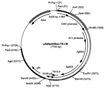
Figure 2: Map of pAdApt35Bsu.TB.LM
Figure 3: Map of pAdApt35Bsu.TB.SM
Figure 4: Map of pAdApt35Bsu.TB.FLM
Figure 5: Map of pAdApt35Bsu.TB.3M
Figure 6: Map of pAdApt35Bsu.TB.4M
Figure 7: Map of pAdApt35Bsu.TB.5M
Figure 8: Map of pAdApt35Bsu.TB.6M
Figure 9: Map of pAdApt35Bsu.TB.7M
Figure 10. Western blot with anti-TB antigen polyclonal on
lysates from A549 cells infected with Ad35 viruses
comprising nucleic acids encoding different sets of TB
CA 02586620 2007-04-27
WO 2006/053871
PCT/EP2005/055984
- 4 -
antigens with the myc-tag (A) and without the myc-tag (B).
(C) similar to (B), with molecular weight markers. See for
notation Table I.
Figure 11. Experimental design of immunization protocol
using 7 different adenoviral vectors (DNA) harbouring
different sets of nucleic acids encoding tuberculosis
antigens.
Figure 12. Percentages of antigen-specific splenocytes that
stain positive for interferon-gamma production (IFNy+) upon
stimulation with no peptide (A: CD4+ cells, B: CD8+ cells).
Figure 13. Percentages of antigen-specific splenocytes that
stain positive for interferon-gamma production (IFNy+) upon
stimulation with a pool of peptides relevant for the Ag85A
antigen (A: CD4+ cells, B: CD8+ cells).
Figure 14. Percentages of antigen-specific splenocytes that
stain positive for interferon-gamma production (IFNy+) upon
stimulation with a pool of peptides relevant for the Ag85B
antigen (A: CD4+ cells, B: CD8+ cells).
Figure 15. Percentages of antigen-specific splenocytes that
stain positive for interferon-gamma production (IFNy+) upon
stimulation with a pool of peptides relevant for the TB10.4
antigen (A: CD4+ cells, B: CD8+ cells).
Figure 16. Overview of percentages of CD4+ and CD8+
splenocytes that stain positive in ICN, in sera obtained
from mice injected with the triple inserts TB-L (A) and TB-
S(B).
Figure 17. Dose response effect using different doses of TB-
S comprising a nucleic acid encoding Ag85A, Ag85B and Tb10.4
antigens. CD4 response towards Ag85A (A), Ag85B (C) and
TB10.4 (E). CD8 response towards Ag85A (B), Ag85B (D) and
TB10.4 (F). (G): CD8 response towards Ag85B with the
CA 02586620 2007-04-27
WO 2006/053871
PCT/EP2005/055984
- 5 -
adjusted peptide pool (see example 6): left graph, upon TB-L
infection; right graph, upon TB-S infection.
Figure 18. CD4 and CD8 responses after priming with BCG and
boosting with different Ad-TB vectors. CD4 response towards
Ag85A (A), Ag85B (C) and TB10.4 (E). CD8 response towards
Ag85A (B), Ag85B (D) and TB10.4 (F).
Figure 19. Nucleotide sequence of TB-LM
Figure 20. Nucleotide sequence of TB-SM
Figure 21. Nucleotide sequence of TB-FLM
Figure 22. Amino acid sequence of TB-LM
Figure 23. Amino acid sequence of TB-SM
Figure 24. Amino acid sequence of TB-FLM
Figure 25. Ag85A stimulation in a BOG prime/Ad35-TB boost
experiment with a long-term read-out. Upper panel: CD4
response, lower panel: CD8 response. Ad35.E = empty Ad35
virus.
Figure 26. Ag85B stimulation in a BOG prime/Ad35-TB boost
experiment with a long-term read-out. Upper panel: CD4
response, lower panel: CD8 response. Ad35.E = empty Ad35
virus.
Figure 27. TB10.4 stimulation in a BOG prime/Ad35-TB boost
experiment with a long-term read-out. Upper panel: CD4
response, lower panel: CD8 response. Ad35.E = empty Ad35
virus.
SUMMARY OF THE INVENTION
The present invention relates to recombinant viral
vectors, preferably replication defective adenoviruses, more
preferably recombinant human adenovirus serotypes Adll,
Ad24, Ad26, Ad34, Ad35, Ad48, Ad49 and Ad50, wherein the
viral vectors comprise a heterologous nucleic acid sequence
encoding for (fusion) polypeptides of at least two antigens
CA 02586620 2013-06-03
from one or more tuberculosis causing bacilli. The encoded
antigens may be directly linked, i.e. forming one single
polypeptide. In one preferred embodiment, the antigens are
present in a precursor polyprotein, in the sense that they
are connected via a linker sequence recognized by a specific
protease that is co-expressed. The heterologous nucleic acid
may comprise the gene encoding the protease. The fusion
proteins with the direct linkages elicit desired immune
responses due to the antigens present in the fusion product,
whereas the proteins comprising the protease sites are
cleaved into separate discrete antigen forms, each
contributing to the desired immune response. The protease is
preferably linked to the antigens by a protease recognition-
site recognized by a cellular protease. Both set-ups provide
additional or even synergistic effects in comparison to
vaccination or therapy in which viral vectors are used that
comprise only a single transgen-encoding unit. More
generally, the invention also relates to viral vectors
comprising a heterologous nucleic acid sequence encoding
multiple antigens separated by protease specific cleavage
sites. It is to be understood that such antigens may be from
a wide variety of sources including, but not limited to,
infectious agents such as viruses, bacteria and parasites,
and are thus according to this aspect of the invention not
limited to antigens from tuberculosis-causing bacilli. The
antigens from Tuberculosis mycobacterium serve as non-
limiting examples of how such multivalent viral vector
vaccines are generated and how, upon entry into the host
cell, the antigens are separated, and they are able to
contribute to the immune response.
CA 02586620 2013-06-03
-6a-
There is provided herein a recombinant nucleic acid
molecule comprising a nucleic acid sequence encoding antigens
from the Ag85A, the Ag85B and the TB10.4 open reading frames
of Mycobacterium tuberculosis, wherein the sequence comprises,
in the 5' to 3' direction, a promoter, and respectively the
nucleic acid sequence encoding Ag85A, Ag85B, and TB10.4
antigens, wherein the antigens are linked so as to form a
fusion protein.
The invention also relates to the use of genetic
adjuvants that are co-expressed from the viral vector. These
CA 02586620 2007-04-27
WO 2006/053871
PCT/EP2005/055984
- 7 -
adjuvants are encoded by a nucleic acid, which is part of
the heterologous nucleic acid sequence introduced into the
viral vector genome. The adjuvant is expressed together with
the specific antigen(s) and is thereby able to stimulate the
immune response towards the antigen(s). Clearly, the
sequence encoding the adjuvant may be linked directly to the
sequence encoding the antigen(s), but is preferably
separated from the sequence encoding the one or more
antigen(s) by the linker sequence encoding the protease
recognition site. In the latter case, the adjuvant is
present in the host separately from the antigen(s) and is
able to provide its immune-stimulatory effects along with
the antigen(s).
DETAILED DESCRIPTION
The present invention relates to multivalent vaccines
comprising a recombinant viral vector. A preferred viral
vector is a recombinant Adenovirus (Ad) vector. The
recombinant adenoviral vector according to the invention
comprises a heterologous nucleic acid sequence encoding at
least two different antigens. The antigens may be within a
single polypeptide. These determinants may be either antigens
from viral, bacterial and parasitic pathogens, or host
antigens, such as, but not limited to, autoimmune antigens or
tumor antigens. In a preferred embodiment, the antigens are
from tuberculosis (TB)-causing bacilli, more preferable from
Mycobacterium tuberculosis, M. africanum or M. bovis or from
a combination thereof. The antigens may be the full-length
native protein, chimeric fusions between the antigen and a
host protein or mimetic, a fragment or fragments thereof of
an antigen that originates from the pathogen, or other
mutants that still elicit a desired immune response. Genes
CA 02586620 2007-04-27
WO 2006/053871
PCT/EP2005/055984
-8 -
encoding TB antigens that may typically be used in the viral
vectors of the present invention include, but are not limited
to: Ag85A (MPT44), Ag85B (MPT59), Ag85C (MPT45), TB10.4
(CFP7), ESAT-6, CFP7A, CFP7B, CFP8A, CFP8B, CFP9, CFP10,
CFP10A, CFP11, CFP16, CFP17, CFP19, CFP19A, CFP19B, CFP20,
CFP21, CFP22, CFP22A, CFP23, CFP23A, CFP23B, CFP25, CFP25A,
CFP26 (MPT51) CFP27, CFP28, CFP29, CFP30A, CFP30B, CWP32,
CFP50, MPT63, MTC28, LHP, MPB59, MPB64, MPT64, TB15, TB18,
TB21, TB33, TB38, TB54, TB12.5, TB20.6, TB40.8, TB10C, TB15A,
TB17, TB24, TB27B, TB13A, TB64, TB11B, TB16, TB16A, TB32,
TB32A, TB51, TB14, TB27, HBHA, GroEL, GroES (WO 95/01441, WO
98/44119, US 6,596,281, US 6,641,814, WO 99/04005, WO
00/21983, WO 99/24577), and the antigens disclosed in WO
92/14823, WO 95/14713, WO 96/37219, US 5,955,077, US
6,599,510, WO 98/31388, US 2002/0150592, WO 01/04151, WO
01/70991, WO 01/79274, WO 2004/006952, WO 97/09428, WO
97/09429, WO 98/16645, WO 98/16646, WO 98/53075, WO
98/53076, WO 99/42076, WO 99/42118, WO 99/51748, WO
00/39301, WO 00/55194, WO 01/23421, WO 01/24820, WO
01/25401, WO 01/62893, WO 01/98460, WO 02/098360, WO
03/070187, US 6,290,969, US 6,338,852, US 6,350,456, US
6,458,366, US 6,465,633, US 6,544,522, US 6,555,653, US
6,592,877, US 6,613,881, US 6,627,198. Antigen fusions that
may be of particular use are those disclosed for the first
time herein (such as Ag85A-Ag85B-TB10.4 and combinations
thereof), but also known fusions such as E5AT-6-MPT59 and
MPT59-E5AT-6 disclosed in WO 98/44119 and in the above-
referenced documents.
One approach for applying multiple antigens may be by
having two or more separate expression cassettes present in
a single vector, each cassette comprising a separate gene of
CA 02586620 2007-04-27
WO 2006/053871
PCT/EP2005/055984
- 9 -
interest. This approach clearly has disadvantages, for
instance related to space availability in the vector:
separate cassettes generally comprise separate promoters
and/or inducers and separate polyadenylation signal
sequences. Such cassettes typically require separate
positions in the viral vector, resulting in more laborious
cloning procedures, whereas a phenomenon known as 'promoter
interference' or 'squelching' (limited availability of
cellular factors required by the promoters to act) may
restrict the expression levels from the different promoters.
As exemplified by the recombinant viral vectors
disclosed herein relating to fusions between multiple TB
antigens, one is now able to make recombinant adenoviral
vectors comprising several nucleic acids encoding more than
one antigen, which viral vector elicits a strong immune
response, whereas the use of single inserts elicit limited
effects. Clearly, these vectors encode recombinant genetic
chimeras, which express the two or more antigens in a single
cistronic mRNA, for example in the form of a fusion protein.
This approach can be effective when DNA vaccines or the
viral vectors are being used to invoke T cell immunity to
the passenger antigens. However, such fusion proteins may
have additional drawbacks that cannot always be envisioned
beforehand, as it was found that such fusions might skew
immunodominant patterns and do not always invoke immunity to
all target antigens with equal potency, whereas a second and
perhaps more significant drawback to expression of genetic
fusions is that the individual components may not fold to a
native conformation, due to the close presence of their
fusion partner or other reasons. As a result of this,
genetic fusions may invoke antibody responses to nonsense
CA 02586620 2007-04-27
WO 2006/053871
PCT/EP2005/055984
- 10 -
epitopes and such antibodies do not recognize native
epitopes displayed by the founder pathogens and may be poor
at combating infection.
The inventors of the present invention have now
developed a system wherein multiple antigens are encoded by
a single heterologous nucleic acid sequence, wherein the
expressed polyprotein is processed into the discrete
antigenic polypeptides. Thus, in one embodiment, the present
invention relates to viral vectors that enable the
expression of multiple antigens that are subsequently
processed into the discrete antigens thus avoiding the
possible limitations associated with genetic fusions, while
also excluding the need for separate expression cassettes.
Heretofore, no compositions or methods have been described
that enable precise processing of viral vector-expressed
genetic fusions into discrete antigens. The expression of
multiple antigens encoded by nucleic acids comprised in a
DNA or viral vector, which antigens are subsequently
processed into discrete antigens is demonstrated by the use
of a protease (PR), such as the viral protease encoded by
Avian Leucosis Virus (ALV; referred to as PR-ALV herein). In
ALV, ALV-PR forms the C-terminal domain of the gag protein,
which is known to catalyse the processing of gag and gag-pol
precursors, a critical step during ALV replication (reviewed
by Skalka. 1989).
A unique ALV-PR directed-processing system was created.
A polyprotein containing ALV-PR and given antigens is
expressed by DNA or viral vectors, in which ALV-PR
preferably forms the N-terminus of a polyprotein followed by
antigen sequences that are linked with ALV-PR digestion
sites. Two different cleavage sites are preferably used in
CA 02586620 2007-04-27
WO 2006/053871
PCT/EP2005/055984
- 11 -
the system. One cleavage site (GSSGPWPAPEPPAVSLAMTMEHRDRPLV;
SEQ ID NO:22) is to release ALV-PR and the other cleavage
site (PPSKSKKGGAAAMSSAIQPLVMAVVNRERDGQTG; SEQ ID NO:21) is
recognized by ALV-PR and used to separate the other encoded
antigens in discrete polypeptides.
Alternatively, the PR and its cleavage sites may be
encoded by or based on other retroviruses such as Human
Immunodeficiency Virus (HIV), murine leukaemia virus, Simian
Immunodeficiency Virus (SIV) and Rous Sarcoma Virus.
According to a preferred embodiment, the invention
discloses recombinant viral vectors comprising nucleic acid
sequences encoding multiple antigens from Mycobacterium
tuberculosis, wherein the different nucleic acid sequences
are separated from each other by sequences encoding the ALV
protease recoginition site. In this the discrete TB antigens
are produced as a polyprotein and subsequently processed
such that they are cleaved into discrete antigenic
polypeptides, each contributing to the immune response. It
is to be understood that the ALV protease system is not to
be limited to the use of TB-specific antigens. A person
skilled in the art will appreciate the possibility that the
system has for applying other antigens, different from or in
combination with TB antigens, and its applicability in other
therapeutic settings such as gene therapy and tumor
vaccination.
Preferably, the viral vector comprising the multiple
antigen-encoding sequences separated by protease sites is an
adenoviral vector. The viral vector may be the viral
particle itself, whereas the term viral vector also refers
to the nucleic acid encoding the viral particle. The
adenoviral vector is preferably a recombinant vector based
CA 02586620 2007-04-27
WO 2006/053871
PCT/EP2005/055984
on, or derived from, an adenovirus species or serotype that
encounters neutralizing activity in a low percentage of the
target population. Such adenoviruses are also sometimes
referred to as 'rare' adenoviruses as they generally do not
regularly circulate within the human population. Preferred
serotypes are therefore Adll, Ad24, Ad26, Ad34, Ad35, Ad48,
Ad49 and Ad50.
As used herein, "antigen" means a protein or fragment
thereof, which when expressed in an animal or human cell or
tissue is capable of triggering an immune response. Examples
include but are not limited to, viral proteins, bacterial
proteins, parasite proteins, cytokines, chemokines,
immunoregulatory agents, and therapeutic agents. The antigen
may be a wild type protein, a truncated form of that protein,
a mutated form of that protein or any other variant of that
protein, in each case capable of contributing to immune
responses upon expression in the animal or human host to be
vaccinated. It is to be understood that when antigens are
directly fused, this fusion is the result of recombinant
molecular biology; thus, a direct fusion of two antigens as
used herein does not refer to two antigenic parts of a single
wild type protein as it occurs in nature. For the sake of
clarity, when two antigenic parts of a single wild type
protein (which two parts are normally directly linked within
the protein) are linked via linkers as disclosed herein (such
as through the ALV protease site, as discussed below), such
fusion is part of the present invention. In preferred
embodiments, the present invention relates to different
proteins (having antigenic activity) that are either directly
linked or that are linked through one or more protease sites.
In a more preferred embodiment, the gene encoding the
CA 02586620 2007-04-27
WO 2006/053871
PCT/EP2005/055984
protease is linked to the protein(s) of interest, even more
preferably through yet another protease site.
The different antigens are not necessarily from one
pathogenic species. Combinations of different antigens from
multiple species, wherein the different antigens are encoded
by nucleic acid sequences within a single vector are also
encompassed by the present invention.
A "host antigen" means a protein or part thereof that is
present in the recipient animal cell or tissue, such as, but
not limited to, a cellular protein, an immunoregulatory
agent, or a therapeutic agent.
The antigen may be encoded by a codon-optimized,
synthetic gene and may be constructed using conventional
recombinant DNA methods.
As mentioned, the antigen that is expressed by the
recombinant viral vector comprising the ALV protease system
can be any molecule that is expressed by any viral,
bacterial, or parasitic pathogen prior to or during entry
into, colonization of, or replication in their animal host.
These pathogens can be infectious in humans, domestic animals
or wild animal hosts.
The viral pathogens, from which the viral antigens are
derived, include, but are not limited to: Orthomyxoviruses,
such as influenza virus; Retroviruses, such as RSV, HTLV-1,
and HTLV-II, Herpesviruses such as EBV; CMV or herpes simplex
virus; Lentiviruses, such as HIV-1 and HIV-2; Rhabdoviruses,
such as rabies virus; Picornaviruses, such as Poliovirus;
Poxviruses, such as vaccinia virus; Rotavirus; and
Parvoviruses, such as Adeno-Associated Viruses (AAV).
CA 02586620 2007-04-27
WO 2006/053871
PCT/EP2005/055984
Examples of viral antigens can be found in the group
including but not limited to the Human Immunodeficiency Virus
(HIV) antigens Rev, Pol, Nef, Gag, Env, Tat, mutant
derivatives of Tat, such as Tat-431-45, T- and B-cell
epitopes of gp120, chimeric derivatives of HIV-1 Env and
gp120, such as a fusion between gp120 and CD4, a truncated or
modified HIV-1 Env, such as gp140 or derivatives of HIV-1 Env
and/or gp140. Other examples are the hepatitis B surface
antigen, rotavirus antigens, such as VP4 and VP7, influenza
virus antigens such as hemagglutinin, neuraminidase, or
nucleoprotein, and herpes simplex virus antigens such as
thymidine kinase.
Examples of bacterial pathogens, from which the
bacterial antigens may be derived, include but are not
limited to, Mycobacterium spp., Helicobacter pylori,
Salmonella spp., Shigella spp., E. coli, Rickettsia spp.,
Listeria spp., Legionella pneumoniae, Fansicella spp.,
Pseudomonas spp., Vibrio spp., and Borellia burgdorferi.
Examples of protective antigens of bacterial pathogens
include the somatic antigens of enterotoxigenic E. coli,
such as the CFA/I fimbrial antigen and the nontoxic
B-subunit of the heat-labile toxin; pertactin of Bordetella
pertussis, adenylate cyclase-hemolysin of B. pertussis,
fragment C of tetanus toxin of Clostridium tetani, OspA of
Borellia burgdorferi, protective paracrystalline-surface-
layer proteins of Rickettsia prowazekii and Rickettsia
typhi, the listeriolysin (also known as "Llo" and "Hly")
and/or the superoxide dismutase (also know as "SOD" and
"p60") of Listeria monocytogenes, urease of Helicobacter
pylori, and the receptor-binding domain of lethal toxin
and/or the protective antigen of Bacillus anthrax.
CA 02586620 2007-04-27
WO 2006/053871
PCT/EP2005/055984
The parasitic pathogens, from which the parasitic
antigens are derived, include but are not limited to:
Plasmodium spp. such as Plasmodium falciparum,
Trypanosome spp. such as Trypanosoma cruzi, Giardia spp. such
as Giardia intestinalis, Boophilus spp., Babesia spp. such as
Babesia microti, Entamoeba spp. such as Entamoeba
histolytica, Eimeria spp. such as Eimeria maxima, Leishmania
spp., Schistosome spp., Brugia spp., Fascida spp.,
Dirofilaria spp., Wuchereria spp., and Onchocerea spp.
Examples of protective antigens of parasitic pathogens
include the circumsporozoite (CS) or Liver Stage Specific
(LSA) antigens LSA-1 and LSA-3 of Plasmodium spp. such as
those of P. bergerii or P. falciparum, or immunogenic mutants
thereof; the merozoite surface antigen of Plasmodium spp.,
the galactose specific lectin of Entamoeba histolytica, gp63
of Leishmania spp., gp46 of Leishmania major, paramyosin of
Brugia malayi, the triose-phosphate isomerase of Schistosoma
mansoni, the secreted globin-like protein of Trichostrongylus
colubriformis, the glutathione-S-transferase of Frasciola
hepatica, Schistosoma bovis and S. japonicum, and KLH of
Schistosoma bovis and S. japonicum.
As mentioned earlier, the recombinant viral vectors
comprising nucleic acids encoding the ALV or ALV-like
protease may encode host antigens, which may be any cellular
protein, immunoregulatory agent, or therapeutic agent, or
parts thereof, that may be expressed in the recipient cell,
including but not limited to tumor, transplantation, and
autoimmune antigens, or fragments and derivatives of tumor,
transplantation, and autoimmune antigens thereof. Thus, in
the present invention, viral vectors may encode tumor,
transplant, or autoimmune antigens, or parts or derivatives
CA 02586620 2007-04-27
WO 2006/053871
PCT/EP2005/055984
thereof. Alternatively, the viral vectors may encode
synthetic genes (made as described above), which encode
tumor-specific, transplant, or autoimmune antigens or parts
thereof. Examples of such antigens include, but are not
limited to, prostate specific antigen, MUC1, gp100, HER2,
TAG-72, CEA, MAGE-1, tyrosinase, CD3, and IAS beta chain.
Clearly, the ALV protease site technology as disclosed
herein is also applicable for gene therapy applications by
introducing multiple polypeptides in a single polyprotein and
having the polyprotein processed into discrete polypeptides
in hosts in need of these multiple (discrete) polypeptides.
As a means to further enhance the immunogenicity of the
viral vectors, expression cassettes are constructed that
encode at least one antigen and an adjuvant, and can be used
to increase host responses to the antigen expressed by said
viral vectors. Such adjuvants are herein also referred to as
'genetic adjuvants' as genes encode the proteins that act as
adjuvant. A preferred use is made of the protease and the
linking protease sites as described above to have the
antigen cleaved from the adjuvant after translation,
although in certain embodiments the adjuvant may also be
directly linked to the antigen.
The particular adjuvant encoded by the viral vectors may
be selected from a wide variety of genetic adjuvants. In a
preferred embodiment, the adjuvant is the A subunit of
cholera toxin (CtxA; examples: GenBank accession no. X00171,
AF175708, D30053, D30052), or functional parts and/or
functional mutant derivatives thereof, such as the Al domain
of the A subunit of Ctx (CtxAl; GenBank accession no.
K02679). Alternatively, any bacterial toxin that is a member
of the family of bacterial adenosine diphosphate-
CA 02586620 2007-04-27
WO 2006/053871
PCT/EP2005/055984
ribosylating exotoxins may be used. Non-limiting examples
are the A subunit of heat-labile toxin (EltA) of
enterotoxigenic E. coli, and the pertussis toxin Si subunit.
Other examples are the adenylate cyclase-hemolysins such as
the cyaA genes of Bordetella pertussis, B. bronchiseptica or
B. parapertussis. Alternatively, the particular ADP-
ribosyltransferase toxin may be any derivative of the A
subunit of cholera toxin (i.e. CtxA), or parts thereof (i.e.
the Al domain of the A subunit of Ctx (i.e. CtxA1), from any
classical Vibrio cholerae strain (e.g. strain 395) or El Tor
V. cholerae (e.g. strain 2125) that display reduced ADP-
ribosyltransferase catalytic activity but retain the
structural integrity, including but not restricted to
replacement of arginine-7 with lysine (R7K), serine-41 with
phenylalanine (S41F) serine-61 with lysine (S61K), serine-63
with lysine (S63K), valine-53 with aspartic acid (V53D),
valine-97 with lysine (V97K) or tyrosine-104 with lysine
(Y104K), or combinations thereof. Alternatively, the
particular ADP-ribosyltransferase toxin may be any derivative
of cholera toxin that fully assemble, but are nontoxic
proteins due to mutations in the catalytic-site, or adjacent
to the catalytic site, respectively. Such mutants are made
by conventional site-directed mutagenesis procedures, as
described below.
In another embodiment, the ADP-ribosyltransferase toxin
is any derivative of the A subunit of heat-labile toxin
(LtxA) of enterotoxigenic Escherichia coli isolated from any
enterotoxigenic E. coli, including but not restricted to E.
coli strain H10407 that display reduced ADP-
ribosyltransferase catalytic activity but retain the
structural integrity, including but not restricted to R7K,
CA 02586620 2007-04-27
WO 2006/053871
PCT/EP2005/055984
S41F, S61K, S63K, V53D, V97K or Y104K, or combinations
thereof. Alternatively, the particular ADP-
ribosyltransferase toxin may be any fully assembled
derivative of cholera toxin that is nontoxic due to
mutations in, or adjacent to, the catalytic site. Such
mutants are made by conventional site-directed mutagenesis
procedures, as described below.
Although ADP-ribosylating toxins are potent adjuvants,
the adjuvant encoded by the viral vectors in the present
invention may also be any bioactive protein from viral,
bacterial or protozoan organisms, immunoregulatory DNA,
double stranded RNA or small inhibitory RNA (herein referred
to as siRNA). The particular bioactive protein can be
selected from but not restricted to the following classes:
Class 1. This class of adjuvants induce apoptosis by
inhibiting Rho, a host small GTPase. Inhibition of Rho has
been clearly associated with the induction of apoptosis.
Induction of apoptosis is useful method to drive bystander T
cell responses and a potent method for the induction of
CTLs. This strategy has not been evaluated in any
experimental system thus far. The active domain of SopE is
contained in amino acids 78-240 and it only requires a 486
bp gene for expression. The catalytic domain of E. coli CNF-
1 is likely to possess similar properties.
Class 2. Bacterial porins have been shown to possess immune-
modulating activity. These hydrophobic homotrimeric proteins
form pores that allow passage of molecules of Mr < 600 Da
through membranes. Examples of porins include the OmpF, OmpC
and OmpD proteins of the Enterobacteriaceae.
Class 3. Double stranded RNA (dsRNA) activates host cells,
including dendritic cells. Expression of an mRNA that
CA 02586620 2007-04-27
WO 2006/053871
PCT/EP2005/055984
- 19 -
encodes an inverted repeat spaced by an intro or ribozyme
will result in the expression of dsRNA.
Class 4. The peptide motive [WYF]xx[QD]xx[WYF] is known to
induce CD1d-restricted NK T cell responses (Kronenberg and
Gapin, 2002). Expression of a peptide with this motif fused
to a T4 fibritin coiled-coiled motif will produce trimeric
peptide that will cross-link TCRs on CD1d-restricted T
cells, thereby activated innate host responses.
Class 5. siRNA can be used to target host mRNA molecules
that suppress immune responses (e.g. kir), regulate immune
responses (e.g. B7.2) or prevent cross presentation (e.g.
Rho).
Class 6. siRNA's can be used for vaccines that target
costimulatory molecules, such as CD80 and CD86. Inhibition
of these molecules will prevent co-stimulation thereby
resulting in T cell anergy.
Surprisingly, as disclosed herein, it has been found
that antigens from TB-causing bacilli, such as the TB10.4
protein may not only act as an antigen in itself, but even
act as an adjuvant towards other TB antigens, for instance
Ag85A: In the case where a triple insert was present (Ag85A-
Ag85B-TB10.4) it was surprisingly found that the presence of
TB10.4 in this construct stimulated the immune response
towards Ag85A, whereas the absence of TB10.4 revealed a
minor effect towards CD8+ splenocytes (see example 4 and
figure 13B). The adjuvant effect of TB10.4 was further
investigated and it was found that TB10.4 indeed stimulated
the activation of CD8 cells directed against Ag85A when
present in a triple construct, whereas an infection with
separate vectors each encoding the separate antigens did not
result in such stimulation, strongly suggesting that the
CA 02586620 2007-04-27
WO 2006/053871
PCT/EP2005/055984
- 20 -
TB10.4 antigen should be present, either in the same vector,
or present within the same translated product.
The present invention also relates to viral vectors
that encode at least one antigen and a cytokine fused by a
protease recognition site. Such vectors are used to increase
host responses to the passenger antigen(s) expressed by said
viral vectors. Examples of cytokines encoded by the viral
vectors are interleukin-4 (IL-4), IL-5, IL-6, IL-10, IL-
12p40, IL-12p70, TGFP, and TNFa.
Recombinant DNA and RNA procedures for the introduction
of functional expression cassettes to generate viral vectors
capable of expressing an immunoregulatory agent in
eukaryotic cells or tissues are known in the art.
Herein, compositions and methods are described for the
construction of viral vectors that express more than one
antigen from a TB-causing bacillus, preferably Mycobacterium
tuberculosis, M. africanum and/or M. bovis. Preferably, the
viral vector is a replication-defective recombinant
adenoviral vector. One extensively studied and generally
applied adenovirus serotype is adenovirus 5 (Ad5). The
existence of anti-Ad5 immunity has been shown to suppress
substantially the immunogenicity of Ad5-based vaccines in
studies in mice and rhesus monkeys. Early data from phase-1
clinical trials show that this problem may also occur in
humans.
One promising strategy to circumvent the existence of
pre-existing immunity in individuals previously infected
with the most common human adenoviruses (such as Ad5),
involves the development of recombinant vectors from
CA 02586620 2007-04-27
WO 2006/053871
PCT/EP2005/055984
-21 -
adenovirus serotypes that do not encounter such pre-existing
immunities. Human adenoviral vectors that were identified
to be particularly useful are based on serotypes 11, 26, 34,
35, 48, 49, and 50 as was shown in WO 00/70071, WO 02/40665
and WO 2004/037294 (see also Vogels et al. 2003). Others
have found that also adenovirus 24 (Ad24) is of particular
interest as it is shown to be a rare serotype (WO
2004/083418). In a preferred embodiment said viral vector is
thus an adenovirus derived from a serotype selected from the
group consisting of: Adll, Ad24, Ad26, Ad34, Ad35, Ad48,
Ad49 and Ad50. The advantage of this selection of human
adenoviruses as vaccine vectors lies clearly in the fact
that humans are not regularly infected with these wild type
adenoviruses. As a consequence, neutralizing antibodies
against these serotypes is less prevalent in the human
population at large. This is in contrast to serotype 5,
because humans are quite regularly infected with this wild
type serotype. The immune responses raised during an
infection with a parental wild-type serotype can negatively
impact the efficacy of the recombinant adenovirus serotype
when used as a subsequent recombinant vaccine vector, such
as a vaccine against malaria in which adenoviruses are
applied. The spread of the different adenovirus serotypes in
the human worldwide population differs from one geographic
area to the other. Generally, the preferred serotypes
encounter a low neutralizing activity in hosts in most parts
of the world, as outlined in WO 00/70071. In another
preferred embodiment, the adenovirus is a simian, canine or
a bovine adenovirus, since these viruses also do not
encounter pre-existing immunity in the (human) host to which
the recombinant virus is to be administered. The
applicability of simian adenoviruses for use in human gene
CA 02586620 2007-04-27
WO 2006/053871
PCT/EP2005/055984
- 22 -
therapy or vaccines is well appreciated by those of ordinary
skill in the art. Besides this, canine and bovine
adenoviruses were found to infect human cells in vitro and
are therefore also applicable for human use. Particularly
preferred simian adenoviruses are those isolated from
chimpanzee. Examples that are suitable include C68 (also
known as Pan 9; US 6,083,716) and Pan 5, 6 and 7 (WO
03/046124); See also WO 03/000851.
Thus, choice of the recombinant vector is influenced by
those that encounter neutralizing activity in a low
percentage of the human population in need of the
vaccination. The advantages of the present invention are
multi-fold. Recombinant viruses, such as recombinant
adenoviruses, can be produced to very high titers using
cells that are considered safe, and that can grow in
suspension to very high volumes, using medium that does not
contain any animal- or human derived components. Also, it is
known that recombinant adenoviruses elicit a dramatic immune
response against the protein encoded by the heterologous
nucleic acid sequence in the adenoviral genome.
The inventors of the present invention realized that a
vaccine comprising multiple antigens would provide a
stronger and broader immune response towards the TB-causing
bacillus. Moreover, despite the fact that a single antigen
could by itself induce protection in inbred strains of mice,
a cocktail comprising several antigens is conceivably a
better vaccine for applications in humans as it is less
likely to suffer from MHC related unresponsiveness in a
heterogeneous population.
However, from a practical standpoint of vaccine
development, a vaccine consisting of multiple constructs
would be very expensive to manufacture and formulate. In
CA 02586620 2007-04-27
WO 2006/053871
PCT/EP2005/055984
-23-
addition to simplifying the manufacturing process, a single
construct may ensure equivalent uptake of the components by
antigen presenting cells and in turn, generate an immune
response that is broadly specific.
In one particular aspect of the invention the
replication-defective recombinant viral vector comprises a
nucleic acid sequence coding for an antigenic determinant
wherein said heterologous nucleic acid sequence is codon-
optimized for elevated expression in a mammal, preferably a
human. Codon-optimization is based on the required amino
acid content, the general optimal codon usage in the mammal
of interest and a number of aspects that should be avoided
to ensure proper expression. Such aspects may be splice
donor or -acceptor sites, stop codons, Chi-sites, poly(A)
stretches, GC- and AT-rich sequences, internal TATA boxes,
etcetera. Methods of codon optimization for mammalian hosts
are well known to the skilled person and can be found in
several places in molecular biology literature.
In a preferred embodiment, the invention relates to a
replication-defective recombinant adenoviral vector
according to the invention, wherein the adenine plus thymine
content in said heterologous nucleic acid, as compared to
the cytosine plus guanine content, is less than 87%,
preferably less than 80%, more preferably less than 59% and
most preferably equal to approximately 45%.
The production of recombinant adenoviral vectors
harboring heterolous genes is well-known in the art and
typically involves the use of a packaging cell line, adapter
constructs and cosmids and deletion of at least a functional
part of the El region from the adenoviral genome (see also
below for packaging systems and preferred cell lines).
CA 02586620 2007-04-27
WO 2006/053871
PCT/EP2005/055984
- 24 -
The vaccines of the present invention are typically
held in pharmaceutically acceptable carriers or excipients.
Pharmaceutically acceptable carriers or excipients are well
known in the art and used extensively in a wide range of
therapeutic products. Preferably, carriers are applied that
work well in vaccines. More preferably, the vaccines further
comprise an adjuvant. Adjuvants are known in the art to
further increase the immune response to an applied antigenic
determinant.
The invention also relates to the use of a kit
according to the invention in the therapeutic, prophylactic
or diagnostic treatment of TB.
The recombinant viral vectors comprising TB antigens of
the present invention may be used in vaccination settings in
which they are applied in combination with BCG. They may
also be applied as a priming agent or a boosting agent,
respectively preceding or following a BOG vaccination to
increase the desired immune responses. It can also be
envisioned that different viral vectors as disclosed herein
are used in prime-boost setups, wherein one vector is
followed by another. Moreover, vectors comprising directly
linked antigens may be combined as such with vectors
comprising the protease-site linked antigens. Prime-boost
settings using one adenovirus serotype as a prime and
another serotype as a boost (selected from the preferred
human, simian, canine or bovine adenoviruses) are also
envisioned. The viral vectors according to the invention may
also be used in combination with vaccines comprising
purified (recombinantly produced) antigens and/or with
vaccines comprising naked DNA or RNA encoding similar or the
same antigens.
CA 02586620 2007-04-27
WO 2006/053871
PCT/EP2005/055984
- 25 -
Thus, the invention relates to a recombinant
replication-defective adenovirus comprising a nucleic acid
sequence encoding two or more antigens from at least one
tuberculosis (TB)-causing bacillus. It is to be understood
that a polypeptide may comprise several antigenic parts or
antigenic fragments (= antigens). Also, a protein itself may
be considered as being an 'antigen'. Preferably, said
recombinant adenovirus is a human or a simian adenovirus.
More preferably, the adenovirus used as a recombinant vector
in the present invention is selected from the group
consisting of human adenovirus serotypes Adll, Ad24, Ad26,
Ad34, Ad35, Ad48, Ad49 and Ad50. The TB-causing bacillus
used for providing the preferred antigen(s) is preferably
Mycobacterium tuberculosis, Mycobacterium africanum and/or
Mycobacterium bovis, and said two or more antigens are
preferably selected from the group consisting of antigens
encoded by the Ag85A, Ag85B, ESAT-6, f72 and TB10.4 open
reading frames of M. tuberculosis. In a highly preferred
embodiment, said nucleic acid sequence encodes at least two
antigens selected from the group consisting of antigens
encoded by the Ag85A, Ag85B, and TB10.4 open reading frames
of M. tuberculosis. In an even more preferred embodiment,
the adenovirus according to the invention comprises a
nucleic acid sequence encoding the full length proteins
Ag85A, Ag85B and TB10.4, wherein it is even more preferred
that these three proteins are encoded by a nucleic acid
comprising a sequence in which the genes encoding the
respective proteins are cloned in that 5' to 3' order
(Ag85A-Ag85B-TB10.4).
The invention relates to a recombinant adenovirus
according to the invention, wherein at least two of said
antigens are expressed from one polyprotein. In one
CA 02586620 2007-04-27
WO 2006/053871
PCT/EP2005/055984
-26-
preferred embodiment at least two of said antigens are
linked so as to form a fusion protein. The linkage may be
direct or via a connecting linker of at least one amino
acid. Where a linker is used to connect two separate
antigens and thus to provide a fusion protein of two or more
antigens according to the invention, preferably one or more
linkers according to SEQ ID NO:23 is used.
The invention also relates to a multivalent TB vaccine
comprising a recombinant adenovirus according to the
invention or a recombinant polynucleotide vector according
to the invention, further comprising a pharmaceutically
acceptable excipient, and optionally an adjuvant. Many
pharmaceutically acceptable recipients and adjuvants are
known in the art.
The invention furthermore relates to a method of
vaccinating a mammal for the prevention or treatment of TB,
comprising administering to said mammal a recombinant
adenovirus, a multivalent TB vaccine or a recombinant
polynucleotide vector according to the invention. In one
aspect, the invention relates to a method of vaccinating a
mammal for the prevention or treatment of TB, comprising the
steps of administering to said mammal a recombinant
adenovirus, a multivalent TB vaccine, or a recombinant
polynucleotide vector according to the invention as a
priming vaccination; and administering to said mammal a
recombinant adenovirus, a multivalent TB vaccine, or a
recombinant polynucleotide vector according to the invention
as a boosting vaccination. The invention also relates to a
recombinant adenovirus, a multivalent TB vaccine, or a
recombinant polynucleotide vector according to the
invention, either one for use as a medicament, preferably in
the prophylactic-, therapeutic- or diagnostic treatment of
CA 02586620 2007-04-27
WO 2006/053871
PCT/EP2005/055984
- 27 -
tuberculosis. The invention also relates to the use of a
recombinant adenovirus, a multivalent TB vaccine, or a
recombinant polynucleotide vector according to the invention
in the preparation of a medicament for the prophylactic-, or
therapeutic treatment of tuberculosis.
In one particular aspect, the invention relates to a
recombinant polynucleotide vector comprising a nucleic acid
sequence encoding two or more antigens and a protease-
recognition site, wherein said antigens are expressed as a
polyprotein, said polyprotein comprising the protease
recognition site separating at least two of the two or more
antigens. Preferably, said polynucleotide vector is a naked
DNA-, a naked RNA-, a plasmid-, or a viral vector. In a
preferred embodiment, said viral vector is packaged into a
replication-defective human or simian adenovirus. It is to
be understood that a viral vector may be seen as two kinds
of entities: the viral DNA encoding the virus may be used as
a nucleic acid vector, while the virus (comprising the viral
vector DNA) may also be used to transfer the nucleic acid of
interest to a host cell through infection of said host cell.
Thus, a 'vector' as used herein refers to a means for
transferring a gene or multiple genes of interest to a host.
This may be achieved by direct injections of the DNA, RNA,
plasmid, or the viral nucleic acid vector, but may also be
achieved by infecting host cells with a recombinant virus
(which then acts as the vector). As exemplified herein,
viruses may be used to immunize mammals (for example mice),
whereas the DNA (for instance in the form of the adapter
plasmid carrying the gene(s) of interest and a part of the
viral DNA) may also be directly injected in the mammal for
immunizing said mammal. Vaccines based on naked DNA, or RNA,
or plasmids are known in the art, whereas vaccines based on
CA 02586620 2007-04-27
WO 2006/053871
PCT/EP2005/055984
-28-
recombinant viruses are also known. For clarity issues, all
entities that deliver a gene or more genes of interest to a
host cell are regarded as a 'vector'.
In one preferred embodiment the nucleic acid present in
said vector comprises a sequence encoding a protease,
wherein it is preferred that said protease upon expression
is expressed as part of the polyprotein and is linked to at
least one of said antigens by a protease-recognition site.
Particularly preferred protease-recognition sites comprise a
sequence according to SEQ ID NO:21 or 22. More preferred is
a recombinant polynucleotide vector according to the
invention, wherein said protease is from an Avian Leukosis
Virus (ALV). In a preferred aspect, the antigens that are
linked through a protease recognition site are from at least
one tuberculosis (TB)-causing bacillus, wherein said TB-
causing bacillus is preferably Mycobacterium tuberculosis,
Mycobacterium africanum and/or Mycobacterium bovis. The two
or more antigens are preferably selected from the group
consisting of antigens encoded by the Ag85A, Ag85B, ESAT-6,
and TB10.4 open reading frames of M. tuberculosis, wherein
said heterologous nucleic acid sequence encodes most
preferably at least two antigens selected from the group
consisting of antigens encoded by the Ag85A, Ag85B, and
TB10.4 open reading frames of M. tuberculosis. Even more
preferred are poluynucleotides according to the invention
wherein the antigens are the full length Ag85A, Ag85B and
TB10.4 polypeptides, of which the encoding genes are cloned
in that 5' to 3' order. Fusion proteins based on these and
other tuberculosis antigens were described in US 5,916,558,
WO 01/24820, WO 03/070187 and WO 2005/061534. However, the
use of the nucleic acids according to the present invention,
encoding the fusion proteins disclosed herein, for
CA 02586620 2007-04-27
WO 2006/053871
PCT/EP2005/055984
- 29 -
incorporation into recombinant adenoviral vectors was not
disclosed.
In yet another aspect, the invention relates to a
recombinant polynucleotide vector comprising a heterologous
nucleic acid sequence encoding an antigen and a genetic
adjuvant. The term 'genetic adjuvant' refers to a
proteinaceous molecule that is encoded by a nucleic acid
sequence. Said antigen and said genetic adjuvant may be
linked directly or in another embodiment linked indirectly,
for instance by a connection comprising a first protease-
recognition site. In another preferred aspect, said
polynucleotide vector is a naked DNA-, a naked RNA-, a
plasmid-, or a viral vector. The viral vector is preferably
packaged into a replication-defective human or simian
adenovirus, wherein said adenovirus is even more preferably
selected from the group consisting of human adenovirus
serotypes Adll, Ad24, Ad26, Ad34, Ad35, Ad48, Ad49 and Ad50.
Also preferred are nucleic acids comprising a sequence
encoding a protease, wherein said protease is preferably
linked to said antigen and/or to said genetic adjuvant by a
second protease-recognition site. The preferred second
protease-recognition site comprises a sequence according to
SEQ ID NO:22, whereas the preferred first protease-
recognition site comprises a sequence according to SEQ ID
NO:21. A preferred protease is a protease from an Avian
Leukosis Virus (ALV), while the antigens are preferably from
at least a tuberculosis (TB)-causing bacillus, more
preferably Mycobacterium tuberculosis, Mycobacterium
africanum and/or Mycobacterium bovis. Preferred antigens are
selected from the group consisting of: Ag85A, Ag85B, ESAT-6
and TB10.4. A most preferred embodiment is a vector wherein
said heterologous nucleic acid sequence encodes at least two
CA 02586620 2007-04-27
WO 2006/053871
PCT/EP2005/055984
- 30 -
antigens selected from the group consisting of M.
tuberculosis antigens Ag85A, Ag85B, and TB10.4, wherein it
is further preferred to have a fusion polypeptide comprising
the full length Ag85A, Ag85B and TB10.4 proteins, in that
order from N- to C-terminus.
As disclosed herein, the TB10.4 has unexpected adjuvant
activity, as it was found that it stimulates the immune
response towards the other (especially Ag85A) antigen
present in the polyprotein. The TB10.4 adjuvant is a
preferred genetic adjuvant. Thus, the invention also
provides a recombinant vector comprising a nucleic acid
encoding the TB10.4 antigen with at least one other antigen,
which antigen is preferably a tuberculosis antigen, more
preferably the Ag85A antigen. In an even more preferred
embodiment, the vector comprises a nucleic acid encoding the
TB10.4 antigen and at least the Ag85A and Ag85B antigens. As
outlined below, the TB10.4 is suggested to increase the
processing of the multiple-antigen translation product
towards the proteosome, resulting in a highly significant
increase in CD8 response. It is very likely that the effect
is not limited to Ag85A and TB10.4 alone, with a wider
applicability of the TB10.4 antigen than limited to
tuberculosis vaccines alone. Thus, the invention, in yet
another embodiment, also relates to a recombinant vector
comprising a nucleic acid encoding TB10.4 and at least one
other antigen, wherein the other antigen is not a
Mycobacterium antigen. The invention discloses the use of
the Mycobacterium TB10.4 antigen as a genetic adjuvant.
Moreover, the invention discloses the use of the TB10.4
antigen in the manufacture of a medicament for the
treatment, diagnosis and/or prophylaxis of a disease other
than tuberculosis, and at least in a disease in which the
CA 02586620 2007-04-27
WO 2006/053871
PCT/EP2005/055984
-31 -
immune response towards the antigen of interest needs to be
stimulated by the action of an adjuvant. So, it is disclosed
that an antigen within TB10.4 of Mycobacterium tuberculosis
can act as an adjuvant towards other antigens, such as
Ag85A. Thus, the invention also relates to the use of the
Mycobacterium antigen TB10.4 as a genetic adjuvant.
Furthermore, in another embodiment, the invention relates to
the use of the Mycobacterium antigen TB10.4 in the
preparation of a medicament for the treatment or prophylaxis
of a disease in which the immune response of a host towards
a certain antigen or therapeutic component of interest needs
to be stimulated. The skilled person would be able to
determine the level of immune response towards a given
antigen of interest and whether an extra stimulation by the
use of an adjuvant, such as a genetic adjuvant, herein
exemplified by TB10.4, might be beneficial in a treated
subject.
The invention further relates to a recombinant
polynucleotide vector according to the invention, wherein
said genetic adjuvant comprises a cholera toxin (CtxAl) or a
mutant derivative thereof, said mutant derivative comprising
a serine to lysine substitution at amino acid position 63
(A1K63) The invention also relates to a multivalent TB
vaccine comprising a recombinant polynucleotide vector
according to the invention, further comprising a
pharmaceutically acceptable excipient, and optionally an
adjuvant.
CA 02586620 2007-04-27
WO 2006/053871
PCT/EP2005/055984
- 32 -
EXAMPLES
Example 1. Construction of Ad35-based adapter plasmids
carrying M. tuberculosis antigens.
Here, the construction of adapter plasmids suitable to
generate El-deleted Ad35-based vectors capable of expressing
single or multiple TB antigens is described. The examples
relate to TB antigens Ag85A (Swissprot #P17944), Ag85B
(Swissprot #P31952) and TB10.4 (Swissprot #053693) as non-
limiting examples of means and methods to generate single-
and multi-antigen vaccine preparations using Adenovirus-
based replication-defective vectors. As already stated
above, the principles applied here can also be applied to
any combination of prophylactic or therapeutic polypeptide.
Construction of pAdApt35Bsu.myc.
Adapter plasmid pAdApt35Bsu is described in applicant's
application WO 2004/001032. This plasmid contains the left
part of the Ad35 genome (including the left Inverted
Terminal Repeat (ITR)), further lacking a functional El
region, and an expression cassette comprising a CMV promoter
inserted into the El region. The adapter also comprises a
functional pIX promoter and a region of Ad35 downstream of
the El region, which region is sufficient for a homologous
recombination event with a cosmid comprising the remaining
part of the Ad35 genome, leading to the generation of a
recombinant replication-defective adenovirus in a packaging
cell, which packaging cell provides all necessary elements
and functions for a functional replication and packaging of
the virus to be produced. The generation of recombinant
adenoviruses using such adapter plasmids is a process well
known to the skilled person.
CA 02586620 2007-04-27
WO 2006/053871
PCT/EP2005/055984
-33-
pAdApt35Bsu was digested with NheI and XbaI and the 5kb
vector-containing fragment was isolated from agarose gel
using the Qiaquick gel extraction kit (Qiagen) according to
manufacturer's instructions. A double-stranded (ds) linker
was prepared from the following single stranded (ss) oligos
(synthesized by Sigma): Myc-oligo 1: 5'- CTA GCA AGA AAA CCG
AGC AGA AGC TGA TCT CCG AGG AGG ACC TGT GAT AAT -3' (SEQ ID
NO:1) and Myc-oligo 2: 5'- CTA GAT TAT CAC AGG TCC TCC TCG
GAG ATC AGC TTC TGC TCG GTT TTC TTG -3' (SEQ ID NO:2). The
two oligonucleotides were mixed using 2 pl of 0.5 jig/p1
stocks in a total volume of 20 pl annealing buffer (10 mM
Tris-HC1 pH 7.9, 10 mM MgC12, 1 mM Dithiotreitol), incubated
at 98 C for 2 min and subsequently cooled down to 4 C at a
rate of 0.6 C per min using a PCR machine. The resulting ds
linker was then ligated with the above prepared pAdApt35Bsu
vector in 3x, 6x or 9x molar excess of the linker. Colonies
were tested for insertion of the linker sequence in correct
orientation by digestion with NheI or XbaI, sites that are
restored only in correct orientation. Sequencing confirmed
that the linker consisted of the expected sequence. The
resulting adapter plasmid is named pAdApt35Bsu.myc (Figure
1).
Below, methods for cloning a large set of different
constructs is provided. An overview of all constructs and
their respective inserts is found in Table I.
CA 02586620 2007-04-27
WO 2006/053871
PCT/EP2005/055984
-34-
Table I. Construct names and their respective inserts
comprising nucleic acids encoding TB antigens. ALV = Avian
Leukosis Virus protease; dig* = a protease recognition site
recognized by a cellular protease; dig = protease
recognition site recognized by the ALV protease; X =
flexible linker, not being a protease recognition site; myc
= myc-tag.
Construct Insert
TB-3 ALV-d ig*-Ag85A-d ig -Ag 85B
TB-3M ALV-d ig*-Ag85A-d ig-Ag85B-myc
TB-4 Ag85A-Ag85B
TB-4M Ag85A-Ag85B-myc
TB-5 Ag 85A
TB-5M Ag85A-myc
TB-6 Ag 85B
TB-6M Ag85B-myc
TB-7 TB10.4
TB-7M TB10.4-myc
TB-S Ag85A-Ag85B-TB10.4
TB-SM Ag85A-Ag85B-TB10.4-myc
TB-L ALV-d ig*-Ag85A-d ig-Ag85B-dig-TB10.4
TB-LM ALV-d ig*-Ag85A-d ig-Ag85B-dig-TB10.4-myc
TB-FL Ag85A-X-Ag85B-X-TB10.4
TB-FLM Ag85A-X-Ag85B-X-TB10.4-myc
Construction of pAdApt35Bsu-based adapter plasmids
containing three TB antigens
The heterologous nucleic acids of the present invention
encode the three M. tuberculosis antigens Ag85A, Ag85B and
TB10.4 as a poly-protein from one mRNA. All fusion sequences
indicated with an `1,4' contain a myc epitope (myc-tag:
SKKTEQKLISEEDL; SEQ ID NO:9) attached to the 3' end of the
CA 02586620 2007-04-27
WO 2006/053871
PCT/EP2005/055984
-35-
sequence to allow analysis of expression using myc-specific
antibodies in case the antibodies specific for the separate
TB antigens do not recognize the fusions properly. Thus, the
'M' in the names of all constructs described below relates
to the myc-tag, whereas also all constructs were made
without a myc-tag.
In a first embodiment (TB-SM) the three antigens are
expressed as a direct fusion polyprotein: Ag85A-Ag85B-
TB10.4-myc (TB-S = Ag85A-Ag85B-TB10.4).
In a second embodiment (TB-LM) the polyprotein precursor
contains a protease, which cleaves the three antigens intra-
cellularly on incorporated digestion sites that separate
them: linker/digestion site sequence: PPSKSKKGGAAAMSSAIQPL
VMAVVNRERDGQTG (SEQ ID NO:21). This digestion occurs through
a sequence-specific protease fused to the N-terminus of the
fusion protein. This protease, derived from the gag gene of
the Avian Leukosis Virus (ALV) is also cleaved resulting in
four separate proteins after protease digestion. The
polyprotein may be as follows: ALV-dig*-Ag85A-dig-Ag85B-dig-
TB10.4-myc (in which 'dig* relates to the digestion site
separating the protease from the antigens [GSSGPWPAPEPPAV
SLAMTMEHRDRPLV; SEQ ID NO:22] of the protease and 'dig'
relates to the digestion site between the antigens, see
above; TB-L = ALV-dig*-Ag85A-dig-Ag85B-dig-TB10.4). Both
protease-cleavable linkers as well as self-cleavage linkers
may be used in the vectors of the present invention and are
encompassed herein. The use of self-processing cleavage
sites has been described in WO 2005/017149.
In a third embodiment the poly-protein (TB-FLM) comprises
the mentioned M. tuberculosis antigens separated by a linker
sequence that is not cleaved (as in the second embodiment
described above) but allows proper and independent folding
CA 02586620 2007-04-27
WO 2006/053871
PCT/EP2005/055984
-36-
of each of the three antigens: Ag85A-X-Ag85B-X-TB10.4-myc
(in which 'X' relates to a flexible linker:
GTGGSGGTGSGTGGSV; SEQ ID NO:23). All these fusion proteins
were also made without the myc-tag (referred to as TB-S, TB-
L and TB-FL respectively) using similar construction methods
(see below).
The desired protein sequences were assembled using the
above indicated published protein sequences for the M.
tuberculosis antigens, the ALV protease PR p15 sequence as
published in Genbank (Acc. No.CAA86524) and the protease
digestion site as in Genbank Acc. No. AAK13202 amino acids
476-500. The three protein sequences were then back
translated to DNA coding sequences optimised for expression
in humans and subsequently synthesized, assembled and cloned
in pCR-Script vectors by Geneart (Germany).
The codon-optimized DNA sequences for TB-LM are
provided (Fig. 19; SEQ ID NO:3), TB-SM (Fig. 20; SEQ ID
NO:4) and TB-FLM (Fig. 21; SEQ ID NO:5), as well as the
protein sequences for TB-LM (Fig. 22; SEQ ID NO:6), TB-SM
(Fig. 23; SEQ ID NO:7) and TB-FLM (Fig. 24; SEQ ID NO:8).
The myc epitope is contained within the C-terminal sequence
SKKTEQKLISEEDL (SEQ ID NO:9) in each of the fusion proteins,
which sequence is not present in the case of TB-L, TB-S and
TB-FL, respectively.
The cloned fusion genes were then digested with
HindIII, XbaI and ApaLI after which the 2.7 kb (TB-L), 2.2
kb (TB-FL) and 2.1 kb (TB-S) fragments were isolated from
agarose gel as described above. ApaLI digestion was done to
digest the plasmid vector in fragments that were better
separable from the inserts. Plasmid pAdApt35Bsu was also
digested with HindIII and XbaI and the vector-containing
fragment was isolated from gel as above. The isolated
CA 02586620 2007-04-27
WO 2006/053871
PCT/EP2005/055984
- 37 -
pAdApt35Bsu vector was ligated in separate reactions to each
of the isolated fragments containing the TB sequences and
transformed into DH5a competent bacteria (Invitrogen).
Resulting colonies were analysed by digestion with HindIII
and XbaI and plasmid clones containing the expected insert
were selected. This resulted in pAdApt35Bsu.TB.LM (Figure
2), pAdApt35Bsu.TB.SM (Figure 3) and pAdApt35Bsu.TB.FLM
(Figure 4), all containing a myc-tag. To generate adapter
plasmids expressing the fusion genes without myc-tag the
inserts are first PCR-amplified using the following primers
and templates:
Fragment TB-L
ALVprot.FW: 5'- GCC CAA GCT TGC CAC CAT GCT GGC CAT GAC CAT
GG -3' (SEQ ID NO:10) and 10.4.RE.stop: 5'- GCT AGT CTA GAT
TAT CAG CCG CCC CAC TTG GC -3' (SEQ ID NO:11) with TB-LM as
template.
Fragment TB-FL and TB-S
85A.FW: 5'- GCC CAA GCT TGC CAC CAT GTT CAG C -3' (SEQ ID
NO:12) and 10.4.RE.stop with TB-FLM or TB-SM as template.
The amplifications were done with Phusion DNA
polymerase (Bioke) according to manufacturer's instructions.
The following program was used: 2 min at 98 C followed by 30
cycles of (20 sec at 98 C, 30 sec at 58 C and 2 min + 30 sec
at 72 C) and ended by 10 min at 72 C. The resulting
fragments were purified using Qiaquick PCR purification kit
(Qiagen) and digested with HindIII and XbaI. The digested
fragments were then again purified over a Qiaquick PCR
purification column as above and ligated with pAdApt35Bsu
digested with the same enzymes and purified over a Qiaquick
PCR purification column. Transformation into competent DH5a
bacteria (Invitrogen) and selection of the clones containing
CA 02586620 2007-04-27
WO 2006/053871
PCT/EP2005/055984
-38-
the correct inserts using HindIII and XbaI as diagnostic
enzymes results in pAdApt35Bsu.TB-L, pAdApt35Bsu.TB-S and
pAdApt35Bsu.TB-FL. These constructs differ from the
constructs presented in Figures 2, 3 and 4 in that they do
not contain the myc epitope at the C-terminal end.
Construction of pAdApt35Bsu-based adapter plasmids
containing two TB antigens
Here, the construction of adapter plasmids containing two TB
antigens using Ag85A and Ag85B as an example is also
described. Obvious to the person with general skill in the
art, other combinations and a different order of M.
tuberculosis antigens can be made using the general strategy
outlined herein. The fusions that are described here are TB-
3M: ALV-dig*-Ag85A-dig-Ag85B-myc and TB-4M: Ag85A-Ag85B-myc,
and the same constructs without the myc-tag. Specific
primers were designed to amplify the Ag85A and Ag85B
sequences from the above described TB-LM and TB-SM fusion
proteins (see below). Fusions are generated with and without
(TB-3 and TB-4 respectively) myc-tag as above. Hereto,
different primer sets and templates are used.
Fragment TB.3M:
ALVprot.FW and 85B.RE myc: 5'- GCC TAG CTA GCG CCG GCT CCC
AGG CTG C -3' (SEQ ID NO:13) with TB-LM as template.
Fragment TB.4M:
85A.FW.TB.L: 5'- GCC CAA GCT TGC CAC CAT GTT CAG CAG ACC CGG
CCT G -3' (SEQ ID NO:14) and 85B.RE myc (see above) with TB-
SM as template.
All reactions were done using Phusion (Bioke) DNA
polymerase with the conditions as described above. PCR
fragments were purified using a Qiaquick PCR purification
kit, digested with HindIII and NheI and again purified using
CA 02586620 2007-04-27
WO 2006/053871
PCT/EP2005/055984
- 39 -
the PCR purification kit. The amplified fragments were
subsequently ligated into plasmid pAdApt35Bsu.myc that was
digested with the same enzymes. After transformation into
competent DH5ot bacteria (Invitrogen) clones were selected
that contained an insert of the correct length. This
resulted in constructs pAdApt35Bsu.TB.3M (Figure 5) and
pAdApt35Bsu.TB.4M (Figure 6).
Fragments TB.3 and TB.4 were generated using the same
forward primers and templates indicated above for fragments
TB.3M and TB.4M but using a different reverse primer named
85B.RE.stop: 5'- GCT AGT CTA GAT TAT CAG CCG GCT CCC AGG CTG
C -3' (SEQ ID NO:15). Amplified fragments were purified as
above, digested with HindIII and XbaI, and again purified as
described intra and cloned into pAdApt35Bsu using HindIII
and XbaI as cloning sites. This gave pAdApt35Bsu.TB.3 and
pAdApt35Bsu.TB.4 that only differ from the constructs in
Figure 5 and 6 in that they have no myc-epitope at the C-
terminus.
Other combinations that may be useful but not described in
detailed cloning procedures herein are:
ALV-dig*-Ag85B-dig-Ag85A-myc
ALV-dig*-Ag85A-dig-TB10.4-dig-Ag85B-myc
ALV-dig*-TB10.4-dig-Ag85A-dig-Ag85B-myc
ALV-dig*-TB10.4-dig-Ag85B-dig-Ag85A-myc
ALV-dig*-Ag85B-dig-Ag85A-dig-TB10.4-myc
ALV-dig*-Ag85B-dig-TB10.4-dig-Ag85A-myc
ALV-dig*-Ag85A-dig-TB10.4-myc
ALV-dig*-Ag85B-dig-TB10.4-myc
ALV-dig*-TB10.4-dig-Ag85A-myc
ALV-dig*-TB10.4-dig-Ag85B-myc
Ag85B-Ag84A-myc
CA 02586620 2007-04-27
W02006/053871
PCT/EP2005/055984
-40-
Ag85A-TB10.4-myc
Ag85B-TB10.4-myc
TB10.4-Ag85A-myc
TB10.4-Ag85B-myc
Ag85A-X-Ag85B-myc
Ag85B-X-Ag85A-myc
Ag85A-X-TB10.4-myc
Ag85B-X-TB10.4-myc
TB10.4-X-Ag85A-myc
TB10.4-X-Ag85B-myc
Ag85A-X-TB10.4-X-Ag85B-myc
Ag85B-X-Ag85A-X-TB10.4-myc
Ag85B-X-TB10.4-X-Ag85A-myc
TB10.4-X-Ag85A-X-Ag85B-myc
TB10.4-X-Ag85B-X-Ag85A-myc
Dig*, dig, myc and X all relate to the same features as
outlined above. It is to be understood that these constructs
may also be produced without the myc-tag.
Construction of pAdApt35Bsu-based adapter plasmids
containing single TB antigens
Here, the Ad35 adapter plasmids containing single TB
antigens are also described. As above, proteins are
expressed with or without myc-tag. Hereto, the appropriate
coding regions were amplified from the TB-L and TB-S
templates using specific primers sets:
Fragment TB.5M:
85A.FW.TB.L and 85A.RE myc: 5'- GCC TAG CTA GCG CCC TGG GGG
G -3' (SEQ ID NO:16) using TB-LM as template.
Fragment TB.6M:
85B.FW: 5'- GCC CAA GCT TGC CAC CAT GTT CAG CCG GCC TGG CCT
G -3' (SEQ ID NO:17) and 85B.RE myc using TB-LM as template.
CA 02586620 2007-04-27
WO 2006/053871
PCT/EP2005/055984
-41 -
Fragment TB.7M:
10.4.FW: 5'- GCC CAA GCT TGC CAC CAT GAG CCA GAT CAT GTA CAA
CTA CCC -3' (SEQ ID NO:18) and 10.4.RE myc: 5'- GCT AGT CTA
GAT TAT CAC AGG TCC TCC TCG -3' (SEQ ID NO:19) using TB-LM
as template.
The fragments without myc-tag were generated with the
same forward primers but with different reverse primers:
85A.RE.stop (for TB.5): 5'- GCT AGT CTA GAT TAT CAG CCC TGG
GGG GCA G -3' (SEQ ID NO:20), 85B.RE.stop (for TB.6), and
10.4.RE.stop (for TB.7). All reactions were done with
Phusion (Bioke) as described above. All amplified fragments
were purified using the Qiaquick PCR purification kit. The
fragments TB-5M and TB-6M were then digested with HindIII
and NheI and after purification as described above, cloned
into pAdApt35Bsu.myc digested with the same enzymes. The
fragments TB-7M, TB-5, TB-6 and TB-7 were digested with
HindIII and XbaI. After purification as above fragments were
ligated into pAdApt35Bsu digested with the indicated
restriction enzymes. This resulted in pAdApt35Bsu.TB.5M
(Figure 7), pAdApt35Bsu.TB.6M (Figure 8), pAdApt35Bsu.TB.7M
(Figure 9), pAdApt35Bsu.TB.5, pAdApt35Bsu.TB.6 and
pAdApt35Bsu.TB.7. The latter three differ only from the ones
in Figure 7, 8 and 9 in that no myc-tag is present at the C-
terminus.
Example 2. Generation of replication-deficient Ad35 viruses
carrying nucleic acids encoding TB antigens
Methods to generate stable replication-defective
recombinant Ad35-based adenoviral vectors carrying
heterologous expression cassettes are well known to the
person of skill in the art and were previously described in
published patent applications WO 00/70071, WO 02/40665, WO
CA 02586620 2007-04-27
WO 2006/053871
PCT/EP2005/055984
- 42 -
03/104467 and WO 2004/001032. This example describes the
generation of Ad35-based TB vectors using PER.C6 cells and
Ad35 viruses comprising the Ad5-derived E4-Orf6 and E4-
Orf6/7 genes replacing the homologous E4-Orf6 and 6/7
sequences in the Ad35 backbone (generally as described in WO
03/104467 and WO 2004/001032). For the purposes of the
invention, PER.C6 cells refer to cells as deposited on
February 29, 1996 as patent deposit under no. 96022940 at
the European Collection of Cell Cultures (ECACC) at the
Centre of Applied Microbiology & Research (CAMR), Porton
Down, Salisbury, Wiltshire, 5P4 OJG United Kingdom.
The generated adapter plasmids described herein,
containing the different TB antigens were digested with Pi-
PspI to liberate the Ad35 sequences and transgene cassette
(adapter fragment) from the plasmid backbone. Construct
pWE.Ad35.pIX-Ec0RV (see WO 03/104467 and WO 2004/001032) was
digested with NotI and EcoRV (fragment 2) and construct
pBr.Ad35.4E3.PR5Orf6 (see WO 03/104467 and WO 2004/001032)
was digested with PacI and NotI (fragment 3). The digested
DNA mixes were incubated at 65 C to inactivate the enzymes.
For each transfection, digested adapter fragment (360 ng),
fragment 2 (1.4 jig) and fragment 3 (1 jig) were mixed to a
(maximum) volume of 15 pl and adjusted to 25 pl with DMEM
(culture medium, Invitrogen). A second mixture was prepared
by mixing 14,4 pl Lipofectamine (Invitrogen) with 10.6 pl
DMEM, after which the two mixes were added together and
mixed by tapping the tube. The resulting DNA-Lipofectamine
mixture was then incubated 30-40 min at room temperature
after which 4.5 ml DMEM was added to the tube. During
incubation, PER.C6 cells that were seeded the day before in
6-well plates at 1.5x10⁶ cells/well in DMEM containing 10%
CA 02586620 2007-04-27
WO 2006/053871
PCT/EP2005/055984
-43-
FBS (Invitrogen/GIBCO) and 10 mM MgC12, were washed with
DMEM. Then, to the first two wells 0.5 ml DMEM was added and
0.5 ml of the incubated transfection mixture. To the second
two wells 0.25 ml medium and 0.75 ml of the transfection
mixture was added. The last two wells received 1 ml of the
transfection mixture. The 6-well plate was then incubated at
37 C and 10% CO2 for 4 h after which an agar overlay was
placed as follows. 30 min before the end of the 4 h
incubation period, a mixture containing 9 ml 2xMEM
(Invitrogen), 0.36 ml FBS, 0.18 ml 1M MgC12 and 1.3 ml PBS
was prepared and placed at 37 C. A sterile pre-made solution
of 2.5% agarose (Seaplaque; Cambrex) in H20 was melted and
also kept at 37 C (at least 15 min prior to use). The
transfection medium was then removed from the cells and
cells were washed with PBS once. Then 7.5 ml of the agar
solution was added to the MEM medium mixture, mixed and 3 ml
was quickly added to each well. The overlay was allowed to
coagulate in the flow after which the plates were incubated
at 37 C/10% CO2 for at least 7 days. When large enough,
single plaques were picked from the wells with the lowest
number of plaques using pipettes with sterile filter tips
(20 'al). The picked plaques were mixed in 200 pl culture
medium each and 100 pl of this was used to inoculate PER.C6
cells in 6-well plates. Upon CPE and after one more
amplification of the viruses on PER.C6 cells in T25 flasks
cells and medium were harvested and freeze/thawed once and
stored as crude lysates. These virus stocks were used to
confirm the presence of the correct transgene by PCR on
isolated virus DNA and to test expression. One of the
amplified plaques was then chosen to generate virus seed
stocks and to produce batches of purified virus according to
CA 02586620 2007-04-27
WO 2006/053871
PCT/EP2005/055984
- 44 -
procedures known in the art using a two-step CsC1
purification method. The concentration of purified viruses
was typically determined by HPLC as described by Shabram et
al. (1997).
Example 3. Analysis of expression of TB antigens upon
infection with Ad35 viral vectors
The expression of the fused TB antigens was determined
by western blotting. Hereto, A549 cells were infected with
the different Ad35 viruses containing the genes encoding the
TB antigens. 48 h post infection, cells were washed twice
with PBS (NPBI), lysed and scraped in lysis buffer (20 mM
Tris-HC1 pH 7.5, 150 mM NaC1, 0.5% DOC, 1% Tween-20 in dH20
supplemented with 1% SDS and Protease inhibitor added as a
pill (Roche)). After 5-10 min in lysis buffer on ice,
lysates were collected and cleared by centrifugation. Equal
amounts of whole-cell extract were fractionated by using 4-
12% Bis-Tris NuPAGE Pre-Cast Gels (Invitrogen). Proteins
were transferred onto Immobilon-P membranes (Millipore) and
incubated with a polyclonal antibody directed to the Culture
Filtrate Protein of M. tuberculosis. This polyclonal serum
was raised in rabbits against an M. tuberculosis culture
comprising secreted proteins. In principle the polyclonal
serum contains antibodies against Ag85A, Ag85B and TB10.4,
which are all secreted proteins. The secondary antibody was
a horseradish-peroxidase-conjugated goat-anti-rabbit
antibody (Biorad). The western blotting procedure and
incubations were performed according to general methods
known in the art. The complexes were visualized with the ECL
detection system (Amersham) according to the manufacturer's
protocol.
CA 02586620 2007-04-27
WO 2006/053871
PCT/EP2005/055984
-45-
Figure 10A shows the results using Ad35 viruses
carrying the TB encoding nucleic acids including the myc
epitope as described herein. The different lanes in figure
10A show the different viral vectors used and Table I
indicates which name refers to what insert. In the same way,
expression of the TB antigens from the Ad35 viruses that do
not contain a myc epitope was measured (figure 10B). Figure
10C shows a similar result, with the molecular weight
indicated on the right hand side. Specific TB (fusion)
proteins expressed from Ad35 viruses are detected by this
method and, in addition, certain cleavage products of TB-3
and TB-L. From figure 10A it can be concluded that the
polyprotein including all three TB antigens is expressed,
since a higher band in lane TB-LM is present as compared to
TB-3M (and the band in land TB-S is higher than the specific
band in TB-4M). Since the TB10.4 is the most C-terminal
polypeptide in the TB-LM and TB-SM polyproteins, this
indicates that the entire polyproteins are translated. It is
also noted that cleavage is not complete, although cleavage
products can be seen in lanes TB-3M and TB-LM. The Ag85A and
Ag85B antigens (lanes TB-5(M) and TB-6(M) respectively) are
expressed. No specific staining is found in lanes TB-7(M)
related to the TB10.4 antigen. It may be that the antigen is
not recognized in a western blot setting by the CFP
polyclonal, whereas it may also be that the protein has run
from the gel or that is poorly expressed in A549 cells when
present in a single expression construct (TB-7M), while
present in a triple construct (as TB-LM, TB-L and TB-S). In
figure 10A, lane TB-LM a slightly shorter band is visible
under the highest (probably non-cleaved) band. This suggests
cleavage of the TB10.4 antigen from the remaining portion of
the polyprotein.
CA 02586620 2007-04-27
WO 2006/053871
PCT/EP2005/055984
-46-
Further experiments should reveal the physical presence
of the protein, although it is clear that the TB10.4 antigen
contributes to the immune response (see below), strongly
indicating that the antigen is present and actively involved
in the immune response.
Example 4. Immunogenicity of vectors encoding M tuberculosis
antigens in mice
First, the immunogenicity of the adapter plasmids as
described in example 1 (DNA constructs) was studied in mice.
The constructs encoded one, two or three TB antigens: Ag85A,
Ag85B and TB10.4. The DNA constructs encoding for the
multiple TB antigens were designed in two ways as described
above, i.e. expressing a polyprotein comprising direct
fusions not containing the myc tag and expressing a
polyprotein comprising a sequence encoding a protease and
the protease recognition sites resulting in the cleavage of
the polyprotein (also not containing the myc tag) into
discrete polypeptides. The following DNA constructs were
used (see example 1):
Single antigen constructs
TB-5 (Ag85A), TB-6 (Ag85B) and TB-7 (TB10.4)
Double antigen constructs
TB-3 (ALV-dig*-Ag85A-dig-Ag85B) and TB-4 (Ag85A-Ag85B direct
fusion)
Triple antigen constructs
TB-L (ALV-dig*-Ag85A-dig-Ag85B-dig-TB10.4) and TB-S (Ag85A-
Ag85B-TB10.4 direct fusion).
The experimental set up is given in Figure 11. Seven
groups of mice were immunized with individual TB DNA
constructs (two experiements, see below). For each
CA 02586620 2007-04-27
WO 2006/053871
PCT/EP2005/055984
- 47 -
immunization, DNA was injected intramuscularly three times
(3 x 50 g) with intervals of 2.5 weeks. As a negative
control, one group of mice received three injections of PBS.
Additional control group received single dose of 6 x 10⁵ cfu
BCG (strain SSI1331) subcutanously.
One week after the last DNA immunization, and six weeks
after the BOG immunization the mice were sacrificed. Spleens
were isolated to serve as a source of cells for cellular
immunological assays. Sera, required for humoral response
analysis, were collected by heart punction and pooled per
group.
The level of specific cellular immune response was
determined using intracellular IFNI, staining (ICS) FACS
assay, by measuring the frequency of IFN7+ 0D4+ and IFN7+
0D8+ splenocytes after in vitro re-stimulation with peptide
pools of corresponding antigens. The immune sera were tested
using immunofluorescence of A549 cells transduced with
adenovirus encoding for corresponding antigen.
Two independent immunization experiments were
performed. For the first experiment, 3 mice per group were
used and the immune response was analyzed for each mouse
individually. For the second experiment, 8 mice per group
were used for DNA immunizations and 4 mice per group for
control immunizations. After in vitro stimulation with
peptides, samples of two-by-two mice from the same group
were pooled and stained for FACS analysis. Similar results
were obtained in both experiments and the data were brought
together for statistical analysis.
The intracellular IFNI, staining (ICS) was performed as
follows. Splenocytes (106 per well of 96-well plate) were
CA 02586620 2007-04-27
WO 2006/053871
PCT/EP2005/055984
-48-
stimulated in duplicate with appropriate peptide pool as
indicated (final concentration 2 jig/ml per peptide), in the
presence of co-stimulatory antibodies: anti-mouse-CD49d and
anti-mouse-CD28 (Pharmingen) in a final dilution of 1:1000.
Peptide pools consisted of 15-mer peptides spanning whole
antigens, with 10-mer (Ag84B) or 11-mer (Ag85A, TB10.4)
overlapping sequences, or adjusted for Ag85B with peptide pl
and p2 from Ag85A, as outlined below in example 6 and 7.
Samples from BCG and PBS immunized mice were stimulated
additionally with CFP (Culture Filtrate Protein; final
concentration 10 jig/ml) and PPD (Purified Protein
Derivative; final concentration 10 jig/ml), which are
antigens commonly used for in vitro stimulation upon BOG
immunization. As a positive control, samples were stimulated
with PMA/ionomycin (final concentrations: 50 ng/ml and 2
g/ml, respectively) whereas the incubation with medium
served as a negative control (no stimulation). After 1 h
stimulation at 37 C, secretion blocker GolgiPlug was added
(Pharmingen; final dilution 1:200) and the incubation was
continued for an additional time period of 5 h. The
corresponding duplicate samples were pooled and processed
for FACS analysis. Briefly, cells were washed with PBS
containing 0.5% BSA and incubated with FcR Blocker
(Pharmingen; dilution 1:50) for 10 min on ice. After a
washing step, the cells were incubated with CD4-FITC
(Pharmingen; dilution 1:250) and CD8-APC (Pharmingen;
dilution 1:50) for 30 min on ice. Upon washing cells were
fixed and permabilized with Cytofix/Cytoperm (Pharmingen)
for 20 min on ice, followed by a washing step with Perm/Wash
buffer (Pharmingen). Intracellular IFNy was stained using
anti-IFM-PE (Pharmingen; dilution 1:100) for 30 min on ice.
CA 02586620 2007-04-27
WO 2006/053871
PCT/EP2005/055984
- 49 -
After final washing steps cells were resuspended in CellFix
(BD) and analyzed using flow cytometer. At least 10,000 CD8+
cells were measured for each individual sample. Results are
expressed as a percentage of CD4+ or CD8+ cells that express
IFNy.
An overview of the in vitro re-stimulation samples is
given in Table II. The results of the ICS are presented in
Figure 12-16.
Table II. Overview of the in vitro re-stimulation samples.
Immunization In vitro antigen stimulation
Ad85A Ad85B TB10A CFP PPD PMA Medium
Ad85A (TB-5) X X X X
Ad85BUB-6) X X X X
TB10.4 (TB-7) X X X
Ad85A.Ad85B (TB-3) X X X X
Ad85A.Ad85B (TB-4) X X X X
Ad85A.Ad85B.TB10.4 (TB-L) X X X X X
Ad85A.Ad85B.TB10.4 (TB-S) X X X X X
BCG X X X X X X X
PBS X X X X X X X
CA 02586620 2007-04-27
WO 2006/053871
PCT/EP2005/055984
- 50 -
Figures 12A and B show that background levels were very
low when the cells were not stimulated. Figure 13A shows a
high frequency of 1FM+ CD4+ splenocytes after stimulation
with peptides of the Ag85A pool. There is a clear cross-
reactivity with CD4+ cells obtained from mice injected with
the construct harboring the Ag85B encoding gene, which is
not unexpected due to the high structural homology between
Ag85A and Ag85B. In contrast to what was found for CD4+
cells, no stimulation of CD8+ splenocytes (see figure 13B)
was detected of cells from mice injected with constructs
encoding either Ag85A alone or in the context of Ag85B
(lanes Ag85A, Ag85B, TB-3L and TB-4S). However, there was a
striking increase in IFN7+ CD8+ splenocytes in mice injected
with the triple constructs TB-L and TB-S, clearly indicating
an important role of the additional antigen (TB10.4) present
in these constructs. Apparently, in this setting, the TB10.4
antigen is able to strongly increase the frequency of CD8+
splenocytes reactive towards the Ag85A peptides, where Ag85A
alone (or in combination with Ag85B) provides no responses.
Figure 14A shows that Ag85B in all settings in which it was
present is able to increase the frequency of IFITy+ CD4+
splenocytes, whereas the effect on IFITy+ CD8+ splenocytes is
minimal (see figure 14B). Also here, cross-reactivity is
found between Ag85B and Ag85A (figure 14A) as discussed
above. Figure 15A shows that the frequency of IFN7+ CD4+
splenocytes responding to the TB10.4 related peptide pool is
present, where no real difference can be found between mice
injected with either a construct with TB10.4 alone or a
construct comprising the triple inserts. However, as shown
in figure 15B, the frequency of IFITy+ CD8+ splenocytes from
mice that were injected with constructs comprising the gene
CA 02586620 2007-04-27
WO 2006/053871
PCT/EP2005/055984
-51 -
encoding the TB10.4 antigen, is dramatically increased upon
stimulation with TB10.4 related peptides, especially in the
context of the triple inserts (Note the y-axis, indicating
that an average of 1.5% of the splenocytes was reactive).
The results are summarized in figure 16A (triple insert in
TB-L: with protease and protease digestion sites) and figure
16B (TB-S: direct linked antigens). Clearly, the different
antigens contribute in different manners to the immune
response: Ag85A induces both CD4 and CD8 responses; Ag85B
only induces a strong CD4 response and hardly any CD8
response. In contrast to Ag85B, the TB10.4 antigen invokes a
strong CD8 response and a minor CD4 response. This indicates
the clear beneficial subsidiary effect of the different
antigens encoded by the sequences present in the triple
inserts.
The BOG immunization did not result in significant ICS
response. However, splenocytes of BOG immunized mice did
produce high levels of IFNI, after 72 h stimulation with CFP
or PPD, as determine using an IFNI, ELISA kit, which
indicates that mice were immunized efficiently (data not
shown).
To determine whether any antigen-specific antibodies
were actually raised in the mice injected with the different
DNA constructs, A549 cells were transduced with Ad35
recombinant adenoviruses encoding the TB antigens in 96 well
plates. The adenoviruses were produced as described in
example 2. For this, 1x10⁴ cells were seeded per well and
viruses were infected with a multiplicity of infection of
5000. Two days after infection cells were fixed with
Cytofix/Cytoperm (20 min at 4 C), followed by a washing step
CA 02586620 2007-04-27
WO 2006/053871
PCT/EP2005/055984
- 52 -
with Perm/Wash buffer. Cells were incubated with immunized
mice sera, diluted 1:2 in Perm/Wash buffer, for 1 h at 37 C.
Upon washing, goat anti-mouse-FITC, diluted 1:5 in Perm/Wash
buffer, was added and incubated for 30 min at 37 C. After a
final wash, cells were analyzed using a fluorescence
microscope.
The immunofluorescence analysis revealed strong antigen
specific staining of cells with sera obtained from mice
immunized with TB-6 (Ag85B alone), TB-3 (ALV-dig*-Ag85A-dig-
Ag85B), TB-4 (Ag85A-Ag85B direct fusion) and TB-L (ALV-dig*-
Ag85A-dig-Ag85B-dig-TB10.4). Weak staining was observed with
sera from mice immunized with TB-S (Ag85A-Ag85B-TB10.4
direct fusion), while sera obtained upon immunization with
TB-5 (Ag85A alone) and TB-7 (TB10.4 alone) did not exhibit
any staining. This indicates that at least some of the
antigens are able to elicit an antibody response. Full
cleavage of the protease from the remaining part of the
polyprotein and expression levels of the separate antigens
was not determined in this experiment.
Example 5. Construction of rAd vectors encoding an antigen
and an adjuvant.
Here, a novel recombinant replication-defective
adenoviral vector is constructed, herein designated Ad35-X-
A1m53, which co-expresses an antigen (referred to as X) and a
mutant derivative of CtxAl that harbors a lysine substitution
at amino acid no. 63 (i.e. herein referred to as A1K63) in
place of the serine that is present in the parental CtxAl.
The construction of adapter plasmids suitable to
generate El-deleted Ad35-based vectors capable of expressing
X and A1K63 is achieved by introducing PCR-amplifyied X using
CA 02586620 2007-04-27
WO 2006/053871
PCT/EP2005/055984
-53-
standard PCR procedures known to persons skilled in the art,
and introducing appropriate cloning restriction sites. The
resultant PCR-generated DNA fragment is digested with the
respective restriction endonuclease(s) and annealed to an
adapter plasmid generally as described above for Ag85A, Ag85
and TB10.4. Additional analysis by restriction endonuclease
digestion, PCR and sequencing of the cloned PCR fragment are
conducted to verify that the DNA was not altered during
construction.
DNA encoding A1K63 is amplified from plasmid pOGL1-A
comprising a copy of CtxAl. The nucleotide sequence of ctxAl
is available in GenBank (Accession # A16422) and modified by
replacing the serine-63 TCA codon (nt 187-189) with a lysine
codon AAA. The mutant derivative is generated using the
QuikChange Site-Directed Mutagenesis Kit (Stratagene). The
site-directed mutagenesis process entails whole-plasmid PCR
using pOGL1-Al DNA as template, forward primer 5'-TGT TTC
CCA CCA___ AAA TTA GTT TGA GAA GTG C-3' (SEQ ID NO:24) and
_
reverse primer 5'- CAA ACT AAT TTT GGT GGA AAC ATA TOO ATC-
3' (SEQ ID NO:25). This procedure modifies nucleotides 187-
189 by replacing the TCA codon with an AAA codon (see
underlined sequences). The resultant PCR-generated plasmid
is digested with DpnI to remove the template DNA and the
digested DNA is introduced into E. coli by chemical
transformation and grown on agar at 30 C for 16 hr. Isolated
colonies are selected and DNA was extracted from overnight
liquid cultures. Plasmid PCR using primers specific for
A1K63, and agarose gel electrophoresis are conducted to
screen for an appropriate derivative. The mutant insert is
cloned thereafter into the same adapter plasmid that
CA 02586620 2007-04-27
WO 2006/053871
PCT/EP2005/055984
_ 54 -
contains X, either upstream or downstream of X and
adenoviruses are produced as described above.
Example 6. Dose response of Ad35.TB vectors in mice
Ad35 recombinant viruses expressing the different
single and fused TB antigens were used to test antigenicity
in mice. Methods to quantify T-cells that produce
interferon-gamma (IFN-y) after stimulation with proteins or
peptide pools are typically performed using methods known in
the art and for example described in WO 2004/037294, Sander
et al. (1991) and Jung et al. (1993). Details are described
below.
The triple insert vector TB-S was used in a dose-
response immunogenicity test. Four different doses of viral
particles (107, 108, 109 and 1010 vp) were injected
intramuscularly in different groups of C57BL/6 mice (5 mice
per group), whereas 3 mice served as a negative control and
were injected with 1010 vp of the empty viral vector. Two
weeks after immunization, the mice were sacrificed and
splenocytes were isolated to serve as the source of cells
for cellular immunological studies. The level of antigen-
specific cellular immune responses was determined using the
intracellular IFNy staining (ICS) FACS assay, by measuring
the frequency of IFNy CD4+ and CD8+ splenocytes as described
above. The results are shown in figure 17. Clearly, the Ad35
based TB-S vector induces an antigen specific immune
response in a dose dependent manner, especially in relation
to the increase in response with Ag85B specific CD4 cells
(figure 17C). No response was found related to TB10.4
specific CD4 cells (figure 17E), and no response was found
related to Ag85B specific CD8 cells (figure 17D). While
hardly any responses were detected with the 107 and 108
CA 02586620 2007-04-27
WO 2006/053871
PCT/EP2005/055984
- 55 -
doses in respect to Ag85A and TB10.4 specific 0D8 cells
(figure 17B and 17F respectively), a marked increase was
found using the 109 and 1010 doses. The 107 dose did not give
any significant effects in any of the settings, while the
108 dose also resulted in an increase in Ag85A and Ag85B
specific 0D4 cells (figure 17A and 17C respectively).
Similar data were found using the TB-L construct (data not
shown).
During an assessment for the 0D8 immunodominant
sequence epitope mapping of M. tuberculosis antigens in
mice, it was found that the peptides referred to as pl
(FSRPGLPVEYLQVPS; SEQ ID NO:26) and p2 (GLPVEYLQVPSPSMG; SEQ
ID NO:27) of Ag85A were the only CD8 immunodominant epitopes
for C57BL/6 mice. The underlined stretch should
theoretically fit in the MHC molecules of C57BL/6 mice. The
sequence of the Ag85A antigen in this region of the protein
(amino acids 1-19: FSRPGLPVEYLQVPSPSMG; SEQ ID NO:28) is
identical to the sequence of Ag85B in the same region.
However, the peptides pl and p2 from the Ag85B pool,
although comprised of the same sequence as peptides from
Ag85A, did not give any CD8 response (see Figure 17D). This
suggests that the peptides pl and p2 from Ag85B were not in
order, perhaps due to production effects or contaminations.
Therefore, an additional dose response experiment was
performed in which the in vitro stimulation peptide pool of
Ag85B was reconstituted with pl and p2 from the Ag85A pool.
The experiment was performed with both TB-S and TB-L
vectors, using doses of 107, 108, 109, and 1010 vp. The T
cell response was determined two weeks after immunization,
generally as described above. As negative controls, one
group of mice was injected with PBS, while one group was
injected with an empty Ad35 virus (1010 vp). The results
CA 02586620 2007-04-27
WO 2006/053871
PCT/EP2005/055984
-56-
with respect to CD8 cells are presented in Figure 17G (TB-L,
left graph, TB-S, right graph). Clearly, CD8 positive cells
were measured upon in vitro stimulation with the adjusted
Ag85B pool, although the peptides from the Ag85A antigen
were identical to the peptides of the Ag85B antigen, which
were originally used and did not provide any positive
results. These observations nevertheless show that also the
Ag85B protein as encoded by the Ad35-based adenoviruses can
induce a CD8 positive T cell response after infection of
said viruses.
Example 7. Ad35 based TB vectors used as a boost upon
priming with BCG
In another experiment Ad35 vectors expressing TB
antigens were tested as a boosting agent for BCG
immunization. Hereto, groups of mice were injected
subcutaneous with BCG vaccine (Bacilli Calmette-Guerin;
reference standard FDA and generally known in the art of
tuberculosis vaccination) according to protocols delivered
by the FDA (standards and testing section CBER).
Four groups of mice (8 mice per group) were primed with
BCG (6x10⁵ cfu/mouse) subcutaneously ten weeks prior to
infection with the adenoviral vectors based on Ad35 carrying
the three directly linked TB antigens (TB-S) or with the
adenoviral Ad35 vectors carrying the following combinations
of antigens:
- TB-4 alone (comprising the Ag85A and Ag85B direct fusion)
- TB-4 + TB-7 (comprising TB10.4 alone)
- TB-5 (comprising Ag85A alone) + TB-6 (comprising Ag85B
alone) + TB-7.
Two control groups (4 mice per group) were primed with PBS
or with BCG, whereafter the PBS group received PBS as mock-
CA 02586620 2007-04-27
WO 2006/053871
PCT/EP2005/055984
- 57 -
immunization, and the BCG primed control group received 109
vp of the empty Ad35 vector. Injections with the Ad35 based
vectors were performed in all cases with 109 vp,
intramuscularly. Four weeks post-infection (14 weeks after
prime), mice were sacrificed and splenocytes were isolated
and used as described above. The results are shown in figure
18. The presence of the Ag85A antigen resulted in a
significant effect towards Ag85A specific 0D4 cells (figure
18A). As expected (see also figure 13B), the triple
construct TB-S induced an Ag85A specific CD8 response, while
the TB-4 vector did not induce such a response (figure 18B).
Similar results were found earlier (figure 13B), indicating
that the presence of Ag85A alone or in combination with
Ag85B does not give a CD8 response, whereas such a response
is found when TB10.4 is present. Interestingly, no effect
was determined when the separate vectors were injected but
in a single shot (TB-4/TB-7 or TB-5/TB-6/TB-7 in figure
18B), indicating that the TB10.4 antigen can not induce an
Ag85A specific CD8 response when co-injected, but rather
that the antigen should be present in the same construct or
at least in the same cell. The mechanism for the adjuvant
effect of TB10.4 is yet unclear.
The effects seen with the Ag85B antigen are in concert
with what was found earlier (figure 18C and D). It must be
noted that the presence of the TB10.4 antigen in the triple
construct TB-S does not give rise to a Ag85B-specific 0D8
response, in contrast to what is found with Ag85A. Both
antigens are well expressed from the constructs, as was
shown in figure 10B. The negative effect may be due to a
corrupted peptide pool used to measure any CD8 response
towards Ag85B (see example 6 and below).
CA 02586620 2007-04-27
WO 2006/053871
PCT/EP2005/055984
-58-
The induction of CD4+ cells using TB10.4 is very low
(figure 18E). The induction of CD8+ cells using TB10.4 in a
separate vector (TB-5/TB-6/TB-7) is significant (note the
scale on the y-axis; see also figure 15B). The induction of
TB10.4 specific CD8 cells using TB-S is very high (figure
18F), with an average of around 12% IFNy positive CD8 cells.
It can be concluded that the TB10.4 antigen is capable
of inducing a 0D8 response towards an antigen, which as a
single construct does not give rise to a 0D8 response
(Ag85A). It is known that activation of CD8 cells requires a
somewhat higher antigenic threshold than the activation of
CD4 cells, which is at least partly due to complex machinery
involved in antigen processing and presentation by MHC class
I molecules (Storin and Bachmann. 2004). Here, it was found
that when TB10.4 was coupled to antigens Ag85A and Ag85B in
a triple-antigen construct, strong 0D8 responses were
triggered, not only against TB10.4 itself but also against
Ag85A. It is likely that the physical presence of TB10.4 in
the construct increases the efficiency of transport of the
fusion protein to the proteosome, which is necessary for the
efficient presentation to and activation of 0D8 cells. The
reason for the higher TB10.4-specific 0D8 cell response is
most likely due to an increased expression level of the
triple construct in comparison to the vector carrying the
TB10.4 antigen alone. Although the CD8 response towards
TB10.4 alone was also significant, no expression levels of
TB10.4 could be determined due to lack of TB10.4 specific
antisera for western blotting.
The increased targeting to the proteosome might be the
result of the presence of specific sites in TB10.4 molecule,
such as sequences involved in binding of ubiquitin (or other
molecules responsible for labeling the proteins destined for
CA 02586620 2007-04-27
WO 2006/053871
PCT/EP2005/055984
- 59 -
processing), or transporter proteins, or sequences that
otherwise increase processing and presentation in the
context of MHC class I molecules (Wang et al. 2004).
Alternatively, the presence of TB10.4 protein in the
construct might physically destabilize the fusion protein,
leading to increased degradation rate of the molecule.
Increased level of antigen processing leads in general to
increased 0D8 cell activation. Furthermore, if much protein
ends up in the proteosome for class I presentation, less
will be present in cytosol and extracellularly and, thus,
not be available for activation of B cells. It has been
reported that an inverse correlation exists between antigen
processing (i.e. 0D8 activation) and antigen specific
antibody titer (Delogu et al. 2000). It is interesting to
mention that a much stronger antigen-immunoflurescence was
observed in sera from mice immunized with double-antigen
constructs rather than from the triple-antigen construct
immunized mice. This finding suggests that our triple-
antigen molecules, containing TB10.4, are highly susceptible
to protesome degradation and CD8 cell activation and, thus,
less available for antibody induction. As a strong T cell
response is a preferable response against tuberculosis, it
is concluded that an Ad35-based triple-antigen vector, which
comprises a nucleic acid encoding the TB10.4 antigen and at
least one other TB antigen, preferably Ag85A and more
preferably, both Ag85A and Ag85B, is very suited to be used
in a vaccine against tuberculosis. The found effects may be
even further increased by using BOG as a priming agent, as
indicated by the results shown in figure 18.
Using the new peptide pool for Ag85B with the peptides
pl and p2 of Ag85A added (as described in example 6), also
CA 02586620 2007-04-27
WO 2006/053871
PCT/EP2005/055984
- 60 -
the prime/boost study with BCG prime, Ad35-TB boost was
repeated, although now the splenocytes were removed from
mice that were sacrificed 26 weeks after prime (16 weeks
after immunization). Mice (8 per group) were immunized with
PBS, Ad35.Empty, Ad35.TB-S, or Ad35.TB-L with either 109 or
10" v p of the respective viral vectors. Results are shown
in Figure 25 (Ag85A stimulation), 26 (Ag85B stimulation) and
27 (TB10.4 stimulation). The results clearly indicate that
significant 0D4 and 0D8 responses can still be measured
after prolonged period of time.
Example 8. Prime-boost-challenge experiment in Guinea pigs
In a subsequent experiment, it was investigated whether
priming with BCG, followed by a boost with Ad35-based TB
vectors, would protect against a Mycobacterium tuberculosis
infection in a challenging set-up.
Guinea pigs were initially primed with BCG typically as
indicated above (6x10⁵ cfu per animal). After 14 weeks, the
animals were either immunized with 10" vp Ad35.TB-S (Ag85A-
Ag85B-TB10.4) or Ad35.TB-4 (Ag85A-Ag85B) recombinant
viruses, or injected with PBS (control group). Eight weeks
later, the animals were challenged with -100 cfu M.
tuberculosis per animal. The animals are monitored up to
approximately 78 weeks post prime for survival. Intermediate
observations suggest that the BCG prime followed by an Ad35-
TB boost ensures a higher survival rate than BCG alone.
CA 02586620 2007-04-27
WO 2006/053871
PCT/EP2005/055984
-61 -
REFERENCES
Delogu G, et al. (2000) DNA vaccination against
tuberculosis: expression of a ubiquitin-conjugated
tuberculosis protein enhances antimycobacterial immunity.
Infect Immun 68:3097-3102
Jung T, et al. (1993) Detection of intracellular cytokines
by flow cytometry. J Immunol Meth 159:197-207
Kaufmann SHE (2000) Is the development of a new tuberculosis
vaccine possible? Nat Med 6:955-960
Kronenberg M and Gapin L (2002) The unconventional lyfestyle
of NKT cells. Nat Rev Immunol 2:557-568
Sander B, et al. (1991) Differential regulation of
lymphokine production in mitogen-stimulated murine spleen
cells. Eur J Immunol 21:1887-1892.
Shabram PW, et al. (1997) Analytical anion-exchange HPLC of
recombinant type-5 adenoviral particles. Hum Gene Ther
8:453-465.
Skalka AM (1989) Retroviral proteases: first glimpses at the
anatomy of a processing machine. Cell 56:911-913
Storin T and Bachmann MF (2004) Loading of MHC class I and
II presentation pathways by exogenous antigens: a
quantitative in vivo comparison. J Immunol 172:6129-6135
CA 02586620 2007-04-27
WO 2006/053871
PCT/EP2005/055984
-62-
Wang J and Xing Z (2002) Tuberculosis vaccines: the past,
present and future. Expert Rev Vaccines 1(3):341-354
Wang Q-M, et al. (2004) Epitope DNA vaccines against
tuberculosis: spacers and ubiquitin modulates cellular
immune responses elicited by epitope DNA vaccine. Scand J
Immunol 60:219-225