Note: Descriptions are shown in the official language in which they were submitted.
CA 02586968 2007-05-09
WO 2006/053159 PCT/US2005/040822
MEDICAL DEVICE FOR DELIVERING BIOLOGICALLY ACTIVE MATERIAL
[0001] This is a continuation-in-part of co-pending U.S. Patent Application
Serial
No. 10/062,794, filed January 31, 2002, which is incorporated herein by
reference.
1. FIELD OF THE INVENTION
[0002] This invention relates generally to medical devices, such as stents,
for
delivering a biologically active material to a desired location within the
body of a patient.
In particular, the invention relates generally to a medical device for
delivering a biologically
active material to a surface of a body lumen. More particularly, the invention
is directed to
a medical device coinprising a plurality of struts and a plurality of non
structural elements
integral with the struts, wherein the struts and the non-structural elements
comprise the
biologically active material. The invention is also directed to a method for
delivering the
biologically active material to body tissue of a patient by inserting this
medical device into
body of the patient, and further a method for designing such medical device.
The invention
is also directed to a medical device comprising a plurality of struts and
having an outer
surface wherein the outer surface which has a middle section and end sections.
The end
sections of the outer surface either (1) contain a greater amount of a
biologically active
material per unit length of the outer surface or (2) have a greater capacity
per unit length to
contain such material than the middle section of the outer surface by having a
greater
surface area per unit length of the outer surface than the middle section or
having a greater
affinity for the biologically active material per unit length of the outer
surface than the
middle section. The struts and the non-structural elements comprise
biologically active
materials. The invention is also directed to a method for delivering the
biologically active
material to the body tissue of a patient by inserting this medical device into
the body of the
patient. Still further, the invention is directed to a method of treating a
body lumen surface
by preventing or treating restenosis or hyperplasia, using the system of the
invention.
Moreover, the invention is directed to a stent having a sidewall which
comprises a middle
section, a first end section and a second end section. The stent also
comprises a band
CA 02586968 2007-05-09
WO 2006/053159 PCT/US2005/040822
comprising a biologically active material. This band is connected to the first
end section
and/or the second end section of the stent.
2. BACKGROUND OF THE INVENTION
[0003] A variety of medical conditions have been treated by introducing an
insertable medical device having a coating for release of a biologically
active material. For
example, various types of biologically active material-coated medical devices,
such as
stents, have been proposed for localized delivery of the biologically active
material to a
body lumen. See, e.g., U.S. Patent No. 6,099,562 to Ding et al. However, it
has been noted
that, with existing coated medical devices, the release profile of a
biologically active
material may not be uniform along the entire length of the medical device.
[0004] For example, even if a biologically active material having a
pharmacological
effect is delivered to a body tissue, such effect may not result if the
concentration of the
biologically active material in the body tissue is below a certain
concentration. Such
concentration is referred to as the minimum effective concentration (C,,,;,,)
of the
biologically active material in the body tissue. Each biologically active
material has
different C,,,;,,. C,,,;,, of a biologically active material also varies
depending on the type of
body tissue to which it is delivered. On the other hand, a biologically active
material
becomes toxic if its concentration is higher than a certain concentration.
Such
concentration is referred to as the maximum effective concentration C,,,a,. In
addition, it is
insufficient that the mean concentration of the biologically active material
delivered through
out the body tissue to be treated is greater than Cmiõ and smaller than Cm,,.
The
concentration of the biologically active material at each and every area
throughout the body
tissue to be treated should be equal to or greater than C,,,;,, but equal to
or smaller than C,,,ax
of the biologically active material. For instance, when a coated stent
comprised of struts,
such as the stent shown in Fig. 1, is used as a medical device for delivering
a hydrophobic
biologically active material, concentrations of the biologically active
material may
significantly differ between the regions of the tissue adjacent to the struts
and the regions of
the tissue farther from the struts. See Hwang et al.,
http://www.circulationaha.org (accepted
in April 2001). Even if the mean concentration of the biologically active
material in the
tissue surrounding the stent is above C,,,iõ of the biologically active
material and at or under
C,,,,,,, the concentrations at certain regions of the tissue to be treated,
which are farther from
-2-
CA 02586968 2007-05-09
WO 2006/053159 PCT/US2005/040822
the struts, may not reach C,,,in. Also, if the amount of the biologically
active material in the
coating is increased to achieve a concentration higher than Cmin at all
regions of the tissue to
be treated, then the concentrations at regions of the tissue adjacent to the
struts may exceed
the toxic levels, as explained below using the figures.
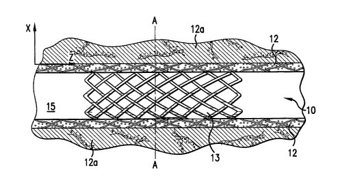
[0005] In Fig. 1, the coated stent 10 is placed in a blood vesse115 having a
vessel
wall 12 to be treated. This vessel wall is surrounded by tissue 12a. The
biologically active
material coated on struts 13 of the stent 10 is released into the vessel wall
12 to be treated.
Fig. 2 is a cross sectional view along line A of the stent 10 in Fig. 1. Fig.
2 also shows the
concentration levels of the biologically active material in each area
surrounding the struts
13 at a certain time after the insertion of the stent into the vessel 15. The
area adjacent to
the struts, i.e., the area between the struts 13 and line 16, has a
concentration level at or
below C,,,,,, which is just below the toxic level. The farther from the struts
13 the tissue to
be treated is located, the lower the concentration of biologically active
material delivered to
the tissue becomes. However, the area between line 18 and line 19 has the
concentration
level at or higher than C,,,in. A concentration of the biologically active
material in the area
outside line 19 is below C,,,in.
[0006] Also, Figs. 2A and 2B clearly show that there are gaps between each
strut 13
wherein the vessel wall to be treated does not receive sufficient biologically
active material
to have Cmin. The areas within line 19, i. e., having concentrations above
Cmin, may be
increased in size to include more area of the vessel wall to be treated 12, if
the amount of
the biologically active material on the struts 13 is increased. However, by
doing so, the
concentration of the biologically active material in the area adjacent to the
struts 13 may
exceed the toxic level. Accordingly, there is a need for a medical device
comprising a
plurality of struts that can achieve the biologically active material
concentration that is
above Cmin and below toxic levels throughout the tissue.
[0007] However, exposure to a medical device which is implanted or inserted
into
the body of a patient can cause the body tissue to exhibit adverse
physiological reactions.
For instance, the insertion or implantation of certain catheters or stents can
lead to the
formation of emboli or clots in blood vessels. Other adverse reactions to
vascular
intervention include endothelial and smooth muscle cell proliferation which
can lead to
hyperplasia, restenosis, i.e., the re-occlusion of the artery, occlusion of
blood vessels,
-3-
CA 02586968 2007-05-09
WO 2006/053159 PCT/US2005/040822
platelet aggregation, and calcification. Restenosis is caused by an
accumulation of
extracellular matrix containing collagen and proteoglycans in association with
smooth
muscle cells which is found in both the atheroma and the arterial hyperplastic
lesion after
balloon injury or clinical angioplasty. Treatment of restenosis often involves
a second
angioplasty or bypass surgery. The drawbacks of such treatment, including the
risk of
repeat restenosis, are obvious.
[0008] When considering treatment using biologically active material eluting
stents,
there are several considerations. Firstly, implantation of a drug eluting
stent requires
precise placement of the stent so that the lesion covered by the stent
includes a sufficient
margin beyond the angiographically identified lesion boundaries. Hence, even
with very
careful placement of the stent, it is possible to miss or undertreat the
lesion. Secondly, even
if a lesion appears to be fully covered by a biologically active material
coated stent, balloon
injury caused during implantation may extend well beyond the ends of the
stent. In the case
where such injury can be visualized by angiography, an additional stent may be
placed to
cover this injury. However, implantation of a second stent may cause further
injury in a
similar fashion to placement of the first stent. Thirdly, even if there is no
evidence of
angiogi-aphic injury, there may be a zone of biological injury that is well
beyond the ends of
the stent.
[0009] Other problems with the current technology, in particular radioactive
stents,
is that restenosis may still occur at the parts of the surface of the body
lumen that are in
contact with the ends of a stent. Closure or constriction of the vessels
commonly occurs
when the vascular cells proliferate around the ends of the stent. This is
known as the
"candy-wrapper effect", also known as edge restenosis or edge effect. Albiero
et al., 2000,
J. Invas. Cardiol. 12(8):416-421; Latchem et al., 2000, Catheter Cardiovasc
Interv.
51(4):422-429; Kim et al., 2001, J. Am. Coll. Cardiol. 37(4):1026-1030. A
schematic
diagram describing this effect is show in Figure 25. Figure 25 shows a cross
section of a
body lumen with a stent implant where restenosis occurred at the opposing ends
of the stent.
The surface 10 of a body lumen 30 at the ends of the implanted stent 40 is
surrounded by
hyperproliferating tissues 20. This appearance is similar to a candy with a
wrapper and thus
the name "candy-wrapper effect". A cause for some types of hyperplasia is that
when a
body lumen is treated with radiation, the radioactive source is usually
targeted towards the
-4-
CA 02586968 2007-05-09
WO 2006/053159 PCT/US2005/040822
center of the stent where the original lesion was situated. In an effort to
minimize
extraneous radiation to healthy vessel tissue, radiation is targeted towards
the center.
Hence, restenosis may still occur at the edge of the stent due to a lower
dosage of radiation
at the ends. The underlying mechanism for this effect is that the radiation
dosage at the
ends is at a level such that it stimulates cell growth as opposed to stopping
it. Clearly, there
remains a great need for therapies directed to the prevention and treatment of
restenosis and
related disorders.
[0010] The edge-effect also can occur with non-radioactive stents. With
existing
coated medical devices, generally, the coating of the biologically active
material is
uniformly applied along the entire length of the device or surface of the
device. For
example, conventional coated stents are coated uniformly along the entire
length of the
surface of the device. The biologically active material-concentration-profile
in the body
lumen along the length of the coated surface may be in the shape of a bell-
curve, wherein
the amount of the biologically active material released at the middle of the
surface causes a
greater tissue concentration than the amount of the biologically active
material released at
the ends of the coated surface. This uneven concentration-profile in the body
lumen along
the length of the coated surface may lead to the application of an inadequate
or sub-optimal
dosage of the biologically active material to the body tissue located at the
ends of the coated
surface. It is possible that such uneven local concentration of the
biologically active
material in the wall of the body lumen along the length of the coated surface
of the medical
device may lead to undesired effects. For example, in the case of a
biologically active
material-coated stent used to prevent or treat restenosis, if the amount of
biologically active
material delivered to the tissue located at the ends of the stent is sub-
optimal, it is possible
that restenosis may occur in such tissue.
[0011] The biologically active material dosage at the tissue located at the
ends of the
coated surface of the medical device can be increased if the concentration or
amount of the
biologically active material is increased along the entire length of the
surface. However, by
increasing the concentration or amount of biologically active material
released along the
entire surface, the dosage delivered to tissue located at the middle of the
surface may be too
great or even at toxic levels.
-5-
CA 02586968 2007-05-09
WO 2006/053159 PCT/US2005/040822
[0012] Thus, there is a need for a medical device that allows precise
placement of
the stent with respect to the lesion, a more uniform concentration-profile for
biologically
active material along the entire length of a coated surface of a medical
device, and provide a
means for therapeutic concentration of biologically active material at and
beyond the
physical ends of an implanted stent. This invention avoids the possibility of
undesired
effects and in particular, preventing intimal hyperplasia and smooth muscle
cell
proliferation which cause stenosis or restenosis of the body lumen caused by
an uneven
biologically active material concentration-profile.
[0013] Moreover, medical devices wherein a biologically active material is
uniformly coated on the entire outer surface of the medical devices that is
exposed to body
tissue are generally used to deliver such biologically active material to
specific parts of such
body tissue. For instance, such devices are used to treat lesions in body
lumen. However,
because the entire outer surface of the device contains the biologically
active material, this
biologically active material will be delivered to healthy body tissue in
addition to the lesion.
Treatment of healthy tissue with the biologically active material is not only
unnecessary but
maybe harinful. Accordingly, there is a need for a medical device that can
realize an
asymmetry release profile of biologically active material to deliver such
material to only a
limited region of the body tissue that requires the biologically active
material.
[0014] Citation of references hereinabove shall not be construed as an
admission
that such references are prior art to the present invention.
3. SUMMARY OF THE INVENTION
[0015] These and otlier objectives are accomplished by the present invention.
To
achieve the aforementioned objectives, we have invented a medical device for
delivering a
biologically active material into a body tissue of a patient; a method for
designing such
device; and a method for delivery of a biologically active material to a body
tissue.
[0016] The medical device of the invention is a medical device for delivery of
biologically active materials to a body tissue of a patient in need of
treatment. The medical
device comprises struts and non-structural elements integral with the struts,
and those struts
and non-structural elements comprise the biologically active material. In an
embodiment,
the non-structural elements project from the struts and are configured in a
shape selected
from the group consisting of a cone, a truncated cone, an oval, a straight
rod, a bent rod, and
-6-
CA 02586968 2007-05-09
WO 2006/053159 PCT/US2005/040822
a rod having heads at the ends. In another embodiment, the non-structural
elements are
configured in a shape selected from the groups consisting of hoops, knots and
bends, which
are located along the stents. In yet another embodiment, the medical device
comprises a
tubular portion having an outer surface, and the non-structural elements are
distributed
throughout the outer surface. In another embodiment, the non-structural
elements are
located in a radially asymmetric distribution on the outer surface. For
example, the non-
structural elements are distributed in a rectangular portion of the outer
surface, or the
rectangular portion is parallel to longitudinal axis of the tubular portion.
The rectangular
portion and the tubular portion may have same length. The surface area of the
rectangular
portion may be from about 25% to about 75% of the entire surface area of the
outer surface.
In yet another embodiment, the outer surface has end sections and a middle
section, and the
end sections comprise a greater number of the non-structural elements per unit
length of the
outer surface than the middle section. In another embodiment, the biologically
active
material is selected from the group consisting of paclitaxel, actinomycin,
sirolimus,
tacrolimus, everoliinus, dexamethasone, halofuginone and hydrophobic nitric
oxide
adducts.
[0017] The present invention is also directed to a method for delivering a
biologically active material to body tissue of a patient which comprises
inserting the above-
mentioned medical device into the body of the patient.
[0018] Further, the present invention is directed to a method for designing
such
medical device, such as a stent, for delivering a biologically active material
to a body tissue
of a patient, wherein the medical device comprises a plurality of struts and a
plurality of
non-structLual elements integral with the struts, wherein the struts and the
non-structural
elements coinprise the biologically active material. The method comprises: (a)
providing a
preliminary medical device comprising struts in a geometric pattern wherein
the struts
comprise the biologically active material; (b) determining a concentration-
profile for the
biologically active material which is released from the preliminary medical
device; and (c)
modifying the geometric pattern of the struts of the preliminary medical
device by
incorporating non-structural elements comprising the biologically active
material that are
integral with the struts to achieve more desired distribution of the
biologically active
material in the body tissue. In an embodiment, the biologically active
material has a
-7-
CA 02586968 2007-05-09
WO 2006/053159 PCT/US2005/040822
minimum effective concentration and a maximum effective concentration for the
body
tissue, and wherein steps (b) and (c) are repeated until the body tissue to be
treated is
substantially free from a concentration of the biologically active material
that is smaller
than the minimum effective concentration and a concentration of the
biologically active
material that is greater than the maximum effective concentration over a
desired time
period. In another embodiment, the biologically active material is selected
from the group
consisting of paclitaxel, actinomycin, sirolimus, tacrolimus, everolimus,
dexamethasone,
halofuginone and hydrophobic nitric oxide adducts.
[0019] The present invention is also directed to a medical device such as a
stent that
is insertable into the body of a patient. The medical device has an outer
surface comprising
struts, and the outer surface has a middle section and end sections. The end
sections have a
greater available surface area per unit length of the outer surface than the
middle section.
In one embodiment, at least a part of each of the middle section and the end
sections have
greater affinity for the biologically active material per unit length of the
outer surface than
the middle section. In yet another embodiment, the end sections have a greater
amount of
the biologically active material per unit length of the outer surface than the
middle section.
Further, in another embodiment, at least a part of each of the middle section
and the end
sections is covered with a coating comprising the biologically active
material, and the
middle section comprises a barrier layer placed over the coating covering the
middle
section. In another embodiment, the end sections have a greater surface area
by having a
more porous surface than struts located at the middle section. The struts
located at the end
sections are comprised of a porous material and the struts located at the
middle section is
comprised of a less porous material. The struts located at the end sections
are covered with
the porous material, and the struts located at the middle section are covered
with the less
porous material. The average diameter of the struts located at the end
sections is greater
than the average diameter of the struts located at the middle section.
[0020] Moreover, the present invention provides another embodiment of the
medical device for treating body tissue. The medical device comprises an outer
surface
comprising struts. The outer surface has a rectangular portion having a
greater capacity for
carrying or containing a biologically active material per unit length of the
outer surface than
the parts of the outer surface that are outside the rectangular portion. In
the alteinative, the
-8-
CA 02586968 2007-05-09
WO 2006/053159 PCT/US2005/040822
rectangular portion may have a greater affinity for the biologically active
material. The
present invention is also directed to a method for delivering a biologically
active material
by inserting the above mentioned medical device comprising the biologically
active
material in such a way that the rectangular portion is in direct contact with
the body tissue
in need of treatment.
[0021] In another embodiment, the end sections have greater affinity for the
biologically active material per unit length of the outer surface than the
middle section. At
least a part of each of the middle section and the end sections of the outer
surface comprise
the biologically active material. The struts located at the end sections
comprise a first
matrix material and the struts located at the middle section comprise a second
matrix
material, and wherein the first matrix material has a greater affinity for the
biologically
active material than the second matrix material. The struts located at the end
sections are
covered with a coating of the first matrix material and the struts located at
the middle
section are covered with a coating of the second matrix material. The end
sections and
middle section fiu=ther comprise the biologically active material.
[0022] At least a part of each of the middle section and the end sections are
covered
with a linking material, and wherein the struts located at the end sections
coinprise a greater
amount of the linking material per unit length of the outer surface than the
struts located at
the middle section. The outer surface comprises the biologically active
material which is
linked to the linking material.
[0023] In yet another embodiment, the end sections have a greater amount of
the
biologically active material per unit length of the outer surface than the
middle section. The
present invention is fixrther directed to a medical device insertable into the
body of a patient,
which coinprises an outer surface, wherein the outer surface has a middle
section and end
sections, wherein at least a part of each of the middle section and the end
sections is
covered with a coating layer comprising a first biologically active material,
and wherein the
end sections carry or contain a larger amount of first biologically active
material per unit
length of the outer surface than the middle section. The medical device may
comprise a
tubular portion that comprises the outer surface. The coating covering the end
sections may
further comprise a coating layer containing a second biologically active
material.
-9-
CA 02586968 2007-05-09
WO 2006/053159 PCT/US2005/040822
[0024] In another embodiment, the present invention provides a medical device
comprising a sidewall and a first band comprising a first biologically active
material. The
sidewall of the medical device has a middle section, a first end section and a
second end
section. The first band is connected to the first end section. In another
embodiment, a
second band is connected to the second end section.
[0025] In another embodiment, the present invention provides a medical device
comprising a middle section, a first and second end sections. The first and
second end
sections each comprise an edge and the first band comprises an inner end. The
first band is
connected to the first end section such that the first band inner end is
adjacent to the first
end section edge.
4. BRIEF DESCRIPTION OF THE DRAWINGS
[0026] Fig. 1 depicts a side view of a stent without non-structural elements
in a
cross-sectioned blood vessel. The stent is coated with a biologically active
material.
[0027] Figs. 2A and 2B depict cross sectional views of the stent and blood
vessel of
Fig. I along line A-A and line B-B (shown in Fig. 2A), respectively. Figs. 2A
and 2B also
show areas of body tissue having different concentration levels of the
biologically active
material.
[0028] Fig. 3 depicts a side view of a stent with non-structural elements in a
cross-
sectioned blood vessel. The stent is coated with a biologically active
material.
[0029] Fig. 4A and 4B depict cross sectional views of the stent and blood
vessel of
Fig. 3 along line C-C and line D-D (shown in Fig 4A), respectively. Figs. 4A
and 4B also
show areas having different concentration levels of the biologically active
material.
[0030] Fig. 5 depicts struts of a conventional expandable stent.
[0031] Figs. 6-14, each depicts struts having non-structural elements integral
with
the struts.
[0032] Fig. 15 depicts wavy struts that have greater surface area per unit
length of
the strut than conventional struts.
[0033] Fig. 16 depicts struts having a greater average diameter per length of
the
strut than the conventional struts.
-10-
CA 02586968 2007-05-09
WO 2006/053159 PCT/US2005/040822
[0034] Fig. 17 depicts a simplified view of a stent having a rectangular
portion of
the outer surface where non-structural elements are located, and the
rectangular portion is
shown by hatching.
[0035] Fig. 18 depicts a perspective view of a stent wherein non-structural
elements
are located only in a rectangular portion of the outer surface.
[0036] Fig. 19 depicts a stent having end sections and a middle section and
comprised of struts, wherein the end sections are comprised of a porous
material and the
middle section is comprised of a less porous material.
[0037] Fig. 20 is a simplified view of a stent which shows the outer surface,
having
end sections and a middle section.
[0038] Fig. 21 is a simplified view of a stent having bands attached to end
sections
of the stent.
[0039] Fig. 22 is a simplified view of a stent having bands attached to end
sections
of the stent.
[0040] Fig. 23 is a simplified view of a stent having bands attached to end
sections
of the stent which is implanted in a lumen.
[0041] Fig. 24 is a simplified view of a stent having bands attached to part
of the
circumference of the end sections of the stent.
[0042] Fig. 25 is a simplified view of a stent implanted in a lumen in which
the edge
effect is illustrated.
5. DETAILED DESCRIPTION OF THE INVENTION
5.1. MEDICAL DEVICE FOR DELIVERING BIOLOGICALLY
ACTIVE MATERIAL WITH DESIRED DISTRIBUTION
5.1.1. NON-STRUCTURAL ELEMENTS
[0043] Even if a biologically active material having a pharmacological effect
is
delivered to a body tissue, such effect may not result if the concentration of
the biologically
active material in the body tissue is below a certain concentration. Such
concentration is
referred to as the minimum effective concentration (C,,,;,,) of the
biologically active material
in the body tissue. Each biologically active material has different C,,,;,,.
C";,, of a
biologically active material also varies depending on the type of body tissue
to which it is
-11-
CA 02586968 2007-05-09
WO 2006/053159 PCT/US2005/040822
delivered. On the other hand, a biologically active material becomes toxic if
its
concentration is higher than a certain concentration. Such concentration is
referred to as the
maximum effective concentration C.. In addition, it is insufficient that the
mean
concentration of the biologically active material delivered through out the
body tissue to be
treated is greater than Cin and smaller than C,,,a, The concentration of the
biologically
active material at each and every area throughout the body tissue to be
treated should be
equal to or greater than CIõin but equal to or smaller than Cma,, of the
biologically active
material.
[0044] When the medical device is comprised of a plurality of struts
comprising a
biologically active material, the body tissue located at or near a center of
each "cell" of the
medical device, i.e., openings between the struts, tends to have the lowest
concentration of
the biologically active material. Such concentration can be below C,,,in. This
is particularly
true when the biologically active material is hydrophobic. When the
concentration of the
biologically active material in the tissue located at the center of each cell
is lower than C,,,i,,,
the concentration can be increased by increasing the amount of the
biologically active
material coated on outer surface of each strut. However, then the
concentration at the tissue
adjacent to the struts may exceed C,,,ax.
[0045] For example, Fig. 1 depicts a coated stent 10 having a conventional
geometric pattern, which is placed in a blood vessel 15 having a vessel wall
12 to be treated.
The biologically active material coated on struts 13 of the stent 10 is
released into the vessel
wall 12 to be treated. Figs. 2A and 2B show cross sectional views along line A-
A and
line B-B (shown in Fig. 2A) of the stent 10 in Fig. 1 and the concentration
levels of the
biologically active material in each area surrounding the struts 13 at a
certain time after the
stent 10 was inserted into the vessel 15. The area adjacent to the struts,
i.e., the area
between the struts 13 and line 16 has a concentration level at or below Cmax,
which is just
below the toxic level. The farther from the struts 13 the area is located, the
lower the
concentration becomes. Thus, the concentration levels gradually decrease from
the area
between lines 16 and 17, the area between 17 and 18, to between 18 and 19. The
area
between line 18 and line 19 has a concentration level at or higher than Cmi,,.
A
concentration of the biologically active material in the area outside line 19
is below Cmin,
-12-
CA 02586968 2007-05-09
WO 2006/053159 PCT/US2005/040822
and thus the pharmacological effects of the biologically active material does
not result in
the area.
[0046] Furthermore, Figs. 2A and 2B clearly show that there are gaps between
each
strut 13, i.e., near the center of cells, wherein the vessel wall to be
treated does not receive
sufficient biologically active material to have Cm;,,. The size of the area
within line 19, i.e.,
the areas having the concentrations above C,,,i,,, may be increased to include
the entire area
of the vessel wall to be treated 12 if the amount of the biologically active
material on the
struts 13 is increased. However, by doing so, the area adjacent to the struts
13 may be also
increased and exceed the toxic level. Therefore, there is a need for a medical
device that
can ensure the concentration of the biologically active material throughout
the body tissue
to be treated is at least C,,,;,, and at most C,,,ax.
[0047] To achieve such a desired distribution of a biologically active
material
throughout the body tissue to be treated, the embodiments of the medical
device of the
present invention comprise a plurality of struts and a plurality of non-
structural elements
integral to the struts. The struts and non-structural elements comprise the
biologically
active material. These non-structural elements are used to adjust the
distribution of the
biologically active material in the body tissue so that the desired
concentration-profile for
the biologically active material released from the medical device into the
body tissue can be
achieved. For instance, the medical device of the present invention can
achieve
concentrations higher than C,,,;,, at the tissue located at the center of
cells without increasing
the local concentration at an area adjacent to the struts higher than Cax.
[0048] An example is shown in Figs. 3, 4A and 4B. Fig. 3 depicts a coated
stent 10'
which is obtained by modifying the conventional geometric pattern of stent 10
shown in
Fig. 1 by incorporating non-structural elements 14 integral to the struts 13.
The stent 10' is
placed in a blood vessel 15 having a vessel wall 12 to be treated. The
biologically active
material coated on struts 13 and non-structural elements 14 of the stent 10'
is released into
the vessel wall 12 to be treated and tissue 12a surrounding the vessel wall
12. Fig. 4A and
4B show cross sectional views along line C-C and D-D (shown in Fig. 4A) B of
the stent
10' in Fig. 3 and the concentration levels of the biologically active material
in each area
surrounding the struts 13 and the nonstructural elements 14 at a certain time
after the stent
10' was inserted in the vessel 15. The area adjacent to the struts, i.e., the
area between the
-13-
CA 02586968 2007-05-09
WO 2006/053159 PCT/US2005/040822
struts 13 or the nonstructural elements 14 and line 16 has a concentration
level from at or
below which is just below the toxic level. The farther from the struts 13 or
the
nonstructural elements 14 the area is located, the lower the concentration
becomes. The
area between line 18 and line 19 has the concentration level at or higher than
C,,,I,,. Fig. 4
clearly shows that the stent 10' can achieve concentrations higher than
C,,,;,, throughout the
entire area of the vessel wall to be treated 12, even at areas located at the
center of cells,
without increasing the concentration at areas adjacent to the struts above
C,,,,,,.
[0049] The term "non-structural element" refers to an element integral with a
strut,
which can project from the strut or can be located along the strut. Such non-
structural
elements have substantially no effect on the mechanical properties of the
struts, such as, for
example, (1) radial strength, (2) longitudinal flexibility, (3) expansion
ratio, (4)
contractibility and (5) profile of a medical device comprising the plurality
of struts. In
embodiments of the medical device of the present invention, the non-structural
elements are
integral with the struts, namely, they are generally made from the same
material as the struts
and are formed as a continuous part of the struts. Preferably, the non-
structural elements
and struts may be manufactured simultaneously; for exatnple, struts having non-
structural
elements can be laser-ablated from a plate of metal or polymer.
[0050] Fig. 5 depicts example of conventional struts without non-structural
element,
and Figs. 6-14 depict examples of non-structural elements integral with the
conventional
struts. Shapes of the non-structural elements include, but not limited to, a
straiglit rod (21
in Fig. 6), a cone (22 in Fig. 7), a truncated cone (not shown), a hoop (23 in
Fig. 8), a knot
(24 in Fig. 9), a bent rod (25 in Fig. 10), an oval (26 in Fig. 11), and a rod
having heads at
its ends (27 in Fig. 12 and 28 in Fig. 13). Bends in the struts (29a and 29b
in Fig. 14) can
be used as non-structural elements so long as they do not affect the
mechanical properties of
the struts.
[0051] This embodiment of the medical device of the present iiivention can be
used
for delivering any kind of biologically active material. Preferably, the
biologically active
material is hydrophobic, e.g., paclitaxel, actinomycin, sirolimus, tacrolimus,
everolimus,
dexamethasone, halofuginone, and hydrophobic nitric oxide adducts. Other
examples of the
biologically active material, coatings containing the biologically active
material, and
examples of the medical device are explained later in this application.
-14-
CA 02586968 2007-05-09
WO 2006/053159 PCT/US2005/040822
5.1.2. DESIGNING MEDICAL DEVICES HAVING
STRUTS AND NON-STRUCTURAL ELEMENTS
[0052] The present invention is directed to a method for designing a medical
device
comprising a plurality of struts and non-structural elements integral with the
struts for
delivering a biologically active material to a body tissue of a patient. As
explained above,
when the struts are placed in a certain geometric pattern, the concentration
of a biologically
active material at a center of each cell may not reach C,,,;,, of the
biologically active
material. However, the method of the present invention provides a geometric
pattern of the
struts in which the concentration of a biologically active material above
C,,,;,, can be
achieved throughout the body tissue to be treated without increasing the
concentration at the
tissue located adjacent to the struts above C,aX.
[0053] In the method of the invention, a preliminary medical device comprising
a
plurality of struts in a geometric pattern is modified by incorporating non-
structural
elements to the struts to improve the concentration-profile for the
biologically active
material released from the device to the body tissue to be treated. Any
medical device
comprising a plurality of struts in a geometric pattern, such as stent, can be
used as a
preliminary medical device for the metliod of the invention provided that the
struts
coinprises a biologically active material.
[0054] In the method of the present invention, a concentration-profile for the
biologically active material delivered to the body tissue from the preliminary
medical
device is determined. From this profile, the areas of tissue in which the
concentration of the
biologically active material is below Q,,;,, can be determined. Such areas are
then correlated
to the parts of the geometric pattern of the struts of the preliminary medical
device that were
in contact with or near such areas.
[0055] The determination of such concentration-profile can be conducted by
actually measuring concentrations using the biologically active material in
vitro with a
tissue model, which is similar to the body tissue to be treated, such as
cannulated animal
arteries with surrounding tissue or an artificial tissue, or in vivo with an
animal model, such
as rabbits, guinea pigs, or pigs. The biologically active material used for
the experiment
may be labeled with a fluorescence, a radioactive material or dye or can be
assayed by
tissue digestion and analyzed by HPLC. Such labeled biologically active
material is coated
on the medical device, and then the coated medical device is inserted into the
tissue model,
-15-
CA 02586968 2007-05-09
WO 2006/053159 PCT/US2005/040822
or artificial tissue, or implanted in an animal. Alternatively, the
biologically active material
may be detected using standard HPLC separation, mass spectroscopy or other
direct
analytical methods. After insertion, the tissue may be appropriately
sectioned, and the
concentration-profile for the labeled biologically active material is measured
by a means
appropriate to the label employed for the experiment. However, a necessary
care should be
taken that the label would not greatly affect the diffusion of the
biologically active material
itself.
[0056] However, the concentration-profile may also be determined by
mathematical
simulation. For example, such simulation can be conducted by using the
following
conditions and equations:
ac a 2c a 2c
a t- Dx a x2 + DZ a z2
[0057] wherein C refers to a concentration of the biologically active material
in the
body tissue, x refers to a distance from the medical device along x axis which
is
peipendicular to a boundary between the medical device and the body tissue, z
refers to a
distance from the medical device along z axis which is parallel to the
boundary, Dx refers
to a diffusion coefficient of the biologically active material in direction
along x axis, Dz
refers to a diffusion coefficient of the biologically active material in a
direction along z axis.
For example, such x axis and z axis are shown in Figs. 1, 2B, 3 and 4B. Dx and
Dz can be
determined by the experiments using the labeled biologically active material
in vitro or in
vivo as described above. C = 0 at t= 0, wherein boundary conditions are as
follows:
[0058] (i) at a common boundary between the struts and the body tissue (at x
0):
Dx ac = hi (C'-Cr)
[0059] wherein Cr refers to a concentration of the biologically active
material in the
struts, and hl refers to a mass transfer coefficient. Value of hl can be
determined by the
same experiments described above or determined by assumption based on the
information
known to one skilled in the art;
[0060] (ii) at a boundary between blood flow and the body tissue (at x= 0):
-16-
CA 02586968 2007-05-09
WO 2006/053159 PCT/US2005/040822
D. ax = h2(C-0)
[0061] wherein h2 refers to another mass transfer coefficient. Value of h2 can
be
determined by the same experiment mentioned above or determined by assuinption
based
on the information known to one skilled in the art;
[0062] (iii) at an adventitial side of vascular wall (at x= L):
Dx C = h3 (C-0)
[0063] wherein h3 is yet another mass transfer coefficient, and L is a width
of a
region of interest. Value of h3 can be determined by the same experiment
nlentioned above
or determined by assumption based on the information known to one skilled in
the art; and
[0064] (iv) "symmetry" (no-flux) boundary conditions at certain cross-sections
perpendicular to z axis:
ac ac
a(z=0)= a (z=Lz)=0
[0065] wherein Lz is the length along z axis of a region of interest.
[0066] Although a simplified model based on two diffusion coefficients of the
biologically active material in two directions, i.e., depth of the tissue
penetration and the
distance diffused, is described above as an example, there are more complex
models can be
also employed for the method of the present invention. Such complex models may
further
account for other variables, such as convection, vessel wall inhomogenetics,
the type of
cells, the lesions, the variabilities brougllt by different coatings or
coating porosity, blood
flow, body temperature, blood pressure, and/or pressure of the implant against
the vessel
wall.
[0067] Subsequent to determining the concentration-profile for the
biologically
active material which is released from the preliminary medical device, the
geometric pattern
of the preliminary medical device is modified by incorporating a plurality of
non-functional
elements that are integral with the struts to achieve more desired
distribution of the
biologically active material in the body tissue to be treated. The non-
structural elements
also comprise the biologically active material. For example, the area of
tissue in which the
-17-
CA 02586968 2007-05-09
WO 2006/053159 PCT/US2005/040822
concentration of the biologically active material is below C,,,;,, is
determined from the
concentration-profile. Then, it is determined which parts of the geometric
pattern of the
struts of the preliminary medical device were in contact with or near such
areas. The non-
structural elements can be incorporated near such parts in the geometric
pattern, so that the
biologically active material released from the non-structural elements would
change the
concentration in those areas.
[0068] For example, a stent 10 having a plurality of struts 13 in a
conventional
geometric pattern in Fig. 1 can be provided as the preliminary medical device.
The struts
13 are coated with a biologically active material. Then, a concentration-
profile in a body
tissue for the biologically active material which is released from the struts
13 is determined.
An example of such profile is shown in Fig. 2A and 2B with the cross-sectional
views of
the stent 10 in the blood vessel 15. The determination of such concentration-
profile can be
conducted by actually measuring concentrations or by mathematical simulation
as
mentioned above. According to the obtained concentration-profile, the
geometric pattern of
the struts 13 of the preliminary stent 10 are modified with non-structural
elements 14, for
example, as shown in Fig. 3. Fig. 4A and 4B show the concentration-profile for
the
biologically active material in the blood wall 12. When the concentration-
profile in the
vessel wall to be treated 12 shown in Figs. 2A-B and 4A-B are compared, in
Figs. 4A-B,
the concentrations generally throughout the entire area of the vessel wall to
be treated 12 are
above C,,,;,, and below C,,,,,. It is clear that the modified stent 10'
achieves a more desirable
concentration-profile in the vessel to be treated 12 with the biologically
active material than
the preliminary stent 10.
[0069] Preferably, after a concentration-profile for the biologically active
material
in the body tissue which is released from the modified preliminary medical
device is
determined, if there is an area of the body tissue having the local
concentration of the
biologically active material lower than C,,,;,,, then the device is modified
again by adding
non-structural elements to the struts. In addition to or instead of merely
adding additional
non-structural elements, the non-structural elements which have been already
added can be
removed or relocated according to the determined concentration-profile.
Consequently, a
medical device having further improved delivery of the biologically active
material is
-18-
CA 02586968 2007-05-09
WO 2006/053159 PCT/US2005/040822
obtained. If necessary, the determination step and the modification step
explained above
can be repeated as many as possible.
5.1.3. MEDICAL DEVICE WITH RADIALLY ASYMMETRIC
AREA HAVING NON-STRUCTURAL ELEMENTS
[0070] The prior sections (section 5.1.1 and 5.1.2) explained how non-
structural
elements can be added to a preliminary medical device to achieve a more
desired
concentration-profile for the biologically active material released from the
device into body
tissue. When the entire outer surface of a medical device, which comprises the
plurality of
struts and non-structural elements, is used to treat body, the non-structural
elements should
be positioned uniformly throughout the entire outer surface of the medical
device.
[0071] However, if the body tissue to be treated is smaller in surface area
than the
entire outer surface of the medical device, then the non-structural elements
do not have to
be positioned throughout the entire surface of the medical device. For
example, the medical
device can comprise a tubular portion comprising an outer surface, such as a
stent, which
comprises a plurality of struts and a plurality of non-structural elements.
The non-structural
elements located in a radially asymmetric distribution, such as shown in Fig.
17 where 33
represents the location of the non-structural element on outer surface of a
simplified figure
of a stent 32. In this figure, the non-structural elements are distributed
only in a rectangular
portion of the outer surface. Fig. 18 depicts a stent wherein non-structural
elements are
provided onto the struts only in a rectangular portion of the outer surface.
Such rectangular
portion may be parallel to longitudinal axis of the tubular portion and may
have the same
length as that of the tubular portion. The rectangular portion is preferably
from about 10 %
to about 90 % of the entire outer surface.
[0072] The present invention is also directed to a method for delivering a
biologically active material to body tissue using the above-mentioned medical
device,
which comprises a tubular portion comprising an outer surface which comprises
a plurality
of struts and a plurality of non-structural elements, and the non-structural
elements are
located in a radially asymmetric distribution on the outer surface. In the
method, the
medical device is inserted into body of the patient. Preferably, the non-
structural elements
are distributed only in a rectangular portion of the outer surface, and the
medical device is
inserted in such a way that the rectangular portion is in direct contact with
the body tissue to
-19-
CA 02586968 2007-05-09
WO 2006/053159 PCT/US2005/040822
be treated. In this way, the body tissue to be treated will receive desired
distribution of the
biologically active material. On the other hand, the body tissue which does
not need to be
treated will be exposed to a lesser amount of the biologically active
material.
5.2. INCREASED CAPACITY OF THE END SECTIONS FOR CARRYING
OR CONTAINING A BIOLOGICALLY ACTIVE MATERIAL
[0073] In other embodiments of the medical device insertable into the body of
a
patient of the invention, the medical device comprises an outer surface
comprising a
plurality of struts, and the end sections of the outer surface have a greater
capacity per unit
length of the outer surface for carrying or containing a biologically active
material than the
middle section of the outer surface. Specifically, in one embodiment of the
medical device,
each strut at the end sections has greater available surface area per unit
length of the outer
surface than the middle section. In another embodiment, the end sections have
a greater
affinity for the biologically active material per unit length of the outer
surface than the
middle section.
[0074] The medical device of the present invention may be manufactured with or
without a biologically active material by a manufacturer. When the medical
device of the
present invention is manufactured without a biologically active material, a
practitioner (e.g.,
a medical doctor or a nurse) can apply the biologically active material to the
medical
device. In either case, since the end sections of the outer surface have a
greater capacity per
unit length of the outer surface for carrying or containing the biologically
active material
than the middle section, the end sections will carry a greater amount of the
biologically
active material when the biologically active material is applied to the
medical device
without needing to change application method of the biologically active
material to the end
sections and the method to the middle section. Therefore, when a practitioner
applies to the
outer surface of the medical device, such as by dipping, a coating composition
containing a
biologically active material, a larger amount of the biologically active
material per unit
length of the outer surface will be deposited at the end sections than the
middle section.
[0075] The term "unit length of the outer surface" refers to the length on an
imaginary straight line along the outer surface drawn between a point on an
edge of the
outer surface and another point on the opposing edge of the outer surface.
Therefore, the
terms, such as "capacity per unit length of the outer surface," "available
surface area per
-20-
CA 02586968 2007-05-09
WO 2006/053159 PCT/US2005/040822
unit length of the outer surface," and "amount per unit length of the outer
surface," refer
respectively to the capacity, available surface area and amount per unit
length of the
imaginary straight line explained above.
5.2.1. INCREASED AVAILABLE SURFACE AREA AT THE END SECTIONS
[0076] As explained above, one of the embodiments of the medical device has
end
sections which have greater available surface area per unit length of the
outer surface than
that of the middle section. The term "available surface area" refers to a
surface area which
is available to be coated by a coating composition comprising a biologically
active material.
[0077] One way of increasing the available surface area of the end sections is
to
fabricate the outer surface of the medical device using more material at its
ends. For
example, when the medical device is comprised of struts, the available surface
area per unit
length of the outer surface in the end sections is increased by adding non-
structural
elements to the struts. The non-structural elements are explained above (see
section 5.1.1).
The end sections comprise a greater nuinber of the non-structural elements per
unit length
of the outer surface than the middle section. The middle section may have
smaller number
of the non-structural elements or no non-structural elements.
[0078] Further, the available surface area can be increased by increasing the
surface
area of the struts themselves. For example, wavy struts 30 shown in Fig. 15
can have more
outer surface area per length than straight struts show in Fig. 5. Also,
struts having greater
average diameter, such as struts wliich are thicker or wider at certain
portion 31 shown in
Fig. 16, have greater outer surface area per length than struts which have
smaller average
diameter. Moreover, the end sections of the outer surface can be made to have
greater
available surface area by roughing the struts' outer surface or providing
indentations or
grooves on the struts' surface. The above-mentioned wavy struts, wider or
thicker struts,
indentations and grooves may have various shapes, so long as such structure
does not affect
stent's structural functions. For example, the above-mentioned structure
should not hinder
self-expansion of a self-expanding stent and should not cause any harm to the
patient body.
The above-mentioned wavy struts, indentations and grooves can be manufactured
by laser
ablation.
-21-
CA 02586968 2007-05-09
WO 2006/053159 PCT/US2005/040822
[0079] In another embodiment in which the capacity of the end sections to
carry or
contain the biologically active material is greater than the capacity of the
middle section, the
end sections of the outer surface are more porous, and the middle section of
the surface is
relatively less porous. The middle section may also be non-porous. For
example, in Fig.
19, the circles 45 and 47 show enlarged portions of the outer surface of the
struts 42 of a
stent 40 in the middle section 44 and end section 46, respectively. The
surface of the struts
in the end section 46 has more pores 48 than the surface of the struts at the
middle section
44. In such embodiment, the end sections 46 have a greater available surface
area per unit
lengtli of the outer surface than that of the middle section 44 since the
pores 48 increase
available surface area.
[0080] The end sections of the outer surface may be made porous by forming the
end sections of the outer surface themselves from a porous material or by
forming the end
sections with a non-porous material and then covering the end sections with a
porous
coating layer. For example, porous metal struts can be prepared by sintering
metal, i.e.,
molding or pressing metal particles into a desired shape and heating them to a
teinperature
sliglitly below the melting point of the metal. Porosity can be changed by
using different
particle sizes and/ar dirnensions and/or different temperatures. Also, porous
metal struts
can be prepared by using metal filaments or fibers. See e.g. U.S. Patent No.
5,843,172
issued to Yan which discloses examples of struts made of porous materials,
which is
incorporated herewith by reference.
[0081] The end sections of the outer surface may be made porous by coated with
a
porous coating layer. A porous coating layer may be prepared, for example, by
applying a
mixture of a polymer, an elutable particulate material and a solvent on a
surface to form a
layer, and then eluting the elutable particulate material from the layer. The
following is a
detailed description of suitable materials and methods useful in producing a
porous coating
layer of the invention.
[0082] Polymer(s) useful for forming the porous coating layer should be ones
that
are biostable, biocompatible, particularly during insertion or implantation of
the device into
the body and avoids irritation to body tissue. Examples of such polymers
include, but not
limited to, polyurethanes, polyisobutylene and its copolyiners, silicones, and
polyesters.
Other suitable polymers include polyolefins, polyisobutylene, ethylene-
alphaolefin
-22-
CA 02586968 2007-05-09
WO 2006/053159 PCT/US2005/040822
copolymers, acrylic polymers and copolymers, vinyl halide polymers and
copolymers such
as polyvinyl chloride, polyvinyl ethers such as polyvinyl methyl ether,
polyvinylidene
halides such as polyvinylidene fluoride and polyvinylidene chloride,
polyacrylonitrile,
polyvinyl ketones, polyvinyl aromatics such as polystyrene, polyvinyl esters
such as
polyvinyl acetate; copolymers of vinyl monomers, copolymers of vinyl monomers
and
olefins such as ethylene-methyl methacrylate copolymers, acrylonitrile-styrene
copolymers,
ABS resins, ethylene-vinyl acetate copolymers, polyamides such as Nylon 66 and
polycaprolactone, alkyd resins, polycarbonates, polyoxyethylenes, polyimides,
polyethers,
epoxy resins, polyurethanes, rayon-triacetate, cellulose, cellulose acetate,
cellulose butyrate,
cellulose acetate butyrate, cellophane, cellulose nitrate, cellulose
propionate, cellulose
ethers, carboxyinetliyl cellulose, collagens, chitins, polylactic acid,
polyglycolic acid, and
polylactic acid-polyethylene oxide copolymers. Since the polymer is being
applied to a part
of the medical device which undergoes mechanical challenges, e.g. expansion
and
contraction, the polymers are preferably selected from elastomeric polymers
such as
silicones (e.g. polysiloxanes and substituted polysiloxanes), polyurethanes,
thermoplastic
elastomers, ethylene vinyl acetate copolymers, polyolefin elastomers, and EPDM
rubbers.
T'lie polymer is selected to allow the coating to better adhere to the surface
of the
expandable portion of the medical device when it is subjected to forces or
stress.
Furthermore, althougli the porous coating layer can be formed by using a
single type of
polymer, various combinations of polymers can be employed.
[0083] The elutable particulate materials which can be incorporated into the
polymer include, but not limited to, polyethylene oxide, polyethylene glycol,
polyethylene
oxide/polypropylene oxide copolymers, polyhydroxyethyl methacrylate,
polyvinylpyrrolidone, polyacrylamide and its copolymers, salts, e.g., sodium
chloride,
sugars, and elutable biologically active materials such as heparin. The amount
of elutable
particulate material that is incorporated into the polymer should range from
about 20% to
90% by weight of the porous coating layer. Furthermore, to increase the
porosity of the
coating layer applied to the end sections of the surface, a larger amount of
the elutable
particulate material can be used to form the porous coating layer at the end
sections than are
used to form the porous coating layer at the middle section. For example, the
amount of the
elutable particulate material may be from about 0 % to about 40 % for the
porous coating
-23-
CA 02586968 2007-05-09
WO 2006/053159 PCT/US2005/040822
layer covering the middle section, and about 50 % to 90% for the porous
coating layer
covering at the end sections. Also, a more porous coating layer can be
realized by using
larger average particle size of the elutable material. For example, the
particles may have an
average particle size from 60-100 microns for porous coating layer covering
the end
sections and from 0 to about 30 microns for the porous coating layer covering
middle
section.
[0084] The solvent that is used to form the mixture or slurry of polymer and
elutable
particulate materials include ones which can dissolve the polymer into
solution and do not
alter or adversely impact the therapeutic properties of the biologically
active material
employed. Exainples of useful solvents for silicone include tetrahydrofiuan
(THF),
chloroform and dichloromethane. The composition of polymer and elutable
particulate
material can be applied to the portion of the medical device in a variety of
ways. For
example, the composition can be spray-coated onto the device or the device can
be dipped
into the composition. One of skill in the art would be aware of methods for
applying the
coating to the device.
[0085] The thickness of the porous coating layer can range from about 25 m to
0.5 mm. Preferably, the thickness is about 30 m to 100 m. After the
composition is
applied to the device, it should be cured to produce a polymer containing the
particulate
material and to evaporate the solvent.
[0086] To elute the particulate material from the polymer, a solvent is used.
The
device can be soaked in the solvent to elute the particulate materials. Other
methods of
eluting the particulate are apparent to those skilled in the art. The choice
of the solvent
depends upon the solubility of the elutable particulate material in that
solvent. For instance,
for water-soluble particulate materials such as heparin, water can be used.
For elutable
particulate materials that can be dissolved in organic solvents, such organic
solvents can be
used. Examples of suitable solvents, without limitation, include ethanol,
dimethyl
sulfoxide, etc.
[0087] Another example of a method for preparing a porous coating is a
catalyst-
free vapor deposition of a coating composition comprising a polyamide,
parylene or a
parylene derivative. See U.S. Patent No. 6,299,604 to Ragheb et ccl., which is
incorporated
herein by reference.
-24-
CA 02586968 2007-05-09
WO 2006/053159 PCT/US2005/040822
[0088] In another embodiment of the present invention, the surface including
the
end sections and middle section are covered with a same porous coating layer
composition;
but the porous coating layer is thicker at the end sections than at the middle
section. For
example, a porous coating layer is applied to the entire surface, and then
another porous
coating layer is applied to the end sections while the middle section is
covered by a sheath.
The thickness of the porous coating layer at the end sections may be from
about 80 m to
about 0.5 mm, and that at the middle section may be from about 10 gm to 40 m.
Since
there is more porous coating at the end sections, the end sections of the
outer surface should
have a greater capacity to carry or contain a biologically active material.
5.2.2. THE END SECTIONS WITH GREATER AFFINITY
FOR THE BIOLOGICALLY ACTIVE MATERIAL
[0089] In another embodiment of the medical device of the present invention,
the
end sections of the outer surface have a greater affinity for the biologically
active material
than the middle section. In particular, the end sections comprise a first
matrix material and
the middle section comprises a second matrix material. The first matrix
material has a
greater affinity for the biologically active material of interest than the
second matrix
material so that the end sections can carry or contain a larger amount of the
biologically
active material per unit length of the outer surface than the middle section.
The end
sections and the middle section of the outer surface may be formed from the
first matrix
material and the second matrix material, respectively. Preferably, the end
sections of the
outer surface and the middle section of the outer surface are formed of
another material and
then are covered with a coating comprising each of the matrix materials.
[0090] Generally, when a biologically active material used is a liydrophilic,
e.g.,
heparin, then a matrix material comprising a more hydrophilic material has a
greater affinity
for the biologically active material than another matrix material that is less
hydrophilic.
When a biologically active material used is a hydrophobic, e.g., paclitaxel,
actinomycin,
sirolimus (RAPAMYCIN), tacrolimus, everolimus, and dexamethasone, then a
matrix
material that is more hydrophobic has a greater affinity for the biologically
active material
than another matrix material that is less hydrophobic.
[0091] Examples of suitable hydrophobic polymers include, but not limited to,
polyolefins, such as polyetliylene, polypropylene, poly(1-butene), poly(2-
butene), poly(1-
- 25 -
CA 02586968 2007-05-09
WO 2006/053159 PCT/US2005/040822
pentene), poly(2-pentene), poly(3-methyl-1 -pentene), poly(4-methyl- 1 -
pentene),
poly(isoprene), poly(4-methyl-l-pentene), ethylene-propylene copolymers,
ethylene-
propylene-hexadiene copolymers, ethylene-vinyl acetate copolymers, blends of
two or more
polyolefins and random and block copolymers prepared from two or more
different
unsaturated monomers; styrene polymers, such as poly(styrene), poly(2-
methylstyrene),
styrene-acrylonitrile copolymers having less than about 20 mole-percent
acrylonitrile, and
styrene-2,2,3,3,-tetrafluoropropyl methacrylate copolymers; halogenated
hydrocarbon
polymers, such as poly(chlorotrifluoroethylene), chlorotrifluoroethylene-
tetrafluoroethylene
copolymers, poly(hexafluoropropylene), poly(tetrafluoroethylene),
tetrafluoroethylene,
tetrafluoroethylene-ethylene copolymers, poly(trifluoroethylene), poly(vinyl
fluoride), and
poly(vinylidene fluoride); vinyl polymers, such as poly(vinyl butyrate),
poly(vinyl
decanoate), poly(vinyl dodecanoate), poly(vinyl hexadecanoate), poly(vinyl
hexanoate),
poly(vinyl propionate), poly(vinyl octanoate),
poly(heptafluoroisopropoxyethylene),
poly(heptafluoroisopropoxypropylene), and poly(methacrylonitrile); acrylic
polymers, such
as poly(n-butyl acetate), poly(ethyl acrylate), poly(1-
chlorodifluoromethyl)tetrafluoroethyl
acrylate, poly di(chlorofluoromethyl)fluoromethyl acrylate, poly(1,1-
dihydroheptafluorobutyl acrylate), poly(1,1-dihydropentafluoroisopropyl
acrylate),
poly(1,1-dihydropentadecafluorooctyl acrylate), poly(heptafluoroisopropyl
acrylate), poly
5-(heptafluoroisopropoxy)pentyl acrylate, poly 11 -
(heptafluoroisopropoxy)undecyl
acrylate, poly 2-(heptafluoropropoxy)ethyl acrylate, and
poly(nonafluoroisobutyl acrylate);
methacrylic polymers, such as poly(benzyl methacrylate), poly(n-butyl
methacrylate),
poly(isobutyl methacrylate), poly(t-butyl methacrylate), poly(t-
butylaminoethyl
methacrylate), poly(dodecyl methacrylate), poly(ethyl methacrylate), poly(2-
ethylhexyl
methacrylate), poly(n-hexyl methacrylate), poly(phenyl methacrylate), poly(n-
propyl
methacrylate), poly(octadecyl methacrylate), poly(1,1-
dihydropentadecafluorooctyl
methacrylate), poly(heptafluoroisopropyl methacrylate),
poly(heptadecafluorooctyl
methacrylate), poly(1-hydrotetrafluoroethyl methacrylate), poly(1,1-
dihydrotetrafluoropropyl methacrylate), poly(1-hydrohexafluoroisopropyl
methacrylate),
and poly(t-nonafluorobutyl methacrylate); polyesters, such a poly(ethylene
terephthalate)
and poly(butylene terephthalate); condensation type polymers such as and
polyurethanes
and siloxane-urethane copolymers; polyorganosiloxanes, i.e., polymeric
materials
-26-
CA 02586968 2007-05-09
WO 2006/053159 PCT/US2005/040822
characterized by repeating siloxane groups, represented by Ra S1O 4_a/2, where
R is a
monovalent substituted or unsubstituted hydrocarbon radical and the value of a
is 1 or 2;
and naturally occurring hydrophobic polymers such as rubber.
[0092] Examples of suitable hydrophilic monomer include, but not limited to;
(meth)acrylic acid, or alkaline metal or ammonium salts thereof;
(meth)acrylamide;
(meth)acrylonitrile; those polymers to which unsaturated dibasic, such as
maleic acid and
fumaric acid or half esters of these unsaturated dibasic acids, or alkaline
metal or
ammonium salts of these dibasic adds or half esters, is added; those polymers
to which
unsaturated sulfonic, such as 2-acrylamido-2-methylpropanesulfonic, 2-
(rneth)acryloylethanesulfonic acid, or alkaline metal or ammonium salts
thereof, is added;
and 2-hydroxyethyl (meth)acrylate and 2-hydroxypropyl (ineth)acrylate.
[0093] Polyvinyl alcohol is also an example of hydrophilic polymer. Polyvinyl
alcohol may contain a plurality of hydrophilic groups such as hydroxyl,
ainido, carboxyl,
amino, ammonium or sulfonyl (-SO3). Hydrophilic polymers also include, but are
not
limited to, starch, polysaccharides and related cellulosic polymers;
polyalkylene glycols and
oxides such as the polyethylene oxides; polymerized ethylenically unsaturated
carboxylic
acids such as acrylic, mathacrylic and maleic acids and partial esters derived
from these
acids and polyhydric alcohols such as the alkylene glycols; homopolymers and
copolymers
derived from acrylamide; and homopolymers and copolymers of vinylpyrrolidone.
[0094] The first matrix material and the second matrix material may be
prepared
using either a hydrophilic polymer or a hydrophobic polymer, or a blend of a
hydrophobic
polymer and a hydrophilic polymer in a chosen ratio. For example, wlien the
biologically
active material is hydrophilic, then the first matrix material may be prepared
by blending
from about 55 % to about 100 % hydrophilic polymer and from about 45 % to
about 0 %
hydrophobic polymer; and the second matrix material may be prepared by
blending from
about 55 % to about 100 % hydrophobic polymer and from about 45 % to about 0 %
hydrophilic polymer. The first matrix material contains a greater amount of
the
hydrophillic polymer than the second matrix material. When the biologically
active
material is hydrophobic, then the first matrix material may be prepared by
blending from
about 55 % to about 95 % hydrophobic polymer and from about 45 % to about 5 %
hydrophilic polymer; and the second matrix material may be prepared by
blending from
-27-
CA 02586968 2007-05-09
WO 2006/053159 PCT/US2005/040822
about 55 % to about 95 % hydrophilic polyiner and from about 45 % to about 5 %
hydrophobic polymer. The first matrix material contains a greater amount of
the
hydrophobic polymer than the second matrix material.
[0095] Again, the outer surface of the medical device of the present invention
is,
covered with each matrix material, i.e., the end sections with a first matrix
material and the
middle section with a second matrix material. A first matrix material
composition may be
prepared and applied by any method to a surface of a medical device to form a
coating, such
as spraying, dipping, rolling, and electrostatic deposition. Likewise, a
second matrix
material composition may be prepared and applied by such methods. The first
matrix
material composition may be applied to the end sections of the outer surface
while the
middle section is covered to prevent coating the middle section witli the
first matrix
material. Then the second matrix material composition may be applied to the
middle
section while the end sections are covered. In another embodiment, the second
material
composition may be applied to the entire outer surface including the middle
section and the
end sections, then the first matrix material composition may be applied to the
end sections
while the middle section is covered.
[0096] After th.. , matrix material compositions are applied to the outer
surface, the
surface should be cured to produce matrix material coatings. The thickness of
the matrix
material coating can range from about 25 m to about 0.5 mm. Preferably, the
thickness is
about 30 m to 100 m.
5.2.3. THE END SECTIONS WITH GREATER AMOUNT OF
CHEMICAL LINKING MATERIAL TO CARRY OR
CONTAIN THE BIOLOGICALLY ACTIVE MATERIAL
[0097] In yet another embodiment of the present invention, the capacity of the
end
sections of the outer surface for carrying or containing a biologically active
material can be
increased relative to that of the middle section by using an increased amount
of chemical
linking material to link the biologically active material to the end sections
of the outer
surface. Specifically, the middle section and end sections of the outer
surface are covered
with a chemical linking material, and the end sections carry or contain a
larger amount of
the linking material per unit lengtli of outer surface than the middle
section. The chemical
linking material allows the biologically active material to attach to the
outer surface.
- 28 -
CA 02586968 2007-05-09
WO 2006/053159 PCT/US2005/040822
"Linking materials" may be any material which can be coupled to a biologically
active
material by any bond that are known in the relevant art including but not
limited to, Van der
Waals force, ionic bond, covalent bond, hydrogen bond or chemical cross-
linking.
[0098] For example, U.S. Patent No. 5,356,433 to Rowland et al., discloses
that
polysaccharides can be immobilized onto metallic surfaces by applying an
organosilane
coating with amine functionality and then applying a polysaccharide using
carbodiimide as
a coupling agent. In the present invention, if the organosilane with amine
functionality is
used as a linking material, the amount of this material per unit length of the
outer surface at
the end sections is greater than that at the middle section. In that way,
larger amount of a
polysaccharide, which is a biologically active material, can coupled to the
end sections.
[0099] Also, U.S. Patent No. 5,336,518 to Narayanan et al. discloses that a
polysaccharide can be immobilized on a surface by applying a coat of
heptafluorobutylmethacrylate (HFBMA) by radiofrequency (RF) plasma deposition,
creating functional groups on the surface by RF plasma with water vapor, and
then applying
the polysaccharide using carbodiimide. In the present invention, larger amount
of HFBMA,
a linking material, is applied to the end sections so that larger amount of a
polysaccharide, a
biologically active material can be coupled to.
5.3. RADIALLY ASYMMETRIC MEDICAL DEVICES
HAVING INCREASED CAPACITY FOR CARRYING OR
CONTAINING A BIOLOGICALLY ACTIVE MATERIAL
5.3.1. MEDICAL DEVICES HAVING NON-STRUCTURAL ELEMENTS
LOCATED IN A RADIALLY ASYMMETRIC DISTRIBUTION
[00100] As explained above, one way to increase the capacity for carrying or
containing a biologically active material of the medical device is to increase
available
surface area. In one embodiment of the medical device of the invention, the
available
surface area is increased in radially asymmetric manner along the entire outer
surface,
instead of only at the end sections. One such embodiment where the surface
area is
increased in a radially asymmetric manner by adding non-structural elements to
the outer
surface (as to non-structural elements, see section 5.1.3). For example, only
a rectangular
portion of the outer surface has the non-structural elements. Such rectangular
portion may
be parallel to longitudinal axis of the tubular portion and may have the same
length as that
-29-
CA 02586968 2007-05-09
WO 2006/053159 PCT/US2005/040822
of the tubular portion. The rectangular portion is preferably from about 25 %
to about 75 %
of the entire outer surface. Please see section 1.3 as to a method for
delivering a
biologically active material to body tissue using such medical device.
5.3.2. MEDICAL DEVICE HAVING RADIALLY ASYMMETRIC
INCREASED AVAILABLE SURFACE AREA OR AFFINITY
[00101] Another embodiment of the medical device of the invention comprises a
tubular portion comprising struts and having an outer surface. A portion of
the outer
surface has increased available surface or affinity for the biologically
active material in such
a way that the available surface area or affinity for the biologically active
material is
radially asymmetric. Please see prior section (section 5.3.1) as to examples
of radially
asymmetric distributions. Increased available surface area or increased
affinity to the
biologically active material can be achieved as explained in the prior
sections (sections
5.2.1 and 5.2.2). Please see section 5.1.3 as to a method for delivering a
biologically active
material to body tissue using such medical device.
5.4. SUITABLE MEDICAL DEVICES
[00102] The medical devices of the present invention are insertable into the
body of'a
patient. Namely, at least a portion of such medical devices may be temporary
inserted into
or semi-perrrianently or permanently implanted in the body of a patient.
Preferably, the
medical devices of the present invention comprise a tubular portion which is
insertable into
the body of a patient. The tubular portion of the medical device need not to
be completely
cylindrical. For instance, the cross-section of the tubular portion can be any
shape, such as
rectangle, a triangle, etc., not just a circle.
[00103] The medical devices suitable for the present invention include, but
are not
limited to, stents, surgical staples, catheters, such as central venous
catheters and arterial
catheters, guidewires, cannulas, cardiac pacemaker leads or lead tips, cardiac
defibrillator
leads or lead tips, implantable vascular access ports, vascular or other
grafts, intra-aortic
balloon pumps, heart valves, cardiovascular sutures, total artificial hearts
and ventricular
assist pumps.
[00104] Medical devices which are particularly suitable for the present
invention
include any kind of stent for medical purposes, which are known to the skilled
artisan.
-30-
CA 02586968 2007-05-09
WO 2006/053159 PCT/US2005/040822
Suitable stents include, for example, vascular stents such as self-expanding
stents and
balloon expandable stents. Examples of self-expanding stents useful in the
present
invention are illustrated in U.S. Patent Nos. 4,655,771 and 4,954,126 issued
to Wallsten and
5,061,275 issued to Wallsten et al. Examples of appropriate balloon-expandable
stents are
shown in U.S. Patent No. 4,733,665 issued to Palmaz, U.S. Patent No. 4,800,882
issued to
Gianturco, U.S. Patent No. 4,886,062 issued to Wiktor and U.S. Patent No.
5,449,373
issued to Pinchasik et al. A bifurcated stent is also included among the
medical devices
suitable for the present invention. In preferred embodiments, the stent
suitable for the
present invention is an Express stent. In specific embodiments, the stent is
ExpressTM stent
and Express "2TM stent (Boston Scientific Corporation, Natick, Mass.).
[00105] The medical devices suitable for the present invention may be
fabricated
from polymeric and/or metallic materials. Examples of such polymeric materials
include
polyurethane and its copolymers, silicone and its copolymers, ethylene vinyl-
acetate,
poly(ethylene terephthalate), thermoplastic elastomer, polyvinyl chloride,
polyolephines,
cellulosics, polyamides, polyesters, polysulfones, polytetrafluoroethylenes,
acrylonitrile
butadiene styrene copolymers, acrylics, polyactic acid, polyclycolic acid,
polycaprolactone,
polyacetal, poly(lactic acid), polylactic acid-polyethylene oxide copolymers,
polycarbonate
cellulose, collagen and chitins. Exanples of suitable metallic materials
include metals and
alloys based on titanium (e.g., nitinol, nickel titanium alloys, thermo-memory
alloy
materials), stainless steel, platinum, tantalum, nickel-chrome, certain cobalt
alloys including
cobalt-chromium-nickel alloys (e.g., Elgiloy7 and Phynox7) and gold/platinum
alloy.
Metallic materials also include clad composite filaments, such as those
disclosed in WO
94/16646.
[00106] The medical devices suitable for the present invention also have an
outer
surface, and the outer surface has end sections and middle section. The term
"outer
surface" refers to a surface of the medical devices which are to be exposed to
body tissue.
For example, the tubular structure shown in Fig. 20 is a simplified view of a
stent 40. The
outer surface of the stent is the surface that is in direct contact with the
body tissue when the
device is inserted into the body. In the case that the medical device is a
stent 40 comprised
of struts 42 as shown in Fig. 19, the "outer surface" of the stent refers to
the surfaces of the
struts which are to directly contact with the body lumen or tissue.
-31-
CA 02586968 2007-05-09
WO 2006/053159 PCT/US2005/040822
[00107] The term "end section" of the outer surface refers to that part of the
surface
which extends from an end or edge of the tubular portion up to about 25%,
preferably from
about 3 % to about 20 % of the entire length of the outer surface. For
example, when the
medical device is a stent 40 as shown in Fig. 19 or 20, the end section 46 of
the outer
surface is a ring-shape portion extending from the edge of the outer surface
of stent having
length e, which is up to 25% of the entire length a of the outer surface of
stent. In Figs. 19
and 20, the end sections are shown as the shaded portions 46.
[00108] The tenn "middle section" refers to the remainder of the outer surface
that is
surrounded by the end sections as defined above. For example, in Fig. 19 or
20, the middle
section 44 is a ring-shape portion having length m, which is surrounded by the
end sections.
5.5. APPLYING BIOLOGICALLY ACTIVE
MATERIAL TO THE OUTER SURFACE
[00109] As discussed earlier, the biologically active material can be applied
to the
embodiments described in sections 2.1 to 2.3 when the device is manufactured
or later on
by a medical professional shortly before the device is inserted into a
patient. The
biologically active material may be applied to the outer surface of the device
obtained as in
sections 1.1-1.3, 2.1-2.3 and 3.1-3.2, alone or in conjunction witli other
materials, such as a
polymeric material. For example, in the embodiment where the end sections have
a greater
available surface area per unit length of the outer surface than the middle
section, the
biologically active material can be applied to the outer surface in a coating
composition
containing the biologically active material and a polymeric material.
Specifically, a coating
composition of biologically active material and polymeric material can be
prepared and
then applied to the outer surface. However, the biologically active material
alone can also
be applied to the outer surface of this embodiment.
[00110] In the embodiments where a portion of the outer surface has a greater
affinity
for the biologically active material or where a portion of the outer surface
contains more
chemical liking material, the biologically active material is preferably
applied alone to the
outer surface. For instance, in the embodiment having a matrix material with
greater
affinity for the biologically active material in a portion of the outer
surface, the biologically
active material can be applied to the matrix material coatings on the outer
surface.
However, the biologically active material can also be applied to the material
along with a
-32-
CA 02586968 2007-05-09
WO 2006/053159 PCT/US2005/040822
polymeric material. Also, the biologically active material can be incorporated
into the
matrix material coating compositions to form matrix material coatings that
already
containing biologically active material.
5.5.1. COATING COMPOSITIONS AND COATING LAYERS
[00111] The coating compositions suitable for the present invention can be
applied
by any metliod to a surface of a medical device to form a coating. Examples of
such
methods are spraying, dipping, rolling, electrostatic deposition and all
modern chemical
ways of immobilization of bio-molecules to surfaces.
[00112] The coating composition used in the present invention may be a
solution of a
biologically active material in an aqueous or organic solvent. Such coating
composition
may be applied to a surface, and the solvent may be evaporated. A biologically
active
material solution may be used when the tubular portion of the medical device
has end
sections having increased surface area or increased affinity as explained
above, especially
when the end sections are porous.
[00113] Furthermore, coating compositions useful for the present invention may
ir.clude a polymeric material and optionally a biologically active material
dispersed or
dissolved in a solvent suitable for the medical device which is known to the
skilled artisan.
The solvents used to prepare coating compositions include ones which can
dissolve the
polymeric material into solution and do not alter or adversely impact the
therapeutic
properties of the biologically active material employed. For example, useful
solvents for
silicone include tetrahydrofuran (THF), chloroform, toluene, acetone,
isooctane, 1,1,1-
trichloroethane, dichloromethane, and mixture thereof.
[00114] A coating of a medical device of the present invention may consist of
various kinds of combination of multiple coating layers. For example, the
first layer and
the second layer may contain different biologically active materials.
Alternatively, the first
layer and the second layer may contain an identical biologically active
material having
different concentrations. In one embodiment, either of the first layer or the
second layer
may be free of biologically active material. For example, when the
biologically active
solution is applied onto a surface and dried (the first layer), a coating
composition free of a
biologically active material (the second layer) can be applied over the dried
biologically
active material.
- 33 -
CA 02586968 2007-05-09
WO 2006/053159 PCT/US2005/040822
[00115] The polymeric material should be a material that is biocompatible and
avoids
irritation to body tissue. Preferably the polymeric materials used in the
coating composition
of the present invention include, but not limited to, polycarboxylic acids,
cellulosic
polymers, including cellulose acetate and cellulose nitrate, gelatin,
polyvinylpyrrolidone,
cross-linked polyvinylpyrrolidone, polyanhydrides including maleic anhydride
polymers,
polyamides, polyvinyl alcohols, copolymers of vinyl monomers such as EVA,
polyvinyl
ethers, polyvinyl aromatics, polyethylene oxides, glycosaminoglycans,
polysaccharides,
polyesters including polyethylene terephthalate, polyacrylamides, polyethers,
polyether
sulfone, polycarbonate, polyalkylenes including polypropylene, polyethylene
and high
molecular weight polyethylene, halogenated polyalkylenes including
polytetrafluoroethylene, polyurethanes, polyortlzoesters, proteins,
polypeptides, silicones,
siloxane polymers, polylactic acid, polyglycolic acid, polycaprolactone,
polyhydroxybutyrate valerate, styrene isobutylene copolymers and blends and
copolymers
thereof. Also, other examples of such polymers includes polyurethane (BAYHDROL
,
etc.) fibrin, collagen and derivatives thereof, polysaccharides such as
celluloses, starches,
dextrans, alginates and derivatives, hyahironic acid, and squalene. Further
examples of the
polymeric materials used in the coating composition of the present invention
include are
selected from the following: polyurethanes, silicones (e.g., polysiloxanes and
substituted
polysiloxanes), and polyesters. Other polymers which can be used include ones
that can be
dissolved and cured or polymerized on the medical device or polymers having
relatively
low melting points that can be blended with biologically active materials.
Additional
suitable polymers include, thermoplastic elastomers in general, polyolefins,
polyisobutylene, ethylene-alphaolefin copolymers, acrylic polymers and
copolymers, vinyl
halide polymers and copolymers such as polyvinyl chloride, polyvinyl ethers
such as
polyvinyl methyl ether, polyvinylidene halides such as polyvinylidene fluoride
and
polyvinylidene chloride, polyacrylonitrile, polyvinyl ketones, polyvinyl
aromatics such as
polystyrene, polyvinyl esters such as polyvinyl acetate, copolymers of vinyl
monomers,
copolymers of vinyl monomers and olefins such as ethylene-methyl methacrylate
copolymers, acrylonitrile-styrene copolymers, ABS (acrylonitrile-butadiene-
styrene) resins,
ethylene-vinyl acetate copolymers, polyamides such as Nylon 66 and
polycaprolactone,
alkyd resins, polycarbonates, polyoxymethylenes, polyimides, polyethers, epoxy
resins,
-34-
CA 02586968 2007-05-09
WO 2006/053159 PCT/US2005/040822
rayon-triacetate, cellulose, cellulose acetate, cellulose butyrate, cellulose
acetate butyrate,
cellophane, cellulose nitrate, cellulose propionate, cellulose ethers,
carboxymethyl
cellulose, collagens, chitins, polylactic acid, polyglycolic acid, polylactic
acid-polyethylene
oxide copolymers, EPDM (etylene-propylene-diene) rubbers, fluorosilicones,
polyethylene
glycol, polysaccharides, phospholipids, and combinations of the foregoing.
[00116] Preferred is polyacrylic acid, available as HYDROPLUS (Boston
Scientific Corporation, Natick, Mass.), and described in U.S. Pat. No.
5,091,205, the
disclosure of which is hereby incorporated herein by reference. In a most
preferred
embodiment of the invention, the polymer is a copolymer of polylactic acid and
plycaprolactone.
[00117] More preferably for medical devices wllich undergo mechanical
challenges,
e.g. expansion and contraction, the polymeric materials should be selected
from elastomeric
polymers such as silicones (e.g. polysiloxanes and substituted polysiloxanes),
polyurethanes, thermoplastic elastomers, etliylene vinyl acetate copolymers,
polyolefin
elastomers, and EPDM rubbers. Because of the elastic nature of these polymers,
the coating
composition adheres better to the surface of the medical device when the
device is
subjected to forces, stress or mechanical challenge.
[00118] A controlled-release coating of a biologically active material may be
prepared by a coating composition comprising an appropriate hydrophobic
polymer. For
example, a controlled-release coating may comprise a coating layer containing
a
biologically active material and a top coating layer comprising a liydrophobic
polymer.
Also, a controlled-release coating may be prepared from a coating composition
containing a
mixture of a hydrophobic polymer and a biologically active material.
[00119] The amount of the polymeric material present in the coatings can vary
based
on the application for the medical device. One skilled in the art is aware of
how to
determine the desired amount and type of polymeric material used in the
coating.
Preferably, the amount of polymeric material ranges from about 1 to about 15
weight % of
the coating composition. Preferably, the amount of polymeric material should
be from
about 1 to about 3 weight % of the coating composition.
[00120] After the composition is applied to the surface, it should be cured to
produce
a polymer containing the particulate material and to evaporate the solvent.
Certain
-35-
CA 02586968 2007-05-09
WO 2006/053159 PCT/US2005/040822
polymers, such as silicone, can be cured at relatively low temperatures, (e.g.
room
temperature) in what is known as a room temperature vulcanization (RTV)
process. More
typically, the curing/evaporation process involves higher temperatures so that
the coated
device is heated in a oven. Typically, the heating occurs at approximately 90
C or higher
for approximately 1 to 16 hours when silicone is used. For certain coatings
the heating may
occur at temperatures as high as 150 C. The time and temperature of heating
will of course
vary with the particular polymer, drugs, and solvents used. One of skill in
the art is aware
of the necessary adjustments to these parameters.
[00121] The thickness of the coating is not limited, but generally ranges from
about
25 m to about 0.5 mm. Preferably, the thickness is about 30 m to 100 m.
5.5.2. SUITABLE BIOLOGICALLY ACTIVE MATERIAL
The term "biologically active material" as used in the present invention
encompasses therapeutic agents, drugs, genetic materials, and biological
materials and can
be used interchangeably with "biologically active material". Non-limiting
examples of
suitable therapeutic agent include heparin, heparin. derivatives, urokinase,
dextrophenylalanine proline arginine chloromethylketone (PPack), enoxaprin,
angiopeptin,
hirudin, acetylsalicylic acid, tacrolimus, everolimus, rapamycin (sirolinius),
amlodipine,
doxazosin, glucocorticoids, betamethasone, dexamethasone, prednisolone,
corticosterone,
budesonide, sulfasalazine, rosiglitazone, mycophenolic acid, mesalamine,
paclitaxel, 5-
fluorouracil, cisplatin, vinblastine, vincristine, epothilones, methotrexate,
azathioprine,
adriamycin, mutamycin, endostatin, angiostatin, thymidine kinase iitliibitors,
cladribine,
lidocaine, bupivacaine, ropivacaine, D-Phe-Pro-Arg chlorornethyl ketone,
platelet receptor
antagonists, anti-thrombin antibodies, anti-platelet receptor antibodies,
aspirin,
dipyridamole, protamine, hirudin, prostaglandin inhibitors, platelet
inhibitors, trapidil,
liprostin, tick antiplatelet peptides, 5-azacytidine, vascular endothelial
growth factors,
growth factor receptors, transcriptional activators, translational promoters,
antiproliferative
agents, growth factor inhibitors, growth factor receptor antagonists,
transcriptional
repressors, translational repressors, replication inhibitors, inhibitory
antibodies, antibodies
directed against growth factors, bifunctional molecules consisting of a growth
factor and a
cytotoxin, bifunctional molecules consisting of an antibody and a cytotoxin,
cholesterol
lowering agents, vasodilating agents, agents which interfere with endogenous
vasoactive
-36-
CA 02586968 2007-05-09
WO 2006/053159 PCT/US2005/040822
mechanisms, antioxidants, probucol, antibiotic agents, penicillin, cefoxitin,
oxacillin,
tobranycin, angiogenic substances, fibroblast growth factors, estrogen,
estradiol (E2), estriol
(E3), 17-beta estradiol, digoxin, beta blockers, captopril, enalopril,
statins, steroids,
vitamins, taxol, paclitaxel, 2'-succinyl-taxol, 2'-succinyl-taxol
triethanolamine, 2'-glutaryl-
taxol, 2'-glutaryl-taxol triethanolamine salt, 2'-O-ester with N-
(dimethylaminoethyl)
glutamine, 2'-O-ester with N-(dimethylaminoethyl) glutamide hydrochloride
salt,
nitroglycerin, nitrous oxides, nitric oxides, antibiotics, aspirins,
digitalis, estrogen, estradiol
and glycosides. In one embodiment, the therapeutic agent is a smooth muscle
cell inhibitor
or antibiotic. In a preferred embodiment, the therapeutic agent is taxol
(e.g., Taxol ), or its
analogs or derivatives. In another preferred embodiment, the therapeutic agent
is paclitaxel,
or its analogs or derivatives. In yet another preferred embodiment, the
therapeutic agent is
an antibiotic such as erythromycin, amphotericin, rapamycin, adriamycin, etc.
The term "genetic materials" means DNA or RNA, including, without limitation,
of
DNA/RNA encoding a useful protein stated below, intended to be inserted into a
human
body including viral vectors and non-viral vectors.
The term "biological materials" include cells, yeasts, bacteria, proteins,
peptides,
cytokines and hormones. Examples for peptides and proteins include vascular
endothelial
growth factor (VEGF), transforming growth factor (TGF), fibroblast growth
factor (FGF),
epidermal growth factor (EGF), cartilage growth factor (CGF), nerve growth
factor (NGF),
keratinocyte growth factor (KGF), skeletal growth factor (SGF), osteoblast-
derived growth
factor (BDGF), hepatocyte growth factor (HGF), insulin-like growth factor
(IGF), cytokine
growth factors (CGF), platelet-derived growth factor (PDGF), hypoxia inducible
factor-1
(HIF-1), stem cell derived factor (SDF), stem cell factor (SCF), endothelial
cell growth
supplement (ECGS), granulocyte macrophage colony stimulating factor (GM-CSF),
growth
differentiation factor (GDF), integrin modulating factor (IMF), calmodulin
(CaM),
thymidine kinase (TK), tumor necrosis factor (TNF), growth hormone (GH), bone
morphogenic protein (BMP) (e.g., BMP-2, BMP-3, BMP-4, BMP-5, BMP-6 (Vgr-1),
BMP-
7(PO-1), BMP-8, BMP-9, BMP-10, BMP-11, BMP-12, BMP-14, BMP-15, BMP-16, etc.),
matrix metalloproteinase (MMP), tissue inhibitor of matrix metalloproteinase
(TIMP),
cytokines, interleukin (e.g., IL-1, IL-2, IL-3, IL-4, IL-5, IL-6, IL-7, IL-8,
IL-9, IL-10, IL-
11, IL- 12, IL- 15, etc.), lymphokines, interferon, integrin, collagen (all
types), elastin,
fibrillins, fibronectin, vitronectin, laminin, glycosaminoglycans,
proteoglycans, transferrin,
-37-
CA 02586968 2007-05-09
WO 2006/053159 PCT/US2005/040822
cytotactin, cell binding domains (e.g., RGD), and tenascin. Currently
preferred BMP's are
BMP-2, BMP-3, BMP-4, BMP-5, BMP-6, BMP-7. These dimeric proteins can be
provided
as homodimers, heterodimers, or combinations thereof, alone or together with
other
molecules. Cells can be of human origin (autologous or allogeneic) or from an
animal
source (xenogeneic), genetically engineered, if desired, to deliver proteins
of interest at the
transplant site. The delivery media can be formulated as needed to maintain
cell function
and viability. Cells include progenitor cells (e.g., endothelial progenitor
cells), stem cells
(e.g., mesenchymal, hematopoietic, neuronal), stromal cells, parenchymal
cells,
undifferentiated cells, fibroblasts, macrophage, and satellite cells.
Other non-genetic therapeutic agents include:
= anti-thrombogenic agents such as heparin, heparin derivatives, urokinase,
and PPack
(dextrophenylalanine proline arginine chloromethylketone);
= anti-proliferative agents such as enoxaprin, angiopeptin, or monoclonal
antibodies
capable of blocking smooth muscle cell proliferation, hirudin, acetylsalicylic
acid,
tacroliinus, everolimus, amlodipine and doxazosin;
= anti-infl.ammatory agents such as glucocorticoids, betamethasone,
dexaniethasone,
prednisolone, corticosterone, budesonide, estrogen, sulfasalazine,
rosiglitazone,
mycophenolic acid and mesalamine;
= anti-neoplastic/anti-proliferative/anti-miotic agents such as paclitaxel, 5-
fluorouracil, cisplatin, vinblastine, vincristine, epothilones, methotrexate,
azathioprine, adriamycin and mutamycin; endostatin, angiostatin and thymidine
kinase inhibitors, cladribine, taxol and its analogs or derivatives;
= anesthetic agents such as lidocaine, bupivacaine, and ropivacaine;
= anti-coagulants such as D-Phe-Pro-Arg chloromethyl ketone, an RGD peptide-
containing compound, heparin, antithrombin compounds, platelet receptor
antagonists, anti-thrombin antibodies, anti-platelet receptor antibodies,
aspirin
(aspirin is also classified as an analgesic, antipyretic and anti-inflammatory
drug),
dipyridamole, protamine, hirudin, prostaglandin inhibitors, platelet
inhibitors,
antiplatelet agents such as trapidil or liprostin and tick antiplatelet
peptides;
= DNA demetllylating drugs such as 5-azacytidine, which is also categorized as
a
RNA or DNA metabolite that inhibit cell growth and induce apoptosis in certain
cancer cells;
-38-
CA 02586968 2007-05-09
WO 2006/053159 PCT/US2005/040822
= vascular cell growth promoters such as growth factors, vascular endothelial
growth
factors (VEGF, all types including VEGF-2), growth factor receptors,
transcriptional
activators, and translational promoters;
= vascular cell growth inhibitors such as anti-proliferative agents, growth
factor
inhibitors, growth factor receptor antagonists, transcriptional repressors,
translational repressors, replication inhibitors, inhibitory antibodies,
antibodies
directed against growth factors, bifunctional molecules consisting of a growth
factor
and a cytotoxin, bifunctional molecules consisting of an antibody and a
cytotoxin;
= cholesterol-lowering agents, vasodilating agents, and agents which interfere
with
endogenous vasoactive mechanisms;
= anti-oxidants, such as probucol;
= antibiotic agents, such as penicillin, cefoxitin, oxacillin, tobranycin,
rapamycin
(sirolimus);
= angiogenic substances, such as acidic and basic fibroblast growth factors,
estrogen
including estradiol (E2), estriol (E3) and 17-beta estradiol;
= drugs for heart failure, such as digoxin, beta-blockers, angiotensin-
converting
enzyme (ACE) inhibitors including captopril and enalopril, statins and related
compounds; and
= macrolides such as sirolimus or everolimus.
Preferred biological materials include anti-proliferative drugs such as
steroids,
vitamins, and restenosis-inhibiting agents. Preferred restenosis-inhibiting
agents include
microtubule stabilizing agents such as Taxol , paclitaxel (i.e., paclitaxel,
paclitaxel
analogs, or paclitaxel derivatives, and mixtures thereof). For example,
derivatives suitable
for use in the present invention include 2'-succinyl-taxol, 2'-succinyl-taxol
triethanolamine,
2'-glutaryl-taxol, 2'-glutaryl-taxol triethanolamine salt, 2'-O-ester with N-
(dimethylaminoethyl) glutamine, and 2'-O-ester with N-(dimethylaminoethyl)
glutamide
hydrochloride salt.
Other suitable therapeutic agents include tacrolimus; halofuginone; inhibitors
of
HSP90 heat shock proteins such as geldanamycin; microtubule stabilizing agents
such as
epothilone D; phosphodiesterase inhibitors such as cliostazole; Barkct
inhibitors;
phospholamban inhibitors; and Serca 2 gene/proteins.
-39-
CA 02586968 2007-05-09
WO 2006/053159 PCT/US2005/040822
Other preferred therapeutic agents include nitroglycerin, nitrous oxides,
nitric
oxides, aspirins, digitalis, estrogen derivatives such as estradiol and
glycosides.
In one embodiment, the therapeutic agent is capable of altering the cellular
metabolism or inhibiting a cell activity, such as protein synthesis, DNA
synthesis, spindle
fiber formation, cellular proliferation, cell migration, microtubule
formation, microfilament
formation, extracellular matrix synthesis, extracellular matrix secretion, or
increase in cell
volume. In another embodiment, the therapeutic agent is capable of inhibiting
cell
proliferation and/or migration.
In certain embodiments, the therapeutic agents for use in the medical devices
of the
present invention can be synthesized by methods well known to one skilled in
the art.
Alternatively, the therapeutic agents can be purchased from chemical and
pharmaceutical
companies.
5.5.3. MEDICAL DEVICES WITH END SECTIONS THAT CARRY OR
CONTAIN A GREATER AMOUNT OF BIOLOGICALLY ACTIVE
MATERIAL THAN THE MIDDLE SECTION _
[00122] In another embodiment of the invention, a more unifonn release-profile
for a
biologically active in.aterial along the length of the outer surface of the
medical device may
be achieved by preparing a medical device having end sections that carry or
contain a
greater amount of a biologically active material than the middle section.
[00123] In section 2, supra, the medical devices of the present invention
having end
sections that have increased capacity for carrying or containing a
biologically active
material were explained. When a coating composition comprising the
biologically active
material is applied to such medical devices by a conventional method, such as
spraying,
dipping, rolling, and electrostatic deposition, the end sections will carry or
contain a greater
amount of the biologically active material per unit length of the outer
surface than the
middle section of the outer surface.
[00124] However, greater amounts of biologically active material at the end
sections
can also be achieved by controlling the amount of the biologically active
material that is
applied to the middle and end sections. For instance, additional coating
composition
containing a biologically active material can be applied to the end sections
so that such
sections have a thicker coating and hence contain more biologically active
material. A
method for preparing such medical device comprises, for example, applying a
first coating
-40-
CA 02586968 2007-05-09
WO 2006/053159 PCT/US2005/040822
composition containing a biologically active material to the end sections and
a middle
section of an outer surface, placing a cover over the middle section, applying
more of the
first coating composition or second coating composition to the end sections of
the outer
surface. The second coating composition may contain the same biologically
active material
as the first coating composition having the same or different concentration or
may contain a
different biologically active material.
[00125] Another example of a method useful in allowing more biologically
active
material to the end sections relative to the middle section involves covering
the middle
section. In particular, a coating composition containing the desired
biologically active
material is applied to the middle section and end sections. The middle section
is then
covered by a sheath or mesh. Such covering can be achieved also by masking
using
photolithography tecliniques. Additional coating composition is then applied
to the end
sections. The covering prevents such additional coating composition from being
applied to
the middle section so that the end sections will contain relatively more
biologically active
material.
[00126] In yet another embodiment of the medical device of the present
invention, a
greater amount of biologically active material can be applied to the end
sections by
applying coating compositions having different concentration of first
biologically active
material to the middle and end sections. For example, applying a coating
composition
containing a first concentration of a biologically active material is applied
to the end
sections while the middle section is covered. Thereafter, a second coating
composition
having a second concentration of biologically active material, which is
smaller than the first
concentration, to the middle section. The sections may be covered using
sheaths or
masking as explained above.
5.5.4. MEDICAL DEVICE COMPRISING A BIOLOGICALLY ACTIVE
MATERIAL IN A RADIALLY ASYMMETRIC DISTRIBUTION
[00127] Yet another embodiment of the medical device of the invention achieves
a
greater amount of release of a biologically active material to a necessary
body tissue. Such
medical device comprises an outer surface comprising the biologically active
material in a
radially asymmetric distribution. For example, a rectangular portion of the
outer surface
has a greater amount of the biologically active material than the rest of the
outer surface.
-41-
CA 02586968 2007-05-09
WO 2006/053159 PCT/US2005/040822
When the medical device comprises a tubular portion, the rectangular portion
may be
parallel to longitudinal axis of the tubular portion. The rectangular portion
may be the same
lengtli as that of the tubular portion. A greater amount of the biologically
active material
can be distributed to a rectangular portion using any of the manners used to
distribute a
greater amount of the biologically active material to the end sections (see
section 5.3,
supra).
5.6. BARRIER LAYER OVER THE MIDDLE SECTION
[00128] In yet another embodiment, there is a barrier layer placed over the
middle
section of the outer surface, so that the end sections of the outer surface
are allowed to
release greater amounts of the biologically active material relative to the
middle section.
The middle and end sections are covered with a coating composition containing
biologically
active material. A covering or barrier layer is then placed over the middle
section to limit
the release of the biologically active material. In this way, the release
ratio of biologically
active material from the end sections is relatively greater than from the
middle section.
[00129] Examples of such barrier layers include, but not limited to, a top-
coating
layer covering the middle section. When the medical device of the present
invention is a
stent, examples of such barrier layers include, but not limited to, a sheath
with or without
apertures or openings. Suitable material for making such barrier layer
include, but not
limited to, hydrophobic polymers listed in section 2.2, supra.
-42-
CA 02586968 2007-05-09
WO 2006/053159 PCT/US2005/040822
5.7. BANDS
[00130] In yet another embodiment of the medical device of the present
invention,
one or more bands are attached to an end section of a stent. A band is a strip-
like piece of
material that when connected to the stent is disposed in a manner concentric
with the stent.
For example, the band can be a cuff that attaches to the inner or outer
surface of the stent
sidewall. Fig. 21 shows a stent with a middle section 44 and end sections 46.
Band 49a
and 49b are attached to the end sections. At the outer end of each end
section, i.e., the end
further away from the middle section is an edge 50. The sidewall of the stent
runs along the
length of the stent. The sidewall may comprise a biologically active material.
[00131] Figs. 21, 22 and 23 show variations of bands attached to end sections
of a
stent. Fig. 21 shows an embodiment in which bands completely surround an outer
circumference of an end section. Alternatively the band can surround less than
the entire
circumference. As shown, the bands may be of any size or width or shape to
best treat the
area of injury. A band may be connected to the end section in a manner such
that the band
covers at most 10%, 20%, 30%, 40%, 50%, 60%, 70%, 80%, 90% or 98% of the outer
surface of the end section.
[00132] As shown in Fig. 21, one band 49a covers only a portion of an end
section
46. Another band 49b has an outer end 52, i.e., the end of the band that is
further away
from the middle section of the stent 44, that extends beyond the outer end of
the end section
upon which the band 49b is disposed. The inner end of the band is the end that
is closer to
the middle section of the stent sidewall.
[001.33] Fig. 22 shows a portion of the band 52 having an outer end that
extends
axially beyond the edge 50 of the end section. The band has a tapered portion
51. This is
beneficial for providing coverage of the distal tip of the expandable balloon
beyond the
edge of the stent. The tapered portion may be sized or shaped in any variation
to best serve
its purpose. A band may be attached to the edge 50 directly. The edge of an
end section is
the extreme end of each end section that is not adjacent the middle section.
In Fig. 22, the
band 52 is connected to the inner surface of the stent and rests along the
inner surface of the
end section. Again, the band may or may not extend axially beyond the edge of
the end
section, and may be of any size and shape.
- 43 -
CA 02586968 2007-05-09
WO 2006/053159 PCT/US2005/040822
[00134] Fig. 23 shows a stent made up of struts having bands 49 attached to
the ends
of the stent. The stent is shown implanted into a body lumen.
[00135] Figs. 24 shows a stent with bands 49 which covers less than the entire
circumference of the end sections 46 of the stent. The bands 49 may be
connected to the
end sections on the outside surface or the inside surface of the end sections.
In one
embodiment, the portions of the end sections 46 may be removed to attach the
bands 49 to
the end sections 46 so that the bands 49 create a smooth continuous surface
with the surface
of the end sections 46. In another embodiment, the bands 49 may have a
textured surface.
[00136] Any suitable materials may be used to make the bands as known in the
art.
Preferably, the bands are made from a polymeric materials, such as, but not
limited to, any
of the polymers listed in section 5.2.1. Preferably, the bands are made from
an elastic
material. Other suitable materials include ceramic and metallic materials. In
addition, the
material must be biocompatible.
[00137] The bands may be made by any suitable process, inchiding, but not
limited
to, molding, extruding, casting, polymerization in molds, cross-linking, or
weaving. Also,
the bands can have a textured surface, the bands may not have a smooth
surface. The
-i:exture can have a pattern such as a series of raised bumps.
[00138] Preferably, the bands comprise a radiopaque material. Any radiopaque
materials may be use to make the bands radiopaque by any suitable method. For
example,
radiopaque materials may be absorbed, polymerized, extruded, or blended into
the materials
used to form the bands. In preferred embodiments, in order to facilitate
detection of the
bands via ultrasound, the bands may also comprise echogenic materials or other
means for
band location. This may or may not be done in combination with echogenic
stents.
[00139] In addition, the bands may include therapeutic agents for treating
tissue that
lies beyond the ends of the stent. The bands provide additional surface area
so that a greater
amount of therapeutic agent may be delivered at the ends of the stent and/or
to provide for
radiopacity to be able to see more precisely the location of the stent during
placement in the
targeted site. In addition, the increased surface area provides added surface
for drug
loading. Moreover, the bands can accommodate a therapeutic agent with a large
therapeutic
index. Thus, a large dose range provide efficacy and have a wide tolerance for
safety.
-44-
CA 02586968 2007-05-09
WO 2006/053159 PCT/US2005/040822
[00140] The bands may have different concentrations of the same therapeutic
agent
or different therapeutic agents in a variety of combinations. The therapeutic
agents may be
incorporated into the bands by any suitable method. For example, the
therapeutic agents
may be coated onto the bonds. Also, a band may be coated with a composition
comprising
a therapeutic agent.
[00141] The bands could overlap the stent ends or be placed in close proximity
to the
stent ends. The amount of overlap may be determined in part by the area of
injury to be
treated. The bands may be of any desired dimensions. Preferably, the bands
have a length
of at least about 1 mm to at least about 10 mm. The length may be varied to
adjust the
amount of therapeutic agent. The length can be calculated in terms of the
amount of
material required to load a selected quantity of therapeutic agent onto the
band. Preferably,
the band extends to the end of the distal balloon so as to be in contact with
any area of the
stent or tissue that receives balloon dilation. In this sense, the band may be
tapered to at
least partially conform to the conical shape of the balloon end at the
proximal end of the
stent. Areas beyond the end of the balloon can be treated by drug diffusion.
This is
particularly helpfiil when the zone of biological injury extends beyond the
reaches of any
effective stent length and cannot be treated by drug delivery immediately
adjacent to all
afflicted areas.
[00142] The bands may be of any desired geometric configurations. For example,
the bands may be cuff-like, or one or more bands may be positioned at the end
of a stent.
The bands may be cast over the end of the stent. Moreover, the bands may be
used to
provide directed diffusion. For example, the band could be formed such that a
first
therapeutic agent on the inner surface of the band could diffuse into the
lumen, and a second
therapeutic agent on the outer surface of the band could diffuse to the wall.
The first and
second therapeutic agents may or may not be the same. In an embodiment, the
first band
comprises an inner surface and an outer surface and that diffusion of a
biologically active
material is inhibited from the inner surface of the band. In another
embodiment, different
amounts of biologically active material may be released from the bands' outer
surface
compared to the bands' inner surface. The bands may comprise a biologically
active
material that is different or the same as the biologically active material of
the sidewall.
- 45 -
CA 02586968 2007-05-09
WO 2006/053159 PCT/US2005/040822
[00143] The band may be constructed of multiple polymer layers so that the
drug is
loaded in a layer with high diffusivity and laminated with an impermeable
barrier. The
drug would only elute from the exposed surfaces of the laminate. The band may
be
constructed so that the drug elutes in a certain direction, such as the
direction of the vessel
wall, blood strearn, or axially. The pH, ionic composition, hydrophobicity or
hydrophilicity
characteristics of the polymers can also be varied. By extending the bands
beyond the ends
of the stent, axial diffusion is facilitated by physically placing the dr-ug
loaded band past the
stent.
[00144] The bands may be attached to the ends of the stent in various manners.
For
example, the band may be placed over the end of the stent or physically
attached to the
struts at the end of the stent with for example an adhesive. Other means of
attachment
could include sutures, thermal bonding, lamination of two layers such that the
struts are
captured between layers, and weaving of the band material into the structure
of the stent.
[00145] The description contained herein is for purposes of illustration and
not for
purposes of limitation. Changes and modifications may be made to the
embodiments of the
description and still be within the scope of the invention. Furtlzerinore,
obvious changes,
inodifications or variations will occur to those skilled in th:, art. Also,
all references cited
above are incorporated herein, in their entirety, for all purposes related to
this disclosure.
-46-