Note: Descriptions are shown in the official language in which they were submitted.
CA 02586980 2007-05-09
WO 2006/053265 PCT/US2005/041077
1
INTERVERTEBRAL SPACER
Background
A large majority of the population will experience back pain at some point in
their
lives that results from a spinal condition. The pain may range from general
discomfort to
disabling pain that immobilizes the individual. The back pain may result from
a trauma to
the spine, be caused by the natural aging process, or may be the result of a
degenerative
disease or condition.
Procedures to remedy these problems may require correcting the spacing between
vertebral members by inserting a spacer. The spacer is carefully positioned
within the disc
space and aligned relative to the vertebral members. The spacer is sized to
position the
vertebral members in a manner to alleviate the back pain.
The spacers often include teeth that extend outward from the body of the
spacer to
maintain the position of the spacer relative to the vertebral members. Various
styles and
shapes of teeth have been used previously to prevent movement after insertion
into the
disc space. Further, the teeth may be located along a single face of the
spacer, multiple
faces, or along limited areas along one or more faces.
The spacer and teeth should also be designed to facilitate insertion into the
disc
space. Teeth designs that limit or prevent movement of the spacer relative to
the vertebral
members may not be practical as they make the spacer too difficult to insert
into the disc
space.
Summary
One embodiment of the present invention is directed to an interbody spacer for
positioning between vertebral members. The spacer may include a body having
anterior
and posterior walls, and opposing first and second faces. The spacer may
further include a
plurality of teeth oriented to allow for inserting the spacer into the space
between the
vertebral members, and prevent or limit movement once the spacer is
positioned.
The teeth may be located on one or both of the first and second faces. The
teeth
may be positioned within a limited section or sections, or across the entirety
of the faces.
CA 02586980 2007-05-09
WO 2006/053265 PCT/US2005/041077
2
Brief Description of the Drawings
Figure 1 is a perspective view of a spacer according to one embodiment of the
present invention;
Figure 2 is a top view of the spacer according to one embodiment of the
present
invention;
Figure 3 is a side view of the spacer according to one embodiment of the
present
invention;
Figure 4 is an enlarged partial perspective view of a plurality of teeth on
the face of
the spacer according to one embodiment of the present invention;
Figure 5 is a cross-sectional view cut along line 5-5 according to one
embodiment
of the present invention;
Figure 6 is a cross-sectional view cut along line 6-6 according to one
embodiment
of the present invention;
Figure 7 is a top view of a spacer according to one embodiment of the present
invention;
Figure 8 is an enlarged partial perspective view of a plurality of teeth on
the face of
the spacer according to one embodiment of the present invention;
Figure 9 is a perspective view of a spacer according to one embodiment of the
present invention;
Figure 10 is an enlarged partial perspective view of a plurality of teeth on
the face
of the spacer according to one embodiment of the present invention; and
Figure 11 is a partial cross-sectional view cut along line 11-11 according to
one
embodiment of the present invention.
Detailed Description
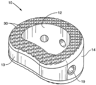
One embodiment of the present invention is directed to an interbody spacer 10
for
insertion between vertebral members. The spacer 10 includes a body having
inferior 18
and superior 17 faces. Teeth 30 are positioned along at least of one of the
faces 17, 18 to
maintain the position relative to the vertebral members. The teeth 30 are
aligned in a
pattern over all or part of at least one of the faces 17, 18.
CA 02586980 2007-05-09
WO 2006/053265 PCT/US2005/041077
3
Figure 1 illustrates one embodiment of the spacer 10 having an annular shape
forming an opening 12. The spacer 10 is formed by opposing posterior and
anterior walls
13, 14 with opposing side walls 15 to complete the ring shape. The anterior
wall 14 has a
greater height than the posterior wall 13 causing the spacer 10 to have a
wedge shape.
One or more apertures 19 may be positioned in the walls. The apertures 19
provide a
means for grasping or moving the spacer 10 with an instrument during the
surgical
process. The apertures 19 may be threaded to mate with the instrument. The
apertures 19
extend through the walls and may further provide an avenue to access the
opening 12,
such as for inserting bone-growth material.
Figure 2 illustrates a top view of the spacer 10 having a generally D-shape.
This
shape is created by the rounded anterior wall 14 and the substantially
straight posterior
wall 13. The interior wall of the opening 12 generally matches the shape of
the outer
walls and also has a generally D-shape. The thickness of the spacer walls
formed between
the outer walls of the opening 12 is generally uniform throughout the spacer
10. It is
understood however that the spacer 10 and opening 12 may have a variety of
different
shapes, and the thickness of the spacer walls may independently vary, each
being
dependent upon the parameters of use.
A transverse plane is defined as being laterally aligned along the spacer and
extending through the side walls 15. The transverse plane is substantially
parallel to the
transverse center line T-T. A longitudinal plane is defined as being aligned
along the
spacer 10 and extending through the anterior and posterior walls 14, 13. The
longitudinal
plane is substantially parallel to a longitudinal center line L-L.
Figure 3 illustrates a side view of the spacer 10 that more clearly
illustrates the
wedge shape formed by the taller anterior wall 14 and shorter posterior wall
13. The
superior face 17 and inferior face 18 are generally convex when the spacer 10
is viewed
from the side. The maximum height is located inward from the anterior wall 14
(i.e., at a
point between the anterior wall 14 and posterior wall 13). In the embodiment
illustrated in
Figure 3, the maximum height is located closer towards the anterior wall 14.
The spacer
10 may be substantially uniform about a side centerline 99. Therefore, a
distance between
the centerline 99 and the inferior face 18 is the same as a corresponding
distance between
the centerline and the superior face 17. A line 98 tangent to the centerline
99 forms an
angle Z. This angle may range from about 2 to about 7.5 . Because the spacer
10 is
CA 02586980 2007-05-09
WO 2006/053265 PCT/US2005/041077
4
uniform about the centerline 99, a complimentary and equal second angle is
formed
between the superior face 17 and the centerline 99 (not shown). Therefore, an
overall
spacer angle formed between tangent lines for the inferior and superior faces
18, 17 may
range from between about 4 to about 15 .
A plurality of teeth 30 are positioned on one or both of the inferior and
superior
faces 18, 17. Figure 4 illustrates a view of one embodiment of teeth 30 each
having two
anterior surfaces 31 that face towards the anterior side 14 of the spacer 10.
The anterior
surfaces 31 are angled relative to each other and connect along a common
anterior edge
36. Each tooth 30 further has two posterior surfaces 32 that face towards the
posterior side
13 of the spacer 10. The posterior surfaces 32 are angled relative to each
other and
connect along a common posterior edge 37. In the embodiment of Figure 4, each
tooth 30
is substantially rectangular and is formed by four sides bounded on each side
by a pathway
34. Each tooth 30 further includes a peak 33 at the intersection of the
anterior and
posterior surfaces 31, 32.
Figure 5 is a cross-sectional view cut along line 5-5 (see Figure 3) in the
transverse
plane. The view cuts through a first row of teeth 30 and illustrates the peaks
33 of both
the first row and an adjacent second row. The adjacent rows of teeth 30 are
offset such
that the peaks 33 of a first row are aligned within valleys of an adjacent
second row. The
bottom of the valleys are located in the pathways 34 extending between the
teeth 30. The
posterior surfaces 32 are curved forming a rounded posterior edge 37.
Figure 6 is a cross-sectional view cut along line 6-6 (see Figure 2) in the
longitudinal plane. The view cuts through a first column of teeth 30 and
illustrates the
peaks 33 of both the first column and the adjacent second column. The adjacent
columns
of teeth 30 are offset such that the peaks 33 of the first column are aligned
within valleys
of the adjacent second column. The bottoms of the valleys are aligned within
the
pathways 34 extending between the teeth 30.
The teeth 30 are ramped in an anterior direction caused by the posterior
surfaces 32
having a lesser angle than the anterior surfaces 31 when viewed along the
longitudinal
plane. Further, the anterior surfaces 32 are curved and undercut the anterior
surfaces 31
causing the peak 33 to face in an anterior direction. This configuration
provides for
inserting the spacer 10 using an anterior approach. The slighter angle of the
posterior
surfaces 32 and anterior angle of the peak 33 do not catch during the
insertion process.
CA 02586980 2007-05-09
WO 2006/053265 PCT/US2005/041077
Once the spacer 10 is inserted in the disc space between the vertebral
members, the greater
angle of the anterior surfaces 31 and the anterior facing peak 33 prevent or
limit anterior
movement of the spacer 10. The embodiments illustrated in Figures 1-6 include
teeth 30
orientated for an anterior insertion approach. Other embodiments may include
the teeth 30
5 oriented in different directions for a different insertion approach.
The pathways 34 extend in a crisscross pattern across the face of the spacer
10.
The pathways include a series of first parallel pathways extending across the
face in a first
direction, and a series of second parallel pathways extending in a second
direction. Each
series of pathways 34 are substantially straight and form an angle relative to
the
longitudinal and transverse planes. In the embodiment of Figure 2, first
pathways are cut
at angle 6 relative to the longitudinal plane. Second pathways are cut at a
mirrored angle
0' relative to the longitudinal plane. A mirrored angle is a negative
equivalent to a first
positive angle. By way of example, the first pathways are formed at an angle
of about 45 ,
and the second pathways are formed at an angle of about -45 , each relative to
the
longitudinal plane.
The pathways 34 may be flat, angled into the face of the spacer 10, or a
combination of both shapes. Specific embodiments include a U-shaped pathway,
and a J-
shaped pathway. In one embodiment, the first pathway has a first shape, and
the second
pathway has a second shape different than the first.
The plurality of teeth 30 and pathways 34 form a uniforin pattern as best seen
in
Figure 2. The teeth 30 are arranged in a series of columns generally aligned
along the
longitudinal plane. A straight line C can be drawn through the peaks 33 of
each of the
teeth 30 in a column. In one embodiment, the line C is substantially parallel
with the
longitudinal centerline L-L. The teeth 30 are also arranged in a series of
rows generally
aligned along the transverse plane. A straight line R can be drawn through the
peaks 33 of
each of the teeth 30 in a row. In one embodiment, the line R is substantially
parallel with
the transverse centerline T-T. Intersections of the first and second pathways
occur along
the edges of the teeth. In the embodiment of Figure 2, the intersections are
aligned with
the lines C and R that extend across the spacer 10.
The teeth 30 are each aligned to face in the same direction with the posterior
surfaces 32 and common posterior edge 37 having the same orientation. This is
best
illustrated in Figure 2. The teeth 30 each work in combination as the spacer
10 is moved
CA 02586980 2007-05-09
WO 2006/053265 PCT/US2005/041077
6
into the disc space. If various teeth 30 across the surface had different
alignments, the
teeth 30 would not work in unison during insertion. By way of example, if the
teeth 30 on
a first section of the face were aligned differently than teeth 30 on a second
section, the
spacer 10 would be more difficult to insert, and may move from the vertebral
space.
A second embodiment of teeth 30 is illustrated in Figures 7 and 8. The same
reference numbers from the first embodiment are used to illustrate like
elements. Each
tooth 30 has a pair of anterior surfaces 31 aligned towards the anterior side
14 of the
spacer 10, and a pair of posterior surfaces 32 aligned towards the posterior
side 13. The
teeth 30 each have a substantially rectangular shape. Each of the anterior and
posterior
surfaces 31, 32 extend outward and are capped by an outer surface 39. Pathways
34
extend in first and second directions and surround the teeth 30. In one
embodiment, each
of the outer surfaces 39 follows the convexity of the surface of the spacer
10. In this
sense, the outer surface 39 is flat relative to the overall convex surface.
Each of the teeth 30 is aligned with the common posterior edges 37 each facing
in
the same direction. Further, each tooth 30 is aligned with the common anterior
edges 36
each facing in the same direction. The teeth 30 may not be parallel to each
other due to
the overall convexity and angle of the superior and inferior faces 17, 18. In
one
embodiment, the spacer 10 is substantially uniform in each direction and can
be used from
a variety of insertion angles.
Figures 9-11 illustrate a third tooth embodiment. Again, the same reference
numbers from the first embodiment are used to illustrate like elements.
Anterior and posterior sides 14, 13 are separated by side walls 15 to form an
opening 12.
Teeth 30 may be positioned on the face in a uniform pattern aligned in a
plurality of
columns such as along line C and rows such as along line R.
Each of the teeth 30 has four sides that form an overall rectangular shape. A
posterior surface 37 is opposed by anterior surface 36 with side surfaces 38
extending
along each side. The posterior 37 and anterior 36 surfaces are substantially
the same
width, as are the side surfaces 38. The posterior surface 37 extends outward
from the
spacer 10 at a lesser angle than the posterior surface 37. In one embodiment,
the posterior
surface 37 has an angle of about 70 , and the anterior surface 36 has an angle
of about 0
(i.e., the anterior surface 36 is substantially perpendicular to the
pathways). In one
embodiment, the side surfaces 38 extend outward from the spacer at about 0 .
In these
CA 02586980 2007-05-09
WO 2006/053265 PCT/US2005/041077
7
embodiments, the angle of the surfaces is determined relative to the
substantially flat
pathways 34.
The teeth 30 are aligned in columns such as depicted by line C of Figure 9.
The
teeth 30 are aligned with no gap being formed between the anterior surface 36
of a first
tooth 30 and the posterior surface 37 of an adjacent second tooth. As
illustrated in Figure
11, this spacing forms a substantially saw-tooth orientation. Each of the
columns on the
face of the spacer 10 is aligned in the same direction. In one embodiment, the
columns are
each substantially parallel with the longitudinal center line L-L. Teeth 30
are also aligned
in row as depicted by line R in Figure 9. In one embodiment, the rows are
substantially
perpendicular to the columns, and may be perpendicular to the longitudinal
center line L-
L.
Pathways 34 are positioned between each of the columns of teeth 30. In one
embodiment, pathways 34 have a width less than the width of the teeth 30
(i.e., less than
the width of the anterior 36 and posterior 37 surfaces).
The teeth 30 of the third embodiment are sized and configured to an anterior
insertion approach. The angle of the posterior surfaces 37 is set to
facilitate movement of
the spacer 10 into the disc space from the anterior side. The anterior surface
36 has a
steeper angle to prevent the spacer 10 from moving out of the disc space after
insertion.
As with the other embodiments, the teeth 30 may be aligned in other
orientations for other
insertion approaches.
The outer edges of the spacer 10 may have a round or curved configuration that
prevent a sharp angle that may make the insertion of the spacer 10 more
difficult, or cause
injury to the patient. In one embodiment as illustrated in Figure 3, the
spacer 10 has a
radius 92 extending along the exterior edges where the inferior and superior
faces 18, 17
meet the side walls 15 and posterior and anterior walls 13, 14. Specific
embodiments
include radiuses of between about 2mm and 3mm. The teeth 30 adjacent to the
edges may
fall within the radius area and be rounded or curved. By way of example as
illustrated in
Figure 7, tooth 30a which slightly overlaps into the edge of the face has a
rounded section
witliin the edge of the spacer 10. Tooth 30b is positioned closer to the edge
and therefore
has a larger rounded section. Tooth 30c is almost completely positioned within
the edge
and therefore is almost completely rounded. Teeth 30 positioned along the
opening 12
may also include rounded sections, as illustrated by tooth 30d.
CA 02586980 2007-05-09
WO 2006/053265 PCT/US2005/041077
8
The spacer 10 may be constructed of a single piece with the teeth 30 formed
into
one or both faces 17, 18. Alternatively, the teeth 30 may be constructed of a
separate
material and attached to the faces 17, 18.
One embodiment of the spacer (e.g., Figure 3) illustrates each of the inferior
and
superior surfaces having a convex shape. Other embodiments may also include
substantially flat surfaces, or concave surfaces. Further, the spacer may have
surfaces of
different shapes with the inferior surface having a first shape and the
superior surface
having a second shape (e.g., convex inferior surface and substantially flat
superior
surface).
The term vertebral member is used generally to describe the vertebral geometry
comprising the vertebral body, pedicles, lamina, and processes. The spacer 10
may be
sized and shaped, and have adequate strength requirements to be used within
the different
regions of the vertebra including the cervical, thoracic, and lumbar regions.
Bone growth material may be positioned within the opening 12 to facilitate
bone
growth through the spacer 10. The bone growth material may include a sponge,
matrix,
and/or other carrier impregnated with a protein such as bone morphogenic
protein (BMP),
LIM mineralization protein (LMP), etc.
The present invention may be carried out in other specific ways than those
herein
set forth without departing from the scope and essential characteristics of
the invention.
The teeth 30 may further be spaced inward from the edges of the faces 17, 18.
A gap 55
may exist along the edge of the faces that does not include any teeth 30. The
spacer 10
may have a variety of shapes and sizes. In one embodiment, the spacer includes
an
interior wall that closes the opening. In another embodiment, the spacer does
not include
an opening 12. In embodiments with teeth 30 on both the inferior and superior
faces 18,
17, the teeth 30 may be aligned in a common direction on each of the faces.
The present
embodiments are, therefore, to be considered in all respects as illustrative
and not
restrictive, and all changes coming within the meaning and equivalency range
of the
appended claims are intended to be embraced therein.