Note: Descriptions are shown in the official language in which they were submitted.
CA 02587011 2007-05-07
WO 2006/052819 PCT/US2005/040158
-1-
MINIMALLY INVASIVE SPINAL FIXATION
GUIDE SYSTEMS AND METHODS
CROSS REFERENCE TO RELATED APPLICATIONS
This application claims priority to U.S. Provisional Patent Application No.
60/626,138 filed on November 9, 2004 and entitled "Minimally Invasive Spinal
Fixation
Guide Systems and Methods," which is hereby incorporated by reference in its
entirety.
BACKGROUND
For a number of known reasons, spinal fixation devices are used in orthopedic
surgery to align and/or fix a desired relationship between adjacent vertebral
bodies.
Such devices typically include a spinal fixation element, such as a relatively
rigid
fixation rod, that is coupled to adjacent vertebrae by attaching the element
to various
anchoring devices, such as hooks, bolts, wires, or screws. The fixation
elements can
have a predetermined contour that has been designed according to the
properties of the
target implantation site, and once installed, the instrument holds the
vertebrae in a
desired spatial relationship, either until desired healing or spinal fusion
has taken place,
or for some longer period of time.
Spinal fixation elements can be anchored to specific portions of the
vertebrae.
Since each vertebra varies in shape and size, a variety of anchoring devices
have been
developed to facilitate engagement of a particular portion of the bone.
Pedicle screw
assemblies, for example, have a shape and size that is configured to engage
pedicle
bone. Such screws typically include a threaded shank that is adapted to be
threaded into
a vertebra, and a head portion having a rod-receiving element, usually in the
form of a
U-shaped slot forined in the head. A set-screw, plug, or similar type of
fastening
mechanism is used to loclc the fixation element, e.g., a spinal rod, into the
rod-receiving
head of the pedicle screw. In use, the shank portion of each screw is threaded
into a
vertebra, and once properly positioned, a rod is seated through the rod-
receiving member
of each screw and the rod is locked in place by tightening a cap or other
fastener
mechanism to securely interconnect each screw and the fixation rod.
CA 02587011 2007-05-07
WO 2006/052819 PCT/US2005/040158
-2-
Recently, the trend in spinal surgery has been moving toward providing
minimally invasive devices and methods for implanting spinal fixation devices.
One
such method, for example, utilizes two percutaneous access devices for
implanting an
anchoring device, such as a spinal screw, into adjacent vertebrae. A spinal
rod is then
introduced through a third incision a distance apart from the percutaneous
access sites,
and the rod is transversely moved into the rod-engaging portion of each spinal
screw.
The percutaneous access devices can then be used to apply closure mechanisms
to the
rod-engaging heads to lock the rod therein. While this procedure offers
advantages over
prior art invasive techniques, the transverse introduction of the rod can
cause significant
damage to surrounding tissue and muscle.
Accordingly, there remains a need for improved methods and devices for
introducing spinal fixation elements, spinal anchors, and/or other spinal
devices into a
patient's spine.
SUMMARY
Disclosed herein are methods and devices for implanting spinal fixation
implants
and devices. In one exemplary embodiment, a method for implanting a spinal
fixation
system includes positioning a spinal fixation element to extend along a
patient's spinal
column adjacent to one or more vertebrae, determining an implant site on at
least one
vertebra, and implanting at least one spinal anchor at the implant site on at
least one
vertebra. In an exemplary embodiment, the spinal fixation element may be
inserted
through a first incision, and each spinal anchor may be inserted through an
incision
separate from the spinal fixation element and one another. Once the spinal
anchor(s) are
implanted, the spinal fixation element may be moved, e.g., approximated,
toward the
spinal anchor(s) to couple, statically or dynamically, the spinal fixation
element to the
anchor(s). In one exemplary method, the spinal fixation element may be locked
to the
spinal anchor(s) to maintain the vertebrae in a fixed position relative to one
another. In
other exemplary embodiments, one or more of the spinal anchors may be
dynamically
coupled to the spinal fixation element to perinit movement of one or more
vertebrae
relative to other vertebrae.
CA 02587011 2007-05-07
WO 2006/052819 PCT/US2005/040158
-3-
Further disclosed herein are various exemplary techniques for positioning the
spinal fixation element along the patient's spinal column. In one embodiment,
the spinal
fixation element may be introduced through a cannula or port and manipulated
to
position the fixation element such that it extends along the patient's spinal
column. An
insertion tool, such as a pivoting implant holder, can be used to introduce
the spinal
fixation element through an incision or through a cannula or port. The
insertion tool
may be effective to insert the spinal fixation element in a first orientation
and to pivot
the spinal fixation element into a second orientation in which the spinal
fixation element
is substantially parallel to a patient's spinal column.
Also disclosed herein are various techniques for determining an implant site
on
each vertebra. In one exemplary embodiment, a targeting member may be
positioned
relative to a target implant site on a vertebra, and the targeting member may
be aligned
relative to the target implant site using an imaging device. The targeting
member may
be part of a guide system having a guide portion that is adapted to be
positioned outside
a patient's body and to extend along a patient's spinal column, and a rod-
engaging
member that is adapted to couple to the spinal fixation element to maintain
the spinal
fixation element in a fixed position within the patient's body extending
adjacent to a
patient's spinal column. The targeting members may be slidably disposed on the
guide
portion to allow each targeting member to be adjusted relative to a target
implant site on
a vertebra.
Various techniques for implanting the spinal anchor(s) are also disclosed
herein.
In one exemplary embodiment, one or more spinal anchors can be percutaneously
delivered to the anchor site through a cannula which may be positioned through
a
minimally invasive pathway to the spinal anchor site. Each cannula may be
attached to
a guide system that is adapted to couple to the spinal fixation element. In
other
exemplary embodiments, the spinal anchors can be delivered through an access
port that
defines a large working channel.
Techniques for coupling the fixation element to the anchor(s) are disclosed
herein. In one exemplary embodiment, the spinal fixation element may be moved,
e.g.,
approximated, toward the spinal anchor(s) by engaging the spinal fixation
element and a
spinal anchor with a grasping tool. In certain exemplary embodiments, one or
more of
the spinal anchors may be a side-loading, top-tightening spinal anchor to
allow the
CA 02587011 2007-05-07
WO 2006/052819 PCT/US2005/040158
-4-
fixation element to be approximated into a side-opening in an anchor and to
allowed a
locking mechanism to be introduced into the top of the anchor to lock the
fixation
element within the anchor.
In yet another exemplary embodiment, a surgical method includes inserting a
spinal rod through a first incision to position the spinal rod adjacent to a
plurality of
vertebrae in a patient's spinal column, determining an implant site on each
vertebra,
making a percutaneous incision at the implant site on each vertebra, creating
a pathway
from the percutaneous incision to the implant site on each vertebra, placing
an anchor
through each pathway to implant an anchor in each vertebra, approximating the
spinal
rod toward the anchors, and locking the spinal rod to each anchor.
Further disclosed herein are guide systems for use in spinal surgery. In one
exemplary embodiment, an exemplary guide system includes a guide portion that
is
adapted to be positioned outside a patient's body and to extend along a
patient's spinal
column, a rod-engaging member that is mated to the guide portion and that is
adapted to
couple to a spinal rod and to maintain the spinal rod in a fixed position
within the
patient's body extending adjacent to a patient's spinal column, and one or
more targeting
member that are slidably coupled to the guide portion. The targeting member
may be
adapted to target an implant site on a vertebra in the patient's spinal
column. The
targeting members can be removably coupled to a support member that is
slidably
disposed on the guide portion, and each support member can also be configured
to mate
to a cannula for providing a pathway to an implant site.
While the guide system can have a variety of configurations, in one embodiment
the rod-engaging member is effective to maintain the rod in a fixed position
that is
spaced a distance apart from the guide portion and substantially parallel to
the guide
portion. The targeting member may be adapted to target an implant site on a
vertebra
that is at a location offset from the spinal rod. In other exemplary
embodiments, the
device can include a locking meclianism coupled to the rod-engaging member and
effective to removably mate a spinal fixation rod to the rod-engaging member.
In yet another exemplary embodiment, a guide system for use in spinal surgery
includes a guide member having a rod-engaging portion that is adapted to
maintain a
spinal fixation element at a fixed position in a patient's body that is
substantially parallel
CA 02587011 2007-05-07
WO 2006/052819 PCT/US2005/040158
-5-
to the guide member, and at least one targeting element that is adapted to
target an
implant site on a vertebra at a location that is offset from the spinal
fixation element.
BRIEF DESCRIPTION OF THE DRAWINGS
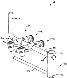
FIG. 1 is a perspective view of an exemplary guide system for implanting a
spinal fixation element and one or more spinal anchors;
FIG. 2A is a side perspective view of an exemplary embodiment of a guide
cannula for use with the methods and devices disclosed herein;
FIG. 2B is a cross-sectional, perspective view of another exemplary embodiment
of a guide cannula for use with the methods and devices disclosed herein;
FIG. 3A is a side view of an exemplary pivoting implant holder having a spinal
fixation element mated thereto and positioned in a first orientation adapted
for
introduction into a patient's spinal column;
FIG. 3B is a side view of the pivoting implant holder shown in FIG. 3A with a
spinal fixation element pivoted into a second orientation;
FIG. 4A is a posterior view of six percutaneous incisions formed in the
thoracolumbar fascia of a patient's back;
FIG. 4B is an end view showing a blunt dissection of the muscles surrounding a
patient's vertebra;
FIG. 4C is an end view of the vertebra in FIG. 4B with a k-wire placed through
the incision and into the patient's vertebra;
FIG. 4D is an end view of the vertebra in FIG. 4C showing an obturator and
several dilators disposed over the k-wire to dilate the tissue and muscles;
CA 02587011 2007-05-07
WO 2006/052819 PCT/US2005/040158
-6-
FIG. 5A is perspective view of a first spinal anchor being implanted in a
vertebra
and having a minimally invasive percutaneous access device coupled thereto and
extending through an incision formed in the patient's tissue surface;
FIG. 5B is a perspective view of the first spinal anchor shown in FIG. 5A
implanted in a vertebra and coupled to a first percutaneous access device that
is coupled
to the guide system shown in FIG. 1, and a second spinal anchor implanted into
an
adjacent vertebra and coupled to a second percutaneous access device that is
coupled to
the guide system shown in FIG. 1;
FIG. 6A is an end view of the vertebra in FIG. 4C showing an access port
defining a working channel extending to the vertebra;
FIG. 6B is a perspective view of the first spinal anchor shown in FIG. 6A
implanted in a vertebra and coupled to a first percutaneous access device that
extends
through a first access port coupled to the guide system shown in FIG. 1, and a
second
spinal anchor implanted into an adjacent vertebra and coupled to a second
percutaneous
access device that extends through a second access port coupled to the guide
system
shown in FIG. 1;
FIG. 7A is a perspective view of an exemplary embodiment of a spinal anchor
and a tool for laterally approximating a spinal fixation element toward the
spinal anchor;
FIG. 7B is a perspective view of another exemplary embodiment of a spinal
anchor and a tool for laterally approximating a spinal fixation element toward
the spinal
anchor;
FIG. 7C is an exploded view of an exemplary polyaxial spinal fixation element
witli an insertion guide;
FIG. 7D is a side, view, partially cutaway, of an exemplary monoaxial spinal
fixation element with an insertion guide;
CA 02587011 2007-05-07
WO 2006/052819 PCT/US2005/040158
-7-
FIG. 8 is a transparent perspective view of another exemplary embodiment of a
tool for laterally approximating a spinal fixation element toward a spinal
anchor;
FIG. 9 is a perspective view of another exemplary embodiment of a guide system
for implanting a spinal fixation element and one or more spinal anchors; and
FIG. 10 is an end view of the guide system shown in FIG. 9.
DETAILED DESCRIPTION
Certain exemplary embodiments will now be described to provide an overall
understanding of the principles of the structure, function, manufacture, and
use of the
devices and methods disclosed herein. One or more examples of these
embodiments are
illustrated in the accompanying drawings. Those skilled in the art will
understand that
the devices and methods specifically described herein and illustrated in the
accompanying drawings are non-limiting exemplary embodiments and that the
scope of
the present invention is defined solely by the claims. The features
illustrated or
described in connection with one exemplary embodiment may be combined with the
features of other embodiments. Such modifications and variations are intended
to be
included within the scope of the present invention.
Disclosed herein are methods and devices for introducing spinal fixation
elements, spinal anchors, and/or other spinal devices into a patient's spine.
A person
skilled in the art will appreciate that, while the methods are described in
connection with
certain spinal instruments and devices, a variety of spinal instruments and
devices can be
used to perform the methods in accordance with the various embodiments
disclosed
herein. Conversely, the instruments and devices disclosed herein can be used
for a
variety surgical procedures. Moreover, a person skilled in the art will
appreciate that
exemplary methods can be performed in any sequence using only some or all of
the
methods.
FIG. 1 illustrates an exemplary embodiment of a guide system 10 that can be
used to position a spinal fixation element, such as a spinal rod, a plate,
and/or a cable or
tether, in a patient's spinal column, to target implant sites on one or more
vertebra, and,
in certain exemplary embodiments, to facilitate implanting a spinal anchor in
a vertebra.
CA 02587011 2007-05-07
WO 2006/052819 PCT/US2005/040158
-8-
As shown, the guide system 10 generally includes a guide portion 12 that is
adapted to
be positioned outside a patient's body and a rod-engaging portion 14 that is
adapted to
couple to a spinal fixation element, such as spinal rod 26, to maintain the
spinal rod 26
in a fixed position within the patient's body such that the rod 26 extends
adjacent to a
patient's spinal column. The rod-engaging portion 14 may be effective to
maintain the
spinal rod 26 in a position that is substantially parallel to, but spaced
apart from, the
guide portion 12 such that guide portion 12 serves as a guide located outside
of the body
to indicate the location of the spinal rod 26 disposed inside the patient's
body. The
guide system 10 can also include one or more targeting instruments 16a, 16b
that are
movably coupled to the guide portion 12 of the system 10. The targeting
instruments
16a, 16b can be adapted to target an implant site on a vertebra in the
patient's spinal
column.
The guide portion 12 of the guide system 10 can have a variety of
configurations.
In one embodiment, for example, the guide system 10 is effective to indicate
the position
of a spinal rod 26 disposed within and extending along a patient's spinal
column. As
shown in FIG. 1, the guide portion 12 has a generally elongate support rod 20
with
opposed first and second ends 20a, 20b. The first end 20a can include a handle
22
formed thereon or mated thereto for facilitating grasping and manipulation of
the system
10. The handle 22 can also optionally be used to couple the guide system 10 to
a
support, such as a table. The second end 20b may be adapted to couple to the
rod-
engaging portion 14. The rod-engaging portion 14 can have virtually any shape
and
size. For example, in the illustrated embodiment, the rod engaging portion 14
extends in
a direction that is transverse to the support rod 20 and it is adapted to
removably engage
a spinal rod 26. As shown in FIG. 1, the first end 14a of the rod-engaging
portion 14
may be mated to the second end 20b of the support rod 20, and the second end
14b of
the rod-engaging portion 14 is in engagement with a spinal rod 26. While not
illustrated,
virtually any technique can be used to removably engage a spinal rod 26,
including, for
example, a clainping mechanism, a threaded engagement, an interference fit,
etc. Some
exemplary techniques for engaging a spinal rod will be discussed in more
detail below
with respect to FIGS. 3A-3B. The rod-engaging portion 14 can also include a
locking
mechanism (not shown) for locking the spinal rod 26 relative to the rod-
engaging
portion 14, and for subsequently releasing the rod 26 from the rod-engaging
portion 14.
CA 02587011 2007-05-07
WO 2006/052819 PCT/US2005/040158
-9-
The guide system 10 can also include one or more targeting instruments coupled
thereto. As shown in FIG. 1, two targeting instruments 16a, 16b are slidably
disposed
on the support rod 20 of the guide portion 12. While a variety of targeting
instruments
and techniques can be employed, in an exemplary embodiment, as shown, one or
more
of the targeting instruments 16a, 16b may include a slidable support 17a, 17b
and a
targeting member 18a, 18b coupled to a terminal end of the support 17a, 17b.
The
targeting members 18a, 18b may be positioned a distance apart from the support
rod 20
as illustrated or at other positions relative to the support rod 20. Spacing
the targeting
members 18a, 18b apart from the support rod 20 allows the targeting members
18a, 18b
to target an implant site on the vertebrae without interference from the
spinal rod 26,
which is located adjacent to the spinal column. In certain exemplary
embodiments, the
targeting members 18a, 18b may be movably coupled to the supports 17a, 17b
such that
the targeting members 18a, 18b can be moved toward and away from the supports
17a,
17b, as well as angularly adjusted relative to the supports 17a, 17b. Such a
configuration allows the targeting instrument 18a, 18b to be properly aligned
with a
target implant site on a vertebra. While one embodiment for targeting members
18a,
18b can be angularly adjustable, one skilled in the art will appreciate that
the members
can also be mounted at a fixed angle. Although not illustrated, guide portion
12 can
initially be attached to rod-engaging portion 14 such that it extends in a
direction
opposite to that shown in FIG. 1 during rod insertion. This configuration can
provide
enhanced visibility and maneuverability during rod insertion. Alternatively, a
handle
(not shown) can be connected to first end 14a of rod-engaging portion 14
during rod
insertion, and the handle can subsequently be replaced with guide portion 12.
FIGS. 9 and 10 illustrate another embodiment of a guide system 10' that can be
used to position a spinal fixation element, such as a spinal rod, a plate,
and/or a cable or
tether, in a patient's spinal column, to target implant sites on one or more
vertebra, and
in certain exemplary embodiments, to facilitate implanting a spinal anchor in
a vertebra.
Similar to the system 10 shown in FIG. 1, the guide system 10' of FIGS. 9 and
10
provide further adjustability options and it generally includes a guide
portion 12' that is
adapted to be positioned outside a patient's body and a rod-engaging portion
14' that is
adapted to couple to a spinal fixation element, such as a spinal rod 26', to
maintain the
spinal rod 26' in a fixed position within the patient's body such that the rod
26' extends
CA 02587011 2007-05-07
WO 2006/052819 PCT/US2005/040158
-10-
adjacent to a patient's spinal column. The rod-engaging portion 14' may be
effective to
maintain the spinal rod 26' in a position that is substantially parallel to,
but spaced apart
from, the guide portion 12' such that guide portion 12' serves as a guide
located outside
of the body to indicate the location of the spinal rod 26' disposed inside the
patient's
body. The guide system 10' can also include one or more targeting instruments
16a',
16b' that are movably coupled to the guide portion 12' of the system 10'. The
targeting
instruments 16a', 16b' can be adapted to target an implant site on a vertebra
in the
patient's spinal column.
Many components of the guide system 10', including support rod 20', rod-
engaging portion 14', and rod 26', are similar to corresponding components
discussed
above with respect to guide system 10. However, the guide system 10' enables
adjustment with additional degrees of freedom, and it is somewhat differently
constructed. While the system 10, shown in FIG. 1, utilizes rod-engaging
portion 14 in
such a way that it extends directly between rod 26 and support rod 20, the
guide system
10' illustrated in FIGS. 9 and 10 indirectly connects the support rod 20' and
the rod-
engaging portion 14'. That is, one end 14b' of the rod-engaging portion 14'
attaches to
rod 26' while the other end 14a' is removably engaged in one of the targeting
instruments
16a', 16b', such as by sleeve 23'. The targeting instrument to which rod-
engaging
portion 14' is attached is connected to support rod 20' by way of slidable
support 17a',
17b' as shown in FIGS. 9 and 10. Such a design enables effective targeting
without
requiring alignment of support rod 26' with rod 20'. For example, a k-wire 11
can be
inserted through the targeting members 18a', 18b' at a wide angle. Further, k-
wire 11, if
used, can be manipulated in such a way that it is rotated relative to the rod
26' to provide
multiple screw trajectories relative to the rod in order to achieve the proper
relationship
between the screw trajectory and the rod.
One skilled in the art will appreciate that a variety of connector mechanisms
can
be used to mate rod-engaging portion 14' to rod 26' and targeting instrument
16a', 16b'.
Further adjustability of the guide system 10' is provided by the embodiment
illustrated in FIGS. 9 and 10 by supports 17a', 17b', which have targeting
members 18a',
18b' coupled to terminal ends thereof. Similar to the embodiment shown in FIG.
1, the
guide system 10' shown in FIGS. 9 and 10 has a slidable support 17a', 17b'
that can be
moved along the longitudinal axis of the support rod 20'. In addition, the
support 17a',
CA 02587011 2007-05-07
WO 2006/052819 PCT/US2005/040158
-I1-
17b' can move (such as by a telescoping movement) in a direction transverse
(i.e.,
laterally) to the support rod 20'. As shown in FIGS. 9 and 10, the targeting
members
18a', 18b' may be positioned a selected distance apart from the support rod
20' thereby
allowing the targeting members 18a', 18b' to target an implant site on the
vertebrae
without interference from the spinal rod 26'. Additionally, as in the
embodiment
illustrated in FIGS. 9 and 10, the targeting members 18a', 18b' can be
angularly adjusted
in the cephalad-candal direction and/or in the medial-lateral direction, such
as by
rotating supports 17a', l7b' about an axis transverse to rod support 20'.
The guide system 10' may also include gauges and/or indicia (not shown) to
indicate angular and/or other spatial positioning of the various adjustable
components of
guide portion 12' with respect to a reference, such as support rod 20'. These
features can
be useful, for example, to construct a system with a preset positioning for
one or more
levels of the spine that may be subject to surgery. The preset positioning can
be
established based on preoperative data (e.g., CT data) or anatomic data
derived from a
population sample. A surgeon could use the preset positioning of the guide
system, if
appropriate, or minor adjustments can be made to the preset positioning based
on the
needs of a patient.
While the targeting members 18a, 18b, 18a', 18b' can have a variety of
configurations, U.S. Publication No. 2003/0187431 of Simonson entitled
"Apparatus
and Method for Targeting for Surgical Procedures," which is incorporated by
reference
herein in its entirety, discloses one such device for targeting an implant
site. A person
skilled in the art will appreciate that a variety of techniques and devices
for targeting an
implant site can be used with the present invention.
The targeting instruments 16a, 16b, 16a, 16b' can also be configured to
facilitate
use of the guide system 10, 10' with other spinal tools and devices. For
example, the
targeting members 18a, 18b, 18a', 18b' can include an inner lumen 19a, 19b
extending
therethrough for receiving spinal tools and devices, such a drill guides,
cannulas, and
access ports. Alternatively, or in addition, the targeting members 18a, 18b,
18a', 18b'
can be removably mated to the slidable support 17a, 17b, 17a', 17b' to allow
each
support member 17a, 17b, 17a', 17b' to mate to a cannula, access port, or
other device or
tool after the targeting members 18a, 18b, 18a', 18b' are removed. Each
support 17a,
17b, 17a', 17b' can thus be used to maintain a cannula, access port, or other
device in a
CA 02587011 2007-05-07
WO 2006/052819 PCT/US2005/040158
-12-
fixed positioned relative to a target implant site, thereby providing a guided
pathway to a
target implant site on a vertebra, as will be discussed in more detail below.
One skilled
in the art will appreciate that they system 10, 10' can be configured for
unilateral rod
placement or for bilateral rod placement as shown in FIGS. 9 and 10.
As previously noted, also disclosed herein are methods for implanting a spinal
fixation system. While the method will be described in connection with guide
system
10, 10, a person skilled in the art will appreciate that the method is not
intended to be
limited to use witli guide system 10, 10' and that a variety of other devices
can be used
to perform the method. In general, an exemplary method includes positioning a
spinal
fixation device, such as the spinal rod 26, 26', to extend along a patient's
spinal column
adjacent to several vertebrae, targeting an implant site on vertebrae,
implanting a spinal
anchor in one or more vertebrae, and coupling a spinal rod to one or more
spinal
anchors. Various exemplary techniques for performing the aforementioned
various steps
are discussed below under the following headings: Rod Introduction, Targeting,
Implanting Spinal Anchors, and Rod Approximation.
Rod Introduction
A variety of techniques can be used to position a spinal rod to extend along a
patient's spinal column adjacent to two or more vertebrae, and the spinal rod
can be
introduced at various locations along the patient's spine. For example, the
spinal rod
can be introduced through the same incision used to introduce a spinal anchor,
or
alternatively the spinal rod can be introduced through an incision that is
separate from
and located a distance apart from the incision(s) used to implant the spinal
anchor(s).
The rod can also either be directly introduced through the incision to extend
up along the
patient's spinal column, or it can be introduced through a cannula, access
port, or other
device for guiding the rod to extend along the patient's spinal column.
Various tools can
also be coupled to the rod to manipulate and facilitate introduction and
positioning of the
rod in the patient's body.
In one exemplary embodiment, referring to FIGS. 1 and 9, the rod 26, 26' is
attached to the guide system 10, 10' and the guide system 10, 10' is
manipulated to insert
the rod 26, 26' through an incision and to bluntly advance the rod 26, 26'
through the soft
tissue until the rod 26, 26' extends along the patient's spinal column,
preferably adjacent
CA 02587011 2007-05-07
WO 2006/052819 PCT/US2005/040158
- 13-
to the pedicles. The proper position of the rod 26, 26' can be determined
using
fluoroscopy. Once properly positioned, the guide system 10, 10' is preferably
fixedly
attached to a support, such as the operating table, using, for exainple, a
retractor arm.
In another exemplary embodiment, the rod can be introduced through a cannula.
FIG. 2A illustrates an exemplary embodiment of a cannula 212 for introducing a
spinal
fixation element, such as a rod. As shown, the cannula 212 is in the form of a
generally
elongate, cylindrical tube having an inner lumen 212c formed therein and
defining a
longitudinal axis L that extends between proximal and distal ends 212a, 212b
thereof.
The cannula 212 has a length l that allows the proximal end 212a of the
cannula 212 to
be positioned outside the patient's body, while the distal end 212b of the
cannula 212
extends into the patient's body to define a pathway for the rod. The cannula
212 also
includes at least one sidewall opening or slot 214, and more preferably two
opposed
sidewall openings (only one opening 214 is shown), forined therein and
extending
proximally from the distal end 212b thereof. The openings 214 allow the spinal
rod to
be rotated from a position coaxial with the cannula 212 to a position in which
the rod
extends along the spinal column.
In another embodiment, shown in FIG. 2B, an exemplary cannula 312 can
include a guide member 320 formed within the distal end 312b of the lumen 312c
to
help guide the spinal rod from the first orientation to the second
orientation. The guide
member 320 is in the form of a sloped shelf formed within the inner lumen 312c
of the
cannula 312 and it is positioned opposite to a sidewall slot 314 formed in the
access
device 312. In use, as the leading end of a spinal rod contacts the shelf 320
and the shelf
320 begins to direct the spinal rod into the second orientation, thereby
causing the spinal
rod to extend in a direction that is substantially transverse to the axis L of
the device
312, and that is preferably substantially parallel to the patient's spinal
column.
Other exemplary techniques for introducing a spinal rod through a cannula or
access device and into a patient's body are described in more detail in U.S.
Patent
Application No. 10/738,130 of Anderson et al. entitled "Methods And Devices
For
Minimally Invasive Spinal Fixation Element Placement," and U.S. Patent
Application
No. 10/737,537 of Anderson et al. entitled "Methods And Devices For Spinal
Fixation
Element Placement." These references are incorporated by reference herein in
their
entirety.
CA 02587011 2007-05-07
WO 2006/052819 PCT/US2005/040158
-14-
FIGS. 3A and 3B illustrate another exemplary embodiment of a technique for
introducing a spinal fixation rod to position the rod to extend along the
patient's spinal
column. In particular, tool 100 can be used to engage a spinal rod and
introduce the rod
through a cannula or an access port, directly through an incision, or through
other
devices known in the art. As shown, the tool 100 generally includes an
elongate shaft
112 having proximal and distal ends 112p, 112d with an inner lumen 112c
extending
therebetween. A pusher shaft (not shown) extends through the elongate shaft
112 and it
preferably includes a proximal end that is coupled to a trigger 118, and a
distal end that
is coupled to a pivoting element 114. The trigger 118 functions to move the
pusher shaft
and thereby rotate a rod 170 coupled to the pivoting element 144 between a
first
orientation, in which the rod 170 is substantially coaxial with the
longitudinal axis L' of
the shaft 112, as shown in FIG. 3A, and a second orientation, in which the rod
170
extends in a direction transverse to the elongate shaft 112, as shown in FIG.
3B. The
tool 100 can also include a mechanism for removably engaging the rod 170 to
allow the
rod 170 to be subsequently released from the tool 100 after it is disposed in
the patient's
body.
In use, the rod 170 is engaged by the tool and it is introduced through an
access
port or an incision in the first orientation shown in FIG. 2A. The trigger 118
can then be
engaged to rotate or pivot the rod 170 into the second orientation, shown in
FIG. 2B,
thereby positioning the rod substantially parallel to the patient's spinal
column. The rod
170 can then be released from the tool 100 and attached to the rod-engaging
member 14,
14' of the guide system 10, 10'. In an alternative embodiment, the tool 170,
or a
variation of the tool 170, can be formed integrally with the guide system 10,
10' such
that the tool 170 functions as the rod-engaging member.
The tool 100, and other embodiments of tools for introducing a spinal rod, are
described in more detail in U.S. Patent Application No. 10/737,538 of Techiera
et al.
filed on December 16, 2003 and entitled "Pivoting Implant Holder," which is
incorporated by reference herein in its entirety. This patent application also
discloses
techniques for engaging a spinal fixation element, and such techniques can
optionally be
incorporated into system 10, 10' to couple a spinal rod to the rod-engaging
member 14,
14'.
CA 02587011 2007-05-07
WO 2006/052819 PCT/US2005/040158
- 15-
Regardless of the technique used to position the rod within the patient's
body,
the rod may be attached to the guide system 10, 10' which is maintained in a
fixed
position, e.g., by attaching the guide system to a support, such as the
operating table.
The rod can thereafter optionally be used to facilitate targeting of the
implant sites.
Targe
Once the rod is in place and attached to the guide system 10, 10', the
targeting
instruments can be used to identify a target implant site on one or more
vertebrae. In
particular, an imaging device can be placed over the targeting members 18a,
18b, 18a',
18b' to align the targeting members 18a, 18b, 18a', 18b' with the target
implant sites on
the underlying vertebra. Once aligned, the targeting members 18a, 18b, 18a',
18b' may
be locked in place relative to the support 20, 20' on the guide system 10,
10'. The
surgeon can then marlc the incision location on the skin below the targeting
members
18a, 18b, 18a', 18b'. Alternatively, a furtlier incision is not needed, and
targeting and
pedicle screw insertion can be effected through the incision through which the
rod is
placed. As previously noted, exemplary methods and devices for targeting an
implant
site are described in more detail in U.S. Publication No. 2003/0187431 of
Simonson
entitled "Apparatus and Method for Targeting for Surgical Procedures," which
is
incorporated by reference herein in its entirety.
Once the implant sites on the vertebrae are targeted, the targeting members
18a,
18b, 18a', 18b' can remain attached to the guide system 10, 10' to allow tools
and devices
to be inserted through the lumens 19a, 19b formed therethrough, or they can be
removed
from the guide system 10, 10' to allow other tools and devices to be attached
to the guide
system 10, 10'.
Imnlanting Spinal AnclzoNs
Once the target implant sites are identified, a spinal anchor can be implanted
at
one or more implant sites. Any type of conventional spinal anchor can be used
to couple
a rod, statically or dynamically, to one or more vertebrae. For illustration
purposes,
however, exemplary methods will be described in connection with a spinal
screw, which
can be a mono-axial screw or a poly-axial screw.
CA 02587011 2007-05-07
WO 2006/052819 PCT/US2005/040158
-16-
In one exemplary embodiment, the spinal anchor can be adapted to receive the
spinal rod laterally. In particular, the spinal anchor can be a side-loading
anchor, such
that the rod can be pulled into an opening in the side of the anchor receiver
head. FIGS.
5A, 5B, 6B, 7A, 7C, 7D and 8 illustrate an exemplary embodiment of a side-
loading
spinal screw 50 having an opening or rod-receiving recess 56 formed in a side
of the
receiver head 52 of the anchor 50. The rod-receiving recess 56 allows a rod to
be
laterally approximated into the receiver head 52, and/or it allows the
receiver head 52 to
be moved to position the rod within the opening 56. The rod can be locked
within the
receiver head 52 by inserting a locking mechanism into the top of the receiver
head 52,
thereby clamping down on the rod. FIG. 7B illustrates another embodiment of a
side-
loading spinal screw 80 having a threaded shaft 84 and a receiver head 82. In
this
embodiment, the receiver head 82 includes a rod-receiving recess 86 that is
offset from
an axis A of the screw 80, such that the rod is maintained at a location
offset from the
screw 80. In one exemplary embodiment, the rod-receiving recess 86 may be open
in a
lateral direction to facilitate capture of the rod within the rod receiving
recess 86 as the
screw 80 is advanced into the vertebra. The threaded shaft 84 of the screw 80
may
include a head 85 that is free to rotate relative to the receiver head 82 such
that the
threaded shaft 84 may be polyaxially oriented relative to the receiver head
82, for
example, in a manner analogous to conventional polyaxial screws. A locking
mechanism, such as a set screw, for example, may be inserted into the receiver
head to
lock the head 85 of the threaded shaft 84 relative to the receiver head 82. A
second
locking mechanism, such as a second set screw, may be inserted to secure the
rod in the
rod receiving recess 86. In this manner, the head 85 of the shaft 84 may be
locked
independent of locking the rod within the recess 86. One skilled in the art
will
appreciate that such a system enables the position of vertebral bodies to be
adjusted
(e.g., in compression or distraction) by moving screws 50, 80 along rod 26. By
way of
example, the proper trajectory of the screw(s) can be determined and they can
be placed
into the vertebra(e). After locking the screw at the desired angle, the
vertebra(e) can be
moved (e.g., compressed or distracted) by moving the screw(s) along the rod.
When the
proper position is attained, the screw(s) can be locked with respect to the
rod.
CA 02587011 2007-05-07
WO 2006/052819 PCT/US2005/040158
-17-
Various techniques can be used to implant the spinal anchors; for example a
minimally invasive percutaneous incision may be made through the tissue at one
or more
of the sites. The location, shape, and size of the incision will depend on the
type and
quantity of spinal anchors being implanted, as well as the technique being
employed to
implant the spinal anchors. By way of non-limiting example, FIG. 4A
illustrates three
midline minimally invasive percutaneous incisions 62a-c formed on one side of
three
adjacent vertebra in the thoracolumbar fascia in the patient's back, and three
additional
midline minimally invasive percutaneous incisions 62d-f formed on the opposite
side of
the three adjacent vertebra in the thoracolumbar fascia in the patient's back.
While not
shown, a guide system 10, 10' can be positioned adjacent to each set of
incisions 62a-c,
62d-f with a targeting member in alignment with each incision.
In certain exemplary embodiments, one or more of the incisions may be
expanded to create a pathway from the incision to proximate a vertebra. For
example,
the incision may be expanded by serial dilation, with a retractor such as an
expandable
retractor, or by any other conventional techniques. In one exemplary
embodiment, blunt
finger dissection can be used, as shown in FIG. 4B, to separate the
longissimus thoracis
and multifidus muscles, thereby exposing the facet and the junction of the
transverse
process and superior articular process.
A spinal anchor may inserted through one or more of the incisions and the
pathways to proximate the vertebra. Any technique for implanting a spinal
anchor can
be used. In one embodiment, for example, a spinal anchor can be implanted over
a
guidewire, such as a k-wire. As shown in FIG. 4C, a guide wire, e.g., a k-wire
64, can
be implanted, either prior to or after formation of the incision, at each
spinal anchor
implant site. The k-wire 64 may extend into the vertebra at the desired entry
point of the
spinal anchor. In certain exemplary embodiments, the k-wire may be advanced
into the
vertebra. In other exemplary embodiments, the k-wire may be positioned
proximate to
or against the vertebra. Fluoroscopy or other imaging may be used to
facilitate proper
placement of the k-wire 64. The incision may be dilated to provide a pathway
for
delivery of a spinal anchor to each implant site, in the manner discussed
above, before or
after placement of the guidewire. For example, FIG. 4D illustrates serial
dilation at one
end of the incision 62 using an obturator 66a having several dilators 66b, 66c
of
increasing size placed there over. The dilators 66b, 66c are delivered over
the obturator
CA 02587011 2007-05-07
WO 2006/052819 PCT/US2005/040158
-18-
66a and k-wire 64 to essentially stretch the skin around the incision 62 and
to expand the
pathway to the anchor site. While not shown, the obturator 66a and the
dilators 66b, 66c
can extend through the targeting members 18a, 18b, 18a', 18b' on the guide
system 10,
10', or alternatively the targeting members 18a, 18b, 18a', 18b' can be
removed from the
guide system 10, 10' and the obturator 66a and dilators 66b, 66c can merely be
guided
along the k-wire.
One skilled in the art will appreciate that a spinal anchor may be advanced to
a
vertebra through the incision without the need for a guidewire.
Once the incision 62 is dilated to the proper size, if necessary, the vertebra
60
may be prepared using one or more bone preparation instruments, such as
drills, taps,
awls, burrs, probes, etc. In certain exemplary embodiments, one or more
cannulae can
be used to provide a pathway from the incision 62 to the anchor site for
insertion of the
bone preparation instruments and/or the anchor. In an exemplary embodiment, a
relatively small cannula (not shown) may be used to introduce bone preparation
instruments into the surgical site. The cannula may be placed through a
targeting
member 18a, 18b, 18a', 18b' on the guide system 10, 10', such that the cannula
is in
alignment with the target implant site. Once the vertebra 60 is prepared, a
spinal anchor
can be delivered along the k-wire, either through the cannula, or after the
cannula is
removed, and implanted in the vertebra. Alternatively, in embodiments not
employing a
guidewire, the spinal anchor may be advanced through the incision, e.g.,
through a
cannula, to the vertebra. A cannula, retractor, or other instrument may be
employed to
guide the spinal anchor to the vertebra.
In another embodiment, shown in FIGS. 5A-5B, a spinal anchor can be
implanted in the vertebra using a minimally invasive technique. Such a
procedure
preferably begins by inserting a guidewire, such as a k-wire, through the
incision and
into the vertebra, dilating the incision to form a pathway, and preparing the
vertebra, as
discussed above. As shown in FIG. 5A, a minimally invasive percutaneous access
device 412, i.e., a cannula, is then inserted through the incision 62,
preferably over the k-
wire (not shown), to the target implant site on the vertebra (60). A spinal
anchor, e.g.,
spinal screw 50, can be attached to the distal end of the cannula 412, as
shown, or the
spinal anchor can be passed through the cannula 412 after the cannula 412 is
positioned
through the incision to extend to the target implant site. In an exemplary
embodiment,
CA 02587011 2007-05-07
WO 2006/052819 PCT/US2005/040158
-19-
as shown in FIG. 5B, each access device 412, 412' is attached to a support
17b, 17a,
17b', 17a' on the guide system 10, 10'. While not shown, the access devices
can
alternatively be inserted through the targeting members 18a, 18b, 18a', 18b'
on the guide
system 10, 10'. Once the screw 50 is positioned adjacent to the vertebra 60, a
driver tool
(not shown) can be positioned through the access device 412 and coupled to a
receiver
head 52 of the spinal screw 50 to drive the screw 50 into the vertebra 60.
This procedure, and other minimally invasive methods and devices for
implanting a spinal anchor, are described in more detail in U.S. Patent
Application No.
10/738,130 of Anderson et al. entitled "Methods And Devices For Minimally
Invasive
Spinal Fixation Element Placement," U.S. Patent Application No. 10/737,537 of
Anderson et al. entitled "Methods And Devices For Spinal Fixation Element
Placement," and U.S. Patent Application No. 10/738,286 filed on December 16,
2003
and entitled "percutaneous access device and bone anchor assembly." These
references
are incorporated by reference herein in their entirety.
In yet another embodiment, shown in FIGS. 6A-6B, a larger cannula or access
port can be used to implant a spinal anchor in each vertebra. Again, as
described above,
the procedure preferably beings by dilating the incision to form a pathway.
The
obturator and dilators can optionally be inserted over a k-wire, as also
previously
described above. Once dilated to the appropriate size, an access port 63 can
be inserted
over the largest dilator, and the dilators and obturator can be removed such
that the
access port 63 defines a working channel 67 extending through tissue to the
target
implant site on the vertebra 60, as shown in FIG. 6A. The vertebra 60 can then
be
prepared using bone preparing tools and devices. A person skilled in the art
will
appreciate that the vertebra can be prepared either before dilation, or during
dilation
using a relatively small cannula, as was also described above. As shown in
FIG. 6B,
each access port 63, 63' can optionally be mated to a support 17a, 17b, 17a',
17b' on the
guide system 10, 10'. While not shown, the access ports 63, 63' can
alternatively extend
through the targeting members 18a, 18b, 18a', 18b' on the guide system 10,
10'.Once the
access port 63 is positioned in relation to the vertebra 60, a spinal anchor
can be inserted
therethrough and implanted in the vertebra 60. The spinal anchor can
optionally be
inserted directly through the access port 63, with or without the use of a
guide wire (not
shown). Or, in another embodiment, shown in FIG. 6B and as previously
described
CA 02587011 2007-05-07
WO 2006/052819 PCT/US2005/040158
-20-
above with respect to FIGS. 5A and 5B, each spinal anchor 50, 50' can be
implanted
using a minimally invasive percutaneous access device 412, 412' coupled
thereto.
One skilled in the art will appreciate that a variety of spinal fixation
elements can
be used with the system described herein. In addition to the spinal fixation
elements
previously described, FIGS. 7C and 7D illustrate a polyaxial pedicle screw 50'
(FIG. 7C)
and a monoaxial pedicle screw 50" (FIG. 7D) in association with an insertion
guide 19.
The polyaxial 50' and monoaxia150" screws are side-loading, such that the rod
26 can be
julled into an opening in the side of the anchor receiver head 52. In
addition, the screws
are top-tightening such that the rod can be locked within the receiver head 52
by
inserting a locking mechanism 27 (e.g., a set screw) into the top of the
receiver head 52,
thereby clamping down on the rod.
This embodiment, and other methods for implanting spinal anchors using an
access port, are described in more detail in U.S. Patent No. 6,159,179 of
Simonson
entitled "Cannula And Sizing And Insertion Method," U.S. Publication No.
2003/0083689 of Simonson entitled "Non Cannulated Dilators," and U.S.
Publication
No. 2003/0083688 of Simonson entitled "Configured And Sized Cannula." These
references are also incorporated by reference herein in their entirety.
A person having ordinary skill in the art will appreciate that the
aforementioned
methods and devices for implanting spinal anchors can be modified depending on
the
type of anchor being implanted, as well as the specific procedure being
employed.
Moreover, other methods and devices known in the art can be used in accordance
with
the present invention. By way of non-limiting example, U.S. Patent Publication
No.
2002/0123668 entitled "Retractor and Method for Spinal Pedicle Screw
Placement," and
U.S. Patent Publication No. 2003/0236447 entitled "Retractor and Method for
Spinal
Pedicle Screw Placement," each describe a surgical retractor and methods for
spinal
anchor placement which can be used with the present invention. These
references are
incorporated herein in their entirety.
Rod Approxin:atiotz
Once the spinal anchors are fully implanted in the vertebrae, the spinal rod
may
be coupled to the anchors. While various techniques can be used to couple the
rod to the
anchors, in an exemplary embodiment, the rod and/or anchors are approximated
toward
CA 02587011 2007-05-07
WO 2006/052819 PCT/US2005/040158
-21 -
one another using a lateral approximator device. The lateral approximator
device can
have virtually any configuration, but it is preferably effective to engage a
portion of the
anchor and engage the rod, thereby allowing the rod and anchor to be moved
toward and
coupled to one another.
FIG. 7A and 7B illustrate exemplary embodiments of a lateral approximator
device 70, 70' that is effective to couple to an anchor 50, 80 and to engage a
rod 26, 26'
to pull the anchor 50, 80 and/or rod 26, 26' toward one another until the rod
26, 26' is
seated within the side opening 56, 86 in the receiver head 52, 82 of the
anchor 50, 80.
More particularly, the device 70, 70' is in the form a pivoting arm that is
coupled to an
elongate tubular member, such as a cannula 41, 41'. In one embodiment, the
cannula 41,
41' can be adapted to attach to the anchor 50, 80 or to fit over a cannula
attached to the
anchor 50, 80. In another embodiment, the cannula 41, 41' can be the
percutaneous
access device 412 shown in FIG. 5A, and the lateral approximator device 70,
70' can be
removably matable to the access device 412. In yet another embodiment, the
cannula
41, 41' can include one or more lumens formed therethrough for guiding tools
and
devices to the anchor 50, 80. For example, the lateral approximator device 70
shown in
FIG. 7A only includes a single lumen formed therein for receiving a driver
tool and a
first locking mechanism, such as a set screw, for locking the receiving head
52 relative
to the shank 54, and for receiving a driver tool and a second locking
mechanism, such as
a set screw, for locking the rod 26, 26' within the receiver head 52. The
lateral
approximator device 80 shown in FIG. 7B, on the other hand, is bifurcated and
includes
first and second lumens A, B formed therein for allowing a first locking
mechanism,
such as a set screw, to be delivered through the first lumen A to lock the
receiver head 82
to the shank 84, while a second locking mechanism, such as a set screw, is
delivered
separately through the second lumen B to lock the rod 26, 26' relative to the
receiver
head 82. Regardless of the configuration of the cannula 41, 41', the lateral
approximator
70, 70' should be effective to pivot relative to the cannula 41, 41' to engage
a rod 26, 26'
and pull the rod 26, 26' and/or anchor 50, 80 toward one another.
FIG. 8 illustrates another embodiment of a lateral approximator device 90. In
this embodiment, the device 90 is in the form of a cannula having an inner
lumen 94
extending therethrough and having a pivoting arm 92 formed thereon or coupled
thereto.
The distal end 91 of the device 90 is effective to engage the receiver head 52
of a spinal
CA 02587011 2007-05-07
WO 2006/052819 PCT/US2005/040158
-22-
anchor 50, and the pivoting arm 92 is effective to pivot to engage a spinal
rod 26, 26' to
pull the rod 26, 26' into the side-opening 56 in the receiver head 52.
A person skilled in the art will appreciate that a variety of other techniques
can
be used to couple a spinal rod to the spinal anchors. Moreover, the spinal rod
does not
need to be directly attached to each anchor, and it can be indirectly attached
to the
anchors using, for example, a band clamp, or slotted or offset connectors.
Once the
spinal rod is fully seated in the receiver head of each spinal anchor, a
closure mechanism
can be applied to each receiver head to retain the spinal rod tlierein.
One skilled in the art will appreciate further features and advantages of the
invention based on the above-described embodiments. Accordingly, the invention
is not
to be limited by what has been particularly shown and described, except as
indicated by
the appended claims. All publications and references cited herein are
expressly
incorporated herein by reference in their entirety.
What is claimed is: