Note: Descriptions are shown in the official language in which they were submitted.
DEMANDES OU BREVETS VOLUMINEUX
LA PRESENTE PARTIE DE CETTE DEMANDE OU CE BREVETS
COMPREND PLUS D'UN TOME.
CECI EST LE TOME 1 DE 2
NOTE: Pour les tomes additionels, veillez contacter le Bureau Canadien des
Brevets.
JUMBO APPLICATIONS / PATENTS
THIS SECTION OF THE APPLICATION / PATENT CONTAINS MORE
THAN ONE VOLUME.
THIS IS VOLUME 1 OF 2
NOTE: For additional volumes please contact the Canadian Patent Office.
CA 02587173 2016-06-06
9851-86
1
POTENT LNA OLIGONUCLEOTIDES FOR THE INHIBITION OF HIF-1A EXPRESSION
FIELD OF THE INVENTION
The present invention provides compositions and methods for inihibiting the
expression of
HIF-la. In particular, this invention relates to LNA oligonucleotides, which
are specifically
hybridisable with nucleic acids encoding HIF-la. The LNA oligonucleotides have
been shown
to inhibit the expression of HIF-1a and pharmaceutical preparations thereof
are disclosed.
BACKGROUND OF THE INVENTION
Solid tumors must establish a blood supply and have enhanced glucose
metabolism to grow
beyond a few millimeters. How they sense hypoxia, and respond by activating
hypoxia-
Inducible genes and secreting angiogenic factors to establish a blood system
is central to
cancer biology. Many tumors contain hypoxlc microenvironments, which have been
associated with malignant progression, metastasis and resistance to
radiotherapy and
chemotherapy.
The discovery of hypoxia-inducible factor-1 (I1IF-1) gave some insight into
the regulation of
hypoxia-inducible genes (US 5,882,914 and WO 96/39426; WO 99/48916). HIF-1 is
composed of two subunits HIF-la (HIF-lalpha; referred to herein as "HIF-la")
and HIF-10
and it binds - 1 hypoxia-response elements (HREs) In enhancers of genes
encoding
anglogenic factors such as VEGF and glycolysis-related proteins such as
glycolytic enzymes
and glucose transporter 1 and 3 (GLU-1 and 3).
It has been demonstrated that engineered down-regulation of HIF-la by
intratumoral gene
transfer of an antisense HIF-la plasmid leads to the down-regulation of VEGA
and decreased
tumor microvessel density (WO 00/76497, Sun X et al, Gene Therapy (2001) 8,
638-645).
The plasmid contained a 320-bp cDNA fragment encoding 5'-end of HIF-la
(nucleotides 152-
454; Genebank AF003698).
WO 2003/085110 shows LNA antisense oligonucleotides which down-regulates human
HIF-la
expression. One compound is named CUR813 (SEQ ID NO. 11).
The present invention disclosed LNA oligonucleotides, which are more potent
than CUR813
(SEQ ID NO. 11). Also the specific LNA oligonucleotides, according to the
invention, induce
CA 02587173 2016-06-06
79851-86
2
apoptosis and inhibit proliferation. Also, the LNA oligonucleotides which have
a 100%
sequence identity to the mouse HIF-la down-regulate the HIF-la expression in
the liver,
colon and kidney in mice.
SUMMARY OF THE INVENTION
The present invention provides compositions and methods for inhibiting the
expression of
IIIF-la. In particular, this invention relates to LNA oligonucleotides over 2
specific motifs
targeting HIF-la. These motifs are disclosed as SEQ ID NOS. 3 and 4.
Specifically preferred
LNA oligonucleotides are SEQ ID NO. 1 and SEQ ID NO. 2. The LNA
oligonucleotides of the
invention are potent inhibitors of HIF-la mRNA expression and protein levels.
More particularly, the present invention provides an LNA ollgonucleotide
consisting of a
sequence selected from the group consisting of
51-(L)G.G.csasasgscsastscscsTõGJ-3' (SEQ ID NO. 3)
and
5'-( ,)T,cTõascstsgsc,c,tst,c,TJA-3' (SEQ ID NO. 4)
wherein capital letters designate a beta-D-oxy-LNA nucleotide analogue, small
letters
designate a 2-deoxynucleotide, underline designates either a beta-D-oxy-LNA
nucleotide
analogue or a 2-deoxynucleotide, subscript "s" designates a phosphorothioate
link between
neighbouring nucleotides/LNA nucleotide analogues, and subscript "x"
designates either a
phosphorothioate link or a phosphorodiester link between neighbouring
nucleotides/LNA
nucleotide analogues, and where the nucleotide units in the bracket, (L), (T)
or (G), (A),
respectively, represent optional units, and
wherein the sequence Is optionally extended by up to five 2-deoxynucleotide
units.
CA 02587173 2016-06-06
' 7b851-86
3
Pharmaceutical compositions comprising the LNA oligonucleotide of the
invention are
also provided. Further provided are methods of inhibiting the expression of
HIF-la in
cells or tissues comprising contacting said cells or tissues with one or more
of the LNA
oligonucleotides or compositions of the invention.
The present invention as claimed relates to:
- an LNA oligonucleotide consisting of a sequence selected from the group
consisting of 51-TxGxGxcsasasgscsastscscsTxGxT-31(SEQ ID NO. 3) and
51-GxTxTxascstsgscscststscsTxTxA-31(SEQ ID NO. 4) wherein capital letters
designate a
beta-D-oxy-LNA nucleotide analogue, small letters designate a 2-
deoxynucleotide,
subscript "s" designates a phosphorothioate link between neighbouring
nucleotides/LNA
nucleotide analogues, and subscript "x" designates either a phosphorothioate
link or a
phosphorodiester link between neighbouring nucleotides/LNA nucleotide
analogues, and
wherein the sequence is optionally extended by up to five 2-deoxynucleotide
units;
- a conjugate comprising an LNA oligonucleotide as described herein and
at least one non-nucleotide or non-polynucleotide moiety covalently attached
to said LNA
oligonucleotide;
- a pharmaceutical composition comprising an LNA oligonucleotide as
described herein or a conjugate as described herein, and a pharmaceutically
acceptable
diluent, carrier or adjuvant;
- a kit comprising (a) a first component containing an LNA oligonucleotide
as described herein or a conjugate as described herein in solid form, and (b)
a second
component containing saline or a buffer solution adapted for reconstitution of
said LNA
oligonucleotides; and
- use of an LNA oligonucleotide as described herein, or a conjugate as
described herein, or a pharmaceutical composition as described herein, or a
kit as
described herein, for inhibiting HIF-la expression.
CA 02587173 2016-06-06
79851-86
3a
.BRIEF DESCRIPTION OF THE FIGURES =
Figure 1A shows an increased stability of SEQ ID NO. 1 and SEQ ID NO. 5 in rat
plasma
(NtacSD male, U-Heparine (Taconic, M8(B)) compared to SEQ ID NO. 6. The
oligonucleotides
were Incubated at 20 pM concentrations at 37 C for 0-, 4-, or 24-hours. No
degradation
fragments of SEQ ID NO. 1 can be detected even after 24 hours digestion.
Figure 1B shows Stability of Full length SEQ ID NO. 1 and SEQ ID NO. 13, a
phosphorothioate
and iso-sequential to SEQ ID NO. 1, in Rat and Human serum. Oligonucleotides
were added
to human or rat serum at a final concentration of 20 pM. The figure shows LNA
oligonucletide
stability up to 1-96 hours in respectively human and rat serum at 37 C. For
rat serum, the
second last panel in Figure 1B demonstrates sustained enzyme activity even
after. 48 hours
and 96 hours. The latter panel function as a negative control demonstrating no
degradation
of SEQ ID NO. 1 and SEQ ID NO. 13 when incubated at 37 C without plasma added.
Figure 1C shows extremely long stability of SEQ ID NO. 1 in human and rat
plasma. The
oligonucleotide was Incubated in human or rat plasma for 1-96 hours and run on
a
.denaturing gel. Following staining with SyBr gold the amount of full length
product was
measured by using a phosphorimager and plotted against time.
Figure 2A shows HIF-la protein down-regulation in LNA oiigonucleotides
transfected 11373
cells. U373 cell were transfected with 2 or 10 nM compound or mock
transfected, incubated
at hypoxia and analysed for HIF-la protein down-regulation by Western
blotting. Tubulin
expression was analysed as control of equal loading.
Figure 2B shows HIF-1alfa protein down-regulation following treatment with SEQ
ID NO. 1 In
1.1373 gliobiastoma cancer cell lines. Pan-actin expression was analysed as
control of equal
loading. Cells were transfected with 0.2, 1 and 10 nM SEQ ID NO. 1 or SEQ ID
NO. 10, which
is a 2bp mm to SEQ ID NO. 1. The lower panel Is a quantification of the gel.
Figure 2C shows down-regulation of HIF-a expression 24 hours following
treatment with the
HIF-la targeting LNA oligonucleotlde, SEQ ID NO. 1, and a LNA containing
scrambled control
oligonucleotlde SEQ ID NO. 8 In U373 cells. The HIF expression is correlated
to either GAPDH
or Beta-actin and related to an untransfected control (mock). Following RNA
purification,
mRNA expression is quantified by QPCR.
CA 02587173 2007-05-08
WO 2006/050734
PCT/DK2005/000721
4
Figure 3A and 3B shows induction of apoptosis measured as a kinetic profile of
induced
Caspase 3/7 activity following 24-72 hours treatment with LNA oligonucleotides
in
glioblastoma cell line U373 at normoxia or hypoxia. SEQ ID NO. 1 is shown to
be a potent
inducer of early apoptosis.
Figure 4A: Induction of early-apoptotic cell stage measured by Annexin V-FITC
and PI flow
cytometry analysis after 48 hours. The U373 cells treated with the LNA
oligonucleotide SEQ
ID NO. 1 were classified as more "early apoptotic" compared to mock and SEQ ID
NO. 12
treated cells.
Figure 4B: Quantification of induction of early apoptosis in U373 cells
following treatment
with SEQ ID NO. 1. Percentage of cells forced into early apoptosis 48 hours
following
treatment of SEQ ID NO. 1 in different dosages. U373 cells were transfected
with SEQ ID NO.
1 or two different scrambled control oligonucleotides SEQ ID NO. 8 and SEQ ID
NO. 12.
Following harvest and incubation with Annexin V ab and PI, the number of cells
in early
apoptosis was measured by Flow cytometry.
Figure 5A and 5B shows compounds transfected glioblastoma cell line U373 cells
24-72 hours
after transfection and incubation at either hypoxia or normoxia. SEQ ID NO. 1
is shown to be
a potent inhibitor of proliferation as measured by MTS assay.
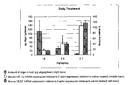
Figure 6A and Figure 6B show in vivo endogenous liver target down-regulation
of two
administration regimens using SEQ ID NO. 1. Measuring mRNA levels of HIF-1a as
well as the
downstream target VEGF shows that SEQ ID NO. 1 is also an effective inhibitor
of said target
Figure 6A: ip injections daily in hairy mice for 14 days. Figure 6B: ip
injections twice weekly
in hairy mice for 14 days.
Figure 6C shows in vivo endogenous kidney HIF-la after down-regulation
administered ip
injections daily in hairy mice for 14 days regimens of SEQ ID NO. 1.
Figure 7A shows that SEQ ID NO. 1 is a potent inhibitor measured by down-
regulation of in
vivo expression of HIF-la in liver following administration of SEQ ID NO. 1.
Different
thiolated versions of SEQ ID NO. 1 (SEQ ID NO. 5 and SEQ ID NO. 6) and SEQ ID
NO. 1
respectively were dosed to hairy mice at 18 or 3.6 mg/kg daily for 14 days and
sacrificed.
Expression of HIF-la was measured at mRNA level by QPCR and normalised to beta-
actin as
described in M&M.
Figure 7B shows that SEQ ID NO. 1 is also a potent inhibitor measured by down-
regulation of
in vivo expression of HIF-la in liver following administration of SEQ ID NO.
1. Different
CA 02587173 2007-05-08
WO 2006/050734
PCT/DK2005/000721
thiolated versions of SEQ ID NO. 1 (SEQ ID NO. 5 and SEQ ID NO. 6) and SEQ ID
NO. 1
respectively were dosed to hairy mice at 50, 10 or 2 mg/kg twice a week for 14
days and
sacrificed. Expression of HIF-la was measured at mRNA level by QPCR and
normalised to
beta-actin.
5 Figure 7C shows down-regulation of in vivo expression of HIF-la in kidney
following
administration of SEQ ID NO. 1. Different thiolated versions of SEQ ID NO. 1
(SEQ ID NO. 5
and SEQ ID NO. 6) were dosed to hairy mice at 18 or 3.6 mg/kg daily for 14
days and
sacrificed. Expression of HIF-la was measured at mRNA level by QPCR and
normalised to
beta-actin.
Figure 8A shows superior in vivo efficacy using SEQ ID NO. 1 compared to SEQ
ID NO. 11
and SEQ ID NO. 12 (a scrambled control) measured by tumor-weight of U373
tumors from
xenograft. SEQ ID NO. 1, SEQ ID NO. 11 and SEQ ID NO. 12 were dosed at 50
mg/kg twice a
week for one week in U373 xenograft mice implanted at the ovaries. 2 days
following the last
dose animals was sacrificed. At sacrifice tumors were weighed and the
individual tumor
weight plus the mean tumor weight (red) was calculated and plotted. A
statistical significant
difference (P=0.005) was found between the Control group (a scrambled control
SEQ ID NO.
12) and the mice treated with a SEQ ID NO. 1.
Figure 8B shows vessel density in U373 tumors from xenograft treated with SEQ
ID NO. 1.
SEQ ID NO. 1 was dosed at 50 mg/kg twice a week for one week in U373 xenograft
mice
implanted at the ovaries. 2 days following the last dose, animals was
sacrificed. Vessel-
density was calculated following CD31 staining and related to the total area.
A statistical
significant difference (P=0.005) was found between the saline group and the
mice treated
with a scrambled control (SEQ ID NO. 12).
Figure 8C shows staining of CD 31 in sections from U373 tumors implanted at
the ovaries
and treated with SEQ ID NO. 1 as described for Figure 8B.
Figure 8D shows HIF-la expression quantified by real-time PCR and normalised
to GAPDH in
U373 tumors implanted at the ovaries and treated with SEQ ID NO. 1, SEQ ID NO.
11, SEQ
ID NO. 12 and PBS as described for Figure 8B.
Figure 9A shows in vivo uptake (in pg per gram tissue) plus target down-
regulation (%
inhibition of HIF-la mRNA expression correlated to (3-actin expression) of
hairy mice following
one i.v. dose of SEQ ID NO. 1 of 25mg/kg. SEQ ID NO. 1 has a half-life of
approximately 46
hours in kidney and 66 hours in the liver. Figure 9B upper panel shows SEQ ID
NO. 1 dosed
at 50 mg/kg once i.p. in hairy mice. Five animals treated with SEQ ID NO. 1 at
50 mg/kg
CA 02587173 2007-05-08
WO 2006/050734
PCT/DK2005/000721
6
were sacrificed following 1, 3, 4, 5 and 8 days after treatment and HIF-la
expression was
analysed and normalised to Beta-actin. Expression of HIF-la was measured at
mRNA level by
QPCR and normalised to beta-actin as described in example 8. In the lower
panel SEQ ID NO.
1 was dosed at 25 or 50 mg/kg once i.v. in hairy mice. Five animals treated
with SEQ ID NO.
1 at 25 or 50 mg/kg were sacrificed following 1, 2, 3, 4, 5 and 8 days after
treatment and
were analysed for full length SEQ ID NO. 1 by HPLC methods as described in
example13.
Data are presented as pg SEQ ID NO. 1/gram tissue.
Figure 9C shows HIF-la expression quantified by real-time PCR and normalised
to GAPDH in
mouse liver in mice receiving one dose of 50 mg/kg i.p. of SEQ ID NO. 1 and
SEQ ID NO. 16
and sacrificed at day 1 and 10.
Figure 10A shows duration of action of SEQ ID NO. 1 inhibiting HIF-la
expression in
xenograft mice dosed 25 mg/kg for 7 days and sacrificed 1 or 5 days after the
last dose.
Figure 10B shows in vivo liver, skin tumor and kidney uptake of fam-labeled
version of SEQ
ID NO. 1 (SEQ ID NO. 7) at 25 mg/kg/day for seven days and sacrificed 5 days
following the
last treatment.
Figure 10C shows target down-regulation (0/0 inhibition of HIF-la mRNA
expression correlated
to GAPDH expression) plus in vivo uptake (in pg per gram tissue) of SEQ ID NO.
7 in the liver
of xenograft mice treated with 5 mg/kg/day SEQ ID NO.7, scrambled control SEQ
ID NO. 20
or saline i.p. on days 7, 10, 13 and 17 after transplantation as described in
example 17.
Figure 10D shows target down-regulation (0/0 inhibition of HIF-la mRNA
expression
correlated top-actin expression) after treatment with SEQ ID NO. 7 or
scrambled control SEQ
ID NO. 20 plus in vivo uptake (in pg per gram tissue) of SEQ ID NO. 7 in mouse
colon treated
as described in example 17.
Figure 10E shows in vivo uptake (in pg per gram tissue) of SEQ ID NO. 7 in
xenograft tumors
HT29 and PC3 treated as described in example 17.
Figure 11 shows in vivo endogenous liver target down-regulation of HIF-la and
VEGF mRNA
after 5 doses of 30 mg/kg every 3rd day of SEQ ID NO. 1 compared to the one
mismatch
control SEQ ID NO. 9.
Figure 12A, Figure 12B, Figure 12C and Figure 12D shows expression of VEGFA
and MMP-2
following treatment with the HIF-la targeting LNA oligonucleotide, SEQ ID NO.
1, and a
scrambled control SEQ ID NO. 8 in U373 cells. A dose -dependent down-
regulation in VEGFA
CA 02587173 2016-06-06
/9851-86
7
and MMP-2 expression (secretion) is observed 48 hours following treatment with
SEQ ID NO.
1 or a scrambled control (SEQ ID NO. 8) in U373 cells. The VEGFA (Figures 12A,
12B and
12C) and MMP-2 (Figures 12D and 12E) expression is related to cell number and
normalized
to mock. In Figures 12A, 12C and 12D VEGFA and MMP-2 expression is measured 48
hours
following treatment, whereas in Figures 12 B and 12E secretion of VEGFA and
MMP-2 is
quantified 24-120 hours following tranfection.
Figure 13 shows down-regulation of HIF-la protein measured by western blot and
disruption
of tube formation of HUVEC cells treated with SEQ ID NO. 1 at 1 and 5 nM
compared to SEQ
ID NO. 8 and untreated control.
Figure 14A Whole body radioluminograms showing the distribution of
radioactivity at 5
minutes a), 4 hours b), 24 hours c) and 18 days d) after a single intravenous
administration
of 3H- labelled SEQ ID NO. 1 in female pigmented mice.
Figure 14B shows the distribution of radioactivity at 5 minutes and 7 days and
that a very
strong retention of the 3H- labelled SEQ ID NO. 1 compound is observed in bone
marrow,
kidney, liver, lung, skin, spleen, urine, gastric mucosa, lymph node, uvea of
the eye and
uterus after 7 days.
Figure 15 shows uptake of a FAM-labelled version of SEQ ID NO. 1 (SEQ ID NO.
7) in
different cell types within bone marrow, spleen and peripheral blood 1 hour
following
administration of SEQ ID NO. 7 compared to untreated cells measured by FACS
analysis.
Figure 16A shows HIF-la expression measured by real-time PCR and normalised to
18S RNA
in the liver and kidney of cynomolgus monkeys treated with 40, 10 and 6mg/kg
SEQ ID NO.
1 twice a week for 4 weeks. Figure 16B shows uptake of SEQ ID NO. 1 in liver
and kidney of
cynomolgus monkeys one day following the last dose or 4 weeks following the
last dose
(recovery animals) treated as described above together with data on recovery
animals (R),
which were left untreated for 4 weeks after end of treatment.
DESCRIPTION OF THE INVENTION
The present invention employs particular LNA oligonucleotides, namely LNA
oligonucleotides
comprising the sequence SEQ ID NO. 3 and SEQ ID NO. 4, for use in inhibiting
the function of
nucleic acid molecules encoding HIF-1 a. In one embodiment, this is
accomplished by
providing antisense LNA oligonucleotides, which specifically hybridise with
nucleic acids
CA 02587173 2016-06-06
p9851-86
8
encoding HIF-la. The inhibition of the expression of HIF-la leads to a
decrease in the number
of functional HIF-1a proteins produced.
The LNA oligonucleotides
More particular, the present invention provides an LNA oligonucleotide
consisting of a
sequence selected from the group consisting of
5'-(10G,Gõcosasgscsast,cscsTõGõ(11-3' (SEQ ID NO. 3)
and
5'-( ,0TõToscstsgscscststscsT,Tx(A1-3' (SEQ ID NO. 4)
wherein capital letters designate a beta-D-oxy-LNA nucleotide analogue, small
letters
designate a 2-deoxynucleotide, underline designates either a beta-D-oxy-LNA
nucleotide
analogue or -a 2-deoxynucleotide, subscript "s" designates a phosphorothioate
link between
neighbouring nucleotides/LNA nucleotide analogues, and subscript "x"
designates either a
phosphorothioate link or a phosphorodiester link between neighbouring
nucleotides/LNA
nucleotide analogues, and where the nucleotide units in the bracket, (L), (T),
or (0õ),(A),
respectively, represent optional units, and
wherein the sequence is optionally extended by up to five 2-deoxynucleotide
units.
The terms "LNA oligonucleotide defined herein", "LNA oligonucleotide according
to the
invention", and the like, refer to the "LNA oligonucleotide" defined above as
well as the
embodiments, variants, salts, prodrugs, etc. provided in the following.
The above-defined LNA oligonucleotides based on SEQ ID NO. 3 and SEQ ID NO. 4
have a
length of 13-20 nucleotide units. The minimal sequence length of 13 is
obtained if the
nucleotide units In the bracket, (10, (T) or (), (A), respectively, are
absent, and the
maximum sequence length of 20 is obtained if the nucleotide units in the
bracket, (1)1 (T) or
(g.,), (A), respectively, are present and if the sequence SEQ ID NO. 3 or SEQ
ID NO. 4 is
extended by five 2-deoxynucleotide units.
In one embodiment, the nucleotide units in the bracket, (1)1 (T) or ( _x),
(A), respectively,
are absent, and in another currently more preferred embodiment, the nucleotide
unit in the
bracket, (1)1 (T) or ( ,,), (A), respectively, are present. Also interesting
are the
CA 02587173 2007-05-08
WO 2006/050734
PCT/DK2005/000721
9
embodiments, where the 5'-terminal optional unit, (Tx) or (a,), respectively,
is present and
where the 3'-terminal optional unit, (T) or (A), respectively, is absent, and
the embodiments
where the 5'-terminal optional unit, (Tx) or (0_), respectively, is absent and
where the 3'
terminal optional unit, (T) or (A), respectively, is present.
The selection of a beta-D-oxy-LNA nucleotide analogue or a 2-deoxynucleotide
for the
underlined nucleotide units in the above SEQ ID NO. 3 and SEQ ID NO. 4 appears
to be less
critical. However, in one embodiment, both of the underlined nucleotide units
designate a 2-
deoxynucleotide. In another currently more preferred embodiment, one or both
of the
underlined nucleotide units designate a beta-D-oxy-LNA nucleotide analogue.
In one variant, the 5'-terminal nucleotide unit in the bracket, (Tx) or (g,),
respectively, is
absent, and the 3'-terminal other underlined nucleotide unit, (T) or (A),
respectively,
designates a 2-deoxynucleotide, or more preferable, a beta-D-oxy-LNA
nucleotide analogue.
In another variant, the 5'-terminal nucleotide unit in the bracket, (Ix) or
(c_x), respectively,
designate a 2-deoxynucleotide, or, more preferable, a beta-D-oxy-LNA
nucleotide analogue,
and the 3'-terminal other underlined nucleotide unit, (T) or (A),
respectively, is absent.
In another variant, the nucleotide units in the bracket are present, and one
or both of the
underlined nucleotide units designate a beta-D-oxy-LNA nucleotide analogue,
i.e. (i) the 5'-
terminal underlined nucleotide designates a beta-D-oxy-LNA nucleotide analogue
and the 3'-
terminal underlined nucleotide units designates a 2-deoxynucleotide, or (ii)
the 3'-terminal
underlined nucleotide designates a beta-D-oxy-LNA nucleotide analogue and the
5'-terminal
underlined nucleotide units designates a 2-deoxynucleotide, or (iii) the 3'-
terminal as well as
the 5'-terminal underlined nucleotides designate a beta-D-oxy-LNA nucleotide
analogue.
In a further variant, the nucleotide units in the bracket, (Tx) or (a,),
respectively, is present,
and both of the underlined nucleotide units designate a 2-deoxynucleotide.
Although the sequences referred to as SEQ ID NO. 3 and SEQ ID NO. 4 (and more
particular
the sequences referred to as SEQ ID NO. 1 and SEQ ID NO. 2 (see further
below)) are
believed to substantially represent the full functionality of the defined LNA
oligonucleotides,
extension of SEQ ID NO. 3 and SEQ ID NO. 4 with up to five 2-deoxynucleotide
units, e.g. 1
unit, 2 units, 3 units, 4 units, or even 5 units, is believed to be possible
without detrimental
effects on the beneficial properties of the base sequences, SEQ ID NO. 3 and
SEQ ID NO. 4.
This being said, the sequence may be extended at the 3'-terminal end, the 5'-
terminal end or
at the 3'-terminal end as well as at the 5'-terminal end, provided that the
total number of 2-
deoxynucleotide units does not exceed 5.
CA 02587173 2007-05-08
WO 2006/050734
PCT/DK2005/000721
Hence, in one embodiment (which may be combined with the foregoing) the LNA
oligonucleotide consists of 15, 16, 17, 18, 19 or 20 nucleotide units selected
from 2-
deoxynucleotides and beta-D-oxy-LNA nucleotide analogues, in particular the
LNA
oligonucleotide consists of 16 nucleotide units selected from 2-
deoxynucleotides and beta-D-
5 oxy-LNA nucleotide analogues. In other embodiments (which may be combined
with the
foregoing) the LNA oligonucleotide consists of 13, 14, 15, or 16 nucleotide
units selected
from 2-deoxynucleotides and beta-D-oxy-LNA nucleotide analogues, in particular
the LNA
oligonucleotide consists of 14 or 15 nucleotide units selected from 2-
deoxynucleotides and
beta-D-oxy-LNA nucleotide analogues.
10 At least for the sake of convenience in the preparation of the LNA
oligonucleotides, it is often
preferred that the sequence is extended by one 2-deoxynucleotide unit at the
3'-end, cf.,
e.g., SEQ ID NO. 1 and SEQ ID NO. 2 below. Most preferable, SEQ ID NO. 3 is
extended by
an adenosine 2-deoxynucleotide unit at the 3'-end, and SEQ ID NO. 4 is
extended by a
cytosine 2-deoxynucleotide at the 3'-end.
As mentioned above, subscript "s" designates a phosphorothioate (-0-P(0,S)-0-)
link
between neighbouring nucleotides/LNA nucleotide analogues, and subscript "x"
designates
either a phosphorothioate (-0-P(0,S)-0-) link or a phosphorodiester (-0-P(0)2-
0-) link
between neighbouring nucleotides/LNA nucleotide analogues. It follows that any
2-
deoxynucletides by which the sequence is extended may be linked by either
either a
phosphorothioate (-0-P(0,S)-0-) link or a phosphorodiester (-0-P(0)2-0-) link.
It is noted that subsequence csasasgscsastscscsT of SEQ ID NO. 3 and
subsequence
ascstsgscscststscsT of SEQ ID NO. 4 are indicated as fully phosphorothiolated,
cf. subscript "s".
Although is it is not currently preferred, it is believed that one, and
possibly also two, of the
phosphorothioate links may be replaced by other links, in particular
phosphorodiester links,
without severely compromising the stability of the LNA oligonucleotide. Thus,
such variants
where one or two of the phosphorothioate links are replaced by, e.g.,
phosphorodiester links
also fall within the intended scope of the present invention.
In one currently preferred embodiment, however, all nucleotide units in the
sequence are
linked by a phosphorothioate group.
One subgroup of particularly interesting LNA oligonucleotides are those
selected from the
group consisting of
5'-T5G5G5c5a5a5g5c5a5t5c5csTsG5Tsa-3' (SEQ ID NO. 1),
CA 02587173 2007-05-08
WO 2006/050734
PCT/DK2005/000721
11
51-TsGsGscsasasgscsastscscsTsGsT-3' (SEQ ID NO. 15), and
5'-GsGscsasasgscsastscscsTsGst-3' (SEQ ID NO. 16).
Among those,
5'-TsGsGscsasasgscsastscscsT,GsTsa-3' (SEQ ID NO. 1)
is currently most preferred.
Another subgroup of particularly interesting LNA oligonucleotides are those
selected from the
group consisting of
5'-GsTsTsascstsgscscststscsTsTsAsc-3' (SEQ ID NO. 2),
5'-GsTsTsascstsgscscststscsTsTsA-3' (SEQ ID NO. 17), and
5'-TsTsascstsgscscststscsTsTsa-3' (SEQ ID NO. 18).
Among those
5'-GsTsTsascstsgscscststscsTsTsAsc-3' (SEQ ID NO. 2)
is currently most preferred.
In the present context, the term "nucleoside" is used in its normal meaning,
i.e. it contains a
2-deoxyribose or ribose unit which is bonded through its number one carbon
atom to one of
the nitrogenous bases adenine (A), cytosine (C), thymine (T), uracil (U) or
guanine (G).
In a similar way, the term "nucleotide" means a 2-deoxyribose or ribose unit
which is bonded
through its number one carbon atom to one of the nitrogenous bases adenine
(A), cytosine
(C), thymine (T), uracil (U) or guanine (G), and which is bonded through its
number five
carbon atom to an internucleoside phosphate group, or to a terminal group.
The term "nucleic acid" is defined as a molecule formed by covalent linkage of
two or more
nucleotides. The terms "nucleic acid" and "polynucleotide" are used
interchangeable herein.
The term "nucleic acid analogue" refers to a non-natural nucleic acid binding
compound.
CA 02587173 2016-06-06
i9851-86
12
The term "LNA monomer" typically refers to a bicyclic nucleoside analogue, as
described In
International Patent Application WO 99/14226 and subsequent applications, WO
00/56746,
WO 00/56748, WO 00/66604, WO 00/125248, WO 02/28875, WO 2002/094250 and WO
=
03/006475.
Beta-D-oxy-LNA Is the LNA nucleotide analogue use in the LNA oligonucleotides
of the
present invention, and the monomer structure (nucleoside) is shown in Scheme
1.
z.
0
Beta-D-oxy-LNA
Scheme 1
In Scheme 1, Z* and Z Indicate the position of a internucleotide linkage to a
neighbouring
nucleoside or a terminal group (I.e. either a 5'-terminal group or a 3'-
terminal group).
One particular example of beta-D-oxy-LNA monomer is the thymidine LNA monomer
(LNA
nucleoside analogue) (15,3R, 4R, 75)-7-hydroxy-1-hydroxymethy1-5-methyl-3-
(thymIn-ly1)-
2,5-dioxa-blcyclo[2:2:1Theptane, i.e. T-beta-D-oxy-LNA.
The term "oligonucleotide" refers, In the context of the present invention, to
an oligomer
(also called oligo) or nucleic acid polymer (e.g. ribonucleic acid (RNA) or
deoxyribonucleic
acid (DNA)) or nucleic acid analogue of those known in the art, preferably
Locked Nucleic
Acid (LNA), or a mixture thereof. This term Includes ollgonucleotides composed
of naturally
occurring nucieobases, sugars and internucleoside (backbone) linkages as well
as
oligonucleotides having non-naturally-occurring portions which function
similarly or with
specific improved functions. =
CA 02587173 2016-06-06
9851-86
12a
As used herein, the terms "target nucleic acid" encompass DNA encoding the HIF-
la,
RNA (including pre-mRNA and mRNA) transcribed from such DNA, and also cDNA
derived from such RNA.
As used herein, the term "gene" means the gene including exons, introns, non-
coding
5' and 3' regions and regulatory elements and all currently known variants
thereof
and any further variants, which may be elucidated.
As used herein, the term "LNA oligonucleotide" refers to an oligonucleotide
binding
by hydrogen bonding to either a target gene "Chimeraplast" and "TFO", to the
RNA
transcript(s) of the target gene "antisense inhibitors", "siRNA", "miRNA",
"ribozymes"
and "oligozymes" or to the protein(s) encoding by the target gene "aptamer",
"spiegelmer" or "decoy".
As used herein, the term "mRNA" means the presently known mRNA transcript(s)
of
a targeted gene, and any further transcripts, which may be identified.
As used herein, the term "targeting" an antisense compound to a particular
target
nucleic acid means providing the antisense oligonucleotide to the cell, animal
or
human in such a way that the antisense compound are able to bind to and
modulate
the function of its intended target.
The LNA oligonucleotides may be designed as siRNA's which are small double
stranded RNA molecules that are used by cells to silence specific endogenous
or
exogenous genes by an as yet poorly understood "antisense-like" mechanism.
CA 02587173 2007-05-08
WO 2006/050734 PCT/DK2005/000721
13
By the terms "unit" and "nucleotide unit" is understood a monomer, i.e. a 2-
deoxynucleotide
or a beta-D-oxy-LNA nucleotide analogue.
The term "at least one" comprises the integers larger than or equal to 1, such
as 1, 2, 3, 4,
5, 6, 7, 8, 9, 10, 11, 12, 13, 14, 15, 16, 17 and so forth.
The term "a" as used about a nucleoside, a nucleoside analogue, a SEQ ID NO,
etc. is
intended to mean one or more. In particular, the expression "a component (such
as a
nucleoside, a nucleoside analogue, a SEQ ID NO or the like) selected from the
group
consisting of ..." is intended to mean that one or more of the cited
components may be
selected. Thus, expressions like "a component selected from the group
consisting of A, B and
C" is intended to include all combinations of A, B and C, i.e. A, B, C, A+B,
A+C, B+C and
A+B+C.
Throughout this specification, the word "comprise", or. variations such as
"comprises" or
"comprising", will be understood to imply the inclusion of a stated element,
integer or step,
or group of elements, integers or steps, but not the exclusion of any other
element, integer
or step, or group of elements, integers or steps.
Preparation of the LNA oligonucleotides
The LNA nucleotide analogue building blocks (p-D-oxy-LNA) can be prepared
following
published procedures and references cited therein, see, e.g., WO 03/095467 Al;
D. S.
Pedersen, C. Rosenbohm, T. Koch (2002) Preparation of LNA Phosphoramidites,
Synthesis 6,
802-808; and WO 2004/069991 A2.
The LNA oligonucleotides can be prepared as described in the Examples and in
WO 99/14226,
WO 00/56746, WO 00/56748, WO 00/66604, WO 00/125248, WO 02/28875, WO
2002/094250 and WO 03/006475. Thus, the LNA oligonucleotides may be produced
using the
oligomerisation techniques of nucleic acid chemistry well-known to a person of
ordinary skill
in the art of organic chemistry. Generally, standard oligomerisation cycles of
the
phosphoramidite approach (S. L. Beaucage and R. P. Iyer, Tetrahedron, 1993,
49, 6123; S.
L. Beaucage and R. P. Iyer, Tetrahedron, 1992, 48, 2223) are used, but e.g. H-
phosphonate
chemistry, phosphotriester chemistry can also be used.
For some monomers, longer coupling time, and/or repeated couplings and/or use
of more
concentrated coupling reagents may be necessary or beneficial.
CA 02587173 2007-05-08
WO 2006/050734
PCT/DK2005/000721
14
The phosphoramidites employed couple typically with satisfactory >95% step-
wise yields.
Oxidation of the phosphorous(III) to phosphorous(V) is normally done with e.g.
iodine/pyridine/H20. This yields after deprotection the native
phosphorodiester
internucleoside linkage. In the case that a phosphorothioate internucleoside
linkage is
prepared a thiolation step is performed by exchanging the normal, e.g.
iodine/pyridine/H20,
oxidation used for synthesis of phosphorodiester internucleoside linkages with
an oxidation
using the ADTT reagent (xanthane hydride (0.01 M in acetonitrile:pyridine 9:1;
v/v)). Other
thiolation reagents are also possible to use, such as Beaucage and PADS. The
phosphorothioate LNA oligonucleotides were efficiently synthesized with
stepwise coupling
yields >= 98%.
Purification of LNA oligonucleotides can be accomplished using disposable
reversed phase
purification cartridges and/or reversed phase HPLC and/or precipitation from
ethanol or
butanol. Capillary gel electrophoresis, reversed phase HPLC, MALDI-MS, and ESI-
MS were
used to verify the purity of the synthesized LNA oligonucleotides.
Salts
The LNA oligonucleotide can be employed in a variety of pharmaceutically
acceptable salts.
As used herein, the term refers to salts that retain the desired biological
activity of the LNA
oligonucleotide and exhibit minimal undesired toxicological effects. Non-
limiting examples of
such salts can be formed with organic amino acid and base addition salts
formed with metal
cations such as zinc, calcium, bismuth, barium, magnesium, aluminum, copper,
cobalt,
nickel, cadmium, sodium, potassium, and the like, or with a cation formed from
ammonia,
N,N-dibenzylethylene-diamine, D-glucosamine, tetraethylammonium, or
ethylenediannine; or
combinations, e.g., a zinc tannate salt or the like.
Such salts are formed, from the LNA oligonucleotide which possess
phosphorodiester group
and/or phosphorothioate groups, and are, for example, salts with suitable
bases. These salts
include, for example, nontoxic metal salts which are derived from metals of
groups Ia, Ib, ha
and IIb of the Periodic System of the elements, in particular suitable alkali
metal salts, for
example lithium, sodium or potassium salts, or alkaline earth metal salts, for
example
magnesium or calcium salts. They furthermore include zinc and ammonium salts
and also
salts which are formed with suitable organic amines, such as unsubstituted or
hydroxyl-
substituted mono-, di- or tri-alkylamines, in particular mono-, di- or tri-
alkylamines, or with
quaternary ammonium compounds, for example with N-methyl-N-ethylamine,
diethylamine,
triethylamine, mono-, bis- or tris-(2-hydroxy-lower alkyl)amines, such as mono-
, bis- or tris-
(2-hydroxyethyl)amine, 2-hydroxy-tert-butylamine or
tris(hydroxymethyl)methylamine, N,N-
CA 02587173 2016-06-06
/9851-86
di-lower alkyl-N-(hydroxy-lower alkyl)amines, such as N,N-dimethyl-N-(2-
hydroxyethyl)-
amine or tri-(2-hydroxyethyl)amine, or N-methyl-D-glucamine, or quaternary
aminonium
compounds such as tetrabutylammonium salts. Lithium salts, sodium salts,
magnesium salts,
zinc salts or potassium salts are preferred, with sodium salts being
particularly preferred.
5 Prodrugs
In one embodiment, the LNA oligonucleotide may be in the form of a pro-drug.
Oligonucleotides are by virtue negatively charged ions. Due to the lipophilic
nature of cell
membranes, the cellular uptake of oligonucleotides is reduced compared to
neutral or
lipophilic equivalents. This polarity "hindrance" can be avoided by using the
pro-drug
10 approach (see e.g. Crooke, R. M. (1998) in Crooke, S. T. Antisense
research and Application.
Springer-Verlag, Berlin, Germany, vol. 131, pp. 103-140). In this approach,
the LNA
oligonucleotides are prepared in a protected manner so that the LNA
oligonucleotides are
neutral when it is administered. These protection groups are designed in such
a way that
they can be removed then the LNA oligonucleotide is taken up be the cells.
Examples of such
15 protection groups are S-acetylthioethyl (SATE) or S-pivaloyithioethyl (t-
butyl-SATE). These
protection groups are nuclease resistant and are selectively removed
intracellulary.
Conjugates
A further aspect of the invention relates to a conjugate comprising an LNA
oligonucleotide as
defined herein and at least one non-nucleotide or non-polynucleotide moiety
covalently
attached to said LNA oligonucleotide.
In a related aspect of the invention, the LNA oligonucleotide of the invention
is linked to
ligands so as to form a conjugate, said ligands intended to increase the
cellular uptake of the
conjugate relative to the antisense oligonucleotides.
In the present context, the term "conjugate" is intended to indicate a
heterogenous molecule
formed by the covalent attachment of an LNA oligonucleotide as described
herein (i.e. an LNA
oligonucleotide comprising a sequence of nucleosides and LNA nucleoside
analogues) to one
or more non-nucleotide or non-polynucleotide moieties.
Thus, the LNA oligonucleotides may, e.g., be conjugated or form chimera with
non-nucleotide
or non-polynucleotide moieties including Peptide Nucleic Acids (PNA), proteins
(e.g.
antibodies for a target protein), macromolecules, low molecular weight drug
substances, fatty
acid chains, sugar residues, glycoproteins, polymers (e.g. polyethylene
glycol), micelle-
CA 02587173 2016-06-06
/9851-86
16
forming groups, antibodies, carbohydrates, receptor-binding groups, steroids
such as
cholesterol, polypeptides, intercalating agents such as an acridine
derivative, a long-chain
alcohol, a dendrimer, a phospholipld and other lipophilic groups or
combinations thereof, etc.,
just as the LNA oligonucleotides may be arranged in dimeric or dendritic
structures. The LNA
oligonucleotides or conjugates of the invention may also be conjugated or
further conjugated
to active drug substances, for example, aspirin, ibuprofen, a sulfa drug, an
antidiabetic, an
antibacterial agent, a chemotherapeutic agent or an antibiotic.
Conjugating in this way may confer advantageous properties with regard to the
pharmacokinetic characteristics of the LNA oligonucleotides. In particular,
conjugating in this
way achieves increased cellular uptake.
In one embodiment, an LNA oligonucleotide is linked to ligands so as to form a
conjugate,
said ligands intended to increase the cellular uptake of the conjugate
relative to the antisense
LNA oligonucleotides. This conjugation can take place at the terminal
positions 573'-OH but
the ligands may also take place at the sugars and/or the bases. In particular,
the growth
factor to which the antisense LNA oligonucleotide may be conjugated, may
comprise
transferrin or folate. Transferrin-polylysine-oligonucleotide complexes or
folate-polylysine-
oligonucleotide complexes may be prepared for uptake by cells expressing high
levels of
transferrin or folate receptor. Other examples of conjugates/ligands are
cholesterol moieties,
duplex intercalators such as acridine, poly-L-lysine, "end-capping" with one
or more
nuclease-resistant linkage groups such as phosphoromonothioate, and the like.
The preparation of transferrin complexes as carriers of oligonucleotide uptake
into cells is
described by Wagner et al., Proc. Natl. Acad. Sci. USA 87, 3410-3414 (1990).
Cellular
delivery of folate-macromolecule conjugates via folate receptor endocytosis,
including
delivery of an antisense oligonucleotide, is described by Low et al., U.S.
Patent 5,108,921.
Also see, Leamon et al., Proc. Natl. Acad. Sci. 88, 5572 (1991).
Pharmaceutical composition
A particularly interesting aspect of the invention Is directed to a
pharmaceutical composition
comprising an LNA oligonucleotide as defined herein or a conjugate as defined
herein, and a
pharmaceutically acceptable diluent, carrier or adjuvant.
CA 02587173 2016-06-06
7'9851-86
17
Directions for the preparation of pharmaceutical compositions can be found in
"Remington: The Science and Practice of Pharmacy" by Alfonso R. Gennaro, and
in
the following.
Pharmaceutically acceptable diluents, carriers or adjuvants are part of the
pharmaceutical composition. Capsules, tablets and pills etc. may contain for
example
the following compounds: microcrystalline cellulose, gum or gelatin as
binders; starch
or lactose as excipients; stearates as lubricants; various sweetening or
flavouring
agents. For capsules the dosage unit may contain a liquid carrier like fatty
oils.
Likewise coatings of sugar or enteric agents may be part of the dosage unit.
The
pharmaceutical composition may also be emulsions of the active pharmaceutical
ingredients (including the LNA oligonucleotide) and a lipid forming a
micellular
emulsion.
An LNA oligonucleotide may be mixed with any material that do not impair the
desired action, or with material that supplement the desired action. These
could
include other drugs including other oligonucleoside compounds.
Pharmaceutical compositions of the present invention include, but are not
limited to,
solutions, emulsions, and liposome-containing formulations. These compositions
may
be generated from a variety of components that include, but are not limited
to,
preformed liquids, self-emulsifying solids and self-emulsifying semisolids.
In one embodiment, the pharmaceutical compositions comprise an LNA
oligonucleotide of the invention (e.g., 0.1 to 90% by weight), or a
physiologically
acceptable salt thereof, mixed with a physiologically acceptable carrier
medium.
Preferred physiologically acceptable carrier media are water, buffered water,
normal
saline, 0.4% saline, 0.3% glycine, hyaluronic acid and the like.
Pharmaceutical compositions of the invention can also comprise conventional
pharmaceutical excipients and/or additives. Suitable pharmaceutical excipients
include stabilizers, antioxidants, osmolality adjusting agents, buffers, and
pH
CA 02587173 2016-06-06
' /9851-86
18
adjusting agents. Suitable additives include physiologically biocompatible
buffers
(e.g., tromethamine hydrochloride), additions of chelants (such as, for
example,
DTPA or DTPA-bisamide) or calcium chelate complexes (as for example calcium
DTPA, CaNaDTPA-bisamide), or, optionally, additions of calcium or sodium salts
(for
example, calcium chloride, calcium ascorbate, calcium gluconate or calcium
lactate).
Pharmaceutical compositions of the invention can be packaged for use in liquid
form,
or can be lyophilized.
For solid compositions, conventional non-toxic solid carriers can be used; for
example, pharmaceutical grades of mannitol, lactose, starch, magnesium
stearate,
sodium saccharin, talcum, cellulose, glucose, sucrose, magnesium carbonate,
and
the like.
The pharmaceutical formulations of the present invention, which may
conveniently be
presented in unit dosage form, may be prepared according to conventional
techniques well known in the pharmaceutical industry. Such techniques include
the
step of bringing into association the active ingredients with the
pharmaceutical
carrier(s) or excipient(s). In general the formulations are prepared by
uniformly and
intimately bringing into association the active ingredients with liquid
carriers or finely
divided solid carriers or both, and then, if necessary, shaping the product.
The compositions of the present invention may be formulated into any of many
possible dosage forms such as, but not limited to, tablets, capsules, gel
capsules,
liquid syrups, soft gels and suppositories. The compositions of the present
invention
may also be formulated as suspensions in aqueous, non-aqueous or mixed media.
Aqueous suspensions may further contain substances which increase the
viscosity of
the suspension including, for example, sodium carboxymethylcellulose, sorbitol
and/or dextran. The suspension may also contain stabilizers.
In preferred embodiments of the pharmaceutical compositions, the LNA
oligonucleotide is formulated in an aqueous carrier, in particular an aqueous
carrier
CA 02587173 2016-06-06
i9851-86
19
comprising a buffer for keeping the pH in the range of 4.0-8.5, and having an
ionic
strength of 20-2000 mM.
The term "aqueous carrier means that the pharmaceutical composition in
question is
in liquid form, and that the liquid carrier predominantly is composed of
water, i.e. that
at least 80% (w/w), or at least 90% (w/w), or even at least 95% (w/w), of the
carrier
consists of water. Other liquid ingredients may also be used, e.g. ethanol,
DMSO,
ethylene glycol, etc.
The aqueous carrier preferably comprises saline or a buffer for keeping the pH
in the
range of 4.0-8.5. Preferably, the buffer will keep the pH in the range of 5.0-
8.0, such
as in the range of 6.0-7.5, such as buffered saline, e.g. phosphate buffered
saline
(PBS).
The ionic strength/tonicity of the pharmaceutical composition is also of
importance.
Thus, typically, the liquid pharmaceutical composition has an ionic strength
of in the
range of 20-2000 mM, such as in the range of 50-1500 mM, or in the range of
100-
1000 mM.
Kits
If the pharmaceutical composition in liquid form is under risk of being
subjected to
conditions which will compromise the stability of the LNA oligonucleotide, it
may be
preferred to produce the finished product containing the LNA oligonucleotide
in a
solid form, e.g. as a freeze dried material, and store the product is such
solid form.
The product may then be reconstituted (e.g. dissolved or suspended) in a
saline or in
a buffered saline ready for use prior to administration.
Hence, the present invention also provides a kit comprising
CA 02587173 2016-06-06
/9851-86
(a) a first component containing an LNA oligonucleotide or a conjugate as
defined
herelnabove in solid form, and
(b) a second component containing saline or a buffer solution (e.g. buffered
saline) adapted
for reconstitution (e.g. dissolution or suspension) of said LNA
oligonucleotide.
5 Preferably said saline or buffered saline has a pH in the range of 4.0-
8.5, and a molarity of
20-2000 mM. In a preferred embodiment the saline or buffered saline has a pH
of 6.0- 8.0
and a molarity of 100-500 mM. In a most preferred embodiment the saline or
buffered saline
has a pH of 7.0-8.0 and a molarity of 120-250mM
For such a kit, the LNA oligonucleotide is preferably selected from the group
consisting of
10 SEQ ID NO. 1, SEQ ID NO. 2, SEQ ID NO. 15, SEQ ID NO. 16, SEQ ID NO. 17,
and SEQ ID
NO. 18. More particular, the LNA oligonucleotide is selected from the group
consisting of SEQ
ID NO. 1 and SEQ ID NO. 2.
The invention is further illustrated in a non-limiting manner by the following
examples.
EXPERIMENTALS
15 Example 1: Monomer synthesis
The LNA monomer building blocks and derivatives thereof were prepared
following published
procedures and references cited therein, see, e.g. WO 03/095467 Al and D. S.
Pedersen, C.
Rosenbohm, T. Koch (2002) Preparation of LNA Phosphoramidites, Synthesis 6,
802-808.
Example 2: Oligonucleotide synthesis
20 Oligonucleotides were synthesized using the phosphoramidite approach on
an Expedite
8900/MOSS synthesizer (Multiple Oligonucleotide Synthesis System) at 1 pmol or
15 pmol
scale. For larger scale synthesis an Akta Oligo Pilot was used. At the end of
the synthesis
(DMT-on), the oligonucleotides were cleaved from the solid support using
aqueous ammonia
for 1-2 hours at room temperature, and further deprotected for 4 hours at 65
C. The
oligonucleotides were purified by reverse phase HPLC (RP-HPLC). After the
removal of the
DMT-group, the oligonucleotides were characterized by AE-HPLC, RP-HPLC, and
CGE and the
molecular mass was further confirmed by ESI-MS. See below for more details.
CA 02587173 2016-06-06
/9851-86
21
Preparation of the LNA-solid support:
Preparation of the LNA succlnyl hemiester
5'-0-Dmt-3'-hydroxy-LNA monomer (500 mg), succinic anhydride (1.2 eq.) and
DMAP (1.2
eq.) were dissolved in DCM (35 mL). The reaction was stirred at room
temperature overnight.
After extractions with NaH2PO4 0.1 M pH 5.5 (2x) and brine (1x), the organic
layer was
further dried with anhydrous Na2SO4 filtered and evaporated. The hemiester
derivative was
obtained In 95% yield and was used without any further purification.
Preparation of the LNA-support
The above prepared hemiester derivative (90 pmol) was dissolved in a minimum
amount of
DMF, DIEA and pyBOP (90 pmol) were added and mixed together for 1 min. This
pre-
activated mixture was combined with LCAA-CPG (500 A, 80-120 mesh size, 300 mg)
in a
manual synthesizer and stirred. After 1.5 hours at room temperature, the
support was
filtered off and washed with DMF, DCM and Me0H. After drying, the loading was
determined
to be 57 pmol/g (see Tom Brown, Dorcas J.S.Brown. Modern machine-aided methods
of
oligodeoxyribonucleotide synthesis. In: F.Eckstein, editor. Oligonucleotides
and Analogues A
Practical Approach. Oxford: IRL Press, 1991: 13-14).
Elongation of the oligonucleotide
The coupling of phosphoramidites (A(bz), G(Ibu), 5-methyl-C(bz)) or T-13-
cyanoethyl-
phosphoramidite) is performed by using a solution of 0.1 M of the 5'-0-DMT-
protected
amidite in acetonitrile and DCI (4,5-dicyanolmidazole) in acetonitrile (0.25
M) as activator.
The thiolation is carried out by using xanthane chloride (0.01 M in
acetonitrile:pyridine 10%).
The rest of the reagents are the ones typically used for oligonucleotide
synthesis. The
protocol provided by the supplier was conveniently optimised.
Purification by RP-HPLC:
Column: Xterra R1318
Flow rate: 3 mlimin
Buffers: 0.1 M ammonium acetate pH 8 and acetonitrile
Abbreviations
*Trade-mark
CA 02587173 2016-06-06
,
7'9851-86
22
DMT: Dimethoxytrityl
DCI: 4,5-Dicyanoimidazole
DMAP: 4-Dimethylaminopyridine
DCM: Dichloromethane
DMF: Dimethylformamide
THF: Tetra hydrofurane
DIEA: N,N-diisopropylethylamine
PyBOP: Benzotriazole-1-yl-oxy-tris-pyrrolidino-phosphonium
hexafluorophosphate
Bz: Benzoyl
Ibu: Isobutyryl
..
CA 02587173 2016-06-06
7'9851-86
23
Example 3: Design of the LNA oligonucleotide
Table 1 ¨ LNA olicionucleotides
SEQ ID NO. 1 5'-TsGsGscsasasgscsastscscsTsGsTsa-3'
SEQ ID NO. 2 5'-GsTsTsascstsgscscststscsTsTsAsc-3'
SEQ ID NO. 3 51-(L)GxGxcsasasgscsastscscsi;Gx(D-3'
SEQ ID NO. 4 s'-(gor.TxascstoscscststscsT,T.(81-31
SEQ ID NO. 5 5'-TGGcsasasgscsastscscsTGTa-3'
SEQ ID NO. 6 5'-TGGcaagcatccTGTa-3'
SEQ ID NO. 7 FAM-TsGsGscsasasgscsastscscsTsGsTsa-3'
SEQ ID NO. 8 5'-CsGsTscsasgstsastsgscsgsAsAsTsc-3'
SEQ ID NO. 9 5'-TsGsGscsasasascsastscscsTsGsTsa-3'
SEQ ID NO. 10 5'-TsGsAscsasasgscsastscscsAsGsTsa-3'
SEQ ID NO. 11 5'-TGGTgsasg5g5cst5g5t5CCGA-3'
SEQ ID NO. 12 5'-TTGCgsgsa5cst5c59595ATGG-3'
SEQ ID NO. 13 5'-tsgsgscsasasgscsastscscstsgstsa-3'
SEQ ID NO. 14 5'-TsTsmCscstsastsgscstsgstsAsTsmCsc-3'
SEQ ID NO. 15 5'-TsGsGscsasasg,csastscscsTsGsT-3'
SEQ ID NO. 16 5'-GsGscsasasgscsastscscsTsGst-3'
SEQ ID NO. 17 S'-GsTsTsascstsgscscststscsTsTsAs-3'
SEQ ID NO. 18 5'-TsTsascstsgscscststscsTsTsa-3'
SEQ ID NO. 19 5'-TsGsGscsasasgscsastscscsTsGst-3'
SEQ ID NO. 20 FAM-CsGsTscsasgstsastsgscsgsAsAsTsc-3'
In Table 1, capital letters designate an 13-D-oxy-LNA nucleotide analogue (13-
D-oxy-LNA),
small letters designate a 2-deoxynucleotide, underline designates either a
beta-D-oxy-LNA
nucleotide analogue or a 2-deoxynucleotide subscript "s" designates a
phosphorothioate link
between neighbouring nucleotides/LNA nucleotide analogues, and no subscript
between
neighbouring nucleotides/LNA nucleotide analogues designates a
phosphorodiester link, and
subscript "x" designates either a phosphorothioate link or a phosphorodiester
link between
CA 02587173 2016-06-06
7'9851-86
24
neighbouring nucleotides/LNA nucleotide analogues, and nucleotide units in a
bracket, e.g.
(L) or ( ...), respectively, represent an optional unit. All LNA-C monomers
are 5-methyl-C
(meC).
Measurement of melting temperature (Tm) of the compounds:
A 3 pM solution of SEQ ID NO. 1 in 10 mM sodium phosphate/100 mM NaCl/ 0.1 nM
EDTA,
pH 7.0 was mixed with its complement DNA/RNA 3 pM in 10 mM sodium
phosphate/100 mM
NaCl/ 0.1 nM EDTA, pH 7.0 at 90 C for a minute and allowed to cool to room
temperature.
The Tm of the duplex was then determined by increasing the temperature 1 C/mm.
from 25 to
95 C. The T, of SEQ ID NO. 1 is shown in Table 2 below:
Table 2
Sequence\ T, DNA RNA
SEQ ID NO. 1 64.2 C 68.4 C
TsGsGscsasasgscsastscscsTsGsTsa
Example 4: Stability of LNA oligonucletides in human or rat plasma
LNA oligonucleotide stability was tested in plasma from human or rats (it
could also be
mouse, monkey or dog plasma). In 45 I plasma, 5 I LNA oligonucleotide is
added (a final
concentration of 20 pM). The LNA oligonucleotides are incubated in plasma for
times ranging
from 0 to 96 hours at 37 'DC (the plasma is tested for nuclease activity up to
96 hours and
shows no difference in nuclease cleavage-pattern). At the indicated time the
sample were
snap frozen in liquid nitrogen. 2 pL (equals 40 pmol) LNA oligonucleotide in
plasma was
diluted by adding 15 pL of water and 3 pL 6x loading dye (Invitrogen). As
marker a 10 bp
ladder (In vitrogen 10821-015) is used. To 1 I ladder 1 I 6x loading and 4
I water is
added. The samples are mixed, heated to 65 0C for 10 min and loaded to a
prerun gel (16%
acrylamide, 7 M UREA, lx TBE, prerun at 50 Watt for 1 h) and run at 50-60 Watt
for 21/2
hours. Subsequently the gel is stained with lx SyBR gold (molecular probes) in
lx TBE for 15
min. The bands were visualised using a phosphoimager from Biorad. (See Figure
1A in rat
plasma & Figure 1B human and rat plasma.)
LNA oligonucleotide stability was tested in plasma from human (it could also
be rat, mouse,
monkey or dog plasma). A final concentration of 20 pM (between 1 or 5 pL) of
LNA
oligonucleotide was add to a total volume of 20 pL plasma and incubated for
the
times ranging from 0 to 24 hours (it could be up to 72 hours - the plasma has
been tested for
CA 02587173 2016-06-06
7b851-86
nuclease activity up to 72 hours and there is no difference in cleavage-
pattern). At the
indicated time the sample were stored at -80 C. 1 pL (equal s 20 pmol) LNA
oligonucleotides
in plasma was diluted 10 x in water and run on a 16% acrylamide, 7 M UREA gel
with a 10 bp
ladder (from In vitrogen (cat no. 10821-015)). The gel was run at
approximately 40 Watt for
5 2-3 hours before it was stained with lx SyBR gold (molecular probes) in
lx TBE for 15 min.
The bands were visualised using a phosphoimager from Biorad. (See Figure 1)
Example 5: In vitro model: Cell culture
The effect of LNA oligonucleotides on target nucleic acid expression can be
tested in any of a
variety of cell types provided that the target nucleic acid is present at
measurable levels.
10 Target can be expressed endogenously or by transient or stable
transfection of a nucleic acid
encoding said nucleic acid.
The expression level of target nucleic acid can be routinely determined using,
for example,
Northern blot analysis, Quantitative PCR, Ribonuclease protection assays. The
following cell
types are provided for illustrative purposes, but other cell types can be
routinely used,
15 provided that the target is expressed in the cell type chosen.
Cells were cultured in the appropriate medium as described below and
maintained at 37 C at
95-98% humidity and 5% CO2. When cultured under hypoxia or anoxia, 02 levels
were kept
at 1-2% or 0-0.5%, respectively. Cells were routinely passaged 2-3 times
weekly.
15PC3: The human prostate cancer cell line 15PC3 was kindly donated by Dr. F.
Baas,
20 Neurozintuigen Laboratory, AMC, The Netherlands and was cultured in DMEM
(Sigma) + 10 /o
fetal bovine serum (FBS) + Glutamax I + gentamicin.
PC3: The human prostate cancer cell line PC3 was purchased from ATCC and was
cultured in
F12 Coon's with glutamine (Gibco) + 10% FBS + gentamicin.
518A2: The human melanoma cancer cell line 518A2 was kindly donated by Dr. B.
Jansen,
25 Section of experimental Oncology, Molecular Pharmacology, Department of
Clinical
Pharmacology, University of Vienna and was cultured in DMEM (Sigma) + 10%
fetal bovine
serum (FBS) + Glutamax I + gentamicin.
U373: The U373 glioblastoma cells were cultured in EMEM (Sigma) containing 10%
fetal
bovine serum plus Glutamax I, NEAA, Sodium Pyruvate and gentamicin at 370C,
95%
humidity and 5% CO2.
CA 02587173 2016-06-06
7.9851-86
26
HeLa: The cervical carcinoma cell line HeLa was cultured in MEM (Sigma)
containing 10%
fetal bovine serum gentamicin at 37 C, 95% humidity and 5% CO2-
MPC-11: The murine multiple myeloma cell line MPC-11 was purchased from ATCC
and
maintained in DMEM with 4mM Glutamax+ 10% Horse Serum.
DU-145: The human prostate cancer cell line DU-145 was purchased from ATCC and
maintained in RPMI with Glutamax + 10% FBS..
RCC-4 +/- VHL: The human renal cancer cell line RCC4 stably transfected with
plasmid
expressing VHL or empty plasmid was purchased from ECACC and maintained
according to
manufacturers instructions.
786-0: The human renal cell carcinoma cell line 786-0 was purchased from ATCC
and
maintained according to manufacturers instructions
HUVEC: The human umbilical vein endothelial cell line HUVEC was purchased from
Camcrex
and maintained in EGM-2 medium.
K562: The human chronic myelogenous leukaemia cell line K562 was purchased
from ECACC
and maintained in RPMI with Glutamax + 10% FBS. U87MG: The human glioblastoma
cell
line U87MG was purchased from ATCC and maintained according to the
manufacturers
instructions.
B16: The murine melanoma cell line B16 was purchased from ATCC and maintained
according to the manufacturers instructions.
LNCap: The human prostate cancer cell line LNCap was purchased from ATCC and
maintained
in RPMI with Glutamax + 10% FBS
Example 6: In vitro model: Treatment with ant/sense oligonucleotide
Cell culturing and transfections: U373 or HeLa cells were seeded in 12-well
plates at 37 C
(5% CO2) in D growth media supplemented with 10% FBS, Glutamax I and
Gentamicin.
When the cells were 60-70% confluent, they were transfected in duplicates with
different
concentrations of oligonucleotides (0.2 - 100 nM) using Lipofectamine 2000
(2.5 - 5 pg/m1).
Transfections were carried out essentially as described by Dean et al. (1994,
JBC 269:16416-
16424). In short, cells were incubated for 10 min. with Lipofectamine in
OptiMEM followed by
CA 02587173 2016-06-06
7b851-86
27
addition of oligonucleotide to a total volume of 0.5 ml transfection mix per
well. After 4
hours, the transfection mix was removed, cells were washed and grown at 37 C
for
approximately 20 hours (mRNA analysis and protein analysis) during either
normoxia or
hypoxia in the appropriate growth medium. Cells were then harvested for
protein and RNA
analysis.
Example 7: in vitro model: Extraction of RNA and cDNA synthesis
Total RNA Isolation
Total RNA was isolated either using RNeasy mini kit (Qiagen cat. no. 74104) or
using the
Trizol reagent (Life technologies cat. no. 15596).
For total RNA isolation using RNeasy mini kit (Qiagen), cells were washed with
PBS, and Cell
Lysis Buffer (RTL, Qiagen) supplemented with 1% mercaptoethanol was added
directly to the
wells. After a few minutes, the samples were processed according to
manufacturer's
instructions.
Tissue samples were homogenised using a Retsch 300MM homogeniser and total RNA
was
isolated using the Trizol reagent or the RNeasy mini kit as described by the
manufacturer.
First strand synthesis
First strand synthesis was performed using either OmniScript Reverse
Transcriptase kit or M-
MLV Reverse transcriptase (essentially described by manufacturer (Ambion))
according to the
manufacturer's instructions (Qiagen). When using OmniScript Reverse
Transcriptase 0.5 pg
total RNA each sample, was adjusted to 12 pl and mixed with 0.2 pl poly
(dT)12_18 (0.5 pg/pl)
(Life Technologies), 2 pl dNTP mix (5 mM each), 2 pl 10x RT buffer, 0.5 pl
RNAguardTM RNase
Inhibitor (33 units/ml, Amersham) and 1 pi OmniScript Reverse Transcriptase
followed by
incubation at 37 C for 60 min. and heat inactivation at 93 C for 5 min.
When first strand synthesis was performed using random decamers and M-MLV-
Reverse
Transcriptase (essentially as described by manufacturer (Ambion)) 0.25 pg
total RNA of each
sample was adjusted to 10.8 pl in H20. 2 pl decamers and 2 pl dNTP mix (2.5 mM
each) was
added. Samples were heated to 70 C for 3 min. and cooled immediately in ice
water and
added 3.25 jtl of a mix containing (2 pl 10x RT buffer; 1 pl M-MLV Reverse
Transcriptase;
CA 02587173 2016-06-06
7b851-86
28
0.25 pl RNAase inhibitor). cDNA is synthesized at 42 C for 60 min followed by
heating
inactivation step at 95 C for 10 min and finally cooled to 4 C.
Example 8: in vitro and in vivo model: Analysis of Oligonucleotide Inhibition
of HIF-la
Expression by Real-time PCR
Antisense modulation of HIF-la expression can be assayed in a variety of ways
known in the
art. For example, HIF-la mRNA levels can be quantitated by, e.g., Northern
blot analysis,
competitive polymerase chain reaction (PCR), Ribonuclease protection assay
(RPA) or real-
time PCR. Real-time quantitative PCR is presently preferred. RNA analysis can
be performed
on total cellular RNA or mRNA.
Methods of RNA isolation and RNA analysis such as Northern blot analysis are
routine in the
art and is taught in, for example, Current Protocols in Molecular Biology,
John Wiley and
Sons.
Real-time quantitative (PCR) can be conveniently accomplished using the
commercially
available IQ Multi-Color Real Time PCR Detection System available from BioRAD.
Real-time quantitative PCR Analysis of HIF-la mRNA Levels
Quantitation of mRNA levels was determined by real-time quantitative PCR using
the iQ Multi-
Color Real Time PCR Detection System (BioRAD) according to the manufacturers
instructions.
Real-time Quantitative PCR is a technique well-known in the art and is taught
in for example
Held et al. Real time quantitative PCR, Genome Research (1996), 6: 986-994.
Platinum Quantitative PCR SuperMix UDG 2x PCR master mix was obtained from
Invitrogen
cat# 11730. Primers and TaqMan probes were obtained from MWG-Biotech AG,
Ebersberg,
Germany
Glyceraldehyde-3-phosphate dehydrogenase (GAPDH), 185 RNA or 3-actin mRNA
quantity
was used as an endogenous control for normalizing any variance in sample
preparation.
The sample content of human GAPDH mRNA was quantified using the human GAPDH
ABI
Prism Pre-Developed TaqMan Assay Reagent (Applied Biosysterns cat. no.
4310884E)
according to the manufacturer's instructions.
CA 02587173 2016-06-06
79851-86
29
For human HIF-la, the PCR primers were: forward primer: 5'-
CTCATCCAAGAAGCCCTAACGTGTT -3 (SEQ ID NO. 21) (final concentration in the
assay; 0.9
pM) reverse primer: 5' -GC1TTCTCTGAGCATTCTGCAAAGC-3' (SEQ ID NO. 22) (final
concentration in the assay; 0.9 pM) and the PCR probe was: 5' FAM -
CCTCAGGAACTGTAGTTCTTTGACTCAAAGCGACA -TAMRA 3' (SEQ ID NO. 23) (final
concentration in the assay; 0.1 pM).
For cynomolgus HIF-la, the PCR primers were: I forward primer: 5'-
GC1TACCATCAGCTATTTGCGTGTG -3' (final concentration in the assay; 0.9 pM) (SEQ
ID NO.
24) reverse primer: 5' - GAACCATAACAAAACCATCCAAGGC -3' (SEQ ID NO. 25) (final
concentration in the assay; 0.9 pM) and the PCR probe was: 5' PAM -
TCATCTTCAATATCCAAATCACCAGCATCCAGAAG -TAMRA 3' (SEQ ID NO. 26) (final
concentration in the assay; 0.1 pM).
For quantification of 18S ribosomal RNA, the TaqMan Eukaryotic 18S rRNA
Endogenous
Control reagent, (PART# 4310875, Applied Biosystems) was used according to the
manufacturers instructions.
For quantification of mouse GAPDH mRNA the following primers and probes were
designed:
Sense primer 5'-AAGGCTGTGGGCAAGGTCATC-3' (SEQ ID NO. 27) (0.3 pM final
concentration),
antisense primer 5'-GTCAGATCCACGACGGACACATT-3 "(SEQ ID NO. 28) (0.6 p.M final
concentration),
TaqMan probe 5'-FAM-GAAGCTCACTGGCATGGCATGGCCTTCCGTGITC-TAMRA-3"(SEQ ID NO.
29) (0.2 p.M final concentration).
Real time PCR using Taciman probes
The cDNA from the first strand synthesis performed as described in example 6
was diluted 2-
20 times, and analyzed by real time quantitative PCR. The primers and probe
were mixed
with 2 x Platinum Quantitative PCR SuperMix UDG (cat. # 11730, Invitrogen) and
added to
3.3 pl cDNA to a final volume of 25 1. Each sample was analysed in
triplicates. Assaying 2
fold dilutions of a cDNA that had been prepared on material purified from a
cell line
expressing the RNA of interest generated standard curves for the assays.
Sterile H20 was
used instead of cDNA for the no template control. PCR program: 50 C for 2
minutes, 95 C for
10 minutes followed by 40 cycles of 95 C, 15 seconds, 60 C, 1 minutes.
CA 02587173 2016-06-06
76851-86
Relative quantities of target mRNA sequence were determined from the
calculated Threshold
cycle using the iCycler iQ Real-time Detection System software. (See Figure
2).
SvBR Green Real Time PCR
To determine the relative mouse HIFla mRNA level cDNA was used in quantitative
PCR
5 analysis using an iCycler from BioRad.
To 8 pl of 5-fold diluted cDNA was added 52 pl of a mix containing 29.5 pl
Platinum qPCR
Supermix-UDG (in-vitrogen), 1030 nM of each primer, 0.57 X SYBR Green
(Molecular probes)
and 11.4 nM Fluorescein (Molecular probes).
Duplicates of 25 pl was used for Q-PCR: 50 C for 120 sec., 95 C for 120 sec.
and 40 cycles
10 [95 C for 30 sec. and 60 C for 60 sec.].
HIF1a mRNA expression was normalized to mouse 13-actin mRNA which was
similarly
quantified using Q-PCR.
Primers:
mHIF1a: 5 '-TGGGACTTTC.i ii __ 1ACCATGC-3.(SEQ ID NO. 30) and 5'-
15 GGAGTGTTTACG1TTTCCTGAAG-3 .(SEQ ID NO. 31)
m13-actin: 5 CCTTCCTTC1TGGGTATGGAA-3.(SEQ ID NO. 32) and 5 '-
GCTCAGGAGGAGCAATGATCT-3 (SEQ ID NO. 33)
mVEGF: 5 .-CACGACAGAAGGAGAGCAGAAGTC-3 (SEQ ID NO. 34) and 5 '-
GTCGGGGTACTCCTGGAAGATGT-3' (SEQ ID NO. 35)
20 BCL-2: forward: 5'-gccctgtggatgactgagta-3' (SEQ ID NO. 36) and reverse:
5'-
cagccaggagaaatcaaacag-3' (SEQ ID NO. 37)
2-fold dilutions of cDNA synthesised from untreated mouse fibroblasts (Ltk
cells) (diluted 5
fold and expressing both HIFla and 0-actin) was used to prepare standard
curves for the
assays. Relative quantities of HIFla mRNA were determined from the calculated
Threshold
25 cycle using the iCycler iQ Real Time Detection System software.
CA 02587173 2016-06-06
,
7b851-86
31
Example 9: In vitro analysis: Western blot analysis of HIF-la protein levels
The in vitro effect of NW-la LNA oligonucleotides on HIF-la protein levels in
transfected cells
was determined by Western Blotting.
Cells were harvested and lysed in 50 mM Tris-HCI pH 6.8, 10% glycerol, 2.5%
SDS, 5 mM
DTI" and 6 M urea supplemented with protease inhibitor cocktail (Roche). Total
protein
concentrations were measured using a BCA protein assay kit (Pierce). 20-100 pg
total protein
was run on 10-12% Bis-Tris gels in MOPS buffer or on 3-8% Tris Acetate gels
and blotted
onto a PVDF membranes according to manufacture's instructions (Invitrogen).
After
overnight incubation in blocking buffer (PBS-T supplemented with 5% low fat
milk powder),
the membranes were incubated overnight with of an anti-HIF-la antibody, BcI-2
antibody
VEGF antibody or antibodies detecting other downstream of HIF-la. As control
of loading,
tubulin or actin were detected using monoclonal antibodies from Neomarker.
Membranes
were then incubated with secondary antibodies and HIF-la were visualized using
a
chromogenic immunodetection kit (Invitrogen) or a chemiluminescens ECL+
detection kit
(Amersham). (See Figure 2A and Figure 2B)
Example 10: In vitro analysis: Antisense Inhibition of Human HIF-1a Expression
using
antisense oligonucleotides and their effect on the downstream targets VEGFA
and MMP-2
The LNA oligonucleotides do also have an effect on the downstream targets
VEGFA and MMP-
2 in media from U373 cells. U373 cells are seeded to 0.3 x 10⁶ cells in T25
flasks (time
study) or 0.6 x 10⁶ cells in T80 flasks (48 hours conc. study). U373 cells is
placed at 37 C
(5% CO2) in growth media supplemented with 10% FBS, Glutamax I and Gentamicin.
The
day after seeding cells were transfected with LNA oligonucleotides in
duplicates or triplicates
using different concentrations of oligonucleotides (0.2 - 10 nM) using
Lipofectamine 2000
(2.5 pg/ml). Transfections were carried out essentially as described by Dean
et al. (1994,
.113C 269:16416-16424). In short, cells were incubated for 10 min. with
Lipofectamine in
OptiMEM followed by addition of oligonucleotide. After 4 hours, the
transfection mix was
removed, cells were washed and grown at 37 C for approximately 20 hours (mRNA
analysis
and protein analysis) during normoxia or hypoxia in the appropriate growth
medium.
Supernatant from cells were harvested at the time indicated. Addition of
protease inhibitors
were added prior to storage at -80 C. Human VEGFA elisa (Cat #DVE-00) and MMP-
2 elisa
(cat # DMP-200) from RD systems was used according to manufacturer. Dependent
on the
time of harvest supernatant was diluted 5-50 fold prior to measurement. See
Figures 12A-E.
CA 02587173 2016-06-06
76851-86
32
Example 11: Apoptosis Induction by LNA oligonucleotides
Culturing of cells
The glioblastoma cell line U373 (ATCC) was cultured in MEM (Sigma)
supplemented with 10%
fetal bovine serum, Glutamax I, NEAA, Sodium Pyruvate and gentamicin at 37 C,
95%
humidity and 5% CO2. When cell reached 60-70% confluency cells were
transfected using
Lipofectamine 2000 (2.5 gimp.
The cervical carcinoma cell line HeLa was cultured in MEM (Sigma) containing
10% fetal
bovine serum gentamicin at 37 C, 95% humidity and 5% CO2. When cell reached 60-
70%
confluency cells were transfected using Lipofectamine 2000 (5 g/m1).
=
Measurement of active Caspase 3/7 activity
U373 cells were seeded to a density of 7000 cells per well In white 96 well
plate (Nunc
136101) in complete MEM the day prior to transfection. The next day cells were
washed once
in prewarmed Opt1MEM followed by addition of 72 I OptiMEM containing 2.5
g/m1
Lipofectamine2000 (In vitrogen). Cells were Incubated for 7 min before adding
18 I
oligonucleotides diluted In OptIMEM. The final oligonucleotide concentration
ranged from 0.2
nM to 100 nM. After 6 hours of treatment, cells were washed in OptIMEM and 100
I DMEM
containing serum was added. Similar 96 well plates with treated U373 cells
were cultured
under normoxia or under Hypoxia/anoxia by placing the 96 well plates in
anaerocult bags
(Merck) until the time of harvest. Plates were equilibrated to room
temperature for 15 min at
. 20 the time Indicated. 100 I of the highly sensitive Caspase 3/7-Gl0TM
Reagent (Promega) was
added directly to the cells in 96well and plates were incubated for 1 hours
min before
recording luminescence (luciferase activity) in Luminoskan Ascent instrument
from Thermo
Labsystems after further 1 min lag period. The luciferase activity is measured
as Relative
Light Units per seconds (RLU/s). The data was processed In the Ascent software
2.4.2. and
graphs of fold induction in relative to mock were drawn in excel.
Transfected cells incubated with the caspase 3/7 inhibitor, which block active
caspase 3/7
activity were used to demonstrate specificity of the apoptotic response.
Moreover,
Staurosporine, camptothecine or taxof induced cells served as positive
control. (See Figure
3A and Figure 38.)
Annexin V-FITC flow cytometrv analysis
*Trade-mark
CA 02587173 2016-06-06
70851-86
33
1 x 10⁶ HeLa cells were seeded in T75 flasks one day prior to transfection. On
the day of
transfection, the cells were washed once in 37 C OptiMEM followed by addition
of 7 ml
OptiMEM containing 2.5 g/m1Lipofectamine2000 (In vitrogen). Cells were
incubated for 7
min before adding 1700 I oligonucleotides diluted in OptiMEM to a final
concentration of 1-
25nM. Mock transfected cells served as control. After 4 hours of treatment,
cells were washed
in OptiMEM and 10 ml culture medium was added. Following oligonucleotide
treatment cells
were allowed to recover for 24-72 hours before they were harvested by scraping
and washed
twice in PBS. 2 x 10⁵ cells were incubated with 5 I Annexin V-FITC and 10 I
propidium
iodide (PI- 10 mg/ml) and incubated for 15 min at room temperature in the
dark. Incubation
of transfected cells with purified recombinant Annexin V (10 g) prior to
adding Annexin V-
FITC were used to demonstrate specificity and selectivity of the staining.
Moreover, TRAIL
(Apo2L) induced HeLa cells (0.5 g/m1) were used as positive control.
0.6 x 10⁶ U373 cells were seeded in T75 flasks one day prior to transfection.
On the day of
transfection, the cells were washed once in 37 C OptiMEM followed by addition
of 7 ml
OptiMEM containing 2.5 p. g / m I Lipofectamine2000 (In vitrogen). Cells were
incubated for 7
min before adding 17000 oligonucleotides diluted in OptiMEM to a final
concentration of 1-
nM. Mock transfected cells served as control. After 6 hours of treatment cells
were washed
in OptiMEM and 10 ml culture medium was added. Following oligonucleotide
treatment cells
were allowed to recover for 24-48 hours before they were harvested by scraping
and washed
20 twice in PBS. 2 x 10⁵ cells were incubated with 5 I Annexin V-FITC and
10 I propidium
iodide (PI- 10 mg/ml) and incubated for 15 min at room temperature in the
dark. Incubation
of transfected cells with purified recombinant Annexin V (10 g) prior to
adding Annexin V-
FITC were used to demonstrate specificity and selectivity of the staining.
Moreover,
Staurosporine (0.2 M) induced U373 cells were used as positive control. (See
Figure 4A and
25 4B.)
Example 12: Proliferation inhibition by LNA oligonucleotides
Cells were treated according to example 11.
Measurement of proliferating viable cells (MTS assay)
U373 cells were seeded to a density of 7000 cells per well in clear 96 well
plate (Scientific
Orange no. 1472030100) in DMEM the day prior to transfection. The next day
cells were
washed once in prewarmed OptiMEM followed by addition of 72 1.d OptiMEM
containing 2.5
g/mILipofectamine2000 (Invitrogen). Cells were incubated for 7 min before
adding 18 I
oligonucleotides diluted in OptiMEM. The final oligonucleotide concentration
ranged from 5 nM
CA 02587173 2016-06-06
76851-86
34
to 100 nM. After 6 hours of treatment, cells were washed in OptiMEM and 100 I
serum
containing DMEM was added. Similar 96 well plates with treated U373 cells were
cultured
under normoxia or under Hypoxia/anoxia by placing the 96 well plates in
anaerocult bags
(Merck) until the time of harvest. Viable cells were measured at the times
indicated by
adding 20 1 the tetrazolium compound [3-(4,5-dimethy1-2-y1)-5-(3-
carboxymethoxypheny1)-
2-(4-sulfopheny1)-2H-tetrazolium, inner salt; MTS] and an electron coupling
reagent
(phenazine ethosulfate; PES) (CellTiter 96 AQueous One Solution Cell
Proliferation Assay,
Promega). Viable cells were measured at 490 nm and 650 nm in a Powerwave
(Biotek
Instruments).
The inhibition of growth rate 6.0D (490-650 nm)/h were plotted against the LNA
oligonucleotide concentration relative to mock, which were set to 100%. (See
Figure 5A and
Figure 5B).
Example 13: In-vivo uptake and target down-regulation of LNA oligonucleotides
Hairy mice were treated either daily or twice a week (5 times) during a 14
days period i.p
injection with saline or SEQ ID NO. 1 and different thiolated versions hereof.
SEQ ID NO. 5 is
partly thiolated (in the gap) whereas SEQ ID NO. 6 has a phosphodiester
backbone. Mice
were treated with a total dose of 10 mg/kg/14 days, 50 mg/kg/14days, or 250
mg/kg/14
days given either daily or twice weekly.
RNA purification and cDNA synthesis from tissue
Approximately 10 mg tissue was homogenized in 400 pl RTL buffer (Qiagen)
supplemented
with 1% mercaptoethanol. Total RNA was isolated using RNeasy mini kit (Qiagen)
according
to manufacture's instructions.
First strand synthesis was performed using random decamers and M-MLV-Reverse
Transcriptase (essentially as described by manufacturer (Ambion)). For each
sample, 0.25 pg
total RNA was adjusted to 10.8 pl in H2O. 2 pl decamers and 2 pl dNTP mix (2.5
mM each)
was added. Samples were heated to 70 C for 3 min. and cooled immediately in
ice water and
added 3.25 I of a mix containing (2 pl 10x RT buffer; 1 pl M-MLV Reverse
Transcriptase;
0.25 pl RNAase inhibitor). cDNA is synthesized at 42 C for 60 min followed by
heating
inactivation step at 95 C for 10 min and finally cooled to 4 C.
Quantitative Real Time PCR analysis
CA 02587173 2016-06-06
7b851-86
To determine the relative mouse HIFla mRNA level In treated and untreated
samples, the
generated cDNA was used in quantitative PCR analysis using an iCycler from
BioRad.
To 8 pl of 5-fold diluted cDNA was added 52 pi of a mix containing 29.5 pl
Platinum qPCR
Supermlx-UDG (in-vitrogen), 1030 nM of each primer, 0.57 X SYBR Green
(Molecular probes)
5 and 11.4 nM Fluorescein (Molecular probes).
Duplicates of 25 pi was used for Q-PCR: 50 C for 120 sec., 95 C for 120 sec.
and 40 cycles
[95 C for 30 sec. and 60 C for 60 sec.].
HIFla mRNA expression was normalized to mouse 13-actin and/or Gapdh mRNA which
was
similarly quantified using Q-PCR.
10 ___________________ mHIF1a: 5 '-TGGGACTTTC.1 ii iACCATGC-3'(SEQ ID NO.
30) and 5 "-
GGAGTGITTAC(.31 I I I __ CCTGAAG-3.(SEQ ID NO. 31)
mfl-actin: 5 CCITCCTICTIGGGTATGGAA-3 .(SEQ ID NO. 32) and 5 '-
GCTCAGGAGGAGCAATGATCT-3' (SEQ ID NO. 33)
mVEGF: 5 '-CACGACAGAAGGAGAGCAGAAGTC-3 (SEQ ID NO. 34) and 5'-
15 GTCGGGGTACTCCTGGAAGATGT-3' (SEQ ID NO. 35)
mGAPDH: 5 AGCCTCGTCCCGTAGACAAAAT-3.(SEQ ID NO. 38) and 5 '-
GTTGATGGCAACAATCTCCACTIT-3 (SEQ ID NO. 39)
mBc1-2: forward: 5'-gccctgtggatgactgagta-3' (SEQ ID NO. 36) and reverse: 5'-
cagccaggagaaatcaaacag-3' (SEQ ID NO. 37)
20 2-fold dilutions of cDNA synthesised from untreated mouse fibroblasts
(Ltk cells) (diluted 5
fold and expressing both HIFla and 13-actin) was used to prepare standard
curves for the
assays. Relative quantities of HIFla mRNA were determined from the calculated
Threshold
cycle using the ICycler IQ Real Time Detection System software.
Extraction of LNA olioonucleotide from tissue
25 Approximately 100 mg tissue was homogenized mechanically In 500 pl
Extraction buffer
(0.5% Igepal CA-630, 25 mM Tris pH 8.0, 25 mM EDTA, 100 mM NaCI containing 1
mg/m1
*Trade mark
CA 02587173 2016-06-06
7b851-86
36
RNAse A) and incubated overnight at 37 C. 500 ml was spiked with reference
=
oligonucleotide and extracted by adding 1 ml phenol-lsoamyl-choloroform
(25:1:24(v/v/v)).
The aqueous phase was transferred to a new tube and extracted again. If
necessary the
extract was lyophilized.
IEX HPLC analysis of extracted LNA oligonucleotides
A sample volume of 50 uL was separated over a DNAPac PA-100 (2x250 mm, Dionex)
column
equipped with a guard column DNAPac PA-100 (2x50 mm, Dionex). The columns were
heated
to 40.C. The flow rate was 0.25 mljmin. and detection wavelength 260 nm. A
gradient of the
mobile phases A: TRIS (20 mM), EDTA (1 mM) and sodiumperchlorate (10 mM) pH:
7.6, B:
TRIS (20 mM), EDTA (1 mM) and sodiumperchlorate (1M) pH: 7.6, (0-13 min.,
A:20%, B:
20%; 14-18 min., A: 40%, B: 60%; 22-28 min., A 0%, B: 100%; 33-38 min., A:
80%, B:
20%).
Figure 6A and Figure 6B show in vivo uptake (in pg per gram tissue) plus
target down-
regulation (0/0 inhibition of HIF-la mRNA expression correlated top-actin
expression relative
to saline treated mice following i.p. administration of SEQ ID NO. 1 either
daily or twice a
week for 14 days (as described above)).
Figure 6C shows in vivo endogenous kidney target down-regulation administered
ip injections
daily In hairy mice for 14 days regimens of SEQ ID NO. 1.
Figure 7A shows that SEQ ID NO. 1 is a potent inhibitor In the liver measured
by Q-PCR on
HIF-la expression upon daily administration.
Figure 7B shows that SEQ ID NO. 1 is also a potent inhibitor in the liver
measured by Q-PCR
on HIF-la expression upon administration twice a week.
Figure 7C SEQ ID NO. 1 Is a potent Inhibitor in the kidney measured by Q-PCR
on HIF-la
expression upon daily administration.
Example 14: In vivo efficacy of SEQ ID NO. 1 in mice bearing U373 xenograft
tumours
The effect of oligonucleotide treatment on growth of tumour xenografts on nude
mice can be
measured using different tumour cell lines. Examples of such cell lines are
human tumour cell
lines U87 (glloblastoma), U373 (glioblastoma), 15PC3 (prostate cancer) , PC3
(prostate
*Trade-mark
CA 02587173 2016-06-06
76851-86
37
cancer), DU145 (prostate cancer), LNCap (prostate cancer and murine tumour
cell line B16
(melanoma).
Treatment of subcutaneous tumour xenografts on nude mice using LNA
oligonucleotides.
Tumour cells were implanted subcutaneously and then serially passaged by three
consecutive
Transplantations. Tumour fragments of 1 mm were implanted subcutaneously with
a krocar
needle in NMRI nude mice. Alternatively, cancer cells typically 10E6 to 10E7
cells suspended
in 300 pi_ matrigel (BD Bioscience), were subcutaneously injected Into the
flanks of NMR1:
nude mice. Mice were treated by intra-peritoneal injections 5 mg/kg/day.
Individual
treatment of the mice started when tumour volume reached 50 mm3. Treatment
with PBS
was initiated when mean tumour volume of the control (saline treated) group
reached 50
mm3. The experiment was terminated when tumours of any group reached maximum
allowed
sizes. The tumour sizes of all mice were measured daily by caliper
measurements. The effect
of treatment was measured as tumour size and tumour growth rate.
In another study using SEQ ID NO. 1, vital tumor pieces from U373 donor mice
are
transplanted onto the fat tissue of the ovaries (day 0) of nude mice. On day
four and nine
after transplantation mice are treated with LNA oligonucleotide at 50 mg/kg
(i.p). Mice are
sacrificed 2 days after the last dose (day 11) and tumor weight plus staining
of tumors with
CD-31 ab is performed (See Figures 8A and BC).
Figures 8B and BC show vessel. density in U373 tumors from xenograft treated
with SEQ ID
NO. 1. Figure 100 shows HIF-la mRNA expression in U373 tumours measured by
QPCR.
SEQ ID NO. 1 was dosed at 50 mg/kg twice a week for one week in U373 xenograft
mice
implanted at the ovaries. 2 days following the last dose animals was
sacrificed. Vessel-
density was calculated following CD31 staining and related to the total area.
A statistical
significant difference (P=0.005) was found between the saline group and the
mice treated
with a scrambled control (SEQ ID NO. 12).
Example 15: Tissue half-life and target knockdown in liver and kidney of SEQ
10 NO. 1
60 NMRI female mice, (app. 25g) was split In groups of 5 and dosed 30mg/kg SEQ
ID NO.1,
i.p. (10 mL/kg 2.5 mg/ml) at day 0, 3, 7, 10 and 14. The groups were taken
down at day 14.
The control groups were dosed with 0.9% saline. Tissue samples was taken and
prepared In
RNA-later.
*Trademark
CA 02587173 2016-06-06
7'9851-86
38
Figure 11 shows in vivo uptake (in pg per gram tissue) plus target down-
regulation (%
inhibition of HIF-la and VEGF mRNA expression correlated to 13-actin
expression) of mice
following 5 i.p. doses of SEQ ID NO. 1 30mg/kg.
Example 16: Duration of action and LNA oligonucleotide uptake in vivo
Duration of Action: 20 Balb/cA-nu, female mice, (app. 25g) PC3, prostate
cancer cell line
(ECACC#90112714) was split in groups of 5 and dosed 25 mg/kg SEQ ID NO. 7,
i.p. (10
mL/kg 2.5 mg/ml) every day from day 7 to day 13. The groups were taken down
one and 5
days after dosing. The control groups were dosed with 0.9% saline. Tissue
samples were
taken and prepared in RNA-later. Figure 10A shows duration of action of mRNA
expression 1
and 5 days post treatment.
LNA oligonucleotide uptake: Following formalin fixation, the tissues were
paraffin embeeded.
The tissue were placed in Holt' s solution (30 g saccharose, 1 g acacia gum,
15mg thymol,
distillied water at 100 ml) over night and frozen. Cryosections at 4 my 's
monted on
coated glass and placed in DAPI solution. The fluorochrome was visualised in
flourescence
microscopy. Figure 10B shows histological results from tissue from liver,
kidney and tumor
are from mice treated with a fam-labeled version SEQ ID NO. 1 at 25 mg/kg/day
for seven
days and sacrificed the 5 days following the last treatment. The picture of
the skin is from
mice treated the same way, however, sacrificed the day after the last
treatment and
overexposed in order to see the weak staining of the basal cells of the skin
(the lower blue
line). These data suggests the following:
Liver: the staining in hepatocytes in mainly located in the cytoplasm
Kidney: Very intensive staining of the proximal tubuli and less staining of
the distal tubuli.
Tumor: Endothelial cell, macrophages are stained (mouse cells).
Skin: An intense staining of the dermis (endothelial cells and macrophages)
and in the
cytoplasma of the basal layer of the epidermis.
Example 17: LNA oligonucleotide uptake and efficacy in vivo
At day 0.3x10.6 cells (PC3 and HT29) were mixed with 300 Imatrixgel and
implanted on
Balb/cA-nu, female mice, (app. 25g). On day 7, 10, 13, 17 mice were treated by
intra-
CA 02587173 2016-06-06
76851-86
39
peritoneal injections 5 mg/kg/day with either saline, a fam labeled version of
SEQ ID NO. 1
(SEQ ID NO. 7) or a fam labeled version of SEQ ID NO. 8 (SEQ ID NO. 20). Three
days (day
20) or 10 days (day 27) after the last dose, the animals were sacrificed. The
saline control
group was dosed with 0.9% saline. Tissue samples were taken and prepared in
RNA-later
until measurement of LNA oligonucleotide content by HPLC analysis or analysis
of HIF-la
mRNA down-regulation. (see Figures 10C-E).
Visualisation of LNA oligonucleotide uptake: Following formalin fixation the
tissues were
paraffin embedded. The tissue were placed in Holt's solution (30 g saccharose,
1 g acacia
gum, 15 mg thymol, distilled water as 100 ml) over night and frozen.
Cryosections at 4 my "s
monted on coated glass and placed in DAPI solution. The fluorochrome was
visualised in
flourescence microscopy (data demonstrating the same biodistribution as in
Figure 10B - data
not shown).
Example 18: In vivo LNA oligonucleotide specificity study of HIF-la and VEGF
Mismatch study: 15 NMRI female mice, (app. 25g) were split in groups of 5 and
dosed 30
mg/kg SEQ ID NO. 1 or SEQ ID NO. 9 i.p. (10 mL/kg, 3.0 mg/ml) over 30 sec day
0, 3, 7,
10, 14. The control groups were dosed with 0.9% saline. The groups were taken
down 3-4
hours after last injection. Tissue samples were taken and prepared in RNA-
later.
Figure 11 shows in vivo endogenous liver target down-regulation of HIF-la and
VEGF mRNA
after 5 doses of 30 mg/kg every 3rd day of SEQ ID NO. 1 compared to the one
mismatch
control SEQ ID NO. 9.
Example 19: In vivo potency of a 14 mer-version of SEQ ID NO. 1.
NMRI female mice (0.025 kg) were treated by intra-peritoneal injections 5
mg/kg/day with
SEQ ID NO. 1. Saline animals served as control animals and were dosed with
0.9% saline.
Five animals were sacrificed 1 day or 10 days after dosing. Tissue samples
were taken and
prepared in RNA-later until measurement HIF-la mRNA expression by QPCR and
normalised
to beta-actin as described in M&M.
Example 20: Preparation of the three-dimensional aortic ring cultures
Angiogenesis was studied by culturing rings of mouse aorta in three-
dimensional collagen
gels with some modifications of the method originally reported for the rat
aorta (Masson et
CA 02587173 2016-06-06
7b851-86
al., 2002 Biol Preoced Online 4(1) p.24-31). Hairy mice were treated once i.v.
with LNA
oligonucleotides at a dose ranging from (10 mg/kg to 50 mg/kg). Three days
after dosing the
thoracic aortas were removed from the mice, sacrificed by cervical dislocation
and
immediately transferred to a culture dish containing ice RPMI Medium
(Invitrogen) containing
5 10% Fetal Calf Serum. The peri-aortic fibroadipose tissue was carefully
removed with fine
microdissecting forceps and iridectomy scissors paying special attention not
to damage the
aortic wall. One millimeter long aortic rings (approximately 15 per aorta - a
max of 1.5 cm of
the aorta) were sectioned and extensively rinsed in 3 consecutive washes of
RPMI with FBS.
Ring-shaped explants of mouse aorta were then embedded in 60 pL of matrigel
(BD
10 biosciences - Matrixgel: 356234) in a well of a 96 well plate. Following
insertion of the aorta
another 40 pL of matrigel is added and left at 37 C for 10 min to solidify.
100 pL of EGM2
(Cambrix) with and without growth factors is added to the wells. As a control,
aorta rings are
additionally covered with EGM2 media contaning 10 pM Ciplatin. The medium was
changed
every second day.
15 Example 21: Quantitative whole body autoradiography study in mice after
single intravenous
administration of 3H-labelled SEQ ID NO. /
Nine female C5761/63 (8 weeks Taconic, DK) mice were given 50 mg/kg of each
test item
intravenously in a tail vein 1.5 mCi/kg 3H-SEQ ID NO. 1.
311-SEQ ID NO. 1 had a specific activity of 155 ptCi/mL.
20 The volume given to each animal was 10 mL/kg of the test formulation.
Individual mice were
killed at 5 min, 15 min, 1 hour, 4 hours, 24 hours, 2 days, 4 days, 7 days and
18 days after
administration of each test item.
For whole body autoradiography, the mice were anaesthetized by isofluran, and
then
immediately immersed in hexane cooled with dry ice to -80 C, ABR-SOP-0130/04.
The frozen
25 carcasses were embedded in a gel of aqueous carboxymethyl cellulose
(CMC), frozen in
ethanol, cooled with dry ice (-80 C) and sectioned sagittaly for whole body
autoradiography,
according to the standard method, ABR-SOP-0131/04. From each animal, 20 pm
sections
were cut at different levels with a cryomicrotome (Leica CM 3600) at a
temperature of about
-20 C. The obtained sections were caught on tape (Minnesota Mining and
Manufacturing Co.,
30 No. 810) and numbered consecutively with radioactive ink. After being
freeze-dried at -20 C
for about 24 hours, selected sections were covered with a thin layer of talcum
powder and
put on imaging plates (Fuji, Japan).
CA 02587173 2016-06-06
76851-86
41
Sections were chosen for phosphor imaging to best represent the tissues and
organs of
interest. Together with a set of 3H calibration standards, the sections were
covered with a
thin layer of talcum powder and put on imaging plates. Due to the low energy
of 3H, talcum
powder was used instead of plastic foil in order to protect the image plate.
The imaging
plates were exposed for 3-7 days at room temperature, enclosed in light tight
cassettes in a
lead shielding box to protect from environmental radiation.
Following exposure the imaging plates were scanned at a pixel size of 50 ion
using BAS 2500
(Fuji Film Sverige AB, Sweden). The tissues and organs of interest were
quantified using
AIDA, version 2.43 (Raytest, Germany).
A water-soluble standard test solution of 3H radioactivity was mixed with
whole blood and
used for the production of the calibration scale. The standard series
consisted of 10 dilutions
from 65.44 to 0.30 nCi/mg. For the purpose of quantification, it was assumed
that all tissues
had similar density and quench characteristics as that of whole blood. The
tissue density was
set to 1 g/ml. The limit of quantification was defined as the mean
concentration value of
eight measurements for background plus three times the standard deviation
value of these
measurements.
The various tissues and organs were identified either on the autoradiograms or
on the
corresponding tissue sections. The term uvea used in this study includes the
retinal pigment
epithelium representing melanin containing structures, choroids and sclera of
the eye. (see
Figures 14A and 14B).
Example 22: Western blot of HUVEC cells transfected with SEQ ID NO. 1
Normal Human Umbilical Vein Endothelial (HUVEC) cells were cultured in Cambrix-
EGM2
medie were transfected as described in example using 2 and 5 nM SEQ ID NO. 1
or 5 nM SEQ
ID NO. 8. Following transfection cells were exposed hypoxia (1% Oxygen) for 16
hours. At
harvest cells were washed in PBS and lysed in a SDS containing lysis buffer
(as described in
example). 50 pg was loaded to Tris-Acetate gels and run at 150 V for 1 hour.
Western
blotting was performed as described in example and the blot was incubated in
anti-human-
HIF-la (1:500) prior to visualisation by enhanced chemiluminescence. A potent
down-
regulation by SEQ ID NO. 1 is seen, whereas the scrambled control SEQ ID NO. 8
does not
down-regulate HIF-la expression in HUVEC cells.
CA 02587173 2016-06-06
76851-86
42
Example 23: In vitro Tube formation/Capillary-Like Structure Formation Assay
Induction of tubulogenesis was performed using Matrigel (Venetsanakos E, Mirza
A, Fanton C
et at. Induction of tubulogenesis in telomerase-immortalized human
microvascular
endothelial cells by glioblastoma cells. Exp Cell Res 2002;273:21-33).
Matrigel was thawed
on ice to prevent premature polymerization; aliquots of 50 pl were plated into
individual wells
of 96-well tissue culture plates (Nunc) and allowed to polymerize at 37 C for
at least 30
minutes. Transfected HUVEC Cells were removed by treatment with trypsin 0.05%-
EDTA. The
cells were washed in serum-containing medium then resuspended to 2-x10E5
cells/ml. Into
each culture welt 100-pi transfected or un-transfected HUVEC cell suspension
in culture media
with growth factors (VEGF, hFGF-B, R3-IGF-1,hEGF with FBS (2%)) and heparin
was added
(n=10). Untreated, mock-transfected as well as HUVEC cells transfected with a
scrambled
control oligo (SEQ ID NO. 8) were used as controls. Dose of control or test
compound was
assayed in 6-10 individual wells and the experiments were performed at least
three times.
For quantification of tube formation the wells was photographed. (See Figure
13)
Example 24: FACS analysis of uptake in cells of the spleen, bone marrow and
peripheral
blood.
NMRI female mice (0.025 kg) were treated with a fam labelled version of SEQ ID
NO. 1, SEQ
ID NO. 7 (50 mg/kg) or an equivalent number of molecules of the Fam amidite
(at 3 mg/kg)
or 0.9% saline. Cells were sacrificed 1 hour post injection and cells from
spleen, Peripheral
Blood (1 ml to which 1 ml PBS containing 0.1% sodium azide + 50 ml heparin
sulfate is
added- place on ice) or Bone marrow is harvested
Spleen
Place spleen in a metal mesh, and wet with lml R10 (R10 tissue culture medium
containing
10% FCS) containing azide. Push the tissue through the mesh and flush through
with a total
of 4m1 R10 + Azide. Remove 0.5 ml of tissue suspension and discard the
remainder. The red
blood cells are lysed in the suspension by adding 50 ml Red Cell Lysis buffer
mix and leave at
RT for 10 min. Spin 2000 rpm 10 mins. If necessary to remove the residual red
cells repeat
this process. Count and block cells.
Spin cells down and resuspend in 1.0 ml FACS buffer containing azide. Assume
cell numbers
5x10⁶ cells per spleen for blocking and add 5 pl of murine CD16/CD32 per
million cells (25 pl
Blocking is added).
CA 02587173 2016-06-06
7 851-86
43
Peripheral Blood
The red cells are lysed by adding 50 ml of Red Cell Lysis Solution. Cells are
spun down and
the process is repeated if necessary. Cells are washed once with PBS,
resuspend and count.
Non-specific antibody binding is blocked by adding murine CD16/CD32 at the
rate 5p1 per
million cells. Leave at RI for 10 min, then proceed to lineage stains.
Bone Marrow
Cut the bone as close to each end as possible using sterile scissors. Draw up
lml of sterile
PBS- into lml syringe fitted with a 25G needle. Insert the needle into one end
of the bone -
usually easiest at the knee - and flush the PBS through the bone. Repeat until
the bone is
clear. Draw the bone marrow up into the needle several times to break up the
marrow. If
concerned about the number of red cells a lysis step can be used as above.
Count the cells and block as above. Place 150,000 cells in a sterile eppendorf
tube on ice for
the Bone Marrow Cultures.
FACS stains
Lineage stains are performed using specific markers. As described:
Stains
1. CD4 APC, CD8 PE FITC, 7AAD T-cells
2. Gr-1 PE, f4/80 APC neutrophils, macrophages
3. Gr-1 PE, Mac-lAPC myelo-monocytic
4. CD34 PE lineage APC stem cells
5. B220 APC, CD19 PE B cells
6. CD11b PE, CD11c APC dendritic cells
Isotypes
7. American hamster IgG1 APC CD11c
8. Rat IgG2a APC CD4, B220
9. Rat IgG2a PE cd8a, CD19, CD34
CA 02587173 2016-06-06
76851-86
44
10. Rat IgG2b PE Gr-1 CD11b
The stains are performed in 96 wells and a total number of 100 pl blocked
cells are stained
with 100 pl stain mix (either isotype controls or specific lineage markers).
The stains are
performed on ice and left for 30 min. The cells are spun for 2000 rpm for 2
min. The
supernatant is sucked off and the cells are washed with 200 pl FACS buffer and
repeat the
centrifugation step. Wash a total of three times. At the end the cells are
resuspended in 200
pl of FACS buffer and add to a FACS tube which already contains 200 pl of FACS
+ 5 pl of
7AAD.
FACS analysis was carried out by using Becton Dickinson FACS Calibur (see
Figure 15).
Endothelial cells, granulocytes and CD4+ lymphocytes and macrophages of
peripheral blood
and dendritic cells and granulocytes of the bone marrow and granulocytes of
the spleen was
shown to stain positive for FAM-labeling five days following administration of
SEQ ID NO. 7.
Example 25: Hif-1a and oligonucleotide content of SEQ ID NO. 1 in cynomolgus
monkey
tissues
In the main toxicity study in cynomolgus monkeys tissues including liver and
kidney samples
were snap frozen and stored at -70 C for subsequent analysis. (see Figure 16A
and 165) The
monkeys had been treated with intravenous injection of 0, 6, 10 and 40
mg/kg/occasion
twice weekly for four weeks In the groups of animals receiving 0, 10 or 40
mg/kg/occasion
some animals were followed for a recovery period of 4 weeks without treatment.
RNA was extracted from samples as described in Example 13 and HIF-la mRNA
content was
measured as described in Example 8 (see Figure 16A). Oligonucleotide content
was
measured as described below (see Figure 1613).
Sample preparation: Extraction from liver and kidney tissues
Chemicals/reagents:
Proteinase K(25.1 mg/m1): Sigma P4850.
Phenol-chloroform-isoamyl-alcohol (25:24:1(v/v/v), saturated with 10mM Tris,
pH: 8.0, 1mM
EDTA: Sigma P2069
CA 02587173 2016-06-06
7b851-86
Igepal CA-630: Sigma, 18896 =
Extraction buffer: 0.5% Igepal CA-630, 25 mM Tris pH 8.0, 25 mM EDTA, 100 mM
NaCI, pH
8.0 (adjusted with 1 N NaOH)
1 mg/ml of Proteinase K in extraction buffer: Prepared before each extraction.
5 Tissues (-100 mg) is weighed off (tissue is kept on dry-ice before and
after weighing). 500p1
extraction buffer containing proteinase K (1 mg/ml) is added. The tissue is
homogenized
mechanically and the homogenate is incubated over night at 37 C.
Reference samples are prepared by dissolving SEQ ID NO. 2 in extraction buffer
at the
relevant concentration range. Exactly 100 mg liver tissue from un-treated
animals is weighed
10 off (kept on dry-ice before and after weighing). Extraction buffer (with
proteinase K, 1
mg/ml) containing the reference material is added to the tissue samples to a
total volume of
0.5 ml. The tissue is mechanically homogenized and is incubated over night at
37 C. The
detection signal of SEQ ID NO. 2 from these samples is used to prepare a
standard curve
covering the lowest and the highest concentrations found in the treated
animals.
15 Tissue samples are transferred to 2 ml microtubes with screw caps. 1 ml
phenol-chloroform-
isoamyl-alcohol (25:24:1(v/v/v)) is added following vigorously shaking for 5
min. Phase
separation is achieved by centrifugation at 4000 RPM for 15 min. The aqueous
phase (upper-
phase) is transferred to a new tube (compatible with the evaporator) and 500
pi Milli-Q-H20
is added to the organic phase (residual from the first extraction). The tubes
are stirred
20 vigorously again for 5 min, following centrifugation at 4000 RPM for 15
min (SAN039 in room
115). The aqueous phases (water phases from 1. extraction and wash) are pooled
and
evaporated to dryness (800C, under nitrogen). The residual is reconstituted in
200 pl Milli-Q-
Water following centrifugation at 4000 RPM for 15 min. The samples are
transferred to HPLC-
vials for analysis.
25 HPLC analysis of oligonucleotide in liver and kidney tissues: Subsequent
to the extraction
SEQ ID NO. 2 is analysed by ion exchange HPLC:
Column:Dionex, DNA pac PA 100: 2 x 50 mm (guard), 2 x 250 mm (analytical)
Column temp: 42 C
Injection vol.: 50 pl
CA 02587173 2016-06-06
76851-86
46
Wash-solvent: Milli-Q-H20
Purge-solvent:Milli-Q-H20
Detection:UV, 260 nm
Solvents:
Buffer A: 1 mM EDTA, 20 mM TRIS-CI, 10 mM NaCI04, pH: 7.6 (1 N NaOH)
Buffer B: 1 mM EDTA, 20 mM TRIS-CI, 1 M NaCI04, pH: 7.6 (1 N NaOH)
Example 26: Duration of action of in vivo treatment using SEQ ID NO.1
Hairy mice were treated with one i.p. injection of 50mg/kg SEQ ID NO. 1. 5
animals in each
group were sacrificed at days 1 and 10 after dosing (see Figure 9C) or at days
1, 2, 3, 4, 5
and 10 after dosing (see Figure 9B). HIF-la mRNA expression was analysed by
real-time
QPCR and normalised to GAPDH.
Example 27: in vivo eye disease corneal model
Mice and anesthesia. BALB/c mice 6-8 weeks of age. Mice were anesthetized
using a
mixture of ketamine and xylazine (120 mg/kg body weight and 20 mg/kg body
weight,
respectively).
Mouse model of suture-induced, inflammatory corneal neovascularization. The
mouse model of suture-induced inflammatory corneal neovascularization (CNV)
was used as
previously described by Streilein 3W, Bradley D, Sano Y, Sonoda Y.
Immunosuppressive
properties of tissues obtained from eyes with experimentally manipulated
corneas. Invest.
Ophthalmol. Vis. Sc!. 1996;37:413-424. Briefly, a 2-mm-diameter corneal
trephine was
placed gently on the central cornea of anesthetized mice solely to mark the
central corneal
area. Three 11-0 sutures were then placed intrastromally with two stromal
incursions each
extending over 1200 of the corneal circumference. The outer point of suture
placement
chosen was halfway between the limbus and the line outlined by the 2-mm
trephine; the
inner suture point was at the same distance from the 2-mm trephine line to
obtain
standardized angiogenic responses. Sutures were left in place for 7 days. Mice
were
euthanized and the cornea with limbus was excised, and flat-mount double-
immunohistochemistry was performed. The presence of inflammatory cells in
normal corneas
CA 02587173 2016-06-06
76851-86
47
and their recruitment into corneas 1 week after suture placement was
quantified in
hematoxylin and eosin-stained serial sections of plastic-embedded corneas
fixed in 10%
paraformaldehyde after enucleation. In addition, for further characterization
of inflammatory
cells recruited to the cornea, double immunohistochemistry was performed on
corneal whole
mounts and frozen sections with the macrophage markers CD11b. The sections was
moreover stained for endothelial cells (vessels by CD31), markers for VEGF,
and VEGFR 's.
Example 28: The corneal micropocket assay
The corneal micropocket assay was performed as previously described (Cao Y, et
al. Vascular
endothelial growth factor C induces angiogenesis in vivo. Proc. Natl. Acad.
Sci. U. S. A.
1998;95:14389-14394). Briefly, corneal micropockets were created using a
modified von
Graefe knife, and a micropellet (0.4 x 0.4 mm) of sucrose aluminum sulfate
coated with
hydron polymer containing 200 ng of VEGF-A164 (R&D) or 200 ng of recombinant
bfgf (RDI,
Flanders, New Jersey, USA) was implanted into each pocket. The pellet was
positioned 0.6-
0.8 mm from the limbus and the site was covered with antibiotic ointment
(erythromycin)
and was left in place for 10 days (n > 5-10 mice each). Hemangiogenic and
lymphangiogenic
responses were quantified as described above using double immunostaining with
CD31/LYVE-
1. The maximal extent of blood versus lymph vessel outgrowth between subjacent
limbus
and pellet was graded semiquantitatively in four categories for both vessel
types: 0, no
outgrowth; 1, outgrowth less than 1/3 of the limbus-pellet distance; 2,
outgrowth between
1/3 and 2/3 of the limbus-pellet distance; 3, vessel reaching pellet.
Example 29: in vivo psoriasis model
In vivo Human Skin/SCID Mouse Chimera
Human skin xenografts were orthotopically transplanted onto 7- to 8-week-old
SCID mice
(Taconic, DK) following previously described procedures by Wrone-Smith T,
Nickoloff BJ:
Dermal injection of immunocytes induces psoriasis. J Clin Invest 1996, 98:1878-
1887.
Briefly, human skin xenografts measuring 1.5 x 1.5 x 0.5 cm were sutured to
the flank of
SCID mice with absorbable 5-0 Vicryl Rapide suture (Ethicon, Somerville, NJ)
and covered
with Xeroform dressings (Kendall Co., Mansfield, MA). Dressings were removed 1
week later
and animals maintained pathogen-free throughout the study. The mice were
treated with
SEW ID NO. 1 and SEQ ID NO. 7 twice a week at 50 mg/kg one-three weeks after
transplantation. Human skin/SCID mouse chimeras were killed following 2-3
weeks of
treatment and 4-mm punch biopsies (Baker's Biopsy Punch, Cummins Derm, Miami,
FL) were
obtained from each xenograft. Biopsies were fixed in neutral-buffered formalin
for paraffin
CA 02587173 2016-06-06
76851-86
48
embedding and/or mounted on gum tragacanth (Sigma Chemical Co., St. Louis,
MO), snap-
frozen in liquid nitrogen-chilled isopentane, and stored at -80 C.
Immunostaining
Cryostat sections of skin were stained for relevant marker including
endotheial cells
(CD31/CD34), macrophages (cd11b) VEGF, VEGFR or HIF-la. The sections were
counter-
stained with hematoxylin and eosin (as described previously). All slides were
examined and
photographed.
DEMANDES OU BREVETS VOLUMINEUX
LA PRESENTE PARTIE DE CETTE DEMANDE OU CE BREVETS
COMPREND PLUS D'UN TOME.
CECI EST LE TOME 1 DE 2
NOTE: Pour les tomes additionels, veillez contacter le Bureau Canadien des
Brevets.
JUMBO APPLICATIONS / PATENTS
THIS SECTION OF THE APPLICATION / PATENT CONTAINS MORE
THAN ONE VOLUME.
THIS IS VOLUME 1 OF 2
NOTE: For additional volumes please contact the Canadian Patent Office.