Note: Descriptions are shown in the official language in which they were submitted.
CA 02587475 2007-05-11
WO 2006/071351 PCT/US2005/039534
IN THE UNITED STATES PATENT AND TRADEMARK OFFICE
A NON-PROVISIONAL PATENT APPLICATION
FOR
SYNERGISTIC EFFECT OF AMLODIPINE AND ATORVASTATIN ON AORTIC
ENDOTHELIAL CELL NITRIC OXIDE RELEASE
Cross-Reference to Related Applications
This continuation-in-part application claims the benefit of and priority to
U.S. Patent
Application No. 09/921,479, filed August 3, 2001 which claims the benefit of
and priority to
U.S. Provisional Patent Application No. 60/223,214, filed on August 4, 2000.
Field of the Invention
This invention relates to the effect of amlodipine and atorvastatin, alone, or
in
combination with one another, or with one another plus a tertiary agent, on
the production and
release of nitric oxide (NO) from endothelial cells.
Background of the Invention
Coronary artery disease (CAD) is the leading cause of mortality in the
developed world,
and is associated with substantial morbidity as well. Typically, the patient
with CAD has several
concomitant conditions, including hypertension, diabetes, and dyslipidemia,
increasing overall
risk for poor outcomes and complicating treatment. A therapeutic goal for the
treatment of CAD
is the development of drugs that can simultaneously target multiple underlying
disease processes
that contribute to atherosclerosis, thereby altering the course of the
disease. Therefore, CAD
CA 02587475 2007-05-11
WO 2006/071351 PCT/US2005/039534
2
therapy may have increased positive outcomes if the use of an antihypertensive
agent and HMG-
CoA reductase inhibitor was combined in a single delivery system.
Free cholesterol is an important structural component of the cell plasma
membrane that
modulates packing of phospholipid molecules, thus regulating lipid bilayer
dynamics and
structure. The cholesterol molecule is oriented in the membrane such that the
long-axis lies
parallel to the phospholipid acyl chains, increasing order in the upper acyl
chain region of the
membrane while decreasing packing constraints at the terminal methyl groups.
During
atherogenesis, however, increasing levels of cellular cholesterol lead to its
abnormal deposition
in the vessel wall and the formation of cholesterol crystals.
In animal models of atherosclerosis, it has been demonstrated that the
cholesterol content
of membranes associated with vascular smooth muscle and macrophage foam cells
becomes
elevated, resulting in the formation of discrete domains. These highly
organized cholesterol
structures, characterized by a unit cell periodicity of 34.0 A, appear to
serve as nucleating sites
for the formation of extracellular crystals. These domains have been
previously described in
model membrane systems. A recent study from our laboratory showed that
cultured mouse
peritoneal macrophage foam cells produced free cholesterol crystals that
extend from
intracellular membrane sites with various morphologies that include plates,
needles and helices.
With the use of x-ray diffraction approaches, the early stages of crystal
formation could be
identified in isolated membranes from these cells. Preventing crystal
formation is an important
goal as cholesterol in this state is practically inert and does not respond
well to pharmacologic
interventions that promote lesion regression.
CA 02587475 2007-05-11
WO 2006/071351 PCT/US2005/039534
3
In addition, the normal production of NO by the endothelium is critical for
maintaining
vascular function. During atherosclerosis, however, endothelial dysfunction
effects a significant
reduction in NO production, resulting in: 1) increased monocyte and LDL
infiltration, 2) loss of
smooth muscle cell fiinction and abnormal proliferation, 3) increased
oxidative stress, and 4)
increased platelet aggregation. Pharmacologic interventions that restore
endothelial function and
NO metabolism have demonstrated benefit in the treatment of various
cardiovascular disorders,
including coronary artery disease.
A pharmaceutical composition that treats both hypertension and hyperlipidemia
would
have several benefits. For example, the multiple risk factors for arterial and
related heart disease
that are often present in an individual patient could be targeted
simultaneously. Additionally, the
ease of taking one combined dosage could significantly enhance patient
compliance with
therapeutic regimens.
Therefore, it is an object of this invention to provide a combination therapy
that will treat
the multiple pathological processes involved in arterial and related heart
disease. These include,
but are not limited to, hypertension and hyperlipidemia. It is also an object
of this invention to
develop useful and convenient dosage levels and forms of such a combination
therapeutic.
!0 Preferably, this pharmaceutical composition would have synergistic effects
on these hallmarks of
arterial and related heart disease, such that the individual effects of the
components of this
composition would be enhanced by their combination.
CA 02587475 2007-05-11
WO 2006/071351 PCT/US2005/039534
4
Thus, this invention encompasses a therapeutic goal for the treatment of CAD
that entails
the development of drugs that can simultaneously target multiple underlying
disease processes
that contribute to atherosclerosis, thereby altering the course of the
disease. Therefore, using this
invention, CAD therapy may have increased positive outcomes if the use of an
antihypertensive
agent and HMG-CoA reductase inhibitor was combined in a single delivery
system.
The clinical manifestations of atherosclerosis, including coronary artery
disease and
stroke, are the leading cause of death and disability in the United States.
Atherosclerosis, in turn,
is causally linked to an impairment of endothelium-dependent relaxations,
characterized by
reduced bioavailability of nitric oxide (NO) produced from endothelial NO
synthase (eNOS).
Indeed, the major risk factors for atherosclerosis such as hyperlipidemia,
diabetes, obesity, heart
failure, hypertension, and smoking are all associated with impaired
endothelium-dependent
relaxation (EDR). Although the underlying mechanisms of the reduced EDR are
multifactorial,
its most important cause is a disruption of the nitric oxide (NO) pathway.
Thus, agents that
enhance and restore the normal production of NO would represent an important
new
development in the treatment of atherosclerosis, and ultimately,
cardiovascular disease. We have
recently discovered that the combination of amlodipine and atorvastatin
synergistically affects
NO bioavailability. There is a current desire to combine these agents with a
third agent that
would further enhance NO bioavailability.
CA 02587475 2007-05-11
WO 2006/071351 PCT/US2005/039534
Summary of the Invention
This invention relates to the effect of amlodipine and atorvastatin, alone, or
in
combination with one another, or with one another plus a tertiary agent, on
the production and
release of nitric oxide (NO) from endothelial cells.
5
One embodiment of the present invention is directed to a pharmaceutical
composition for
enhancing NO production comprising therapeutically effective amounts of
amlodipine,
atorvastatin and a NO enhancing tertiary compound. In one aspect of this
embodiment, the
atorvastatin can be either atorvastatin itself or its hydroxylated metabolite.
In yet another aspect,
the NO enhancing tertiary agent can be, for example, L-arginine,
tetrahydrobiopterin, an ACE-
inhibitor, an antioxidant, a(3-blocker, an angiotensin II type 1-receptor
antagonist and alike.
In yet another embodiment, a method of synergistically increasing nitric oxide
production
by endothelial cells comprising administering a therapeutically effective
amount of a
combination of amlodipine, an atorvastatin compound, and an NO enhancing
tertiary agent is
described.
In still another embodiment, a method of treating arterial and related heart
disease
comprising administering a therapeutically effective amount of a combination
of amlodipine, an
atorvastatin compound, and an NO enhancing tertiary agent is described.
Another embodiment of the present invention is directed to a method of
lowering blood
pressure and systemic lipid concentrations comprising administering a
therapeutically effective
CA 02587475 2007-05-11
WO 2006/071351 PCT/US2005/039534
6
amount of a combination of amlodipine, an atorvastatin compound, and an NO
enhancing
tertiary agent.
Brief Description of the Drawings
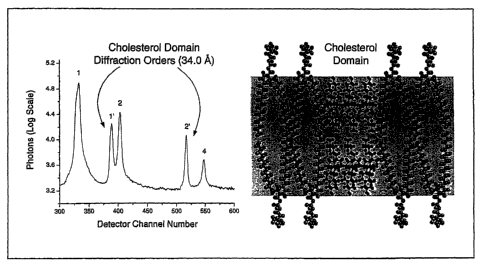
Figure 1 shows the X-ray diffraction pattern and corresponding molecular model
for
cholesterol-enriched membrane bilayer. Diffraction peaks corresponding to
sterol-rich and -poor
domains can be clearly distinguished at 87% relative humidity at 20 C. The
peaks labeled 1' and
2' correspond to the sterol-rich domain (d = 34.0 A) while the surrounding
sterol-poor area of
the membrane had a d-space value of 60.7 A, corresponding to peaks labeled 1,
2 and 4. The
corresponding molecular model demonstrates cholesterol bilayer domain with a
dimension of
34.0 A (each individual cholesterol monohydrate molecule is 17.0 A) that is
highlighted by the
shaded region of the figure.
Figure 2 shows the differential effects of temperature (Figure 2A) and
relative humidity
(Figure 2B) on the molecular dimensions of cholesterol monohydrate domains
versus
surrounding sterol-poor membrane regions for samples containing verapamil. The
membrane
width, as measured in A units by x-ray diffraction analysis, represents the
distance from the
center of one membrane to the next, including surface hydration. In Fig. 2A,
the effect of
temperature on membrane width was evaluated at a constant 93% relative
humidity while in Fig.
2B the effect of relative humidity was measured at a constant temperature of
20 C. These data
demonstrate that the structure of the cholesterol monohydrate crystalline
domains (34.0 A) are
unaffected by changes in temperature or humidity, as compared to the
surrounding sterol-poor
region of the membrane.
CA 02587475 2007-05-11
WO 2006/071351 PCT/US2005/039534
7
Figure 3 shows the X-ray diffraction pattern from oriented membrane lipid
bilayers
containing elevated levels of cholesterol (1.1:1 and 1.2:1 cholesterol to
phospholipid mole ratios)
prepared in the absence or presence of the AML/AT combination at 5 C. At a
1.1:1 cholesterol
to phospholipid mole ratio, peaks labeled 1, 2 and 4 correspond to d-space
values of 54.2 A and
53.0 A, respectively, for the control and drug-containing samples. At a 1.2:1
cholesterol to
phospholipid mole ratio, peaks labeled 1 and 2 corresponded to d-space values
of 55.5 A and
53.5 A, respectively, for the control and drug-containing samples. This figure
demonstrates that
at a low concentration (30 nM), the combination of AML and AT completely
blocked the
aggregation of cholesterol into discrete cholesterol domains.
Figure 4 shows the X-ray diffraction patterns from oriented membrane lipid
bilayers
containing elevated levels of cholesterol (1.2:1 cholesterol to phospholipid
mole ratio) prepared
in the absence or presence of AML alone, AT alone, AML/AT combination,
AT/nifedipine
combination, and AML/lovastatin combination at 5 C. The peaks labeled 1, 2 and
4 correspond
to the sterol-poor region of the membrane while peaks labeled 1' and 2'
correspond to the
structure of cholesterol monohydrate domains within the membrane (34.0 A). The
dimensions
of the surrounding sterol-poor regions were as follows: control (55.5 A), AML
alone (57.8 A),
AT alone (56.8 A), AML/AT (53.5 A), AT/nifedipine (56.5 A) and AML/lovastatin
(54.4 A).
These experiments demonstrated that the ability of the AML/AT combination to
interfere with
membrane cholesterol domain formation could not be reproduced by the drugs
separately or
other CCB/statin combinations.
CA 02587475 2007-05-11
WO 2006/071351 PCT/US2005/039534
8
Figure 5 shows the X-ray diffraction patterns from oriented membrane lipid
bilayers
containing elevated levels of cholesterol (1.1:1 cholesterol to phospholipid
mole ratio) prepared
in the absence or presence of AML alone, AT alone, and AML/AT combination at 5
C. The
peaks labeled 1, 2 and 4 correspond to the sterol-poor region of the membrane
while peaks
labeled 1' and 2' correspond to the structure of cholesterol monohydrate
domains within the
membrane (34.0 A). The dimensions of the surrounding sterol-poor regions were
as follows:
control (52.4 A), AML alone (54.4 A), AT alone (55.8 A), and AML/AT (53.9 A).
These
experiments demonstrated that the AML/AT combination was able to interfere
with membrane
cholesterol domain formation in a manner that could not be reproduced by the
drugs separately.
Figure 6 shows the dose response curves for NO release stimulated by
amlodipine,
atorvastatin (Compound T), and a mixture of amlodipine with varying
concentrations of
atorvastatin (Compound T).
Figure 7 depicts the effect of amlodipine, atorvastatin either alone or in
combination on
NO synthesis.
Detailed Description of the Invention
This invention relates to the effect of amlodipine and atorvastatin, alone, or
in
10 combination with one another, or with one another plus a tertiary agent, on
the production and
release of nitric oxide (NO) from endothelial cells.
CA 02587475 2007-05-11
WO 2006/071351 PCT/US2005/039534
9
One embodiment of the present invention is directed to a pharmaceutical
composition for
enhancing NO production comprising therapeutically effective amounts of
amlodipine,
atorvastatin and a NO enhancing tertiary compound. In one aspect of this
embodiment, the
atorvastatin can be either atorvastatin itself or its hydroxylated metabolite.
In yet another aspect,
the NO enhancing tertiary agent can be, for example, L-arginine,
tetrahydrobiopterin, an ACE-
inhibitor, an antioxidant, a(3-blocker, an angiotensin II type 1-receptor
antagonist and alike.
Studies were conducted to examine the effect of combining amlodipine and
atorvastatin.
The protocol and results are setforth below.
Preparation of reconstituted membrane samples. Porcine cardiac phospholipid
dissolved
in HPLC-grade chloroform (10.0 mg/ml) was obtained from Avanti Polar Lipids
Inc. (Alabaster,
AL) and stored at -80 C. The fatty acid composition of the phosphatidylcholine
lipids was
determined by gas-liquid chromatographic analysis. The overall ratio of
saturated to unsaturated
fatty acids was 0.8:1, with the primary constituents being 18:21inoleic acid
(30%), 16:0 pahnitic
acid (22%), 18:1 oleic acid (13%), and 20:4 arachidonic acid (11%).
Cholesterol powder was
also purchased from Avanti Polar Lipids Inc. Amlodipine besylate (AML) was
obtained from
Pfizer Central Research (Groton, CT) while atorvastatin calcium (AT) was
provided by Parke
Davis (Ann Arbor, MI).
The effects of the drugs on membrane cholesterol organization and structure
were
assessed in well-defmed lipid vesicles containing equimolar levels of
cholesterol and
phospholipid: This reconstituted membrane system was used for the following
reasons: 1) this
CA 02587475 2007-05-11
WO 2006/071351 PCT/US2005/039534
system reproduces changes in membrane structure observed in cholesterol-
enriched,
atherosclerotic macrophage and smooth muscle cell membranes, 2) the membrane
preparation
does not contain calcium channels, and 3) these samples can be prepared in a
highly reproducible
fashion. Lipid vesicles were formed from phospholipid and cholesterol
dissolved in chloroform
5 at a fixed molar ratio and added to individual glass 13 x 100-mm test tubes.
The chloroform
solvent was removed by shell-drying under a steady stream of N2 gas. Residual
solvent was
removed under vacuum while the samples were shielded from light. Membrane
vesicles were
produced for diffraction analysis by rapidly mixing the dried lipids at room
temperature
following addition of buffered saline (0.5 mmol/L HEPES and 154.0 mmol/L NaCI,
pH, 7.2).
10 The final phospholipid concentration was 5.0 mg/mL. Membrane samples were
oriented for
diffraction analysis by centrifugation and then placed in hermetically sealed
canisters that
controlled temperature and relative humidity, as previously described.
Small angle x=ray diffraction analysis. Small-angle x-ray diffraction
approaches were
used to directly examine the effects of the various drugs on the organization
of cholesterol in the
membrane. X-ray diffraction experiments were conducted by aligning the samples
at grazing
incidence with respect to a collimated, nickel-filtered monochromatic x-ray
source (CuKa = 1.54
A) produced by a high-brilliance rotating anode microfocus generator (Rigaku
Rotaflex RU-200,
Danvers, MA). The diffraction data were collected on a one-dimensional,
position-sensitive
electronic detector (Innovative Technologies, Newburyport, MA) placed at a
distance of 150 mm
from the sample. In addition to direct calibration of the detector system,
cholesterol
monohydrate crystals were used to verify the calibration, as previously
described. The unit cell
CA 02587475 2007-05-11
WO 2006/071351 PCT/US2005/039534
11
periodicity, or d-space, of the membrane lipid bilayer is the measured
distance from the center
between one bilayer to the next, including surface hydration, and calculated
from Bragg's Law.
NO release measurements. All measurements presented were recorded in vitro. NO
release was measured directly from a single endothelial cell in the rabbit
aorta. Measurements
were done in Hank's balance solution at 37 C. A porphyrinic sensor (diameter
0.2 0.1 m) was
placed near the surface (10 5 m) of the endothelial cells using a computer
controlled
micromanipulator. The sensor operated with a three-electrode system [sensor
working electrode;
platinum wire (0.1 mm) counter electrode, and saturated calomel electrode (SCE
- reference
electrode)]. The three electrodes were connected to a potentiostat/galvanostat
PAR273. Data
were acquired with the use of an IBM computer with custom software. The
current proportional
to NO concentration was measured by porphyrinic sensor, which operated in
amperometric mode
at constant potential of 0.63 V vs. SCE.
The release of NO was initiated by the injection of potential agonists of
endothelial NO
synthase (eNOS) using a temtoinjector placed in the controlled distance from
the endothelial cell.
Two different agonists were tested: amlodipine and atorvastatin. The different
concentrations of
these two compounds applied simultaneously were also tested.
Atherosclerotic-like membranes have distinct crystalline-like sterol domains:
Membrane
sterol-rich domains may represent an important nucleating site for free
cholesterol crystal
formation, an important feature of the unstable plaque. The separate and
combined effects of
AML and AT on cholesterol monohydrate formation in membranes reconstituted
from native
CA 02587475 2007-05-11
WO 2006/071351 PCT/US2005/039534
12
phospholipids isolated from cardiac tissue was evaluated. Phospholipid
composed of
heterogeneous acyl chains was used for these analyses. This membrane system
reproducibly
formed discrete sterol-rich domains at levels previously observed in
atherosclerosis studies under
similar experimental conditions.
X-ray diffraction analysis of oriented, cholesterol-enriched membranes
produced strong,
reproducible diffraction orders that correspond to structurally distinct
sterol-rich and -poor
membrane regions. The d-space measurement refers to the average distance from
the center of
one membrane bilayer to the next, including surface hydration. The d-space of
the sterol-rich
region was 34.0 A, indicative of a cholesterol bilayer structure as a single
cholesterol
monohydrate molecule has a long axis of 17 A (Fig. 1). The surrounding sterol-
poor regions,
meanwhile, had an average width of 65.9 A at 20 C and 93% relative humidity.
The much larger
width (>90 %) of the sterol-poor domains is attributed to the abundance of
phospholipid in the
surrounding membrane region. The cholesterol domains were invariably present
over a wide
range of temperatures (5-37 C) and relative humidity levels (74-93%),
consistent with previous
x-ray diffraction analyses on atherosclerotic-like membrane samples.
In Fig. 1, diffraction peaks corresponding to the sterol-rich and -poor
domains can be
clearly distinguished at 20 C. The peaks labeled 1' and 2' correspond to the
sterol-rich domain
(d = 34.0 A) while the surrounding sterol-poor area of the membrane had a d-
space value of 60.7
A, corresponding to peaks labeled 1, 2 and 4. The peaks that describe the
cholesterol
monohydrate phase are very sharp, as expected for a crystalline-like
structure. In every sample
that was evaluated, it was observed that the dimensions of the sterol-poor
region of the
CA 02587475 2007-05-11
WO 2006/071351 PCT/US2005/039534
13
membrane was modulated by temperature and relative humidity due to its
heterogeneous
chemical composition and the dynamic mobility of the phospholipid-cholesterol
binary mixture.
At 93% relative humidity, for example, the d-space of the sterol-poor region
decreased by 5.5 A
(9%) as sample temperature was increased from 15 C (64 A) to 40 C (58.5 A),
consistent with
increased trans-gauche isomerizations (Fig. 2). Over this same temperature
range, however, the
cholesterol monohydrate phase remained unchanged at 34.0 A, as expected for a
crystalline-like
structure. In addition, the highly reproducible 34.0 A structure was
unaffected by large changes
in relative humidity (52 to 93%) at 20 C while the sterol-poor region changed
by 19% or 10 A
(52 to 62 A) over this same range.
Synergistic inhibition of sterol domain formation with amlodipine and
atorvastatin: The
addition of both AML and AT to cholesterol-enriched membrane samples prevented
sterol
domain formation in a synergistic fashion. At an aqueous buffer concentration
of 30 nM, the
combination of AML and AT completely blocked the formation of cholesterol
domains in
membrane samples containing cholesterol and phospholipid at 1.1:1 and 1.2:1
cholesterol:phospholipid mole ratios. In the presence of the two drugs, only
peaks corresponding
to the phospholipid bilayer could be observed under a variety of experimental
conditions, as
compared to control (Fig. 3). At a 1.1:1 mole ratio, the d-space values for
the control and drug
combination-containing samples were 54.2 and 53.9 A, respectively, at 74%
relative humidity
and 5 C. At a 1.2:1 mole ratio, the d-space values for the control and drug
combination-
containing samples were 55.5 and 53.5 A, respectively, at 74% relative
humidity and 5 C.
CA 02587475 2007-05-11
WO 2006/071351 PCT/US2005/039534
14
When AML or AT were added separately to the membrane samples, cholesterol
domains
could be clearly detected under identical conditions with small angle x-ray
diffraction
approaches. Moreover, the combination of AML and AT with other drugs had no
inhibitory
effect on cholesterol crystal formation. Both the combination of AML with the
HMG-CoA
reductase inhibitor lovastatin and the combination of AT with the CCB
nifedipine failed to
interfere with cholesterol domain formation, as compared to control samples
(Fig. 4).
Cholesterol domains were very prominent in these samples with a unit cell
periodicity of 34.0 A.
These discrete structures coexist with the surrounding sterol-poor region of
the membrane. At
5 C and 74% relative humidity, the surrounding sterol-poor region of the
membrane samples had
the following d-space values: control (55.5 A), AML/lovastatin (54.4 A), and
AT/nifedipine
(56.5 A). Finally, when AML and AT were added separately to the cholesterol-
enriched
membrane samples, they did not interfere with domain formation.
The synergistic effect of AML and AT on cholesterol domain formation was also
observed at a lower concentration of cholesterol. At a cholesterol to
phospholipid mole ratio of
1.1:1, the drug combination effectively interfered with cholesterol
crystallization within the
membrane samples (Fig. 5). By contrast, when used separately, the drugs had no
effect on
domain formation, even at this lower level of membrane cholesterol. At 5 C and
74% relative
humidity, the surrounding sterol-poor region of the membrane samples had the
following d-space
values: control (55.5 A), AML alone (54.4 A), AT alone (55.8 A), and AML/AT
(53.9 A).
An explanation for the synergistic effect of AML and AT on the organization of
cholesterol may be their chemical properties. AML has very high lipophilicity
as compared to
CA 02587475 2007-05-11
WO 2006/071351 PCT/US2005/039534
other CCBs and a formal positive charge at physiologic pH. An electrostatic
interaction between
AML and AT as well as the phospholipid headgroup region of the membrane
contributes to the
high affinity of this agent for the lipid bilayer. Moreover, the charged amino-
ethoxy function of
AML directs the drug to a region of the membrane that overlaps the steroid
nucleus of
5 cholesterol molecules, an effect that may directly lead to a disruption in
the self-association of
cholesterol molecules in the membrane. Likewise, it has been observed that AT
partitions to a
similar location in the membrane as AML.
The key finding was the observation that the combination of AML and AT
inhibited the
10 formation of separate cholesterol domains in atherosclerotic-like membranes
in a synergistic
fashion. This biophysical effect of the drug combination was directly
characterized with small
angle x-ray diffraction approaches using lipid membranes enriched with
cholesterol. As
cholesterol aggregates within the membrane may serve as nucleating sites for
extracellular free
cholesterol crystal formation in the vessel wall, the ability of the AML/AT
combination to block
15 such sterol domain formation indicates a novel antiatherosclerotic
mechanism of action. This
observed effect appears to be distinct for these drugs as other combinations
failed to reproduce
this change in the aggregation properties of free cholesterol.
In atherosclerosis, the incidence of lesion rupture and thrombosis is affected
by the lipid
composition of the atherosclerotic plaque. The lipid component of
atherosclerotic lesions
consists primarily of cholesterol and phospholipid, with lesser amounts of
fatty acid and
triacylglycerol. Over time, cholesterol forms crystalline structures in the
human atheroma, an
event that contributes to overall lesion mass and plaque instability. Once
crystallized,
CA 02587475 2007-05-11
WO 2006/071351 PCT/US2005/039534
16
cholesterol within the lesion is essentially inert and cannot be effectively
removed by lipoprotein
acceptors in the plasma. By contrast, non-crystallized cholesterol associated
with foam cell
membranes or intracellular stores can be depleted by plasma HDL and
pharmacological
interventions, leading to lesion regression.
Recent reports indicate that the cellular membrane is a cellular site for free
cholesterol
accumulation, leading to discrete sterol-rich domains and eventually crystal.
In macrophage
foam cells, for example, a critical mass of cholesterol is achieved following
lipoprotein (native or
oxidized) uptake and/or phagocytosis of lipid released from neighboring
necrotic foam cells.
Ultimately, a nucleating event will occur at a critical concentration of
cholesterol enrichment,
leading to cholesterol domain development within the membrane. By interfering
with the
formation of highly organized cholesterol aggregates within the membrane, the
combination of
AML and AT may significantly slow or even prevent subsequent crystal
development in the
vessel wall, and thereby block the progression of an otherwise irreversible
step in
atherosclerosis. Moreover, these agents may work synergistically with HDL and
lipid-lowering
therapy in reducing the accumulation of cholesterol crystals in the wall of
the diseased artery by
maintaining cholesterol in a non-crystalline or dynamic state in cellular
membranes.
The mechanism by which AML and AT interfere with the aggregation of
cholesterol into
discrete domains may be related to its their molecular membrane interactions.
At physiologic
pH, more than 90% of the amino ethoxy function associated with the #2 position
of the
dihydropyridine ring of AML is in the charged state. This positive charge
contributes to specific
electrostatic interactions of AML with phosphate groups associated with the
phospholipid bilayer
CA 02587475 2007-05-11
WO 2006/071351 PCT/US2005/039534
17
surface. The results of previous small-angle x-ray diffraction, differential
scanning calorimetry
and nuclear magnetic resonance analyses support a molecular model that places
the charged
amino function of AML near oppositely charged groups in the phospholipid
headgroup region.
Simultaneously, the hydrophobic portion of the dihydropyridine molecule is
buried within the
membrane hydrocarbon core, adjacent to the headgroup region. These biophysical
measurements indicate that the time-averaged location of the ring structure
for AML overlaps the
sterol nucleus of cholesterol in the membrane, where it can then modulate
certain biophysical
effects of the molecule, and interfere with its self-association. Likewise,
small-angle x-ray
diffraction approaches demonstrated that AT partitioned to a discrete location
in the membrane
bilayer.
Thus, this unexpected, synergistic effect can be attributed to the molecular
interactions of
these compounds with membrane lipid constituents. This fmding has important
relevance for the
treatment of coronary artery disease (CAD) as this disorder is characterized
by the abnormal
accumulation of free cholesterol into separate, membrane domains (d-space of
34.0 A). These
domains disrupt cellular function and lead to extracellular crystal formation,
an important feature
of the unstable atherosclerotic plaque. Small angle x-ray diffraction analyses
demonstrated, for
the first time, that the combination of AML and AT blocked the aggregation of
free cholesterol
into crystalline-like domains at low, nanomolar concentrations. By cointrast,
the combination of
these agents with other related drugs showed no inhibitory effect on
cholesterol crystal
formation. These fmdings indicate that the combination of AML and AT produces
a novel anti-
atherosclerotic effect by disrupting cholesterol crystal formation in
atherosclerotic-like
membranes. By disrupting the formation of cholesterol crystals in the vessel
wall, the AML/AT
CA 02587475 2007-05-11
WO 2006/071351 PCT/US2005/039534
18
combination would reduce plaque instability while facilitating cholesterol
efflux to sterol
acceptor particles, such as HDL. This new anti-atherosclerotic mechanism of
action for the
AML/AT combination would complement the separate activities of these agents in
the effective
treatment of cardiovascular disease.
NO Release, from Aortic Endothelial Cells: Figure 6 shows dose response curves
for NO
release stimulated by amlodipine, atorvastatin, and the mixture of 5 mol/L of
amlodipine and
variable concentrations (from 1 - 5 mol/L) of atorvastatin. Based on the data
depicted in Fig. 6,
there is a significant synergistic effect observed after stimulation of NO
release from endothelial
cells by the combination of amlodipine and atorvastatin over a range of doses.
Therefore, the results of these analyses demonstrated a powerful synergistic
effect forthe
combination of amlodipine and atorvastatin on the inhibition of cholesterol
crystal formation and
nitric oxide release from rabbit aortic endothelial cells. The results of this
study provide
compelling scientific support for the combined use of AML and AT in the
treatment of
cardiovascular disorders. These novel antiatherosclerotic effects of the
AML/AT combination
complement the separate activities of these agents in the treatment of
cardiovascular disease,
including CAD.
The present invention describes methods for synergistically increasing nitric
oxide (NO)
release present in a subject's vasculature by administering an effective
amount of amlodipine and
atorvastatin metabolite with at least one other NO enhancing tertiary agent
that enhances NO
bioavailability from endothelial cells.
CA 02587475 2007-05-11
WO 2006/071351 PCT/US2005/039534
19
Nitric oxide (NO) is produced by the enzymatic conversion of the amino acid L-
arginine
to L-citrulline by the enzymatic action of an NADPH-dependentNO synthase
(NOS). The NOS
enzyme requires Ca2i'/calmodulin, FAD, FMN, and tetrahydrobiopterin (BH4) as
cofactors
(Moncada and Higgs, 1993, N. Engl J Med. 329:2002-2012; Nathan and Xie, 1994,
J Biol Chem.
269:13725-28, the entire teachings of which are incorporated herein by
reference). Inthe blood
vessels, NO is produced from the endothelium by constitutive expression of the
endothelial
isoform of NOS (eNOS), which is activated by mechanical stress such as blood
shear-stress and
stimulation with agonists such as bradykinin and acetylcholine. NO has a
variety of functions,
but its action as the endothelium-derivedrelaxing factor (EDRF) is the most
important for the
maintenance of vascular homeostasis (Moncada and Higgs, 1993).
An impairment of endothelium-dependentrelaxations (EDR) is present in
atherosclerotic
vessels even before vascular structural changes occur and represents the
reduced eNOS-derived
NO bioavailability. Endothelial dysfunction as characterized by an impairment
of EDR, and
thereby reduced eNOS-derived NO bioactivity, is the critical'step for
atherogenesis. Among
various mechanisms responsible for the impaired EDR, the increased NO
breakdown by
superoxide is important, and there is augmented production of superoxide in
atherosclerotic
vessels. Under certain circumstances, eNOS becomes dysfunctional and produces
superoxide
rather than NO. The pathophysiological role of dysfunctional eNOS has
attracted attentions in
vascular disorders, including atherosclerosis.
CA 02587475 2007-05-11
WO 2006/071351 PCT/US2005/039534
As previously mentioned, under normal conditions, NO is generated by vascular
endothelium nitric oxide synthase (eNOS) in response to activation of
mechanochemical
receptors associated with increased vascular flow and natural agonists such as
acetylcholine,
bradykinin and substance P. Endothelial dysfunction, including loss of normal
NO production, is
5 associated with various cardiovascular disorders including atherosclerosis,
hypertension, heart
failure, and diabetes mellitus (see, Drexler H, Hayoz D, Munzel T, Homig B,
Just H, Brunner
HR, Zelis R., Endothelial function in chronic congestive heart failure, Am. J.
Cardiol.
1992;69:1596-1601; Gilligan DM, Panza JA, Kilcoyne CM, Waclawiw MS, Casion PR,
Quyyumi AA., Contribution of endothelium-derived nitric oxide to exercise-
induced
10 vasodilation. Circulation. 1994;90:2853-2858; Panza JA, Quyyumi AA, Brush
JE, Epstein SE.
Abnormal endothelium-dependent vascular relaxation in patients with essential
hypertension. N.
Engi. J. Med. 1990;323:22-27; Cardillo C, Kicoyne CM, Quyyumi AA, Cannon RO,
Panza JA.
Selective defect in nitric oxide synthesis may explain the impaired
endothelium-dependent
vasodilation in patients with essential hypertension. Circulation. 1998;97:851-
856; Drexler H,
15 Hornig B. Endothelial dysfunction in human disease. J. Mol. Cell. Cardiol.
1999;3:51-60, the
entire teachings of which are incorporated herein by reference.)
In patients with documented hypertension, decreased NO production results in
loss of
normal vasodilation. During the development of heart failure, endothelial
dysfunction results in
20 maladaptive changes in the peripheral vasculature and skeletal muscle,
leading to symptoms of
exercise intolerance (Drexler H, Hayoz D, Munzel T, Homig B, Just H, Brunner
HR, Zelis R.
Endothelial function in chronic congestive heart failure. Am. J. Cardiol.
1992;69:1596-1601;
Gilligan DM, Panza JA, Kilcoyne CM, Waclawiw MS, Casion PR, Quyyumi AA.
Contribution
CA 02587475 2007-05-11
WO 2006/071351 PCT/US2005/039534
21
of endothelium-derived nitric oxide to exercise-induced vasodilation.
Circulation. 1994;90:2853-
2858, the entire teachings of which are incorporated herein by reference).
Production of NO appears to be an essential activity of the endothelium for
maintaining a
smooth, nonthrombogenic surface. During atherosclerosis, however, a deficiency
in NO
synthesis has adverse consequences on vascular hemodynamics and inflammation
(Libby P.
Changing concepts in atherogenesis. J. Intern. Med. 2000;247:349-358; Ross R.
Atherosclerosis
-- An inflammatory disease. N. Engl. J. Med. 1999;340:115-126, the entire
teachings of which
are incorporated herein by reference). These deleterious effects include: 1)
increased free
radical damage, 2) platelet aggregation, 3) increased hyperadhesiveness of
leukocytes, 4)
enhanced vasoconstriction, and 5) increased production of the vasoconstrictor,
endothelin. Thus,
a deficiency in NO availability could be a key early event that promotes
atherogenesis in the
human vasculature.
Pharmacologic agents that enhance NO synthesis have favorable effects on
patients with
hypertension and atherosclerotic disease (i.e., coronary artery disease) by
increasing constitutive
levels of eNOS (Wiemer G, Linz W, Hatrik S, Scholkens BA, Malinski T.
Angiotensin-
converting enzyme inhibition alters nitric oxide and superoxide release in
normotensive and
hypertensive rats. Hypertension. 1997;30:1183-1190; Treasure CB, Klein JL,
Weintraub WS,
Talley JD, Stillabower ME, Kosinski AS, Zhang J, Boccuzzi SJ, Cedarholm JC,
Alexander RW.
Beneficial effects of cholesterol-lowering therapy on the coronary endothelium
in patients with
coronary artery disease. N. Engl. J. Med. 1995;332:481-487, the entire
teachings of which are
incorporated herein by reference). Surprisingly, the combination of amlodipine
and atorvastatin
CA 02587475 2007-05-11
WO 2006/071351 PCT/US2005/039534
22
enhances NO production from human endothelial cells in a highly synergistic
fashion. This
finding has broad implications for the use of these agents in the treatment of
cardiovascular
diseases.
In one aspect, methods for increasing nitric oxide (NO) release present in a
subject's
vasculature by administering an effective amount of amlodipine and
atorvastatin metabolite with
at least one other agent that enhances NO bioavailability from endothelial
cells are described.
Examples of suitable enhancing NO tertiary agents include, but are not limited
to, L-arginine
(substrate for NOS), tetrahydrobiopterin (BH4, a co-factor of NOS), ACE-
inhibitors (ramipril,
enalapril, quinapril), antioxidants (e.g., vitamin E, probucol, vitamin C), (3-
blockers (nebivolol,
carvedilol, metoprolol) and angiotensin II type 1(AT1)-receptor antagonists
(irbesartan,
candesartan, valsartan, losartan).
One aspect of the present embodiment is directed toward administering an
effective
amount of amlodipine/atorvastatin metabolite with a peroxisome proliferator
activated receptor
(PPAR7) agonists (e.g., rosiglitazone). These agents are used for the
treatment of diabetes by
enhancing sensitivity of cells to insulin. However, these agents have shown
additional vascular
benefits beyond genomic regulation, resulting in improved blood pressure and
vessel function
consistent with endothelial improvement (Ryan et al. 2004 Hypertension, 43:661-
666, the entire
teaching of which is incorporated herein by reference).
A particular aspect of the present embodiment is directed toward a method for
treating a
subject that has an endothelial cell dysfunction. The endothelial cell
dysfunction causes or
CA 02587475 2007-05-11
WO 2006/071351 PCT/US2005/039534
23
contributes to one or more cardiovascular disorders. In a further aspect, the
cardiovascular
disorder is selected from the group consisting of atherosclerosis,
hypertension, dyslipidemia,
diabetes mellitus, heart failure, obesity, smoking and renal failure. These
subjects can be
administered an effective amount of a combination of amlodipine, atorvastatin,
and a third agent,
such as those described above.
Any of the identified compounds of the present invention can be administered
to a
subject, including a human, by itself, or in pharmaceutical compositions where
it is mixed with
suitable carriers or excipients at doses therapeutically effective to prevent,
treat or ameliorate a
variety of disorders, including those characterized by that outlined herein. A
therapeutically
effective dose further refers to that amount of the compound sufficient result
in the prevention or
amelioration of symptoms associated with such disorders. Techniques for
formulation and
administration of the compounds of the instant invention may be found in
Goodman and
Gilman's The Pharmacological Basis of Therapeutics, Pergamon Press, latest
edition.
The compounds of the present invention can be targeted to specific sites by
direct
injection into those sites. Compounds designed for use in the central nervous
system should be
able to cross the blood-brain barrier or be suitable for administration by
localized injection.
Pharmaceutical compositions suitable for use in the present invention include
compositions wherein the active ingredients are contained in an effective
amount to achieve its
intended purpose. More specifically, a therapeutically effective amount means
an amount
effective to prevent development of or alleviate the existing symptoms and
underlying pathology
CA 02587475 2007-05-11
WO 2006/071351 PCT/US2005/039534
24
of the subject being treating. Determination of the effective amounts is well
within the capability
of those skilled in the art.
For any compound used in the methods of the present invention, the
therapeutically
effective dose can be estimated initially from cell culture assays. For
example, a dose can be
formulated in animal models to achieve a circulating concentration range that
includes the ICso
(the dose where 50% of the cells show the desired effects) as determined in
cell culture. Such
information can be used to more accurately determine useful doses in humans.
A therapeutically effective dose refers to that amount of the compound that
results in the
attenuation of symptoms or a prolongation of survival in a subject. Toxicity
and therapeutic
efficacy, of such compounds can be determined by standard pharmaceutical
procedures in cell
cultures or experimental animals, e.g., for determining the LD50 (the dose
lethal to 50% of a
given population) and the ED50 (the dose therapeutically effective in 50% of a
given population).
The dose ratio between toxic and therapeutic effects is the therapeutic index
and it can be
expressed as the ratio between LD50 and ED50. Compounds which exhibit high
therapeutic
indices are preferred. The data obtained from these cell culture assays and
animal studies can be
used in formulating a range of dosage for use in human. The dosage of such
compounds lies
preferably within a range of circulating concentrations that include the ED50
with little or no
toxicity. The dosage can vary within this range depending upon the dosage form
employed and
the route of administration utilized. The exact formulation, route of
administration and dosage
can be chosen by the individual physician in view of a patient's condition.
Dosage amount and
CA 02587475 2007-05-11
WO 2006/071351 PCT/US2005/039534
interval can be adjusted individually to provide plasma levels of the active
moiety which are
sufficient to maintain the desired effects.
In case of local administration or selective uptake, the effective local
concentration of the
5 drug may not be related to plasma concentration.
The amount of composition administered will, of course, be dependent on the
subject
being treated, on the subject's weight, the severity of the affliction, the
manner of administration
and the judgment of the prescribing physician.
The pharmaceutical compositions of the present invention can be manufactured
in a
manner that is itself known, e.g., by means of conventional mixing,
dissolving, granulating,
levigating, emulsifying, encapsulating, entrapping or lyophilizing processes.
Pharmaceutical compositions for use in accordance with the present invention
thus can be
formulated in conventional manner using one or more physiologically acceptable
carriers
comprising excipients and auxiliaries which facilitate processing of the
active compounds into
preparations which can be used pharmaceutically. Proper formulation is
dependent upon the
route of administration chosen.
For injection, the agents of the invention can be formulated in aqueous
solutions,
preferably in physiologically compatible buffers such as Hank's solution,
Ringer's solution, or
physiological saline buffer. For transmucosal administration, penetrants
appropriate to the
CA 02587475 2007-05-11
WO 2006/071351 PCT/US2005/039534
26
barriers to be permeated are used in the formulation. Such penetrants are
generally known in the
art.
For oral administration, the compounds can be formulated readily by combining
the
active compounds with pharmaceutically acceptable carriers well known in the
art. Such carriers
enable the compounds of the invention to be formulated as tablets, pills,
dragees, capsules,
liquids, gels, syrups, slurries, suspensions and the like, for oral ingestion
by a subject to be
treated. Pharmaceutical preparations for oral use can be obtained solid
excipient, optionally
grinding a resulting mixture, and processing the mixture of granules, after
adding suitable
auxiliaries, if desired, to obtain tablets or dragee cores. Suitable
excipients are, in particular,
fillers such as sugars, including lactose, sucrose, mannitol, or sorbitol;
cellulose preparations
such as, for example, maize starch, wheat starch, rice starch, potato starch,
gelatin, gum
tragacanth, methyl cellulose, hydroxypropylmethylcellulose, sodium
carboxymethylcellulose,
and/or polyvinyl-pyrrolidone (PVP). If desired, disintegrating agents can be
added, such as the
cross-linked polyvinyl pyrrolidone, agar, or alginic acid or a salt thereof
such as sodium alginate.
Dragee cores are provided with suitable coatings. For this purpose,
concentrated sugar
solutions can be used, which can optionally contain gum arabic, talc,
polyvinyl pyrrolidone,
carbopol gel, polyethylene glycol, and/or titanium dioxide, lacquer solutions,
and suitable
organic solvents or solvent mixtures. Dyestuffs or pigments can be added to
the tablets or dragee
coatings for identification or to characterize different combinations of
active compound doses.
CA 02587475 2007-05-11
WO 2006/071351 PCT/US2005/039534
27
Pharmaceutical preparations which can be used orally include push-fit capsules
made of
gelatin, as well as soft, sealed capsules made of gelatin and a plasticizer,
such as glycerol or
sorbitol. The push-fit capsules can contain the active ingredients in
admixture with filler such as
lactose, binders such as starches, and/or lubricants such as talc or magnesium
stearate and,
optionally, stabilizers. In soft capsules, the active compounds can be
dissolved or suspended in
suitable liquids, such as fatty oils, liquid paraffin, or liquid polyethylene
glycols. In addition,
stabilizers can be added. All formulations for oral administration should be
in dosages suitable
for such administration.
For buccal administration, the compositions can take the form of tablets or
lozenges
formulated in conventional manner.
For administration by inhalation, the compounds for use according to the
present
invention are conveniently delivered in the form of an aerosol spray
presentation from
pressurized packs or a nebulizer, with the use of a suitable propellant, e.g.,
dichlorodi-
fluoromethane, trichlorofluoromethane, dichlorotetrafluoromethane, carbon
dioxide or other
suitable gas. In the case of a pressurized aerosol the dosage unit can be
determined by providing
a valve to deliver a metered amount. Capsules and cartridges of e.g., gelatin
for use in an inhaler
or insufflator can be formulated containing a powder mix of the compound and a
suitable powder
base such as lactose or starch.
The compounds can be formulated for parenteral administration by injection,
e.g., by
bolus injection or continuous infusion. Formulations for injection can be
presented in unit
CA 02587475 2007-05-11
WO 2006/071351 PCT/US2005/039534
28
dosage for, e.g., in ampoules or in multidose containers, with an added
preservatives. The
compositions can take such forms as suspensions, solutions or emulsions in
oily or aqueous
vehicles, and can contain formulatory agents such as suspending, stabilizing
and/or dispersing
agents.
Pharmaceutical formulations for parenteral administration include aqueous
solutions of
the active compounds in water-soluble form. Additionally, suspensions of the
active compounds
can be prepared as appropriate oily injection suspension. Suitable lipohilic
solvents or vehicles
include fatty oils such as sesame oil, or synthetic fatty acid esters, such as
ethyl oleate or
triglycerides, or liposomes. Aqueous injection suspensions can contain
substances which
increase the viscosity of the suspension, such as sodium carboxymethyl
cellulose, sorbitol, or
dextran. Optionally, the suspension can also contain suitable stabilizers or
agents which increase
the solubility of the compounds to allow for the preparation of highly
concentrated solutions.
Alternatively, the active ingredient can be in powder form for constitution
with a suitable
vehicle, e.g., sterile pyrogen-free water, before use.
The compounds can also be formulated in rectal compositions such as
suppositories or
retention enemas, e.g., containing conventional suppository bases such as
cocoa butter or other
glycerides.
In addition to the formulations previously described, the compounds can also
be
formulated as a depot preparation. Such long acting formulations can be
administered by
CA 02587475 2007-05-11
WO 2006/071351 PCT/US2005/039534
29
implantation (e.g., subcutaneously or intramuscularly) or by intramuscular
injection. Thus, for
example, the compounds can be formulated with suitable polymeric or
hydrophobic materials
(e.g., as an emulsion in an acceptable oil) or ion exchange resins, or as
sparingly soluble
derivatives, e.g., as a sparingly soluble salt.
A pharmaceutical carrier for the hydrophobic compounds of the invention is a
co-solvent
system comprising benzyl alcohol, a non-polar surfactant, a water-miscible
organic polymer, and
an aqueous phase. Naturally, the proportions of a co-solvent system can be
varied considerably
without destroying its solubility and toxicity characteristics. Furthermore,
the identity of the co-
solvent components can be varied.
Altenatively, other delivery systems for hydrophobic pharmaceutical compounds
can be
employed. Liposomes and emulsions are well known examples of delivery vehicles
or carriers
for hydrophobic drugs. Certain organic solvents such as dimethylsulfoxide also
may be
employed, although usually at the cost of greater toxicity. Additionally, the
compounds can be
delivered using a sustained-release system, such as semipermeable matrices of
solid hydrophobic
polymers containing the therapeutic agent. Various of sustained-release
materials have been
established and are well known to those skilled in the art. Sustained-release
capsules can,
depending on their chemical nature, release the compounds for a few weeks up
to over 100 days.
Depending on the chemical nature and the biological stability of the
therapeutic reagent,
additional strategies for protein stabilization can be employed.
CA 02587475 2007-05-11
WO 2006/071351 PCT/US2005/039534
The pharmaceutical compositions also can comprise suitable solid or gel phase
carriers or
excipients. Examples of such carriers or excipients include, but are not
limited to, calcium
carbonate, calcium phosphate, various sugars, starches, cellulose derivatives,
gelatin, and
polymers such as polyethylene glycols.
5
Many of the compounds of the invention can be provided as salts with
pharmaceutically
compatible counterions. Pharmaceutically compatible salts can be formed with
many acids,
including but not limited to hydrochloric, sulfuric, acetic, lactic, tartaric,
malic, succinic, etc.
Salts tend to be more soluble in aqueous or other protonic solvents that are
the corresponding
10 free base forms.
Suitable routes of administration can, e.g., include oral, rectal,
transmucosal, transdermal,
or intestinal administration; parenteral delivery, including intramuscular,
subcutaneous,
intramedullary injections, as well as intrathecal, direct intraventricular,
intravenous,
15 intraperitoneal, intranasal, or intraocular injections.
Alternatively, one can administer the compound in a local rather than systemic
manner,
e.g., via injection of the compound directly into an affected area, often in a
depot or sustained
release formulation.
Furthermore, one can administer the compound in a targeted drug delivery
system, e.g.,
in a liposome coated with an antibody specific for affected cells. The
liposomes will be targeted
to and taken up selectively by the cells.
CA 02587475 2007-05-11
WO 2006/071351 PCT/US2005/039534
31
The compositions can, if desired, be presented in a pack or dispenser device
which can
contain one or more unit dosage forms containing the active ingredient. The
pack can, e.g.,
comprise metal or plastic foil, such as a blister pack. The pack or dispenser
device can be
accompanied by instruction for administration. Compositions comprising a
compound of the
invention formulated in a compatible pharmaceutical carrier can also be
prepared, placed in an
appropriate container, and labeled for treatment of an indicated condition.
Suitable conditions
indicated on the label can include treatment of a disease such as described
herein.
EXAMPLE
The following is an experiment that demonstrates the combination of amlodipine
and
atorvastatin stimulated nitric oxide production from human endothelial cells
in a synergistic
fashion as compared to control. These data demonstrate a synergistic effect of
this unique
combination of compounds in treating the disease state of atherosclerosis,
which is the
underlying disease process for various cardiovascular disorders, including
coronary artery
disease and heart failure. As discussed above, a deficiency in nitric oxide
production is
associated with endothelial dysfunction, a major cause of hypertension and
atherosclerosis.
The protocol employed is set forth below.
Nanosensor Measurements of Nitric Oxide:
1. Nanosensors were prepared from carbon fibers. The size of the tip of carbon
fiber was
reduced from 6 gm to less than 1 m by temperature controlled burning. The
sensors were made
CA 02587475 2007-05-11
WO 2006/071351 PCT/US2005/039534
32
sensitive to NO by deposition of electrically conductive polymeric porphyrin
and covered with a
thin layer of Nafion according to the procedures previously described
(Malinski T, Taha Z. Nitric
oxide release from a single cell measured in situ by a porphyrinic-based
microsensor. Nature.
1992;358:676-678, the entire teaching of which is incorporated herein by
reference).
2. Measurements of NO were made in the growth medium solution. The nanosensor
was
positioned at a distance of about 5 2 m from the surface of endothelial cell
with a help of a
motorized computer micromanipulator. The nanosensor operates as a component of
a three-
electrode system: nanosensor (working electrode), saturated calomel electrode
(reference
electrode) and platinum wire (counter electrode, 0.5 mm diameter).
The nanosensor operates at a constant potential of 0.68 V versus saturated
colomel
electrode.
Amperograms (current vs. time curves) were recorded with a Guniry FAS 1
Femtostat
(Warminster, PA).
3. HUVEC cells were obtained from American Type Culture Collection (Manassas,
VA)
and grown in Ham's F 12K medium with 2 mM L-glutamine adjusted to contain 1.5
g/L sodium
bicarbonate and supplemented with 0.1 mg/ml heparin and 0.03 - 0.05 mg/mL
endothelial cell
growth supplement (ECGS) + 10% fetal bovine serum. The HUVEC cells were kept
in the
atmosphere of elevated CO2 concentration (5%).
CA 02587475 2007-05-11
WO 2006/071351 PCT/US2005/039534
33
4. For the measurements cell wells were transferred to a Faraday cage and,
with the help of
inverted microscope (Leica Microsystems, Wetzlar, Germany) and
micromanipulator, the
nanosensor was positioned near the surface of HUVEC. The baseline was
stabilized after about
20 seconds.
5. Amlodipine, Atorvastatin or the mixture of the two drugs was injected with
the help of a
nanoinjector. The NO concentration was measured for about 60 seconds.
6. The nanosensor for NO was calibrated using saturated solution
(concentration 1.82
mmol/L verified with the coulometric method).
7. Prepared stock solutions:
A) Amlodipine:
Weight = 51.5 mg, MW = 567.1
Stock Solution: 10 M in ethanol
take 5.7 mg and dissolve in 1 mL of ethanol.
B) Atorvastatin:
Weight = 53.6 mg, MW = 585.68
Stock Solution: 10 M in methanol
take 5.9 mg and dissolve in 1 mL of methanol.
8. Sample solutions of Amlodipine and Atorvastatin were prepared as follows.
Nine
separate concentrations of Amlodipine and Atorvastatin were tested: 0.25;
0.75; 1.00; 1.50; 2.00;
CA 02587475 2007-05-11
WO 2006/071351 PCT/US2005/039534
34
2.50; 3.00 and 5.00 M. The working solutions were prepared by dilution of
stock solutions
with distilled water.
The Pipetting Scheme was a follows:
A) Amlodipine and Atorvastatin (both M stock)
Table 1: Amlodipine and Atorvastatin (both M stock)
Concentration ( M) Concentration ( M) l of Stock l of Water
Vial Final
50.0 0.25 5 995
150.0 0.75 15 985
200.0 1.00 20 980
300.0 1.50 30 970
400.0 2.00 40 960
500.0 2.50 50 950
600.0 3.00 60 940
1000.0 5.00 100 900
B) The working solutions had a concentration 200 x times higher than required
(final) as the
cell well volume was 2 mL while the injected volume was 10 L (200 x
dilution).
CA 02587475 2007-05-11
WO 2006/071351 PCT/US2005/039534
9. The synergistic effect was tested at a constant concentration (5 M) of
Amlodipine (A)
and variable concentrations of Atorvastatin (T). The next series of
experiments tested this effect
at constant ratios of both compounds according to formulations (A:T):
1 MofA: 1 MofT;2 MofA:2 MofT;2.5 MofA: 2.5 MofT; 3.0 MofA: 3.0
5 M of T; 5.0 M of A: 5.0 M of T.
10. Peak of maximal NO concentration was calculated.
11. Area under current vs. time curve (amperogram) was integrated (coulometry)
and
10 amount of NO detected by the nanosensor was calculated.
The following HUVEC samples were analyzed in triplicate at 37 C. The method
used
was described above.
Table 2: Amlodipine
0 M - Control #1
0 M - Control #2
0 [tM - Control #3
0.25 M-1of3
0.25 M - 2 of 3
0.25 M-3 of 3
0.75 M-1of3
0.75 M-2of3
0.75 M-3of3
1.0 M - 1 of 3
1.0 M-2of3
CA 02587475 2007-05-11
WO 2006/071351 PCT/US2005/039534
36
1.0 M - 3 of 3
1.5 M-1of3
1.5 M-2of3
1.5 M-3of3
2.0 M - 1 of 3
2.0 M-2of3
2.0 M-3 of3
2.5 M-1of3
2.5 M-2of3
2.5[tM-3of3
3.0 M - 1 of 3
3.0 M-2of3
3.0 M-3of3
5.0 M - 1 of 3
5.0 M - 2 of 3
5.0 M-3of3
Atorvastatin
The Atorvastatin data were recorded in a similar manner as Amlodipine data.
Table 3: Mixture: Amlodipine (5 M) + Atorvastatin (varies)
Atorvastatin
0.25 M-1of3
0.25 M-2of3
0.25 M-3 of3
0.75 M - 1 of 3
0.75 M-2of3
0.75 M-3 of3
CA 02587475 2007-05-11
WO 2006/071351 PCT/US2005/039534
37
1.0 M-1of3
1.0 M-2of3
1.0 M-3 of3
1.5 M-1of3
1.5 M-2of3
1.5 M-3of3
2.0 M-1of3
2.0 M - 2 of 3
2.0 M-3of3
2.5 M-1of3
2.5 M-2of3
2.5 M - 3 of 3
3.0 M-1of3
3.0 M-2of3
3.0 M - 3 of 3
5.0 M-1of3
5.0 M-2of3
5.0 M - 3 of 3
Table 4: Mixture (same ratios, in equimolar concentrations)
Amlodipine Atorvastatin sample
1.0 M 1.0 M 1 of 3
1.0 M 1.0 M 2 of 3
1.0 M 1.0 M 3 of 3
CA 02587475 2007-05-11
WO 2006/071351 PCT/US2005/039534
38
2.0 M 2.0 M 1 of 3
2.0 M 2.0 M 2 of 3
2.0 M 2.0 M 3 of 3
2.5 M 2.5 [,M l of 3
2.5 M 2.5 M 2 of 3
2.5 M 2.5 M 3of3
3.0 M 3.0 M 1 of 3
3.0 M 3.0 M 2 of 3
3.0 M 3.0 M 3 of 3
5.0 M 5.0 M 1 of 3
5.0 M 5.0 M 2 of 3
5.0 M 5.0 M 3 of 3
The data were presented as mean tSEM for each of the triplicate measurements.
The
data (calculation and plotting) were transferred to Microcal Origin Software
(OriginLab Corp.,
Northampton, MA).
CA 02587475 2007-05-11
WO 2006/071351 PCT/US2005/039534
39
Table 5:NO Peak Measurements
Substance Injected NO Concentration, mean ~ SEM
(concentration, M) (concentration, nM)
Amlodipine (0.25) 24.21 +3.11
Amlodipine (0.75) 48.44 +5.83
Amlodipine (1.00) 53.50 0.39
Amlodipine (1.50) 58.47 +11.00
Amlodipine (2.00) 72.25 +8.20
Amlodipine (2.50) 121.30 24.11
Amlodipine (3.00) 151.26 +18.00
Amlodipine (5.00) 158.00 +19.81
Atorvastatin (0.25) 0.50 +0.02
Atorvastatin (0.75) 1.11 +0.12
Atorvastatin (1.00) 2.31 f0.53
Atorvastatin (1.50) 5.20 +1.21
Atorvastatin (2.00) 8.12 +3.10
.Atorvastatin (2.50) 9.85 +3.00
Atorvastatin (3.00) 15.61 +2.19
Atorvastatin (5.00) 48.69 +2.48
Amlodipine (5.00) + Atorvastatin (0.25) 182.25 21.14
Amlodipine (5.00) + Atorvastatin (0.75) 242.20 f24.00
Amlodipine (5.00) + Atorvastatin (1.00) 274.94 22.06
Amlodipine (5.00) + Atorvastatin (1.50) 271.33 15.20
Amlodipine (5.00) + Atorvastatin (2.00) 247.00 6.11
Amlodipine (5.00) + Atorvastatin (2.50) 231.60 +7.80
Amlodipine (5.00) + Atorvastatin (3.00) 208.71 30.74
Amlodipine (5.00) + Atorvastatin (5.00) 130.50 +15.12
Amlodipine (1.00) + Atorvastatin (1.00) 126 18
Amlodipine (2.00) + Atorvastatin (2.00) 17817
Amlodipine (2.50) + Atorvastatin (2.50) 201 +11
Amlodipine (3.00) + Atorvastatin (3.00) 219 +6
Amlodipine (5.00) + Atorvastatin (5.00) 160 71
CA 02587475 2007-05-11
WO 2006/071351 PCT/US2005/039534
Figure 7 depicts the separate and combined effects of amlodipine (open
squares),
atorvastatin (shaded circles), on NO release (nM) from human endothelial cells
as a function of
drug concentration ( M). At equimolar concentrations of amlodipine and
atorvastatin, a
pronounced synergistic effect was observed over a range of micromolar
concentrations (1.0
5 through 3.0 M). The release of NO was measured electrochemically with a
sensitive
porphyrinic sensor placed in close proximity to the cultured cell surface. The
drug combination
caused the release of NO from the human endothelial cells at levels that
exceeded the expected
additive effects of the drugs, and thus, indicated a clear synergistic effect.
10 It will now be apparent to those skilled in the art that other embodiments,
improvements,
details, and uses can be made that are consistent with the letter and spirit
of the foregoing
disclosure and within the scope of this patent and the appended claims.