Note: Descriptions are shown in the official language in which they were submitted.
CA 02587570 2007-05-14
WO 2006/055448 PCT/US2005/041028
OFF-AXIS ANCHOR GUIDANCE SYSTEM
CROSS REFERENCE TO RELATED APPLICATIONS
This application claims the benefit of U.S. patent application serial No.
10/990,272,
entitled "An Implant Assembly and Method For Use In An Internal Structure
Stabilization System" filed on November 16, 2004; and U.S. patent application
serial
No. 10/991,845, entitled "Off-axis Anchor Guidance System" filed on November
18,
2004.
TECHNICAL FIELD
io This application relates generally to the field of medical implant devices
and more
particularly to systems and methods for inserting and guiding bone anchoring
devices.
BACKGROUND INFORMATION
Orthopedic injuries, deformities, and degenerative diseases often require
intervention in the form of surgery for placing implants to stabilize an
internal
structure, promote healing, and relieve pain. In the area of spinal surgery,
for
example, a common procedure includes placement of pedicle screws that are
joined
by a connecting rod spanning between these screws. Once placed, the rod must
be
firmly secured to the bone securing elements to provide a stable construct
which
2o effectively immobilizes or creates a controlled dynamic motion to a
corresponding
portion of the spine.
One problem when connecting the rods to the pedicle screws is to position the
rods
in place as quickly as possible without doing more damage to the surrounding
tissue
and muscle of the patient. In order to reduce this damage, procedures have
been
developed that allow the physician to secure the pedicle screws in the bony
portion
of the spine and to then connect the rods or brace between the pedicle screws.
Techniques have been developed to allow the surgeon to perform this procedure
in a
minimally invasive manner, utilizing a percutaneous method, inserting screws
through small ports and avoiding an open approach.
1
CA 02587570 2007-05-14
WO 2006/055448 PCT/US2005/041028
In one such procedure, a surgeon identifies the desired vertebral level and
pedicle
positions via standard techniques. Once the target vertebrae are identified, a
small
incision is made through the patient's skin and a tracking needle (or other
device) is
inserted to pinpoint exactly where each pedicle screw is to be placed. A
fluoroscope,
or other x-ray technique, is used to properly position the tracking needle.
Once the
proper position is located, a guide wire is positioned with its distal end
into the
pedicle of vertebrae. The surgeon then slides a series of continuing larger
sized
dilators down the guide wire. The surgeon may also slide a hole tapping
instrument
over the guide wires. The hole tapping instrument may be used to tap a hole in
the
lo pedicle. After the hole is tapped, a cannulated pedicle screw and a
modified screw
driver may be inserted down the guide wire until the screw reaches the desired
position. The position may be again checked with fluoroscopic techniques. For
purposes of this application, a cannulated pedicle screw is defined as a
pedicle
screw that contains a cannulation centered and running entirely through its
is longitudinal axis.
After the position of the cannulated pedicle screw has been confirmed, the
surgeon
is ready to screw the cannulated pedicle screw into the vertebrae. After the
cannulated pedicle screw has been inserted, this procedure may be repeated for
each additional level. When one or more pedicle screws are in place, a brace
or rod
20 may be positioned by techniques known in the art. Under current practice,
the
physician then must work the brace, or other supporting device, so that each
brace
end is positioned properly with respect to the preplaced pedicle screws, and
tighten
the brace to each pedicle screw to complete assembly.
Once a patient recovers and become active, the brace may be subject to
relatively
25 large structural forces. These forces are applied to the shanks of the
cannulated
pedicle screws. Consequently, it is the shanks of the cannulated pedicle
screws that
resist the applied forces. To be more specific, it is the portion of the screw
shank
that is positioned within the pedicle of the vertebral body (approximately two-
thirds of
the length of the screw from the distal tip of the screw towards the proximal
end of
30 the screw) (the highest stress region is that region of the pedicle screw
that is
nearest the entry point of the pedicle, which tends to be about two thirds up
from the
distal tip of the pedicle screw).
2
CA 02587570 2007-05-14
WO 2006/055448 PCT/US2005/041028
When conventional pedicle screws are cannulated, a significant portion of
their
cross-sectional area is removed to create the cannulation. The cannulation,
therefore, causes higher stress in the remaining portions of the shank which
is
subject to the applied forces. This causes a significant weakening of the
screw.
This weakening can cause failure of the pedicle screw which means that the
patient
would have to undergo additional surgery to have the pedicle screws replaced.
In order to minimize the reduction in strength of the screws, the cannulations
are
made as small as possible. This means that the guides wires must also be
small,
which may lead to advancement, kinking, breakage, or other problems during
io surgery. Inadvertent advancement of the guide wire is a critical concern to
clinicians.
If the guide wire becomes bent through off-angle manipulation by the surgeon,
as the
tap or screw is inserted, the tap or screw pushes the guide wire forward. This
unwanted guide wire advancement could cause the guide wire to push forward
through the anterior wall of the vertebral body, causing trauma to the
patient.
is What is needed, therefore, is a device and system which will allow for
anchors to be
guided and inserted into patients while maintaining the structural integrity
and safety
of the anchor and/or the guide wire.
SUMMARY
In response to these and other problems, in one embodiment, there is a bone
2o anchor characterized by: a shank having a proximal end and a distal end,
wherein
the shank has a bore extending through the distal end of the shank to a point
located
along a side of the shank.
In some embodiments, the shank has an exterior helical thread adapted to
mate with the bone.
In some embodiments, the bone anchoring device may be characterized by a head
coupled to the proximal end of the shank.
In some embodiments, the bone anchoring device may be characterized in that
the
3o head is bulbous.
3
CA 02587570 2007-05-14
WO 2006/055448 PCT/US2005/041028
In some embodiments, the bone anchoring device may be characterized in that
the
head has an exterior helical thread adapted to mate with a rod receiving
device.
In some embodiments, the bone anchoring device may be characterized in that
the
thread is a reverse thread.
In some embodiments, the bone anchoring device may be characterized in that
the
head has at least one longitudinal spline configured to couple with a ring
within a rod
receiving part of an implant system.
In some embodiments, the bone anchoring device may be characterized in that
the
head is generally U-shaped.
Additionally, in some embodiments, there may be system for guiding bone
anchors,
the system characterized by: a bone anchor according to any of the claims I to
8; a
guide wire having a diameter sized to slidingly engage the bore of the bone
anchor;
and a dilator having a longitudinal slot wherein the longitudinal slot allows
the guide
wire to extend through the longitudinal slot when the anchor is positioned
within the
dilator.
In some embodiments, the system may be further characterized by a driving
device
which is adapted to engage the proximal end of the anchor.
In some embodiments, the system may be characterized in that the driving
device
further comprises: an outer shaft, an inner shaft rotatedly disposed within
the outer
shaft having an distal end and proximal end, and a removable handle coupled to
the
proximal end.
In some embodiments, the system may be characterized in that the outer shaft
is
3o adapted to couple with a rod receiving portion of a medical implant device
and the
distal end of the inner shaft is adapted to engage the anchor.
4
CA 02587570 2007-05-14
WO 2006/055448 PCT/US2005/041028
Additionally, in some embodiments there may be a system for positioning a bone
anchor into a bone, the system characterized by: a bone anchor according to
any of
the embodiments above; a means for positioning the bone anchor; and a dialator
having a longitudinal slot for allowing the means for positioning the bone
anchor to
extend through the dialator.
In some embodiments, the system may be further characterized by a means for
enlarging an access space to allow the dialator to access a bone anchor site.
io In some embodiments, the system may be further characterized by a means for
checking the position of the guide wire with fluoroscopic techniques.
Thus various aspects of this disclosure allows the shanks of wire guided
anchors to resist larger forces than conventional cannulated screws because
the
offset bores do not cause a cannulation through the entire length of the
anchor.
Furthermore, the offset bore may be of a relatively larger diameter when
compared
to the conventional cannulation of conventional cannulated pedicle screws. The
larger diameter bore results in a larger diameter guide wire which: increases
the
strength of the guide wire; reduces kinking; allows a surgeon to have tactile
feedback
2o regarding the placement and location of the guide wire; and allows the
surgeon to
maintain hold of the proximal end of the guide wire at all times throughout
the
procedure. Furthermore, the relative short length of the offset bore reduces
the
friction between the instruments and the guide wire, thereby reducing the
likelihood
of guide wire advancement.
These and other features and advantages will be more clearly understood
from the following detailed description taken in conjunction with the
accompanying
drawings. It is important to note the drawings are not intended to represent
the only
aspect of the invention.
Although the present invention and its advantages have been described in
3o detail, it should be understood that various changes, substitutions and
alterations
can be made herein without departing from the invention as defined by the
appended
claims. Moreover, the scope of the present application is not intended to be
limited
5
CA 02587570 2007-05-14
WO 2006/055448 PCT/US2005/041028
to the particular embodiments of the process, machine, manufacture,
composition of
matter, means, methods and steps described in the specification. As one will
readily
appreciate from the disclosure, processes, machines, manufacture, compositions
of
matter, means, methods, or steps, presently existing or later to be developed
that
perform substantially the same function or achieve substantially the same
result as
the corresponding embodiments described herein may be utilized. Accordingly,
the
invention is intended to encompass within its scope such processes, machines,
manufacture, compositions of matter, means, methods, or steps.
BRIEF DESCRIPTION OF THE DRAWINGS
io Fig 1. illustrates an isometric view of an example medical implant device
which incorporates one or more aspects of the present invention.
Fig. 2 illustrates a cross-section view of an illustrative embodiment of an
anchoring device which incorporates one or more aspects of the present
invention.
Fig. 3a illustrates a front view of an illustrative embodiment of an
is anchoring device which incorporates one or more aspects of the present
invention.
Fig. 3b illustrates a front view of an alternative embodiment of an
anchoring device which incorporates one or more aspects of the present
invention.
Fig. 3c illustrates a front view of yet another alternative embodiment of an
anchoring device which incorporates one or more aspects of the present
invention.
20 Fig. 4 illustrates a driving device coupled to an anchor which incorporates
one or more aspects of the present invention.
Fig. 5 illustrates an isometric view of an illustrative embodiment of a
dilator
which incorporates one or more aspects of the present invention.
Fig. 6 illustrates one step in an illustrative embodiment of a procedure for
25 implanting a guided anchoring device.
Fig. 7 illustrates one step in an illustrative embodiment of a procedure for
implanting a guided anchoring device.
6
CA 02587570 2007-05-14
WO 2006/055448 PCT/US2005/041028
Fig. 8 illustrates one step in an illustrative embodiment of a procedure for
implanting a guided anchoring device.
Fig. 9 illustrates one step in an illustrative embodiment of a procedure for
implanting a guided anchoring device.
Fig. 10 illustrates one step in an illustrative embodiment of a procedure for
implanting a guided anchoring device.
Fig. 11 illustrates one step in an illustrative embodiment of a procedure for
implanting a guided anchoring device.
Fig. 12 illustrates an example stabilization device configuration which may
io result from the procedure described in reference to Figs. 6 to 11.
DETAILED DESCRIPTION
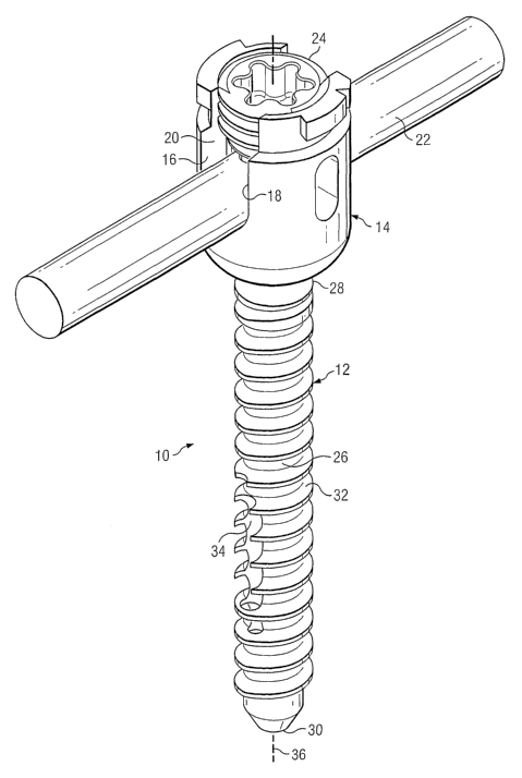
Turning now to Fig. 1, there is presented one illustrative embodiment of an
anchoring
system showing certain aspects of the present invention. As shown, a medical
implant device 10 includes an anchor 12 which may be coupled to a rod
receiving
part 14. In some embodiments, the rod receiving part 14 may include
noncontiguous
walls 16 and 18 which form a channel 20 for receiving a rod 22. In some
embodiments, there is a closure member 24 which engages the walls 16 and 18
and
thus applies pressure to the rod 22 to effectively clamp about the rod 22,
thereby
positionally securing the rod 22 relative to the anchor 12. Such a closure
member is
more fully described in a co-pending and commonly assigned U.S. Patent
Application Serial No. 10/805,967 filed on March 22, 2004 entitled "CLOSURE
MEMBER FOR A MEDICAL IMPLANT DEVICE" (hereafter "the '967 patent
application), which is hereby incorporated by reference.
In the illustrated embodiment, the anchor 12 has a shank 26 having a proximal
end
28 and a distal end 30. In this example embodiment, the anchor 12 illustrates
a
screw and thus has a helical thread 32 positioned about the shank 26. It is
important
to note that although a screw is illustrated, the anchor 12 could be any
suitable
anchor having any suitable surface. For example, the anchor 12 could be a ring
shank fastener, a barb, a nail, a brad or a trocar. Furthermore, the anchor 12
may
7
CA 02587570 2007-05-14
WO 2006/055448 PCT/US2005/041028
also have an expandable diameter which allows the anchor to "lock" into the
bone
after placement.
Proximal to the distal end 30, there may be a bore 34 the center of which may
be
rotatedly offset from a longitudinal axis 36 of the shank 26. As illustrated,
the bore
34 extends from the distal end 30 to the side of the shank 32.
Fig 2. illustrates a cross sectional view of one aspect of the anchor 12. In
this view,
it can be seen that the anchor 12 has a head 38 coupled to the proximal end 28
of
the shank 26. In this embodiment, the bore 34 begins at the distal end 30 and
exits
through a side 40 of the shank 26. In other words, in relation to the
longitudinal axis
io 36 of the shank 26, the bore 34 is angularly rotated about the distal end
30 of the
shank forming an acute angle a with the longitudinal axis. For the purposes of
this
application, the term "off-axis" shall be taken to mean a bore having a center
longitudinal axis which is non-concentric in relation to the center
longitudinal axis of
the shank. Thus, an off-axis bore may run either laterally parallel to the
longitudinal
is axis of the shank or be angularly rotated about the distal end of a shank
as illustrated
in Fig. 2.
As previously described, anchors, such as anchor 12, are typically subjected
to
relatively large forces. The large external forces and the overall placement
of such
anchors result in localized regions of higher stresses which may cause the
anchor to
2o break in such regions. A typical region of higher stress is illustrated as
region 42.
The region 42 is generally located along the shank 26 below the proximal end
28 of
the shank 26. Note that in this embodiment, the cross sectional area of the
shank 26
in region 42 has not been reduced. Thus, the full cross-sectional area of the
shank
26 is available in this region to resist the applied forces. Furthermore, the
use of the
25 full cross-sectional area (without cannulation) reduces the stress in the
region 42
which may greatly increase the strength of the anchor 12.
This arrangement is in contrast to conventional cannulated pedicle screws
which
have a cannluation extending entirely through the screw along their
longitudinal
center axes. The cannulation causes a reduction in cross sectional area at
high
30 stress locations which contributes to a failure of the cannulated screw.
8
CA 02587570 2007-05-14
WO 2006/055448 PCT/US2005/041028
The bore 34 may receive a guide wire (not shown). Because the bore does not
cause a cannulation through the entire length of the anchor, the bore may be
of a
relatively larger diameter when compared to conventional cannulated pedicle
screws.
The larger diameter bore allows the guide wire to also have a larger diameter,
which
increases the strength of the guide wire and reduces wire advancement and
kinking.
Furthermore, the larger diameter increases the strength of the guide wire and
may
allow a surgeon to have tactile feedback regarding the placement and location
of the
guide wire.
By having the guide wire exit the side of the anchor, the surgeon can keep
hold of
io the proximal end of the guide wire at all times throughout the procedure.
This may
help ensure that the guide wire does not advance as the screw is slid down the
wire.
Additionally, the relative short length of the bore 34 (compared to a
conventional
cannulated screw) will tend to reduce the friction between the instruments and
the
guide wire, thereby reducing guide wire advancement. Guide wire advancement
will
is also be reduced because the guide wire is pulled out prior to advancing the
anchor
so there is no guide wire advancement during screw insertion as with
conventional
cannulated systems.
Turning now to Fig. 3a, there is illustrated a front view of one embodiment of
the
anchor 12. As illustrated, the bore 34 (Fig. 2) forms a generally elongated
opening
2o 46 with the front side of the shank 26. In some embodiments, there may be a
series
of smaller openings 46a formed by the threads and the bore. In this
illustrative
embodiment, the head 38 of the anchor 12 may include an external helical
thread 50.
In some embodiments, the helical thread 50 may be a "reverse" screw thread
which
may be adapted to engage a corresponding reverse screw thread of the rod
25 receiving part 14 (Fig. 1). For purposes of this application, a reverse
screw thread is
a thread designed to engage a corresponding thread in an opposite rotational
direction when compared to conventional threads. The head 38 may include
various
recesses and/or protrusions 52 to engage a driving device (not shown) that may
be
used to drive the anchor 12 into the bone (not shown). In some embodiments,
the
3o driving device may also be used to remove an installed anchor from a bone.
9
CA 02587570 2007-05-14
WO 2006/055448 PCT/US2005/041028
Fig. 3b illustrates a front view of an off-axis pedicle screw 56 incorporating
various
aspects of the present invention. As illustrated, a bore 57 forms a generally
elongated opening 55 with one side of a shank 59. The off-axis pedicle screw
56
has an alternative embodiment of a head 58. In this illustrative embodiment,
the
head 58 may include one or more splines, for example splines 60a, 60b, and
60c.
The splines 60a, 60b, and 60c may be equally spaced circumferentially around
the
head 58. In some head embodiments, the splines 60a, 60b, and 60c may be spaced
at unequal distances circumferentially around the head 58. The splines 60a,
and
60b, and 60c may include surface protrusions, recesses and/or texturing to
enhance
io coupling of the off-axis pedicle screw 56 with a ring of a bone fastener
assembly (not
shown). In some embodiments, sides of the splines 60a, 60b, and 60c may have a
tapering so that the splines form a dovetail connection with a ring. In some
embodiments, the spline width may be tapered so that a good interference
connection is established when the bone screw is coupled to a ring. Splines
60a,
60b and 60c may include one or more projections (not shown) to facilitate
coupling
the head 58 to an inner surface of a ring which may be part of the rod
receiving part
assembly.
Turning now to Fig. 3c, there is illustrated a front view of another
alternative
embodiment of an off-axis pedicle screw 62. In some embodiments, the off-axis
pedicle screw 62 may be a have a head 64 which is adapted to be a fixed angle
fastener as depicted in Fig. 3c. Such fixed angle fastener heads are well
known in
the art.
Fig. 4 illustrates the anchor 12 coupled to a guide wire 72. As will be
explained in
detail below, once the guide wire 72 is in place, the guide wire 72 may be
slipped
through the bore 34 (not shown) of the anchor 12 as illustrated in Fig. 4. The
anchor
may also be coupled to a driving device 70.
As illustrated in Fig. 4, a distal end of driving device 70 is positioned in
an external
sleeve 74. In some embodiments, the sleeve 74 may be coupled to the rod
receiving
part 14 (Fig. 1) of the medical implant device 10. The driving device 70 may
include
3o an outer shaft 76, an inner shaft 78, and removable handle 80. The outer
shaft 76
may include a textured portion 82. In some embodiments, the textured portion
82
CA 02587570 2007-05-14
WO 2006/055448 PCT/US2005/041028
may facilitate rotation of the outer shaft 76 without the use of the removable
handle
80.
In some embodiments, the distal end of inner shaft 78 (not shown) may be
coupled
to the anchor 12 during use. The proximal end 81 of the inner shaft 78 may be
coupled to the removable handle 80. Thus, during the anchor placement, the
inner
shaft 78 may be rotatable relative to outer shaft 76 so that anchor 12 can be
inserted
into a bone. In some embodiments, a proximal portion of the inner shaft 78 may
include a coupling portion (not shown) which is adapted to mate with the
removable
handle 80. The removable handle 80 may also be adapted to fit other
instruments
io which may be used in the procedure such as a bone awl and/or a bone tap
(not
shown).
Fig. 5 illustrates an isometric view of an illustrative embodiment of a
dilator 84 which
may be used with various aspects of the present invention. As will be
explained
below, dilators are typically used in spinal procedures. The dilator 84 has a
is longitudinal slot 86 which allows the guide wire 72 to extend outside of
the dilator
during use.
Referring now to Figs. 6 to 11, the manner of using certain aspects of the
present
invention will now be described. The surgeon identifies the desired vertebral
levels
and pedicle positions via standard techniques. Once the target vertebrae are
20 identified, a small incision is made through the skin and a tracking needle
(or other
device) is inserted to pinpoint exactly where each anchor is to be placed. A
fluoroscope, or other x-ray technique, may be used to properly position the
tracking
needle. Once the proper position is located, the guide wire 72 may be
positioned
with its distal end against the pedicle, in this case pedicle 86 of vertebrae
L4 as
.25 illustrated in Fig. 6.
As shown in Fig. 7, the surgeon may then slide a series of continuing larger
sized
dilators 88a, 88b, 88c, and 88d down the guide wire 72. Approximately four or
five
dilators are used until a diameter suitable for passing the anchor and its
extensions
is achieved. In some embodiments, the last dilator used will be the slotted
side
3o dilator 84 discussed with reference to Fig. 5. Once slotted dilator 84 is
in place, the
other dilators 88a through 88d may be removed. In some embodiments, a bone awl
11
CA 02587570 2007-05-14
WO 2006/055448 PCT/US2005/041028
and/or bone tap may inserted over the guide wire to tap a hole into the
pedicle in
preparation for receiving the anchor 12, which in this case may be a pedicle
screw.
This tap will usually be a size slightly smaller than the pedicle screw thread
size
selected for that patient and that level.
After the hole is tapped and the inner dilators, such as dilators 88a-88d are
removed,
the surgeon is ready to introduce the anchor 12 into the vertebrae. As shown
in Fig.
8, prior to inserting the anchor 12 (e.g., pedicle screw), the guide wire 72
is placed
through the off-axis bore 34 (not shown) of the anchor 12. The anchor 12 may
be
coupled to the driving device 70 as previously described. The driving device
70
io engages the proximal end of the anchor 12. As the anchor 12 and the distal
end of
the driving device 70 enters the slotted dilator 84, the slot 86 of the
dilator allows the
guide wire 72 to extend beyond the passage of the dilator 84 as illustrated in
Fig. 9.
Once the anchor 12 is in position, which may be verified by flourscopy
techniques,
the guide wire 72 may be removed. It may also be desirable at this stage to
also
remove the dilator 84. To accomplish this, the removable handle 80 may be
removed in order to allow the dilator 84 to slip over the driving device 70 as
shown in
Fig. 10. Once the anchor 12 is in position, the driving device 70 may then be
rotated
into a proper position, as shown in Fig. 11. The surgeon may then screw the
anchor
12 into the pre-tapped hole in vertebrae L4. Pressure on the driving device 70
forces the anchor to be in-line with the external sleeve 74. A similar
procedure may
be repeated for each additional level, in this case level L5.
Once the pedicle screws are in place, an assembly may be coupled to the
pedicle
screws. For instance, Fig. 12 shows an example medical implant device 100. A
similar medical implant device 100 is described further in co-pending and
commonly
assigned U.S. Patent Application Serial No. 10/690,211 filed October 21, 2003
titled
"SYSTEM AND METHOD FOR STABILIZATION OF INTERNAL STRUCTURES"
(hereafter "the '211 patent application). More specifically, medical implant
device
100 may be a stabilization device that may include pedicle screws (or
"anchors")
102a and 102b that are inserted into vertebrae of a patient's spine, such as
vertebrae L4 and L5, respectively. The pedicle screws have off-axis bores 103a
and
103b which have been used in conjunction with guide wires (not shown) to guide
the
12
CA 02587570 2007-05-14
WO 2006/055448 PCT/US2005/041028
screws to the proper location. Assemblies 104a and 104b may be coupled to
pedicle
screws 102b and 102a, respectively. Such assemblies 104a and 104b each form a
receiving member for receiving closure member (e.g., set screw 106a or 106b).
Generally, such receiving member formed by assemblies 104a and 104b is a
noncontiguous (e.g., open-back member) having at least two walls, such as
walls
108a and 110a, that are separated by slots.
In this illustrative embodiment, closure member 106a and walls 108a and 110a
are
formed to have complementary threads that are formed in a manner that aids in
preventing splaying of the receiving members. In the specific implementation
shown,
io closure member 106 and walls 108a and 110a of the receiving member are
dovetail
configurations, such as described in the '967 patent application. Of course,
other
interlocking configurations, may be used in alternative implementations. As
further
shown in Fig. 12, a brace (or "rod") 112 extends from assembly 104a to
assembly
104b, and closure members (e.g., set screws) 106 are used for securing a first
end
is 114 of the brace 112 to the pedicle screw 102a and the other end 116 of the
brace
112 to pedicle screw 102b.
Thus, the medical implant device 100 may be installed using various aspects of
the
present invention. As previously described, the anchors 102a and 102b are able
to
resist larger forces than conventional cannulated screws. Furthermore, because
the
2o bores 103a and 103b do not cause a cannulation through the entire length of
the
anchors, the bores may be of a relatively larger diameter when compared to
conventional cannulated pedicle screws. The larger diameter bore allows the
guide
wire to also have a larger diameter, which increases the strength of the guide
wire,
reduces kinking, allows a surgeon to have tactile feedback regarding the
placement
25 and location of the guide wire, and allows the surgeon can keep hold of the
proximal
end of the guide wire at all times throughout the procedure. The relative
short length
of the bore reduces the friction between the instruments and the guide wire,
thereby
reducing guide wire advancement.
The foregoing description of the embodiments of the invention has been
presented
30 for the purposes of illustration and description. It is not intended to be
exhaustive or
to limit the invention to the precise form disclosed. Many modifications and
variations
13
CA 02587570 2007-05-14
WO 2006/055448 PCT/US2005/041028
are possible in light of the above teaching. It is intended that the scope of
the
invention be limited not by this detailed description, but rather by the
claims. For
instance, various embodiments of the present invention could be integrated
into
various navigational systems, such as the GE EM Tracking system.
Additionally, there may be additional embodiments such as a system for guiding
bone anchors, the system comprising a guide wire; an anchor comprising of a
shank
having a proximal end and a distal end, wherein the shank has an offset bore
at the
distal end which is adapted to allow the guide wire to slide through the bore;
a dilator
having a longitudinal slot wherein the longitudinal slot allows the guide wire
to extend
io through the longitudinal slot when the anchor is positioned within the
dilator; and a
driving device which is adapted to engage the proximal end of the anchor.
In some embodiments, the bore extends through the distal end of the shank to a
point located along a side of the shank.
In some embodiments, the bore is not concentric to a longitudinal center axis
of the
shank. i
In some embodiments, the driving device further comprises: an outer shaft, an
inner
shaft rotatedly disposed within the outer shaft having an distal end and
proximal end,
and a removable handle coupled to the proximal end.
In some embodiments, the outer shaft is adapted to couple with a rod receiving
portion of a medical implant device and the distal end of the inner shaft is
adapted to
engage the anchor.
Additionally, there may be a method for positioning a bone anchor into a bone,
the
method comprising: making an incision in the skin to allow access to the bone;
positioning a distal end of a guide wire to a desired location within the bone
to
slideably receive the dilators, and bone anchor; sliding a dialator having a
longitudinal slot over the guide wire; sliding the guide wire into a bore at
the distal
end of the anchor and causing the guide wire to exit the bore at a side
surface
opening; positioning the anchor into the dialator such that the guide wire
extends
through the longitudinal slot; and positioning the anchoring device at the
desired
location as defined by the distal end of the guide wire.
14
CA 02587570 2007-05-14
WO 2006/055448 PCT/US2005/041028
In some embodiments the method also includes sliding a series of continuing
large
dialator over the guide wire to increase the visual area of a surgical site.
In some embodiments the method also includes preparing a bore to receive the
anchoring device.
In some embodiments the method also includes rotating the anchoring device to
drive the anchoring device into the bone.
In some embodiments the method also includes checking the position of the
guide
wire with fluoroscopic techniques.
Additionally, there may be a guided bone anchoring device comprising: a head;
a
io shank having a proximal end and a distal end, wherein the proximal end is
coupled
to the head; and a bore extending through the distal end of the shank to a
point
located along a side of the shank.
In some embodiments, the head is bulbous.
In some embodiments, the head has an exterior helical thread adapted to mate
with
is a rod receiving device.
In some embodiments, the thread is a reverse thread.
In some embodiments, the shank has an exterior helical thread adapted to mate
with
the bone.
In some embodiments, the head has at least one longitudinal spline configured
to
20 couple with a ring within a rod receiving part of an implant system.
In some embodiments, the head is generally U-shaped.
Additionally, in some embodiments, there may be a guided bone anchoring device
comprising: a head; a shank having a proximal end, a distal end, and a
longitudinal
center axis running between the proximal and distal end; and a passage for
receiving
25 a guide wire, the passage having a longitudinal center axis which is not
concentric to
the longitudinal center axis of the shank.
CA 02587570 2007-05-14
WO 2006/055448 PCT/US2005/041028
In some emaoaiments, tne neaa is qUIpoUS ana nas an exterior neiicai tnreaa
adapted to mate with a rod receiving device.
In some embodiments, the thread is a reverse thread.
16