Note: Descriptions are shown in the official language in which they were submitted.
CA 02587684 2012-10-29
SOFT LINEAR MAPPING CATHETER WITH STABILIZING TIP
FIELD OF INVENTION
[0001]
The present invention relates to an improved
mapping catheter that is particularly useful for mapping
electrical activity in a wall region of or near the heart.
BACKGROUND OF INVENTION
[0002] Atrial fibrillation is a common sustained
cardiac arrhythmia and a major cause of stroke. Atrial
fibrillation results in a fast and irregular cardiac rhythm
which often leads to palpitations and a deterioration of
cardiac function with cardiac output decreasing by an average
of 30%. There is also an increased incidence of intra cardiac
thrombosis (blood clotting) which can potentially lead to
embolic events such as strokes.
Consequently, 20 to 35% of
cerebrovascular accidents (CVAs) are related to paroxysmal or
chronic atrial fibrillation.
[0003]
This condition is perpetuated by reentrant
wavelets propagating in an abnormal atrial-tissue substrate.
Various approaches have been developed to interrupt wavelets,
including surgical or catheter-mediated atriotomy. Atrial
fibrillations can also be treated by pulmonary vein isolation
which proves to be insufficient in 30 to 50% of paroxysmal
atrial fibrillation patients and 90% of permanent atrial
fibrillation.
In such cases, it may be necessary to ablate
and perform linear lesions in addition to pulmonary vein
isolation, in the right and left atriums.
These linear
lesions have been done using RF ablation catheters for about a
decade.
Each lesion should ideally be transmural and
continued with adjacent lesions so as to obtain a final linear
lesion blocking electrical activity between two natural areas
of block. The most common locations of these lines are the
mitral isthmus in the left atrium, with a lesion extending
-1-
CA 02587684 2012-10-29
from the mitral annulus to the left inferior pulmonary vein.
Other possible locations include the roof of the left atrium,
with a lesion connecting the ostium of the superior right
pulmonary vein to the left superior vein. However, because
conventional catheters generally treat tissue in a localized
manner, numerous repeated applications of the catheter are
typically needed to form a linear lesion.
Thus, while the
formation of linear lesions is possible, it can be a time-
consuming, labor-intensive procedure.
[0004] Prior to
treating the condition, one has to
first determine the location of the wavelets. Various
techniques have been proposed for making such a determination.
None of the proposed techniques, however, provide sufficient
assistance in guiding the formation of the linear lesion or
easing the linear line assessment process, particularly for
regions of the mistral isthmus and the left atrium roof.
SUMMARY OF THE INVENTION
[0005]
A catheter adapted for mapping near a tubular
region of a heart, has an elongated tubular catheter body
having proximal and distal ends, an intermediate section
distal of the catheter body, and a mapping assembly at the
distal end of the intermediate section.
The electrode-
carrying mapping assembly has a generally circular main
segment with a support member having shape-memory, and a
generally linear proximal segment which has greater
flexibility than either the intermediate section or the
generally circular main segment. In accordance with the
present invention, the generally circular main segment is
adapted to releasably anchor itself in the tubular region and
to map circumferentially around the tubular region and the
generally linear segment is adapted to contact generally along
its length heart wall tissue near an ostium of the tubular
region. Advantageously, the mapping assembly is adapted to
-2-
,
CA 02587684 2012-10-29
conduct mapping of said wall tissue along a linear pattern
extending radially from the ostium.
Moreover, the mapping
assembly is adapted to be rotated about the ostium to perform
mapping of said wall tissue along different radially-extending
linear patterns about the ostium. To that end, the generally
linear segment has a proximal portion that is generally devoid
of electrodes and the catheter includes a control handle to
deflect the catheter along the intermediate section.
[0006]
In another embodiment of the present invention,
a catheter adapted for mapping near a tubular region of a
heart, has an elongated tubular catheter body having proximal
and distal ends and a mapping assembly at the distal end of
the catheter body.
The electrode-carrying mapping assembly
has a generally circular main segment with a support member
having shape-memory, and a generally linear proximal segment
which has greater flexibility than either of the catheter body
and the generally circular main segment. In accordance with
the present invention, the generally circular main segment is
adapted to releasably anchor itself in the tubular region and
to map circumferentially around the tubular region and the
generally linear segment is adapted to contact generally along
its length heart wall tissue near an ostium of the tubular
region. Advantageously, the mapping assembly is adapted to
conduct mapping of said wall tissue along a linear pattern
extending radially from the ostium.
Moreover, the mapping
assembly is adapted to be rotated about the ostium to perform
mapping of said wall tissue along different radially-extending
linear patterns about the ostium. To that end, the generally
linear segment has a proximal portion that is generally devoid
of electrodes and the catheter includes a control handle to
deflect the catheter along the generally linear segment of the
mapping assembly.
-3-
I
,
CA 02587684 2012-10-29
. .
[0007]
In another embodiment, electrodes are carried
on both the generally linear segment and the generally
circular segment.
In a more detailed embodiment, the
generally linear segment has a length of about 30 mm and the
generally circular main segment has an outer diameter of about
25 mm. Moreover, both the generally linear segment and the
generally circular main segment may each carry at least five
ring electrode pairs.
[0008]
The present invention also includes a method
for mapping electrical activity of wall tissue near a tubular
region of or near the heart, the method using a catheter in
accordance with the present invention. In one embodiment, the
method includes inserting the generally circular segment of a
catheter in accordance with the present invention into a
tubular region of or near the heart, releasably anchoring the
generally circular segment in the tubular region near its
ostium, contacting the generally linear segment of the
catheter generally along its length with wall tissue near the
ostium, and
mapping the electrical activity of the wall
tissue along a linear pattern extending radially from the
ostium.
The method may also include rotating the mapping
assembly about the ostium and mapping the electrical activity
of the wall tissue along a different linear pattern extending
radially from the ostium. The tubular region is selected from
the group consisting of pulmonary veins, the coronary sinus,
the superior vena cava, and the inferior vena cava.
BRIEF DESCRIPTION OF THE DRAWINGS
[0009]
These and other features and advantages of the
present invention will be better understood by reference to
the following detailed description when considered in
conjunction with the accompanying drawings wherein:
[0010]
Fig. 1 is a side view of an embodiment of the
catheter of the present invention.
-4-
i
CA 02587684 2012-10-29
. .
[0011] Fig. 2 is a side cross sectional view of the
catheter body of Fig. 1, including the junction between the
catheter body and the intermediate section.
[0012] Fig. 3 is a side cross sectional view of the
catheter body of Fig. 1, including the junction between the
intermediate section and the mapping assembly.
[0013] Fig. 4 is a schematic perspective view of an
embodiment of the mapping assembly according to the present
invention.
[0014] Fig. 5 is a schematic perspective view of the
mapping assembly of Fig. 4 with its distal portion releasably
anchored in a tubular region and its proximal portion
generally lying against tissue surrounding the tubular region.
[0015] Fig. 6 is a side cross sectional view of an
embodiment of the junction between the proximal region and the
distal region of the mapping assembly.
[0016] Fig. 6a is a cross sectional view taken along
6A-6A in Fig. 6.
[0017] Fig. 6b is a side cross sectional view of an
embodiment of the junction between the proximal region and the
distal region of the mapping assembly, having lead wires for
electrodes on the distal region.
[0018] Fig. 7 is a side view of an embodiment of the
mapping assembly according to the present invention in a
clockwise formation.
[0019] Fig. 8 is a side view of the mapping assembly
of Fig. 7 in a counterclockwise formation rotated 90 .
[0020] Fig. 9 is a schematic view of an embodiment of
the mapping assembly according to the present invention.
[0021] Fig. 10 is a schematic view of the mapping
assembly according to the present invention depicting the
relationship between two electrodes.
-5-
I
CA 02587684 2012-10-29
. .
[0022] Fig. 11 is a schematic view of an alternative
embodiment of the mapping assembly according to the present
invention.
[0023] Fig. 12 is a schematic view of another
alternative embodiment of the mapping assembly according to
the present invention.
[0024] Fig. 13 is a schematic view of yet another
alternative embodiment of the mapping assembly according to
the present invention.
[0025] Fig. 14 is a side view of an alternative
embodiment of the catheter of the present invention.
[0026] Fig. 15 is a schematic perspective view of an
embodiment of the mapping assembly of Fig. 14.
[0027] Fig. 15a is a cross sectional view taken along
line 15A-15A in Fig. 15.
[0028] Fig. 16 is a side cross sectional view of the
catheter body of Fig. 1, including the junction between the
catheter body and the mapping assembly.
[0029] Fig. 17 is a side cross sectional view of an
alternative embodiment of the junction between the proximal
and distal regions of the mapping assembly, with lead wires
for electrodes carried on the distal region.
[0030] Fig. 17a is a cross sectional view taken along
lines 17a-17a in Fig. 17.
DETAILED DESCRIPTION OF THE INVENTION
[0031] In a disclosed embodiment of the invention,
there is provided a catheter 10 having a mapping assembly at
its distal end. As shown in FIG. 1, the catheter comprises an
elongated catheter body 12 having proximal and distal ends, an
intermediate section 14 at the distal end of the catheter
body, a control handle 16 at the proximal end of the catheter
body, and a mapping assembly 17 mounted at the distal end of
the catheter to the intermediate section 14.
-6-
CA 02587684 2012-10-29
[0032] With reference to FIG. 2, the catheter body 12
comprises an elongated tubular construction having a single,
axial or central lumen 18. The catheter body 12 is flexible,
i.e., bendable, but substantially non-compressible along its
length. The catheter body 12 can be of any suitable
construction and made of any suitable material. A presently
preferred construction comprises an outer wall 20 made of
polyurethane or PEBAX. The outer wall 20 comprises an imbedded
braided mesh of stainless steel or the like to increase
torsional stiffness of the catheter body 12 so that, when the
control handle 16 is rotated, the intermediate section 14 of
the catheter 10 will rotate in a corresponding manner.
[0033] The outer diameter of the catheter body 12 is
not critical, but is preferably no more than about 8 french,
more preferably 7 french. Likewise the thickness of the outer
wall 20 is not critical, but is thin enough so that the
central lumen 18 can accommodate a puller wire, lead wires,
and any other desired wires, cables or tubings. If desired,
the inner surface of the outer wall 20 is lined with a
stiffening tube (not shown) to provide improved torsional
stability. A disclosed embodiment, the catheter has an outer
wall 20 with an outer diameter of from about 0.090 inch to
about 0.94 inch and an inner diameter of from about 0.061 inch
to about 0.065 inch.
[0034] With further reference to Fig. 2, the
intermediate section 14 comprises a short section of tubing 22
having three lumens. The first lumen 30 electrode carries lead
wires 50 and the second lumen 32 carries a puller wire 64.
There may also be third lumen 34. The tubing 22 is made of a
suitable non-toxic material that is preferably more flexible
than the catheter body 12. A suitable material for the tubing
22 is braided polyurethane, i.e., polyurethane with an
embedded mesh of braided stainless steel or the like. The size
-7-
CA 02587684 2012-10-29
of each lumen is not critical, but is sufficient to house the
lead wires or the puller wire.
[0035] The useful length of the catheter, i.e., that
portion that can be inserted into the body excluding the
mapping assembly 17, can vary as desired. In one embodiment,
the useful length ranges from about 110 cm to about 120 cm.
The length of the intermediate section 14 is a relatively
small portion of the useful length, and preferably ranges from
about 3.5 cm to about 10 cm, more preferably about 4cm to
about 8cm, and still more preferably about 6.5 cm.
[0036] A preferred means for attaching the catheter
body 12 to the intermediate section 14 is illustrated in FIG.
2. The proximal end of the intermediate section 14 comprises
an outer circumferential notch 26 that receives the inner
surface of the outer wall 20 of the catheter body 12. The
intermediate section 14 and catheter body 12 are attached by
glue or the like.
[0037] If desired, a spacer (not shown) can be located
within the catheter body between the distal end of the
stiffening tube (if provided) and the proximal end of the
intermediate section. The spacer provides a transition in
flexibility at the junction of the catheter body and
intermediate section, which allows this junction to bend
smoothly without folding or kinking. A catheter having such a
spacer is described in U.S. Pat. No. 5,964,757.
[0038] Within the intermediate section 14, the puller
wire 64 extends into the second lumen 32. Preferably the
puller wire 64 is anchored at its distal end to the distal end
of the intermediate section 14, as shown in FIG. 3.
Specifically, a T-shaped anchor is formed, which comprises a
short piece of tubular stainless steel 80, e.g., hypodermic
stock, which is fitted over the distal end of the puller wire
64 and crimped to fixedly secure it to the puller wire. The
-8-
CA 02587684 2012-10-29
distal end of the tubular stainless steel 80 is fixedly
attached, e.g., by welding, to a cross-piece 82 formed of
stainless steel ribbon or the like. The cross-piece 82 extends
through a hole formed in the outer wall and because the cross-
piece 82 is larger than the hole and, therefore, cannot be
pulled through the hole, the cross-piece 82 anchors the distal
end of the puller wire 64 to the distal end of the
intermediate section 14. Within the second lumen 32 of the
intermediate section 14, the puller wire 64 extends through a
plastic, preferably Teflon , puller wire sheath (not shown),
which prevents the puller wire 64 from cutting into the wall
of the intermediate section 14 when the intermediate section
is deflected.
It is understood that the puller wire 64
enables the catheter to deflect generally along the length of
the intermediate section 14.
[0039]
Longitudinal movement of the puller wire 64
relative to the catheter body 12, which results in deflection
of the intermediate section 14 and generally the mapping
assembly 17, is accomplished by suitable manipulation of the
control handle 16. Examples of suitable control handles for
use in the present invention are disclosed, for example, in
U.S. Pat. Nos. Re 34,502 and 5,897,529.
[0040]
At the distal end of the intermediate section
14 is a mapping assembly 17, as shown in FIGS. 4 and 5. The
mapping assembly comprises a more flexible, generally straight
proximal region 38 and a less flexible but pre-shaped distal
region 40 having a straight proximal segment 42, a
transitional segment 44 and a generally circular main segment
46.
[0041] The
proximal region 38 is mounted on the distal
end of the intermediate section 14, as described in more
detail below, so that its axis is generally parallel to the
axis of the intermediate section, and preferably has an
-9-
CA 02587684 2012-10-29
exposed length, e.g., not contained within the intermediate
section 14, ranging from about 20 mm to about 70 mm, more
preferably about 25 mm to about 50 mm, still more preferably
about 42 mm, but can vary as desired.
[0042] As
illustrated in Figs. 3 and 6, the proximal
region 38 comprises a tubing 39 which can be made of any
suitable material that is flexible and biocompatible and
preferably plastic, such as polyurethane or PEBAXTM.
The
tubing 39 may have any cross-sectional shape and may have a
single lumen or multiple lumens and is generally free of any
interior support members although its lumen is occupied by
lead wires 50 or other electrical connections for electrodes
or any other electrical or electromagnetic elements that may
be mounted on the mapping assembly 17. A preferred means for
attaching the tubing 39 to the intermediate section 14 is
illustrated in FIG. 3. The proximal end of the tubing 39
extends over and overlaps with the distal end of the tubing
22. Glue or the like is applied between the contacting inner
surface of the tubing 39 and outer surface of the tubing 22.
Additional glue may be applied immediately proximal of the
proximal end of the section 38 to form a seal 41.
[0043]
As shown in Figs. 4 and 5, a series of paired
ring electrodes 36 are mounted on the tubing 39 of the
proximal region 38. The ring electrodes 36 can be made of any
suitable solid conductive material, such as platinum or gold,
preferably a combination of platinum and iridium, and mounted
onto the tubing 39 with glue or the like. Alternatively, the
ring electrodes can be formed by coating the tubing 39 with an
electrically conducting material, like platinum, gold and/or
iridium. The coating can be applied using sputtering, ion beam
deposition or an equivalent technique.
[0044]
In a preferred embodiment with reference to
Fig. 6, each ring electrode 36 is mounted by first forming a
-10-
CA 02587684 2012-10-29
hole in the tubing 39. An electrode lead wire 50 is fed
through the hole, and the ring electrode 36 is welded in place
over the lead wire and tubing 39. The lead wires 50 extend
through the tube 39. The proximal end of each lead wire 50 is
electrically connected to a suitable connector 37 (Fig. 1),
which is connected to a source of RF energy (not shown).
[0045] The number of the ring electrodes 36 on the
assembly 17 can vary as desired. Preferably, the number of
ring electrodes ranges from about six to about twenty,
preferably from about eight to about twelve. In a disclosed
embodiment, the assembly carries ten ring electrodes forming
five pairs. The pairs of ring electrodes 36 are preferably
approximately evenly spaced along the proximal region 38, as
best shown in FIG. 4. In a disclosed embodiment, a distance of
approximately 5 mm is provided between each pairs of the ring
electrodes 36, which each electrode within a pair separated by
a distance of about 1 mm. Advantageously, the proximal region
38 includes a proximal segment 38a which is generally devoid
of electrodes such that the mapping assembly 17 can generally
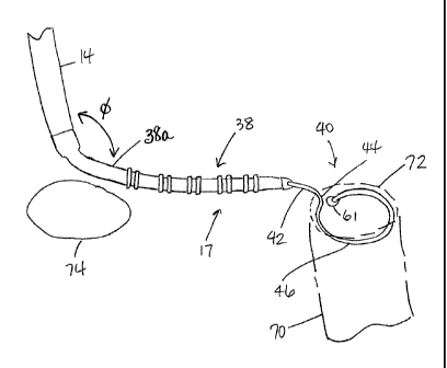
lay against wall tissue, as shown in Fig. 5. In
particular,
the proximal segment 38a enables the distal end of the
intermediate section 14 and the proximal end of the mapping
assembly 17 (e.g., the proximal segment 38a) to define an
angle phi therebetween ranging between about 45 and about
315 .
The proximal segment 38a may have a length ranging
between about 0.5 inch and about 2.0 inches, and more
preferably about 1.0 inch.
[0046] As for the distal region 40 of the assembly 17,
the straight segment 42 is mounted on the distal end of the
proximal region 38, as described in more detail below, so that
its axis is generally parallel to the axis of the proximal
region 38 and preferably has an exposed, length, e.g., not
-11-
CA 02587684 2012-10-29
contained within the proximal region 38, ranging from about
10-20, more preferably about 15 mm, but can vary as desired.
[0047]
The generally circular main segment 46 is
generally traverse to the catheter body 12 and is preferably
generally perpendicular to the catheter body 12.
The
generally circular main segment need not form a flat circle,
but can be very slightly helical, as shown in FIGS. 4, 7 and
8. The main segment 46 has an outer diameter preferably
ranging to about 10 mm to about 35 mm, more preferably about
15 mm to about 30 mm, still more preferably about 25 mm. The
transition segment 44 between the segments 42 and 46 is
slightly curved and formed such that, when viewed from the
side with the segment 42 at the top of the circular main
segment 46 as shown in FIG. 7, the proximal segment 42 (along
with the proximal region 38) forms an angle u with the
circular main segment 46 ranging from about 75 to about 95 ,
preferably from about 83 to about 93 , more preferably about
87 . The main region segment 46 can curve in a clockwise
direction, as shown in FIG. 7 or a counterclockwise direction,
as shown in FIG. 8. When the assembly 17 is turned 90 , as
shown in FIG. 8, so that the transition segment 44 is near the
center of the main segment 46, the proximal segment 42 (along
with the proximal region 38) forms an angle g with the main
circular segment 46 ranging from about 90 to about 135 ,
preferably from about 100 to about 110 , more preferably
about 105 .
[0048]
As illustrated in Fig. 6, the distal region 40
of the mapping assembly 17 is formed from a support member 54
covered by a non-conductive covering 56. The support member 54
is made of a material having shape-memory, i.e., that can be
straightened or bent out of its original shape upon exertion
of a force and is capable of substantially returning to its
original shape upon removal of the force. A suitable material
-12-
,
CA 02587684 2012-10-29
for the support member 54 is a nickel/titanium alloy. Such
alloys typically comprise about 55% nickel and 45% titanium,
but may comprise from about 54% to about 57% nickel with the
balance being titanium. A preferred nickel/titanium alloy is
nitinol, which has excellent shape memory, together with
ductility, strength, corrosion resistance, electrical
resistivity and temperature stability. The non-conductive
covering 56 can be made of any suitable material, and is
preferably made of a biocompatible plastic such as
polyurethane or PEBAX.
[0049] A means for attaching the distal region 40 to
the proximal region 38 is illustrated in FIG. 6. At the
proximal end of the distal region 40, a stainless steel tubing
71 is welded onto the distal end of the support member 54 at
their respective contacting surface 75. A proximal end of the
tubing 71 is flattened or otherwise shaped to form a spade 73
with an elongated cross-section (Fig. 6A) which anchors the
proximal end of the distal region 40 in the distal end of the
proximal region 38. In particular, the spade 73 sit within a
cored-out distal end of the tubing 39 forming a notch 84. Glue
or the like is applied to the distal end of the tubing 39 to
form a plug 53 sealing the region of attachment. As such, the
elongated cross-section of the spade 73 anchors the distal
region 40 against rotational movement about the axis of the
support member 54 relative to the proximal region 38.
Moreover, a base 86 of the spade 73 anchors the distal region
40 against distal movement relative to the proximal region 38.
[0050] The proximal end of the support member 54 and
the nonconductive covering 56 terminate a short distance
within the lumen of the tubing 39 of the proximal section 38,
approximately about 5 mm, so as not to adversely affect the
flexibility of the proximal section 38.
-13-
CA 02587684 2012-10-29
. .
[0051]
Because the proximal region 38 is generally
without any internal structure other than lead wires 50 from
the electrodes or perhaps a second puller wire for changing
the diameter of the circular segment 46, the proximal region
38 is more flexible than either the tip section 14 or the
distal region 40. In that regard, the tubing 39 has a Young's
Modulus or flexibility durometer rating lesser than either the
intermediate section 14 or the distal region 40 and ranging
between about 35D to 60D (22.8 MPA to 88.0 MPA), more
preferably about 55D (67.2 MPA).
The lesser flexibility of
the tip section 14 and the distal region 40 relative to the
proximal region 38 (due to the underlying tubing structure
and/or internal structures or wires extending therethrough)
enables the user to manipulate the mapping assembly 17 to
reach the target site, and further to manipulate the circular
segment 46 to enter into and releasably anchor itself in a
tubular region, e.g., a pulmonary vein.
With a greater
flexibility, the proximal region 38 can then be manipulated to
generally lie flat against wall tissue around an ostium of the
tubular region, as shown in FIG. 5.
In accordance with a
feature of the present invention, the proximal region 38 has
greater softness, floppiness and/or flexibility relative to
the intermediate section 14 and the distal region 40 of the
mapping assembly 17.
[0052] If desired, additional electrodes 58 could be
mounted along the circular segment of the distal region distal
region 40. FIG. 9 shows one electrode arrangement for the
circular segment 39. As explained above, the generally
circular main segment 39 is very slightly helical, although
FIGS. 9 and 11 depict the main segment 39 as a flat circle, as
it would generally appear when viewed from the distal end of
the catheter. Referring to both Figs. 9 and 10, a first ring
electrode 58a is provided, which is the electrode that is on
-14-
,
CA 02587684 2012-10-29
the generally circular main segment 46 closest to the
transitional segment 44. A second electrode 58b is provided,
which is the electrode that is on the generally circular main
segment 46 adjacent its tangent location 43 (Fig. 10).
Preferably, the first electrode 58a is positioned along the
circumference of the generally circular main segment 46 at a
distance 0 of no more than about 55 from the tangent location
43, more preferably no more than about 48 from the tangent
location, still more preferably from about 15 to about 36
from the tangent location. Preferably the second electrode 58b
is positioned along the circumference of the generally
circular main segment at a distance 0 of no more than about
55 from the tangent location, more preferably no more than
about 48 from the tangent location 43, still more preferably
from about 15 to about 36 from the tangent location.
Preferably the first electrode 58a is positioned along the
circumference of the generally circular segment at a distance
y of no more than 100 from the second electrode 58b,
preferably no more than 80 from the second electrode, still
more preferably from about 30 to about 75 from the second
electrode. There is also shown an electrode 58c in Fig 9.
which is longer than the other ring electrodes, preferably
having a length ranging from about lmm to about 1.5mm. The
longer ring electrode provides a signal to the user when the
catheter is being viewed under fluoroscopy.
By having one
ring electrode, such as the electrode 58c, sized differently
from the other ring electrodes, the user has a reference point
when viewing the catheter under fluoroscopy.
[0053]
Fig. 11 shows another electrode arrangement for
the main segment 46 where generally the single ring electrodes
58 have been configured into electrode pairs 57.
It is
understood that lead wires 50b for the electrodes 58 may
extend parallel with the support member 58 through the
-15-
CA 02587684 2012-10-29
. .
nonconductive covering 58 of the distal region 40 and through
the lumen of the tubing 39 of the proximal region 38, as shown
in Fig. 6b.
[0054]
As shown in the embodiment of Figs 1-11, the
distal end of the generally circular segment 46 may be capped,
preferably with polyurethane glue, to form an atraumatic cap
61 (Figs. 4 and 5) and to prevent body fluids from entering
the mapping assembly 17.
[0055]
In an alternative design as shown in Figs. 12
and 13, the mapping assembly 17 includes a generally straight
distal segment 48 which forms a tangent relative to the
generally circular segment and contacts the main segment at
the tangent location. The generally straight distal segment 48
is provided with an atraumatic design to prevent the distal
end of the mapping assembly 17 from penetrating tissue. In the
depicted embodiment, the distal segment comprises a tightly
wound coil spring 44 made, for example, of stainless steel,
such as the mini guidewire commercially available from Cordis
Corporation (Miami, Fla.) or a coil having a 0.0045 inch wire
size and a 0.009 inch inner diameter, such as that
commercially available from Microspring. The coil spring 44 is
mounted at its proximal end in a short piece of tubing 55 with
polyurethane glue or the like, which is then glued or
otherwise anchored within the non-conductive covering. The
tubing 55 is less flexible than the nonconductive covering 56
but more flexible than that support member 54 to provide a
transition in flexibility along the length of the mapping
assembly 17. The distal end of the distal segment 40 is
capped, preferably with polyurethane glue 65, to prevent body
fluids from entering the mapping assembly 17.
[0056] In the depicted embodiment, the generally
straight distal segment 48 has a length of about 0.5 inch, but
can be any desired length, for example, ranging from about
-16-
,
CA 02587684 2012-10-29
. .
0.25 inch to about 1.0 inch. The generally straight distal
segment 48 is preferably sufficiently long to serve as an
anchor for introducing the catheter into a guiding sheath, as
discussed in more detail below, because the mapping assembly
17 must be straightened upon introduction into the sheath.
Without having the generally straight distal segment 48 as an
anchor, the mapping assembly 17 has a tendency to pull out of
the guiding sheath upon its introduction into the guiding
sheath. Any other atraumatic tip design that prevents the
distal end of the mapping assembly from penetrating tissue
could be provided. An alternative design in the form of a
plastic ball is described in copending U.S. Patent No.
6,371,955, entitled "ATRIAL BRANDING IRON CATHETER AND METHOD
FOR TREATING ATRIAL FIBRILLATION". Additionally, if desired,
the distal segment 48 can be formed, at least in part, of a
radiopaque material to aid in the positioning of the mapping
assembly 17 under fluoroscopy. A suitable and similar distal
segment is disclosed in U.S. Pat. No. 6,711,428.
[0057] The lead wires 50 attached to the ring
electrodes 36 extend through the lumen of the tubing 39 of the
proximal region 38 (Fig. 6), through the first lumen 30 of the
intermediate section 14 (Fig. 3), through the central lumen 18
of the catheter body 12 (Fig. 2), and the control handle 16,
and terminate at their proximal end in the connector 37 (Fig.
1). The portion of the lead wires 50 extending through the
central lumen 18 of the catheter body 12, control handle 16
and proximal end of the intermediate section 14 are enclosed
within a protective sheath 62 (Fig. 2), which can be made of
any suitable material, preferably polyimide. The protective
sheath 62 is anchored at its distal end to the proximal end of
the intermediate section 14 by gluing it in the first lumen 30
with polyurethane glue or the like.
-17-
,
CA 02587684 2012-10-29
=
[0058] The puller wire 64 is provided for deflection
of the intermediate section 14. The puller wire 64 extends
through the catheter body 12 (Fig. 2) and the second lumen 32
of the intermediate section 14 (Fig. 3). The puller wire 64
is anchored at its proximal end to the control handle 16, and
is anchored at its distal end to the intermediate section 14.
The puller wire 64 is made of any suitable metal, such as
stainless steel or Nitinol, and is preferably coated with
Teflon , or the like. The coating imparts lubricity to the
puller wire 64. The puller wire 64 preferably has a diameter
ranging from about 0.006 to about 0.010 inch.
[0059] As shown in Fig. 2, a compression coil 66 is
situated within the catheter body 12 in surrounding relation
to the puller wire 64. The compression coil 66 extends from
the proximal end of the catheter body 12 to the proximal end
of the intermediate section 14. The compression coil 66 is
made of any suitable metal, preferably stainless steel. The
compression coil 66 is tightly wound on itself to provide
flexibility, i.e., bending, but to resist compression. The
inner diameter of the compression coil 66 is preferably
slightly larger than the diameter of the puller wire 64. The
Teflon , coating on the puller wire 64 allows it to slide
freely within the compression coil 66. The outer surface of
the compression coil 66 is covered by a flexible, non-
conductive sheath 68, e.g., made of polyimide tubing.
[0060] The compression coil 66 is anchored at its
proximal end to the outer wall 20 of the catheter body 12 by
proximal glue joint 70 and at its distal end to the
intermediate section 14 by distal glue joint 72. Both glue
joints 70 and 72 preferably comprise polyurethane glue or the
like. The glue may be applied by means of a syringe or the
like through a hole made between the outer surface of the
catheter body 12 and the central lumen 18. Such a hole may be
-18-
CA 02587684 2012-10-29
. ,
formed, for example, by a needle or the like that punctures
the outer wall 20 of the catheter body 12 which is heated
sufficiently to form a permanent hole. The glue is then
introduced through the hole to the outer surface of the
compression coil 66 and wicks around the outer circumference
to form a glue joint about the entire circumference of the
compression coil.
[0061] In use, a suitable guiding sheath is inserted
into the patient with its distal end positioned at a desired
mapping location, for example, the left atrium of the heart.
An example of a suitable guiding sheath for use in connection
with the present invention is the PrefaceTM. Braiding Guiding
SheathTM, commercially available from Cordis Webster (Diamond
Bar, Calif.). The distal end of the sheath is guided into one
of the atria. A catheter in accordance with the present
invention is fed through the guiding sheath until its distal
end extends out of the distal end of the guiding sheath. As
the catheter is fed through the guiding sheath, the mapping
assembly 17 is straightened to fit through the sheath. Once
the distal end of the catheter is positioned at the desired
mapping location, the guiding sheath is pulled proximally,
allowing the deflectable intermediate section 14 and mapping
assembly 17 to extend outside the sheath, and the distal
region 40 of the mapping assembly 17 returns to its original
shape due to the shape-memory of the support member 54. The
distal region 40 of mapping assembly 17, in particular, the
generally circular main segment 46 (with or without the distal
segment 48) is then inserted into a pulmonary vein 70 (Fig. 5)
so that the outer circumference of the generally circular main
segment 46 of the assembly is in contact with a circumference
inside the tubular region. Preferably at least about 50%, more
preferably at least about 70%, and still more preferably at
least about 80% of the circumference of the generally circular
-19-
CA 02587684 2012-10-29
main segment 46 is in contact with a circumference inside the
tubular region. As such, the circular segment 46 is therefore
releasably anchored in the tubular region, e.g., a pulmonary
vein 70 which enables the more flexible proximal region 38
carrying the electrodes 36 to contact and lay flat against
wall tissue near and surrounding an ostium 72 or extending
between the ostium 72 and another ostium 74 of another
pulmonary vein.
Consequently, a user can bridge the linear
gap between pulmonary veins for mapping and/or ablation
purposes with one placement of the catheter instead of
multiple placements. Benefits thereof include the ability to
guide burns to locations that do not show yet a complete
lesion and the ability to obtain a complete linear lesion with
fewer burns. In particular, the configuration, including the
length, of the proximal region 38 enables the proximal region
38 to serve as a generally linear template or guide against
which another catheter tip can be moved along.
[0062] The releasable anchoring and stabilization
provided by the circular segment 46 generally enables the
distal region 40 to remain relatively stationary in the
tubular region while the proximal region 38 can be manipulated
to rotate about the ostium 72 so as to sweep a circular region
around the ostium 72.
For example, if an angle zero is
defined by an axis extending between the ostia 72 and 74, the
proximal region 38 may be manipulated to sweep out 360 around
the ostium 72.
With the generally linear mapping
configuration of the electrodes 36 carried on the proximal
region 38, a multitude of radially extending linear mappings
can be accomplished about the ostium 72 as the proximal region
38 is rotated about the ostium 72. Moreover, when such linear
mappings are completed, the circular segment 46 can be
inserted into the ostium 74 where a multitude of radially
-20-
CA 02587684 2012-10-29
extending linear mappings can be accomplished about the ostium
74.
[0063] Where the circular segment 46 carries the
electrodes 58, the circular arrangement of the electrodes 58
permits measurement of the electrical activity at that
circumference of the tubular structure so that ectopic beats
between the electrodes 58 can be identified. The size of the
generally circular main segment 46 permits measurement of
electrical activity along a diameter of a pulmonary vein or
other tubular structure of or near the heart because the
circular main segment has a diameter generally corresponding
to that of a pulmonary vein or the coronary sinus.
Additionally, because the main segment 46 need not form a flat
circle, but can be somewhat helical, it is easier for the user
to guide the mapping assembly 17 into a tubular region.
[0064]
In an alternative embodiment of the present
invention, the catheter 10 of Figs. 14-17, where similar
components are designated by similar reference numerals,
generally except as discussed herein, the distal end of the
catheter body 12 is joined with the proximal end of a mapping
assembly 17' having a proximal region 38' and a distal region
40. The useful length of the catheter may range between about
110 cm and about 120 cm, and more preferably about 115 cm.
[0065]
In the illustrated embodiment, the proximal
region 38' is more flexible than either the catheter 12 and
the mapping assembly 17' and includes an elongated proximal
segment 38a' that is generally devoid of electrodes serving
generally the same function as describe hereinabove in
relation to the segment 38a.
The proximal region 38'
comprises a tubing 39' having a length ranging between about
60mm and about 70mm and preferably about 65mm having at least
three lumens 130, 132 and 134, which may or may not of equal
size but may be about 0.025 inches in diameter.
There may
-21-
CA 02587684 2012-10-29
. .
also be a fourth lumen 136 which may be occupied by other
wires or tubing. In one embodiment, the tubing 39' comprises
PellathaneTM and barium sulfate. In particular, the tubing 39'
comprises PellathaneTM of two different durometer rating and
barium sulfate. In a particularly preferred embodiment, the
tubing 39' comprises about 53% PellathaneTM of about 55D
durometer, about 10% PellathaneTM of 80A durometer (where A is
a lower level hardness scale than D, which defines 80A as
softer than 55D), about 36% barium sulfate and about 1% color
and other components for use in the extrusion of the tubing
39'.
It is understood that the barium sulfate is used for
radio-opacity.
In general, the proximal region 38' is less
flexible than the aforementioned proximal region 38 in the
first embodiment.
Surrounding the tubing 39' may be a
stainless steel braid tubing 100 for increasing torque and
stiffness in the tubing 39'.
[0066]
Extending through the lumen 130 of the tubing
39' are lead wires 50a for the ring electrodes 36 on the
proximal region 38'. The ring electrode pairs on the proximal
region 38' are generally spaced apart a distance of about 5
mm, with each electrode within a pair separated by a distance
of about 1.0 mm.
Extending through the lumen 132 is the
puller wire 64 whose distal end is anchored to the distal end
of the proximal tubing 39' by means of the tubular stainless
steel 80 and cross-piece 82. Accordingly, in this embodiment,
it is understood that the puller wire 64 enables the catheter
to deflect generally along the length of the proximal region
38'. Extending through the lumen 134 are lead wires 50b for
the electrodes 57 on the generally circular segment 46 of the
distal region 40.
The lead wires 50b extend alongside the
support member 54 and the spade 73 and inside the covering 56
and then through the lumen 134 of the tubing 39. The lead
wires then may extend further proximally through a
-22-
CA 02587684 2012-10-29
nonconductive sheath 62b whose distal end terminates at the
proximal end of the tubing 39. Any other additional wires
(such as a contraction wire for the segment 46), or tubing
(such as an irrigation tubing) may extend through the lumen
136.
[0067]
It is understood by one of ordinary skill in
the art that the distal region 40 may assume other embodiments
and configurations.
For example, other suitable anchoring
mechanisms may include balloons, deflectable tips, expanding
mechanisms or needle-type anchoring mechanisms. There may be
a pre-curve set in the distal portion of the catheter to allow
the floppy proximal region 38 to flop in a desired plane. In
that regard, a passive bend shape is added to the catheter by
cooking it at high temperature(but below melting temperature)
while bent in the desire shape. It allows for easier catheter
placement in specific anatomy, if the pre-curve is optimized
for that anatomy, and it also makes the catheter pre-disposed
to bending in a particular manner during active deflection.
[0068]
If desired, two or more puller wires can be
provided to enhance the ability to manipulate the intermediate
section 14. In such an embodiment, a second puller wire and a
surrounding second compression coil extend through the
catheter body and into an additional off-axis lumen in the
intermediate section. The first puller wire is preferably
anchored proximal to the anchor location of the second puller
wire. Suitable designs of catheters having two or more puller
wires, including suitable control handles for such
embodiments, are described, for example, in U.S. Patent Nos.
6,123,699, 6,171,277, 6,183,463, and 6,198,974.
[0069] Moreover,
the control handle can be configured
with a contraction wire to manipulate a contraction of the
circular segment 46. The support member 54 is pre-shaped with
a curvature ranging between about 340 and 380 , and more
-23-
CA 02587684 2012-10-29
. .
preferably about 3600, between the proximal end of the
circular segment 46 (at the junction with the distal end of
the transition segment 44) and the distal end of the circular
segment 46.
With manipulation of the contraction wire, the
diameter of the circular segment is contracted to increase the
degree of curvature . The distal end of the circular segment
46 is drawn toward the proximal end by the contraction wire
whose distal end is attached to the distal end of the circular
segment 46 and whose proximal end is in the control handle. A
suitable contraction wire and controlling mechanism are
disclosed in U.S. Patent Nos. pending application US Serial
Nos. 7,142,903 and 6,987,995.
[0070]
The preceding description has been presented
with reference to presently preferred embodiments of the
invention. Workers skilled in the art and technology to which
this invention pertains will appreciate that alterations and
changes in the described structure are possible and that the
drawings may not be to scale.
[0071]
Accordingly, the foregoing description should
not be read as pertaining only to the precise structures
described and illustrated in the accompanying drawings, but
rather should be read consistent with and as support to the
following claims.
-24-