Note: Descriptions are shown in the official language in which they were submitted.
CA 02587828 2007-05-10
1
NUCLEAR MAGNETIC RESONANCE DEVICE
FIELD OF THE INVENTION
This Invention refers to the VPEPN/H-201 "Zero Series Prototype" Apparatus and
associated Method that allow to Quantify the Diagnosis and to Selectively
manipulate
parameters of the Non-ionizing electromagnetic emission, such as: Frequency,
Power and
Polarity, with the objective of using the aforementioned emission as a
Personalized
Therapy, to treat, in principle, pathologies generically identified as Cancer
and HIV/AIDS.
Meaning that the Apparatus integrates in itself the modalities of Diagnosis
and
Therapy, uses
exclusively Non-ionizing electromagnetic radiation and is based
theoretically and functionally on the Physical Principle of Resonance and in
particular on
the principle known as Nuclear Magnetic Resonance (NMR), contributing the
following
important innovative elements: the Quantification of the Diagnosis and the
Selective
Manipulation of some of the parameters that intrinsically form part of the
electromagnetic
emissions of subject, and that, we reiterate, for the modality of personalized
therapy
specifically.
We want to make known as a special explanation, that the aforementioned
VPEPN/H-201 Apparatus, represents what would be a "Zero Series Prototype" of a
Nuclear Magnetic Resonance (NMR) Apparatus of imprecise origin (manufacturer)
which,
assigned to the conceptual and functional benefits of this invention, is able
to operate as
required, in the modalities identified as Quantitative Diagnosis and
Personalized Therapy.
That the manufacturers can implement the Invention in the production lines in
use,
generating as a consequence, new Apparatuses with this innovative functional
duality of
Quantitative Diagnosis and Personalized Therapy, that has been improved, in
addition; but
the Invention can also be implemented, to the compatible technological methods
in use,
CA 02587828 2007-05-10
_
2
-
whether adding the option of Quantitative Diagnosis only, or adding to
the previous one,
the Personalized Therapy proposed in the Invention.
BACKGROUND OF THE INVENTION
In the process of mummification, the Egyptians used, perhaps empirically, a
combination of Complementary Radiations and Chemical Compounds to make the
preservation of bodies more effective; in the 18th century, around 1774, the
German
Professor, Franz Anton Mesmer (1734-1815), developed his own theory that
generated a
current of followers of what was known at the time as "Mesmerism", using
Magnetism as a
Therapeutic Method for different types of pathologies; by 1845, Michael
Faraday
(England), researched the Magnetic Properties of dry blood; by 1897, the
physicist J. J.
Thomson, of Cambridge University (England), discovered the Electron. During
the
following two decades a series of outstanding physicists among which Max
Planck, Ernest
Rutherford, Niels Bohr, Erwin Schrodinger and Werner Heisenberg are included,
based
their research on the work of each other to advance in the study of the
structure and
properties of the Atom and the Atomic Particles, with this, they
revolutionized Physics and
elaborated a new theory and language known as Quantum Mechanics; in the early
20th
century, the Russian Engineer George Lahkhovsky, used Non-Ionizing
Electromagnetic
radiations without Complementary Substances, in order to treat different types
of Tumors;
in 1936 Linus Pauling and Charles D. Coryell discovered that the Magnetic
State of
hemoglobin changed depending on its state of oxigenation; in 1937 Isidor Isaac
Rabi and
his colleagues developed the Molecular Beam Magnetic Resonance by passing a
beam of
lithium chloride molecules through a Magnetic field and then subjecting it to
Radio waves;
in 1945 and only three weeks apart, the research groups directed by Edward
Purcell and
Felix Bloch independently proved the phenomenon known as "Nuclear Magnetic
Resonance of Condensed Matter"; in 1948 Nicolaas Bloembergen, Edward Purcell
and
Robert Pound published a paper on "Nuclear Magnetic Relaxation"; in 1949 Erwin
Hahn
CA 02587828 2007-05-10
3
discovered the Spin Echo Phenomenon in the measurements of Nuclear Magnetic
Resonance (NMR); in the late 1950's, Russell Varian, of Varian Associates,
proposed a
new Impulse Method called Nuclear Magnetic Resonance (NMR) with Fourier
Transform.
Practically at the same time, Irving Lowe and Richard Norberg, both from the
University of
Washington in Saint Louis, theoretically and experimentally demonstrated how
to obtain
all the available results of the experiments with Continuous Wave through the
Mathematical Manipulation of the Signals produced in an experiment with
Impulses.
However, at that moment this Mathematical Process necessary to analyze the
data of the
Impulses (a technique called Fourier Transformation) was not practical
(Viable) due to the
limitations of the computer equipment of the time; in the 1960's Richard Ernst
and Weston
Anderson applied the Fourier Analysis to the Impulse Signals to increase the
sensitivity of
Nuclear Magnetic Resonance; in the second half of the 20th century , Antoine
Priore, an
Italian electronic technician, developed a therapeutic proposal using Non-
Ionizing
Electromagnetic Radiation without using Complementary Substances to treat
different
types of Tumors; in 1969 Raymond Damadian, a physician at the Medical
Downstate
Center of Brooklyn (New York), began to devise the way of using this technique
to detect
the first signs of Cancer in the body. In an experiment made in 1970, he
extirpated a series
of fast growing Tumors that had been implanted in laboratory rats and proved
that the
Nuclear Magnetic Resonance (NMR) of the Tumors was different to NMRs of normal
tissue. In 1971 he published the results of his experiments in "Science"
magazine although
the Clinical Reliability of his Method had not yet been proved for the
Detection or
Diagnosis of Cancer; in 1971 Godfrey Hounsfield built the first Computerized
Tomography
Scanner, which is the base of almost all the imaging systems used nowadays; in
1972 Paul
Lauterbur combined the idea of the Gradient with the one of the Computerized
Tomography Scanner to make several projections and to reconstruct them to
obtain the first
Magnetic Resonance image (MRI); in 1976 Peter Mansfield conceived the
Echoplanar
Technique, which can explore all the brain in a few seconds and in the same
year he and his
English colleagues, published the first image of a human finger obtained by
Magnetic
CA 02587828 2007-05-10
4
- Resonance (MR); in the 1980's Russian Scientists used the combination
of Non-Ionizing
Electromagnetic radiations with Complementary Substances denominated "Target"
Substances as a Therapy against Tumors; in 1990 Seiji Ogawa detected
variations in the
oxigenation of the local tissue using contrast media that depended on the
oxygen level of
blood; in the 1990's German Scientists developed the Proposal of the Russian
Academicians, using "Nano-particles" as "Target" Substances in combination
with Non-
Ionizing electromagnetic radiations to treat different types of Tumors; by the
late of 1990's
Professor Panos Pappas (Greece) presented a therapeutic proposal denominated
"PAPIMI"
in which he used Non-Ionizing Electromagnetic Radiations without using
Complementary
Substances to treat different types of Tumors; in 1992 John W. Belliveau,
Peter Bandettini
and Seiji Ogawa independently published their studies on the cerebral answer
to sensorial
stimulation, for which images obtained by Magnetic Functional Resonance (MFR)
were
used and in October, 2003 Paul Lauterbur (American) and Peter Mansfield
(British) were
awarded the Nobel prize in Medicine for their fundamental discoveries related
to the use of
Nuclear Magnetic Resonance (NMR), which lead to the development of a modern
system
that produces three-dimensional images of the organs inside the human body.
In the state of the technique, there are several Apparatuses and/or Methods
for the
Diagnosis and Therapy of both Cancer and HIV/AIDS. In the case of Cancer,
there are
problems of Empiricism associated to the Diagnosis. The Apparatuses and
Methods
associated to the Diagnosis of Cancer through Images, only provide a "mute"
Image of the
pathology object of study; i. e., they provide a Qualitative Diagnosis, that
does not express
anything specifically, thus the level of Empiricism in the clinical
interpretation of the
images obtained continues and through Biopsies can a Diagnosis with a high
index of
certainty be obtained. For VIH/AIDS we consider that the available options of
Diagnosis in
use at the moment are adequate and reliable.
In the field of Cancer Therapy, there are different approaches such as
Chemotherapy that uses chemical compounds generically identified as Cytostatic
and Non-
CA 02587828 2007-05-10
Cytostatisc drugs, Radiotherapy that uses Ionizing radiations in more than 90%
of the
therapeutic applications and, in the field of No-Ionizing radiations, that
area of research is
practically and mainly in an experimental phase; in both cases (Ionizing and
No-Ionizing
radiations) the levels of Selectivity and/or Differentiation are practically
null, thus, the
5 index of Empiricim is high.
For Patentability effects, the Registration of the Linear Particle Accelerator
¨an
apparatus whose fundamental contribution is to eliminate certain limitations
of the
radiations used in "Conventional Radiotherapy" (with Ionizing Electromagnetic
radiation),
such as: low propagation speed , insignificant penetrability, etc.¨ was
accepted by all the
International Scientific Community and by institutions such as the World
Intellectual
Property Organization (WIPO).
The aforementioned apparatus and its associated method "somehow" manipulates
the above mentioned Ionizing radiation, but only at the level of the total
amount of
radiations that form part of the emission as a whole the emission in a given
moment, but
NOT in a Selective and separate way, which is the particular and distinctive
case of this
Invention.
Our Scientific and Technical proposal in the sense Selectively manipulating
the
parameters that conform the electromagnetic emissions of therapeutic interest
such as
Frequency, Power and Polarity, is without doubt of a greater Conceptual and
Practical
scope and it is duly guaranteed with the Prospective Integral Functional
Profile that our
proposal unobjectionably has in opposition of other Magnetic Resonance Systems
and/or
Apparatuses not related to the medical profile and the indistinct and/or
respective Methods
associated to the same.
Surgery is another additional or complementary therapeutic option, with the
well-
known adverse effects surgery has in itself.
In the case of the abovementioned therapeutic options destined to Cancer, we
want
to make known that these options are applied separately and/or in combination,
depending
CA 02587828 2007-05-10
6
- on each particular case, and that for the specific case of HIV/AIDS
the totality of
therapeutic options offered at the moment are based on Vaccine Candidates
(preferably
through the administration of so called Vaccine Cocktails which are deemed
more
effective) and other pharmacological candidates such as interferons,
monoclonal
antibodies, etc. that can be combined or not with vaccines, and this, in
direct function of the
clinical state of each particular case.
In the group of Apparatuses, patent request no. 1361418 of China, published on
July 31, 2002 , by Qiu Jianqin and collaborators, which protects a control
panel for a
Nuclear Magnetic Resonance (NMR) Apparatus that operates using Radio
frequencies (RF)
and makes a multiple direct sampling of the Variable Frequency; the design of
the United
States of America USD 457885 S, for Matsumura Kiyoshi and collaborators,
published on
May 28, 2002, for an exploratory Apparatus for tomography by means of Nuclear
Magnetic Resonance (NMR); another Apparatus is protected by patent no.
5.736.858 of the
United States of America, for Katznelson and collaborators, published on April
7, 1998,
that deals with a complete body of gradient coils that obtains ultra-fast
images; another
similar patent is U.S. no. 5.530.355, for Doty and collaborators, published on
June 25,
1996, that protects a transversal gradient coil Apparatus that provides a
Magnetic
Resonance (MR) image.
For the Diagnosis, there is the use of imaging contrast agents in the state of
technique, as is the case of international application WO 03013616, for
Mainero Valentino
and collaborators, published on February 20, 2003; the United States of
America patent
application no. US2002121898, for Brevard Christian and collaborators,
published on
September 5, 2002, contains an excitation circuit and a test that measures and
recovers the
return signal emitted by the Nuclear Magnetic Resonance (NMR) Apparatus;
patent no.
5.590.656 of the United States of America, for O'Dorisio and collaborators,
published on
January 7, 1997, describes and protects the use of radio tracers as markers of
the tissues of
pathologies, this Method includes the administration of a radio tracer and
also a test of
CA 02587828 2007-05-10
7
. radiation detection; patent no. 5.596.992 of the United States of
America, for Haaland and
collaborators, published on January 28, 1997, covers the classification of
cancerous tissue
by infrared radiation; patent no. 5.579.773 of the United States of America,
for Vo-Dinh
and collaborators deals with an Apparatus and Method for the Diagnosis of
Cancer using a
laser; patents nos. 5.420.510, 5.404.882 and 5.281.917 of the United States of
America, and
European patent EP 0234524, talk about Methods that use images to diagnose
Cancer.
For the Diagnosis of pathologies like Cancer, there are documents in the state
of the
technique or state of the art that report the use of the Apparatus and Method
to improve the
Diagnosis of Cancer; for example, the patent of the United States of America
no.
6.535.755, for Ehnholm, published on March 18, 2003, that protects the
Apparatus and
Method to increase the signal/noise ratio of a Nuclear Magnetic Resonance
(NMR) signal,
with which the temperature of the operation of the treatment of Cancer is
controlled; patent
of the United States of America no. 5.885.547, for Gray and collaborators,
published on
March 23, 1999, describes a Cancer Therapy that uses internal radionuclides
that emit beta
and gamma radiation; patent of the United States of America no. 5.596.619 for
Carol,
published on January 21, 1997, describes an Apparatus and Method for a Therapy
using a
spatially modulated beam that crosses the tumor; international application WO
9620733,
for Freytag Svend and collaborators, published on July 11, 1996 describes a
Cancer
Therapy that uses selective markers that are exposed to radiation, obtaining
in this way a
combined Method; patent of the United States of America no. 5.528.652, for
Smith and
collaborators, published on June 18, 1996, describes a Cerebral Therapy
Method, using
Ionizing radiation; application for international patent WO 9412240, for
Laustsen Torben
and collaborators, published on June 9, 1994, refers to a Cancer Therapy that
uses infrared
radiation with optical fibers; European patent EP 0562644, for Nunan Craig and
collaborators, published on September 29,1993, refers to an Apparatus that
produces
irregular radiation in which the Power of the incident Ionizing radiation is
adjusted; patent
of the United States of America no. 5.231.984, for Santana-Blank, published on
August 13,
CA 02587828 2007-05-10
...
8
1993, describes an Apparatus and Method to perform a Laser Therapy on
cancerous tissues
-
of the skin; European patent EP 0406454, for Albini Domenico and
collaborators,
published on January 9, 1991, refers to an Apparatus that performs
photochemistry with the
aid of a laser; patents of the United States of America numbers 4.815.448 and
4.815.447,
for Mills, published on March 28, 1989, protect a Therapy that uses Selective
radiation of
Frequencies combined with agents used in Chemotherapy; patent of the United
States of
America no. 4.690.130, for Mire11 Stuart, published on September 1, 1987,
protects an
electromagnetic control system for Chemotherapy; Japanese patent JP 57185220,
for
Endou Hiroshi and collaborators, published on November 15, 1982, proposes a
Therapy of
radiation with visible light with the aid of an agent that contains an active
component and a
chlorophill derivate.
During Ionizing or Non-Ionizing Therapy (and only when the Resonance is
obtained), an region of Hyperthermia in the area surrounding the radiated
pathology
appears; thus, Hyperthermia is a problem associated to all radiation Therapies
used
nowadays; however, there are documents, in the state of the technique or in
the state of the
art, that refer to the problem, for example, patents of the United States of
America numbers
5.441.532 and 5.251.645 for Fenn, published on August 15, 1995 and October 12,
1993,
respectively, protect an assembly of elements in phase with the incident
Radiofrequency
that causes the heating, the Power of the incident radiation is controlled to
avoid excessive
heating where it is not needed; US 4.819.642 patent for Ndersen and
collaborators,
published on April 11, 1989 does the same; as well as US 4.702.262 patent for
the same
inventor, published on October 27, 1987, in which they basically locate the
Hyperthermia
area.
We can additionally refer as patents of reference of interest, to the
following: EPO
patents No. US 5 690 109 A, WO 91/07132 A, EP 0695560, WO 9519841, EP 0198257,
WO 8804414, EP 0252118, and WO 8703798; from Spain patents No. 0240990,
0305008,
0284542, 0355750, 0340005, 0705603, 0400940, 0512981, 0650601, and 0711121;
from
CA 02587828 2007-05-10
,
9
Switzerland patents No. CH 681356 and CH 669733; from Australia patents No.
563137,
528476 y 534533; from Japan patents No. 04102465 and from the United States of
America patents No. 4515165, 4524779, 4691712, 4935631, 5079698, 5168514,
5442675,
5464445 and 5609816.
In all the therapeutic options based on Ionizing and Non-Ionizing radiations
currently in use, the radiation parameters are not Selectively manipulated
with the purpose
of responding to determined specific requirements that the therapeutic
interests, the Bio-
Energetic Balance, and/or the particular clinical state of the patient at a
determined time
may require, due to the existing Conceptual Limitations and current state-of-
the-art; in our
Scientific proposal we do Selectively and Indistinctly manipulate parameters
such as
Frequency, Power, and Polarity of Radiation, since they are essential to
adequately
guarantee the resolution of conceptual, functional and technological
limitations, among
others, such as the ones mentioned before".
Furthermore, it is very important, to duly recognize the Viability of this
Invention
for the following reasons:
a) In the Scientific order because all the Principles of Work that support it
are
perfectly demonstrable at theoretical level.
b) In the Technical order because we have necessary and sufficient means to
integrally implements it in the practical order.
c) In the Commercial order because the Demand is adequately identified, for an
Offer
of the scope and making as the one that is the subject of this Invention.
The Innovative Functional Profiles exposed above can only be made real through
the
VPEPN/H-201 "Zero Series Prototype" Apparatus and its associated Method, which
we
mention as an example, thanks to the integral conception of our Scientific-
Technical
Proposal in the Conceptual and Functional orders.
CA 02587828 2007-05-10
_
. BRIEF DESCRIPTION OF THE INVENTION
An objective of this Invention is the exclusive use of Non-Ionizing
Electromagnetic
Radiation to which parameters such as Frequency, Power and Polarity are
selectively
manipulated, and the latter, exclusively for the (Personalized) Therapy
Modality that shall
5 be applied, in principle, to pathologies such as Cancer and HIV/AIDS.
It is another objective of the present Invention, to eliminate the level of
Empiricism
that today distinguishes the Diagnosis and Therapy Modalities, since exact
indexes of
Selectivity and/or Differentiation are obtained when the Resonance Frequency
is quantified
for the first time and therefore, the Diagnosis of the Pathology. This
achievement
10 unequivocally conditions the ability to knowledgably apply a
Personalized Therapy with a
scope and making that has no precedents in the medical practice.
It is still another objective of this Invention, to eliminate the harmful
effects
Hyperthermia causes in healthy tissue and other tissues that surround the
pathology treated
with Electromagnetic radiations, whether they are Ionizing or Non-Ionizing.
It is yet another objective of this Invention to totally avoid the use of the
so-called
"Target" Substances, avoiding the problems associated to the possible
Evacuation of these
substances.
It is also an objective of this Invention to totally eliminate the use of
Ionizing
radiation both for Diagnosis as well as for Theray Modalities.
It is an objective of this Invention to avoid the collateral damage caused by
Chemotherapy and/or Radiotherapy to the, Nervous and Immune Systems.
It is another objective of the Invention to decrease the use of Surgery as a
therapeutic option.
It is also an objective of this Invention to decrease the time that passes
between
Diagnosis, Therapy and Rehabilitation of the patient.
CA 02587828 2007-05-10
11
. It is also an objective of this Invention to provide an
Apparatus and associated
Method that ca be quickly implemented in the so-called Health Market.
It is also an objective of this Invention to decrease the high Costs
specifically
associated to Research and Development "R&D" related to the search of
therapeutic
options for pathologies like Cancer and HIV/AIDS, among others.
It is another objective of the Invention, to provide a Personalized Therapy
for the
pathologies generically identified as Cancer and HIV/AIDS, among others.
It is also an objective of the this Invention to definitively annul the
harmful effects
and consequences that cause Cellular Mutation and overlap, Biological conducts
that
distinguish, as if they were "Biological Standards", pathologies such as
Cancer and
HIV/AIDS, among many others.
BRIEF DESCRIPTION OF THE ILLUSTRATIONS
With the purpose of promoting the understanding of the Theoretical and
Scientific-
Technical Principles of the Invention, we will now refer to the modalities
shown in the
illustrations and a specific language will be used to describe them. However,
it shall be
understood that it is not our intention to limit in Any way the scope of the
Invention, any
alteration and modification of the VPEPN/H-201 "Zero Series Prototype"
Apparatus and
its associated Method, as well as other Systems and/or Devices that use the
Physical
Principle of Resonance in profiles of use or application different to the ones
described in
this Invention shall have to be considered to be under the protection of the
scope and spirit
of this proposal; thus, this very special mention is made with the express
intention of
making it clear that in this application we have followed the preferred
modality to
illustratively and decriptively exemplify the application of the Invention of
subject. That
said other applications made under the Work, Manufacture and Integral
Conception
Principles of our Scientific-Technical Proposal (of the Principles of the
Invention) are
. CA 02587828 2013-01-10
12
illustrated in the Proposal and are completed and exposed in the same way as
would normally
be, so that someone skilled in the art to which this Invention refers may
reproduce them, as
well as by specialists of any other potential application profiles different
to the subject
Investigation as already described.
FIGURE 1
Ionizing Radiations
It can be seen that in the area of influence of the path (4) of Radiation (R),
the
Radiation Ionizes (6) the matter it crosses (2), be this Biological or not,
aside from generating
harmful side effects in the Area Surrounding the Pathology (1) as a product of
Hyperthermia
(3).
FIGURE 2
Non-Ionizing Radiation WITHOUT "Target" Substance
In this illustration it can be seen the two possible behaviors that can take
place:
2 a) WITHOUT Hyperhermia, in case there is NO hyperthermia caused by Radiation
(R), not in Resonance with the Pathology of reference (1) in all the matter it
crosses (2).
2 b) In the Surrounding Area WITH Hyperthermia (3), if the Radiation (R)
Resonates
with the Pathology (1) then there is Resonance and the harmful side effect
caused by
Hyperthermia can be seen on the surrounding Healthy Biological tissue (2).
FIGURE 3
Non-Ionizing Radiations WITH "Target" Substance
Hyperthermia (3) caused by Radiation (R) can be seen in the area surrounding
the
Pathology (1), this Hyeprthermia causes harmful side effects aside from the
problem of
Evacuating the Target Substance (7) used from the healthy tissue (2).
CA 02587828 2007-05-10
13
FIGURE 4
Areas of "Conflict"
Area affected by the Pathology (1), healty Surrounding Area, (2) Healty
Surrounding Area affected by Hyperthermia (3), Tissue and area possibly
affected by
Radiation (R) in the Area of influence (4). The potential Evacuation (5) of
the "Target"
Substance (7) is visualized.
FIGURE 5
Pathologic Area
The Pathologic Area (1) is represented being crossed by the Non-Ionizing
Electromagnetic radiation (R) with a Resonance Frequency (f1) and the
respective and
necessary Power value (P1) that will destroy it. In this case a Thermal
Dissipation (8)
(irradiation) is generated in the Healty Surrounding Area (2) produced by
Hyperthermia. In
this hypothetical case a Positive Polarity (Poll) is assumed as an indication
of Energy
Contribution.
FIGURE 6
Surrounding Area of Healthy Tissue with Hyperthermia
It represents the Healty Surrounding Area (2) affected by Hyperthermia (3)
being
passed through by the Non-Ionizing Electromagnetic Radation (R) in the
Resonance
Frequency (f3) and its respective Power value (P3) that shall evacuate the
Excess Heat
yielded by the Pathological Area (1). In this hypothetical case a Negative
Polarity (Pol3) is
assumed as an indication of Energy Absorption.
CA 02587828 2007-05-10
,
' 14
- FIGURE 7
Healthy Tissie Perimetral Zone
The Pathology (1) is represented and the Healthy Surrounding Area (2) not
affected
by Hyperthermia (3) being crossed by the Non-Ionizing Electromagnetic
Radiation (R) in
the Resonance Frequency (f2) and its respective and necessary Power value (P2)
and
Alternate Polarity (Pol2) (in this hypothetical case), which shall Contribute
or Yield Energy
to this area (2) and this, in direct function with the Therapeutic interests
and/or the
Bioenergetic Balance.
FIGURE 8
Integration of the Emissions
It is the visual integration of ILLUSTRATIONS 5, 6 and 7 in order to have an
approximate idea of the concept of "Sequence of Emission Pattern", in this
specific
hypothetical case; where R1 is the Radiation that acts on the healthy tissue
(2), R2 is the
Radiation that acts on the Pathology (1) and R3 is the Radiation that solves
the problem of
Hyperthemia (3). The R-3 Complex Therapy Modality, operates on three different
frequencies integrated in the chart of Radiation (R), and this is what better
illustrates the
Main or Basic Concept of the Scientific Proposal.
Where the behavior or ratio in values of the Frequency (f) and Power (p)
Variables
would be:
f1 #f2 =f3
P1 > P2 = P3
Polarity (Pol), where: Poll is positive (+)
Po12 can be positive (+) or negative (-)
Po13 is negative (-)
CA 02587828 2007-05-10
FIGURE 9
Hypothetical Charts of the Main or Basic Principle of the Integral Operation
of the
Apparatus; in this particular case, for the of R-3 Complex Therapy Modality
Where R1 is the Radiation that acts on the healthy tissue, R2 is the Radiation
that
5 acts on the Patology and R3 is the Radiation that solves the problem of
Hyperthermia and
the behavior or ratio in values of Frequency (f) and Power (p) Variables would
be:
f2 = f3
P1 > P2 = P3
Polarity (Pol), where: Poll is positive (+)
10 Pol2 can be positive (+) or negative (-)
Pol3 is negative (-)
FIGURE 10
"Conventional" Hypothetical Emission
15 It represents an Emission of Conventional Radiofrequency that passes
through
Biological Substances (2) and some of the parameters that compose it, such as
Frequency,
Power and Polarity, are shown since they were considered of high-priority in
our studies
and analysis. The Radiations RI, R2 and R3 form a beam that cannot be
functionally
divisible and in this example the ratio between the fundamental parameters is:
f1#f2#f3
P1 = P2 = P3
POli = P012 = Pol3 (Indeterminate for this example)
CA 02587828 2007-05-10
16
FIGURE 11
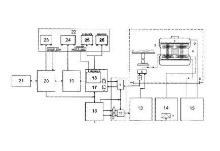
Simplified Functional Scheme of the VPEPN/H-201 "Zero Series Prototype"
Apparatus
In this illustration the fundamental Devices and/or Tehcnologic Parts that
make this
Invention Viable are shown conceptually and Functionally integrated:
a) Radiofrecuency Resonating Antenna (4)
b) Low-Signal Radiofrequency Processor/Modulator (10)
c) Radiofi-equency Pulse Amplifier (13)
d) Manual Control Digital Filter/Selector(18)
e) Central Pulse Control (16)
I) Frequency Matrix Monitor (25)
g) Frequency Image Monitor (26)
h) Control Panel (28)
It is important to note that without the Conceptual and Functional integration
of the
abovementioned Devices and/or Parts (Technological Innovations) it would not
be
possible to attain the objectives and goals achieved with this Invention.
That the Devices and/or Parts listed in paragraphs (d, f, g, h) are essential
to operate
the Apparatus in the Quantitative Diagnosis Modality, and that to operate the
Apparatus in
the Personalized Therapy Modality the former plus the ones listed in
paragraphs (a, b, c, e)
would be essential.
DETAILED DESCRIPTION OF THE INVENTION
This Invention is represented to "exemplify", through the VPEPN/H-201 "Zero
Series Prototype" Apparatus and associated Method which allows to Selectively
manipulate parameters such as Frequency, Power and Polarity of the radiation
that will be
CA 02587828 2007-05-10
17
used as a Therapy and that will affect an area previously identified and
localized as an area
of Threapeutic interest; the Apparatus aside from Quantifying the Resonance
Frequency of
different cellular compositions, shall then guarrantee a Quantitative
Diagnostic which
undeniably conditions the application of a Personalized Therapy that has no
precedents in
the medical practice, as well as significatively improving Diagnosis as an
independent
modality.
The procedure begins by using the technique traditionally known as Nuclear
Magentic Resonance (NMR) in use at present, obtaining in this way an image of
the area
affected by the pathology (Qualitative Diagnosis), once the image generated by
the area
studied is obtained in its phase of return to equilibrium and having
established with it the
adequate interconnection to the Frequency Processor (17) of Illustration 11,
the Frequency
Matrix Monitor (25) that provides reliable and exact information of the
specific values of
the Resonance Frequencies localized in the different areas studied, in this
Monitor, we can
accurately see the specific values of the Resonance Frequencies, the Frequency
Image
Monitor (26) with which the images are obtained in function of the specific
net Frequency
of each tissue, organ and or system being studied, we shall then get with the
aforementioned device, and only at that moment, a graphic Profile of the
Frequencies of
the studied area, aside from the traditional "mute" images that can be
obtained to
(Qualitatively) Diagnose due to the limited options given by the state of the
technique or
the state of the art at this time.
The Conventional Image Monitor (24) only provides information at a Qualitative
level, which implies an Empirical Diagnosis, but this is also a valuable
information that
provides images that are a direct function of the density of tissues that
would be integrated
to the other previously mentioned data (visual and value graphs).
From the previous, we can infer that the information obtained is an
Electromagnetic
Profile of Resonance Frequencies that provides accurate information both for
Diagnosis
CA 02587828 2007-05-10
= 18
and for Therapy, this procedure which favors the present medical practice for
treating
pathologies generically identified as Cancer and HIV/AIDS, in principle.
A Modality of Quantitative Diagnosis is proposed, since using the Frequency
Matrix Monitor (25) the Specific, Particular and Respective Values of the
Resonance
Frequencies of the areas of interest are accurately known and the Frequency
Image Monitor
(26) provides images that are in direct function of the aforementioned
Frequencies which
provide a graphic, and visual profile of the Frequencies of the studied area
and through the
Conventional Image Monitor (24), images that are in direct function with the
density of the
tissues are provided, then under the terms and conditions mentioned before, an
Electromagnetic Profile of Integral Resonance Frequencies that conditions the
accurate and
effective application in sequence of Diagnosis and Therapy, and in this way, a
Personalized
Therapy that has no precedents in today's medical practice can be implemented.
With the Control Panel (28) located in the Operator's Console (22) the Manual
Control Digital Filter/Selector is manipulated (18) to make a step by step
scanning and
analysis depending on the therapeutic requirements of the studied area, to
accurately
determine and define the Useful Work Resonance Frequencies (UWRF) and their
respective Power and Polarity values, being the aforementioned parameters
essential for
obtaining first the Quantitative Diagnosis, and second to guarantee the later
application of
the aforementioned Personalized Therapy, as established by the Methodology
proposed in
the Invention.
The above mentioned parameters (Frequency, Power and Polarity), already
selected
and modified; i. e. Selectively manipulated in function of the therapeutic
interests, are
programmed in the Central Computer (20) (fig 11) with the aid of the Keyboard
(27)
located in the Operator's Console (22), to give the precise instructions to
the Central
Computer (20) which will send the information to the Central Pulse Control
(16) to be
analogically processed by the Low Radiofrequency Signal Processor/ Modulator
(10) and
amplified in the Radiofrequency Pulse Amplifier (13).
CA 02587828 2007-05-10
19
-
The Central Computer (20), the Central Pulse Control (16), the Digital-
Analogical
Converters (12), the Low Radiofrequency Signal Processor/Modulator (10), the
Radiofrequency Pulse Amplifier (13) and the Radiofi-ecuency Resonating Antenna
(4),
must be in operation and must have the necessary and sufficient Functional
Capacity
(Operational) to then handle the "New Personalized Parameters" (modified,
previous
Selective Manipulation) of Frequency, Polarity and, very specially, Power to
next proceed
to perform a new of Radiofrequency emission with the aforementioned parameters
emitted
with the specific objective of using them as a Personalized Therapy. As shown
in the
disposition of the elements in Illustration 11, the Conventional Image Monitor
(24), the
Frequency Matrix Monitor (25) and the Frequency Image Monitor (26) help the
operator
see the evolution of the treatment in the area object of Therapy.
Once this step is finished, a second Diagnosis is made using the aforesaid
traditional procedure, where the generated Radiofrequency emission would be
again
characterized by the fact that the parameters of the emission such as
Frequency, Power and
Polarity would be integrated as a whole, and exclusively circumscribed to the
functional
criteria and design ranges of the Apparatus object of this Invention; i. e.,
that the
Radiofrequency emission behaves as an Indivisible and Unifunctional
Electromagnetic
beam, in which the parameters that form said electromagnetic beam as a whole
are not
partially or totally variable at any moment.
It is evident that the course of action and the specific treatment for the
patient will
be defined, depending on the results that shall be evaluated by specialized
medical
personnel.
In principle, a "re-arrangement", by means of the Non-Ionizing Electromagnetic
Radiation, of the cells that were catalogued as malignant is achieved,
guarranteeing the
absorption and/or yield of energy by the healthy and diseased cells, following
a case-
specific criteria associated to the specific and particular modification of
some of the work
parameters that form said radiations, acquiring in this way, Specific and
Selective effects
CA 02587828 2007-05-10
on healthy or diseased cells. Beams of Non-Ionizing electromagnetic radiations
with
specific objectives independent from each other are used.
By maintaining this cellular "re-arrangement" and its respective level of
Bioenergetic Balance, the cells catalogued as malignant cannot reach
(gradually or not) the
5
appropriate energy levels to guarrantee their existence. By maintaining their
Bioenergetic
Levels, the healthy cells will have the real possibility of revitalizing their
functions up to
certain limits and thus, they may play their specific biological role in a
more efficient way,
which in itself implies a recovery and therefore, the conditions for what we
call cellular
"re-arrangement" are established.
10 The
basis of the Scientific-Technical proposal of this Invention is the
application of
the Physical Principle of Resonance and in particular the one identified as
Nuclear
Magnetic Resonance (NMR) by means of the VPEPN/H-201 "Zero Series Prototipe"
Apparatus and associated Method since it is only under the condition of
Resonance in
which the transference of energy in one way or another is materialized; i. e.,
the yield or
15
absorption of energy. The unequivocal difference in the values of the
Resonance
Frequencies is the basis for the recognition of the different molecules that
form the tissues,
and such is precisely the basis of Nuclear Magnetic Resonance (NMR) applied
with
medical purposes.
By obtaining the value of the Frequency in which the malignant cellular focus
20
Resonates and also knowing the Resonance Frequency value of the cells of the
tissue that
surround it, a Dual Electromagnetic Field (R-2 Simple Therapy) is applied in
principle,
where a thermal contribution that destroys them shall be conditioned,
modifying the values
of the Power of the electromagnetic emission that affects the tissue
identified as malignant
above the normal limits of tolerance and for the case of the surrounding
tissue, the Power is
maintained in the range of values used for the Diagnosis Modality. The
Modality identified
as R-2 Simple Therapy is suggested to treat pathologies such as HIV/AIDS and
Leukemia.
R-2 Simple Therapy is shown in illustrations 5 and 7.
CA 02587828 2007-05-10
_ 21
_ For the case of tumors, the R-3 type Complex Therapy is
suggested, this therapy is
shown in Illustration 9.
Depending on the therapeutic interests and in face of the possibility of other
pathologies associated and/or concurrent to the main pathology, Therapies that
may be
necessary and advisable such as R-4 Therapy, R-5 Therapy, among others, can be
implemented.
The evaluation and selection of the Polarity (positive or negative) of the
incident
radiation is very important and significant since, depending on the Polarity
as well as on
the interests and objectives desired with respect to the Bioenergetic Balance
or
Equilibrium, there may be energy absorption or yield. As mentioned before, a
Selective
application of radiation is needed for each kind or group of malignant cells
in a specific and
particular way, thus the principle of application of this Invention is the use
of different
beams of radiation, with precise, exact and specific sequencies and doses,
where each is
designed and intended for a specific objective (goal), being also independent
from the rest.
An example is shown in Illustration 8.
The Electromagnetic Field aimed to the malignant cells, will be of a
Continuous
and Non-Pulsating nature; in this way, the harmful effect with a greater range
is
guarranteed and the cells to which it is destined have few or no possibilities
of survival.
According to the technological criteria hereby proposed the viral nucleus of
HIV/AIDS can be precisely and accurately entered, this nucleus is in most
cases very
protected; hence, until now, it has been immune to the different treatments to
which it has
been put through.
Illustration 1, shows the effect caused by an incident beam of Ionizing
radiation of
the previous technique, where it can be seen that the area of action of the
Ionizing radiation
affects the pathogenic tissue and in the same way the surrounding healthy
tissue is affected
where a harmful area of Hyperthermia is also generated around the pathology.
The
CA 02587828 2007-05-10
_
_
22
_
treatment with Ionizing radiation affects all the tissue it crosses
generating harmful side
effects of different range and nature.
Illustration 2a, shows the effect of a No-Ionizing radiation without "Target"
Substance but it does not inhibit or affect the tumoral tissue since the
Frequency of the
incident radiation does not make the diseased tissue Resonate and in
consequence an area
of Hyperthermia is not generated around it.
In illustration 2b, the Frequency of the incident radiation interacts with the
tumoral
tissue, making it resonate, and as a result a surrounding area of Hyperthermia
is generated
with the consequent harmful side effects on the surrounding healthy tissue.
In illustration 3, another Modality of Therapy is represented where a "Target"
substance is used, the harmful side effect of Hyperthermia can be seen in this
illustration,
with the addition of the problem of the Evacuation of the "Target" substance
(represented
by the dispersed spots in the healthy tissue) from the body.
In llustration 4, shows the areas of interest in Therapy with radiation of an
area
affected by a tumor, where the reference (1) represents the Pathological Area;
area (2) is a
Healthy Tissue that is partially affected by Hyperthermia pictured inside the
dotted circle
(3); reference (4) indicates the route of the incident radiation in the tissue
that is potentially
affected by it; reference (5) represents the potential Evacuation of the
denominated
"Target" Substance.
In llustration 5, represents a pathological area that is crossed by a Non-
Ionizing
Electromagnetic Radiation that makes the pathology resonate and absorb the
necessary and
sufficient amount of energy to guarantee its destruction; the Hyperthermia
generated by the
abovementioned concept, will be solved a posteriori. The radiation that makes
the
pathogenic tissue has a Frequency (f1), Power (P1) and Polarity (Poll). The
Frequency (f1)
is specific for that pathogenic tissue. In this Invention said Resonance
Frequency is exactly
measured and once it has been identified, this Invention also provides the
necessary and
CA 02587828 2007-05-10
_
= 23
_ sufficient technological resources that allow the modification of
Power and Polarity of the
incident radiation at will, depending on the therapeutic interests, in such a
way that it is
possible to rehabilitate or destroy the area identified as pathogenic.
In llustration 6, represents the incident radiation beam in the area of
Hyperthermia
with Frequency (13), Power (P3) and Polarity (Po13); which drags the excess
heat emitted
by the tumoral tissue that in a way is being rehabilitated or destroyed
depending on the
case, and consequently cooling the surrounding area affected by Hyperthermia.
In this
hypothetical case a negative Polarity of the incident radiation as an
indication of absorption
of energy is assumed. It is clarified that the Frequency (13) was previously
identified with
the technique proposed in this Invention, as well as the rest of the procedure
to be applied
to Selectively modify or not Power and Polarity depending on the therapeutic
interests.
In addition, with the knowledge of this Invention, the Resonance Frequency of
a
distant healthy surrounding area that has not been affected by Hyperthermia
(Ill. 7) can be
identified and a Non-Ionizing Electromagnetic Radiation can be aimed, with a
Resonance
Frequency (f2) and its respective values of Power (P2) and Polarity (Po12);
this radiation
will emit or absorb energy to this area depending on the therapeutic
requirements and/or
with the purpose of maintaining the Bio-energetic Balance in this area. In
this hypothetical
case an alternate Polarity of the incident radiation is assumed.
It is important to note that the Sequence of the Emission Pattern of the Non-
Ionizing Electromagnetic Radiation must guarantee: the harmful effect with the
highest
range allowed on the pathogenic tissue so that the malignant cells that
compose it have
slight or no possibilities of survival; that it can adequately solve the
Hyperthermia
problem, and avoid all types of harmful side damage on the healthy tissue
surrounding the
pathology object of Therapy, as well as on the Nervous and Immune Systems.
Illustration
8 represents the visual integration of the beams of illlustrations 5, 6 and 7
that form a single
Emission Sequence and where the behavior or ratio of the values of Frequency
(1), Power
(p) and Polarity (Pol) fulfill the following ratios:
CA 02587828 2007-05-10
24
fi# f2 = fi
P1> P2 = P3
Polarity (Pol)
Poll is positive (+)
Pol2 is positive (+) or negative (-)
Pol3 is negative (-)
Illustration 9, represents the hypothetical graphs of the Basic or Main
Principle of
Work of this Invention, note the continuous beam emission sequence used in
this particular
case in the Modality of R-3 Complex Therapy where the behavior or ratio of
parameters
such as Frequency, Power and Polarity, fulfill the following ratios:
fi# f2 = f3
P1> P2 = P3
Polarity (Pol)
Poll is positive (+)
Po12 is positive (+) or negative (-)
Po13 is negative (-)
Conventional Nuclear Magnetic Resonance equipment (NMR) emits a
Radiofrequency beam in which parameters such as Frequency, Power and Polarity
that
compose it are constant in their respective values; i. e., the subject beam is
displayed as a
set of indivisible and "unifunctional" radiations as well. Illustration 10
shows three (3)
beams with different frequencies but equal Power and an indeterminate Polarity
of equal
signs. The ratio of the parameters is as follows:
f2 f3
P1 = P2 = P3
CA 02587828 2007-05-10
= 25
Poll Indeterminate
Po12 Indeterminate
Pol3 Indeterminate
Illustration 11, shows the Simplified Functional Diagram of the Nuclear
Magnetic
Resonance (NMR) system of this invention, called VPEPN/H-201 "Zero Series
Prototype"
Apparatus, where the List/Description of the Devices and/or Parts would be the
following:
1) Magnet with incorporated cooling system and Isolation for the Assembly
2) Shim coil system
3) Gradient system coil
4) Radiofi-equency resonator antenna
5) Patient table
6) Transmit ¨ Receive Selector
7) Preamplifier
8) Radio frequency and magnetic-proof leadthroughs
9) Radiofrequency low signal processor / Quadrature demodulator with low-pass
filters
10) Radiofi-equency low signal processor / Modulator
11) Analog-Digital Converter (ADC)
12) Digital -Analog Converter (DAC)
13) Radiofi-equency pulse amplifier
14) Gradient amplifiers X, Y, Z with Digital-Analog Converters
15) Magnet power supply
16) Central pulse control
17) Frequency Processor
18) Manual Control Digital Filter/Selector
19) Image Processor
CA 02587828 2007-05-10
26
20) Central Computer
21) Image Storage
22) Operator's Console
23) Protocol Monitor
24) Conventional-Image Monitor
25) Frequency Matrix Monitor
26) Frequency Image Monitor
27) Keyboard
28) Control Panel.
The proposed VPEPN/H-201 "Zero Series Prototype" Apparatus works exclusively
with Non-Ionizing Electromagnetic Radiations, which are selectively
manipulated for their
application in the Modality of Personalized Therapy only; this Therapy does
not generate
harmful side effects of any kind to the surrounding healthy tissues. It can be
applied to the
different presentations and/or manifestations of pathologies in different
organs, tissues
and/or systems whether in a so-called latent state or in the phase of evident
clinical
manifestation.
With the use of this Invention, exact indexes of Selectivity and/or
Differentiation
that allow making a Quantitative Diagnosis and an effective and Customized
Therapy are
achieved; it annuls the so-called Hyperthermia effect, caused by all types of
radiation used
in the state of the technique or in the state of the art for the Therapy
Modality, and because
it is applied in "Real Time" it nulifies the effects of Overlapping and
Cellular Mutability
inherent in all living beings as a natural mechanism for survival. The
presentation of the so-
called "drug resistance" as a natural mechanism for survival of certain
pathologies is
categorically discarded, since this Invention does not propose the use of
drugs as a
fundamental part of or as a complement to this Therapy and it only uses Non-
Ionizing
Electromagnetic Radiations which are adjusted on-line to the evolutive stage
of the
pathology being treated. In the innovating proposal of this Invention, Non-
Ionizing
CA 02587828 2007-05-10
27
electromagnetic radiations are used instead of Ionizing Radiations, the so-
called "Target"
Substances are also not used, thus avoiding the problems associated with the
Evacuation of
these substances from the body.
The Therapy is innocuous as a whole since it does not cause harmful side
effects
(for example, it does not affect in any way the Nervous and Immune Systems);
it
significatively reduces the time that passes between Diagnosis, Therapy and
Recovery of
the patient; the Empiricism that at the moment characterizes the Diagnosis and
Therapy
Modalities is annulled, except for those Diagnoses where physical-chemical
analysis are
used and that complement the biopsies and the different test Modalities used
to diagnose
the HIV/AIDS.
It significatively reduces the Costs of Research and Development (R+D), as
well as
those of welfare services.
The Dosimetry Calculation procedures are significantly simplified and they are
now
exact.
It is a non-invasive application that considerably limits the use of Surgery
as a
therapeutic modality.
The following Table establishes a comparison, at a qualitative level, between
the
Existing Technologies (state-of-the-art) and the Technology contained in our
Invention so
that the potential scope of our Invention can be accurately appraised.
PREVIOUS TECHNOLOGY
PROPOSED TECHNOLOGY
CHARACTERISTICS
TO ANALIZE AND DIAGNOSTIC
THERAPY DIAGNOSTIC THERAPY
COMPARE
CANCER HIV/Aids CANCER HIV/Aids CANCER HIV/Aids CANCER HIV/Aids
Solved
Solved Solved
Introduce
Selectivity and/or Only on Solved With
NO With With With
1
Quantitative
Differentiation Qualitative level Empiricism Quantitative
Quantitative Quantitative
Modality
,
Modality Modality Modality
2 Cellular Overlap not covered
not covered Not Solved Not Solved Solved Solved Solved
Solved
3 Cellular Mutability not covered
not covered Not Solved Not Solved Solved Solved Solved
Solved
Physical-
Only on a
4 Types of Radiations employed Non-Ionizing
Chemical Ionizing >>> Non-Ionizing Pharmacological Non-
Ionizing Non-Ionizing Non-Ionizing
Non-Ionizing
+ existent
Analysis
basis
"Target Substance"
,
Eventually No In some cases No NO NO NO NO
o EVACUATION
H
,
I 6
to
Eventually, with Very Low
o Hyperthermia Eventually
No Yes No Eventually Solved
1
low impact impact
N
o
0
CV 00 7 Drug Resistance
No No Yes
Yes (100%)
NO NO NO NO
l'4 (in function of # 3) Very High
co
CV .
CO
N Harmful Side Effects Yes
OD 8 Relative Undefined
Undefined NO NO NO NO
in (in function of # 1) Very
Important
CV
0
4 Index of Empiricism
o 9 Very High Solved
High Very High NO NO NO NO
(in function of # 1)
Surgery
Eventually No Frequently No NO NO NO NO
(See implications)
11 "R+D" Costs High Relative
Low Very High Excessively Very Low Very Low Very Low
Very Low
High
,
Implementation on the Health
On Short
12 On Long Term On
Long Term On Long Term On Long Term On Short Term On
Short Term On Short Term
Market
Term
,
On Short
13 Operational Phase of the Project On
Long Term On Long Term On Long Term On Long Term On
Short Term On Short Term On Short Term
Term
,
CA 02587828 2013-01-10
29
Specific Modalities of this Invention have been illustrated and described, it
will be
obvious for those experts in the technique and/or the art that several
modifications or
changes can be made without leaving the scope of the Invention. We shall
attempt to cover
the aforementioned, within the aggregated claims so that all the changes and
modifications
fall within the scope of this Invention.
Although the Invention has been illustrated and described in detail in the
drawings
herein attached and in this document, the same has to be considered as
illustrative yet non-
restrictive and/or limitative in character. It is understood that only the
preferred modality
has been shown and described; in consequence, we wish to protect all the
changes and
modifications that are included in the scope of the claims, as construed in
view of the
specification as a whole.