Note: Descriptions are shown in the official language in which they were submitted.
CA 02587857 2013-11-05
STEERABLE DEVICE FOR ACCESSING A TARGET SITE AND METHODS
BACKGROUND OF THE INVENTION
[0003] Field of the Invention. This invention relates generally to a design of
devices and
systems for safely and effectively accessing tissue. The invention provides a
device and
system that can be easily steered through tissue within a patient from a
location outside the
patient's body. The system also provides a platform for delivery of materials
and devices to a
target site or anatomic location within a body.
[0004] Description of related art. A variety of needles, lancets, trocars,
stylets, cannulas,
devices and systems for examining, diagnosing, treating, or removing tissue
from a patient are
known in the art. See, U.S. Patents 4,013,080 entitled Cannula Connector and
Director
Indicator Means for Injection System (Froning); 4,769,017 entitled Self-
Sealing Infusion
Manifold and Catheter Connector (Fath et al); 5,240,011 entitled Motorized
Biopsy Needle
Positioner (Assa); 5,526,821 entitled Biopsy Needle with Sample Retaining
Means
(Jamshidi); 5,660,185 entitled Image-Guided Biopsy Apparatus with Enhanced
Imaging and
Methods (Shmulewitz); 5,735,264 entitled Motorized Mammographic Biopsy
Apparatus
(Siczek et al); 6,315,737 B1 entitled Biopsy Needle for a Biopsy Instrument
(Skinner);
6,328,701 Bl entitled Biopsy Needle and Surgical Instrument (Terwilliger);
6,402,701 Bl
entitled Biopsy Needle Instrument (Kaplan); 6,464,648 B1 entitled Biopsy
Device and Remote
Control Device Therefor (Nakamura); 6,485,436 Bl entitled Pressure-Assisted
Biopsy Needle
Apparatus and Technique (Truckai et al); 6,558,337 B2 entitled Positioner for
Medical
Devices such as Biopsy Needles (Dvorak et al); 6,709,408 B2 entitled Dual
Action Aspiration
Biopsy Needle (Fisher); 6,908,440 B2 entitled Dual Action Aspiration Biopsy
Needle
(Fisher); and 6,918,881 B2 entitled Biopsy Needle with Integrated Guide Pin
(Miller et al).
U.S. Patent Publications US 2004/0133168 Al entitled Steerable Needle
(Salcudean et al.); as
well as PCT Publications WO 00/13592 Al entitled Device for Receiving and
Actuating a
Biopsy Needle (Heinrich); WO 03/077768 Al entitled Biopsy Needle and Biopsy
Needle
Module that Can be Inserted into the Biopsy Device (Heske et al); WO
2004/062505 Al
entitled Flexible Biopsy Needle (Bates et al.); and WO 2004/086977 Al entitled
Coaxial
Cannula Provided with a Sealing Element (Heske et al.).
- 1 -
CA 02587857 2013-11-05
10005] For example, biopsy needles are used in the medical field to remove
tissue, cells or
fluids from a body for examination and diagnostic testing. Biopsy needles can
form part of a
biopsy system. Currently, there are three main types of procedures that are
used to obtain a
biopsy, or tissue sample. First, a surgeon can use a scalpel, or other
suitable cutting
instrument, to make an incision in a patient that is large enough for the
surgeon to access the
tissue to be tested. One or more large pieces of a target site, such as a
tumor, lesion, cells or
fluid, are then removed and tested for malignancy. This procedure is typically
performed
under general anesthesia.
[0006] Another technique, the core tissue biopsy procedure, uses a large bore
needle to cut
or shear away one or more visible pieces of a tumor or lesion. The pieces of
tissue obtained
using a large bore needle are visible to the
- la-
CA 02587857 2007-05-15
WO 2006/058195
PCT/US2005/042705
irlo Ir. "'Jr .fff fF IF Cf:' frit ..e` II 17 if
Ir. lj,õõ I) 4-õ,p71 f ffõ,F1
unaided eye and may require further processing to view through a microscope
(i.e., due to the size and thickness of
the tissue pieces obtained).
[0007] Yet another technique is the use of fine needle aspiration (FNA)
needles with small bores to obtain tissue
samples. A needle is used with a syringe to access the target site. Negative
pressure is created in the syringe, and as
a result of the pressure difference between the syringe and the mass, cellular
material can be drawn into the syringe
and removed. Typically, the needle is moved in and out in order to facilitate
obtaining enough tissue or material to
examine and make a diagnosis.
[0008] There are many medical conditions for which a physician might wish to
obtain access to a target site or
obtain a sample of tissue or material from a patient. For example, pulmonary
disorders affect millions of
Americans, and many more individuals worldwide, each year. While some
pulmonary disorders are chronic (e.g.,
chronic obstructive pulmonary disease (COPD)), many are acute and deadly. For
example, lung cancer is the
leading cause of death attributable to cancer for both men and women. More
people die of lung cancer, than die of
breast, prostate and colon cancer combined. It is estimated that in the United
States alone, over 170,000 new cases
of lung cancer are diagnosed each year. Of those people diagnosed with lung
cancer, the prognosis is grim: 6 of 10
will die within one year of being diagnosed and between 7 and 8 will die
within two years of diagnosis.
[0009] Most lung cancers start in the lining of the bronchi (plural for
bronchus), although lung cancer can start in
other parts of the lung as well. Since it generally takes many years for lung
cancer to develop, there can be areas of
pre-cancerous changes in the lung long before the formation of lung cancer.
With currently available technology, the
pre-cancerous changes are often not detected because the changes cannot be
seen on an x-ray and do not cause
symptoms early on that would cause a patient to seek medical attention. It is
for this reason that most people with
lung cancer are not diagnosed during the critical early stages of the disease.
[0010] Taking chest x-rays and checking sputum under a microscope for the
appearance of cancer cells had been
performed for screening but was found to be unreliable, and thus is not even
recommended screening for persons of
high risk (e.g., those people who smoke). Recently, spiral CT scanning has
shown promise as a potential screening
tool for finding lung cancer at an early stage. However, at this juncture it
is not known whether the use of spiral CT
scans improves the prognosis for long-term survival by increasing the early
detection of the disease. Even with a
scan indicating the possible presence of pre-cancerous tissue, the ability to
take a biopsy for testing is difficult
without causing the lungs to collapse, which can result in a required hospital
stay.
[0011] Each condition where access to tissue for examining or diagnosing a
condition, or where obtaining a biopsy
would be desirable, presents its own challenges. The, lung, however, presents
a useful platform for understanding
issues relating to accessing and treating target sites as well as obtaining
biopsies.
[0012] In the lung, any time a procedure requires an instrument to be inserted
through an incision in the chest wall,
the pleural layers surrounding the lung are pierced or compromised. As a
result of the propensity for transthoracic
procedures to cause, for example, pneumothorax, there is a limitation on the
outer diameter of the instruments that
are used for these procedures. This is a significant drawback for procedures
such as percutnaeous transthoracic lung
tissue biopsy, where the interventionalist introduces a biopsy needle through
the chest wall. Other procedures which
are limited when applied to transthoracic procedures include percutaneous
transthoracic needle aspiration (PTNA),
mediastinoscopy, thorascopy and drainage of pleural effusions. Air leaks and
bleeding frequently occur either
during insertion or removal of the device through the opening in the pleural
lining of the chest cavity. Even when
using small needles of 19-23 gauge, the incidence of pneumothorax is
relatively high, being in the range of 30-40%
and the incidence of hemothorax is 25%. Because of the anatomical challenges
and physiological mechanics of the
lung, accessing the target site or anatomic location on a first attempt is
very important.
-2-
CA 02587857 2016-09-26
[0013] Even during the biopsy process currently practiced, multiple tissue
samples or cores
may be taken through the smallest gauge needle possible in an effort to
increase biopsy
efficacy while decreasing the likelihood of, for example, pneumothorax.
However, each time
the needle is reinserted, the chances for pneumothorax or bleeding increase.
Additionally, due
to the small size of the multiple samples, the pathologist may not have the
benefit of a sample
size large enough to improve the accuracy of diagnosis.
[0014] Thus, there exists a need for devices and methods that provide
minimally invasive
access to a target site or anatomic location, such as lung tissue, for
diagnostics and treatment
which are able to access the target site more accurately. In the context of
the lung, there is a
need for such a device that does not increase the risk of causing the lung to
collapse, or air or
blood entering the pleural space. The present invention satisfies these needs
and provides
related advantages as well.
SUMMARY OF THE INVENTION
[0015] A variety of steerable needles, lancets, trocars, stylets, cannulas,
devices and systems
are provided for examining, diagnosing, treating, or removing tissue, cells or
fluid. The
steerable needles, lancets, trocars, stylets, cannulas, devices and systems
also provide a
platform for delivery of target materials, such as therapeutics, biologies,
polymers, glues, etc.,
to a target site within a patient.
[0016] An embodiment of the invention comprises a steerable-device for use in
accessing a
target site in a patient comprising: a steerable member having a proximal end
and a sharp
distal end with a longitudinal length therebetween, the steerable member
having a lumen
extending between the proximal end and the distal end, the distal end having a
closed
sharpened tip adapted to penetrate tissue so as to form a path through the
tissue, wherein the
steerable member defines an axial trajectory along the direction of the
longitudinal length and
extending distally from the distal end; and a steering mechanism axially
affixable within the
lumen of the steerable member so that the steering mechanism is advanceable
with the
steerable member along the path, a distal end of the steering mechanism
remaining proximal
to the distal end of the steerable member, the steering mechanism adapted to
be operated by a
user from a proximal end to apply a bending force to bend the steerable member
when the
- 3 -
CA 02587857 2016-09-26
steering mechanism is axially affixed in the lumen of the steerable member,
the bending force
configured to impose curvature in the steerable member between the proximal
end and the
distal end so as to reorient the path while the user advances the longitudinal
length of the
steerable member and the steering mechanism distally along the path such that
subsequent
distal tissue penetrating advancement of the axially coupled steerable member
and steering
mechanism together the path away from the trajectory suitably to access the
target site;
wherein the steering mechanism is removable from the distally advanced
steerable member;
and wherein the steerable member further comprises a port positioned along the
longitudinal
length of the steerable member and extending between an outer surface of the
steerable
member and the lumen of the steerable member.
[0017] Another embodiment of the invention comprises a steerable device for
use in
accessing a target site in a patient comprising: a metal bendable needle
having a proximal end
and a sharp distal end with a longitudinal lumen therebetween, the distal end
adapted to
penetrate tissue so as to form a path through the tissue, wherein the needle
defines an axial
trajectory in the direction of the longitudinal lumen and extending distally
from the distal end;
and a steering mechanism comprising a proximal knob and a shaft receivable in
and
removable from the lumen, engagement between the steering mechanism and the
needle
axially coupling the steering mechanism relative to the needle so that the
steering mechanism
is advanceable axially with the needle along the path when the proximal end is
pushed
distally, a distal end of the steering mechanism remaining proximal to the
distal end of the
steerable member, the steering mechanism operationally coupling the knob to
the needle when
the steering mechanism is disposed therein so that actuation of the knob by a
user applies a
bending force to bend the needle, the bending force configured to impose
curvature in the
needle between the proximal end and the distal end so as to reorient the path
when the user
advances the longitudinal length of the needle distally along the path such
that subsequent
distal tissue penetrating advancement of the axially coupled needle and
steering mechanism
together within the tissue angles away from the trajectory to access the
target site; wherein the
steering mechanism is removable from the distally advanced bendable needle.
[0019] In any of these embodiments of the invention, mechanisms can be
provided that are
adapted to apply a bending force that increases the strain on the steerable
member to induce
- 3a -
CA 02587857 2016-09-26
curvature. Moreover, the steerable member can be further adapted in the
embodiments to
create a path to the target site during operation. The steerable device can be
adapted to
penetrate tissue directly or indirectly, i.e., by being positioned within a
device that is adapted
to penetrate tissue.
[0020] In still other embodiments, an outer sheath can be provided. For the
embodiments
having an outer sheath, relative positions of a distal end of the steerable
member and a distal
end of the outer sheath can be adapted to remain the same, or substantially
the same, upon
application of the bending force.
[0021] In yet other embodiments, the steerable device can have a steering
mechanism with at
least one pull wire, or a plurality of differential wires or pull wires. For
other embodiments,
the steerable member can be configured to comprise coaxial members. For
embodiments with
a coaxial member, the coaxial members can comprise an outer needle and a
lancet device
disposed within the needle and adapted to be bent by the steering mechanism.
Thus, for
example, the coaxial members can be configured to comprise a lancet device in
a first
configuration and an aspiration device in a second configuration. Other
combinations and
configurations are also possible. The device can also be used to guide another
instrument to
the target site.
- 3b -
CA 02587857 2007-05-15
WO 2006/058195
PCT/US2005/042705
tj r5: ) 1)1 if.
*hi] stili anottier emixicriment ofthe invention includes a steerable device
for use in accessing target site or
anatomic location in a patient comprising: an outer sheath; a steerable member
positioned within the outer sheath
having a deformable control wire adapted to engage a first end of the
steerable member and a second end of the
steerable member; and a control mechanism adapted to provide control of a
distal end of the steerable device from a
proximal end adapted to provide access to a target location of a subject
through an access lumen in the patient.
[0023] Another embodiment of the invention includes a steerable device for use
in accessing target site or
anatomic location in a patient comprising: an outer sheath having a flange
with an optional position indicator
marked on the flange; a steerable member positioned within the outer sheath;
and a control mechanism having at
least one position indicator on a proximal surface of the control mechanism
and which is adapted to provide control
of a distal end of the steerable device from a proximal end adapted to provide
access to a target location of a subject
through an access lumen in the patient.
[0024] Yet another embodiment of the invention includes a steerable device for
use in accessing a target site or
anatomic location in a patient comprising: an outer sheath; a steerable member
positioned within the outer sheath
having a plurality of control wires adapted to engage a first end of the
steerable member and a second end of the
steerable member; and a control mechanism adapted to provide control of a
distal end of the steerable device from a
proximal end adapted to provide access to a target location of a subject
through an access lumen in the patient.
[0025] Still another embodiment of the invention includes a steerable
percutaneous device for use in accessing
target site in a patient comprising: an outer sheath; a steerable member
positioned within the outer sheath having a
steering wire housed within a notched control member; and a control mechanism
adapted to provide control of a
distal end of the steerable system from a proximal end adapted to provide
access to target site of a subject through
an access hole in the patient. Access can be made percutaneously, if desired,
or by other mechanisms as discussed
herein.
[0026] Any of the embodiments can also include an outer sheath that is formed
of a flexible material. Additionally,
embodiments can provide for an outer sheath with a flange at a proximal end.
The flange can further be provided
with position indicators. In still other embodiments of the invention, the
outer sheath can form a cup at a proximal
end for engaging a spring, or axial control mechanism, used to control
movement of the steerable member in at least
one axis.
[0027] Embodiments of the device also contemplate use of an external control
device that is accessible from a
remote location either wired or wirelessly. Such a control mechanism can be
configured to engage the steerable
member, the outer sheath, the control mechanism, or combinations thereof.
Remote access can be from another
room, another location, or a position within the room where the patient is not
in physical contact with the
interventionalist controlling the device.
[0028] The control mechanisms of each of the embodiments described enable
movement of a distal end of the
steerable percutaneous device up to 360 about a first axis, and/or up to
1800, or more, about a second axis.
[0029] Embodiments of the invention include appropriate control mechanisms,
such as handles, knobs, thumb
screws, thumb wires, ball controls and/or joysticks.
[0030] The steerable devices can be cannulated. The steerable devices can also
be adapted to remove target tissue,
cells or fluid, deliver therapy to a target site (including tissues, cells or
fluid) or diagnose a target site. In some
embodiments, it may be desirable to adapt and configure the steerable member
to make it removable from the lumen
of the outer sheath, such as once the device has been advanced to the target
site. Once removed, the steerable
member can be replaced with a member adapted to remove target site, deliver
therapy to target site or diagnose
target site.
-4-
CA 02587857 2007-05-15
WO 2006/058195
PCT/US2005/042705
11 Itt:::' ItI it .r" It .. 7:1t .r' It II
=õõii ¨rjr,õõ
[0031] Yet another aspect of the invention provides a biopsy needle whose
sampling tip can be more easily steered
from outside the patient. Still another aspect of the invention provides a
biopsy needle whose sampling tip can be
steered and controlled from a position remote from an imaging radiation field.
Another aspect of the invention is a
steerable biopsy needle whose position can be held in place during imaging.
The biopsy needles are adapted and
configured to remove tissue, cells or fluids from the target site.
[0032] Another aspect of the invention is a steerable needle, lancet, trocar,
stylet, cannula, device and/or system
that can be easily steered from outside the patient to: a) guide a needle
towards an intended target site or target
sample; b) guide devices that provide or extract energy to kill or remove
cancer cells; and c) guide ports to extract or
infuse fluids, solids or glues in or out of body cavities that require
assistance to access. The steerable needle, lancet,
trocar, stylet, cannula, device and/or system may be removable or integral
with any of these devices to simplify use
and allow the user to steer at any time during the procedure. Devices that
incorporate aspects of the steerable aspect
of the invention include, for example:
a. Co-axial dual members: wherein an outer needle is guided by a steerable
needle, lancet, trocar,
stylet, cannula, device and/or system and the device can be replaced by a
second inner device that
is used to aspirate a target tissue, such as cancer cells, for biopsy and
diagnostic characterization.
The outer needle can be left in place to be used as a guide for the inner
needle to harvest multiple
sequential samples.
b. Steerable needle, lancet, trocar, stylet, cannula, device and/or system
that can steer a flexible
cannula to regions in a patient's body that cannot otherwise be accessed or
present anatomical
challenges in accessing. The steerable needle, lancet, trocar, stylet,
cannula, device and/or system
may be removed to increase the port lumen size to enhance drainage or infusion
of liquids, solids
or materials that solidify such as glues.
c. A steerable needle, lancet, trocar, stylet, cannula, device and/or
system comprised of or made to
guide a tissue removing device. Such an embodiment would include a device that
may use stored
energy to shear tissue in order to sample and examine its condition. The
device can be used to
sample or extract cancerous tissue or entire tumors. The device may also use
radio frequency
waves to simultaneously cut tissue and coagulate blood that could otherwise
cause bleeding
complications.
d. A steerable needle, lancet, trocar, stylet, cannula, device and/or
system may be devised to extract
heat energy in order to freeze and kill pathologic tissue.
e. A steerable needle, lancet, trocar, stylet, cannula, device and/or
system may be devised to deliver
energy in order to heat and kill pathologic tissue. The energy can be
delivered to the tissue in the
form of light or magnetic energy such as radio frequency, microwave,
ultrasound, laser derived
light, or radiation wave forms such as x-ray energy. A device that delivers
any combination of
cryoablation and the other forms of energy can be configured to kill tissue
with different levels of
intensity and depth. This adaptability is useful for widespread dense tumors.
The steering feature
allows for convenient and quick use of the different energy modalities to be
applied to different
regions within the patient.
[0033] In an embodiment of the methods of the invention, a method is provided
for delivering a device to a target
site in a patient comprising: penetrating tissue with a steerable member; and
applying a bending force after
penetrating the tissue to bend the steerable member to deliver the device to
the target site.
-5-
CA 02587857 2007-05-15
WO 2006/058195
PCT/US2005/042705
itib34] fanblei embodiment a '1T& 'methods of the invention, a method is
provided for delivering a device to a
target site in a patient comprising: penetrating tissue with a steerable
member; and actively changing a shape of the
steering member after penetrating the tissue to delivery the device to the
target site.
[0035] In still another embodiment of the methods of the invention, a method
is provided for delivering a device to
a target site in a patient comprising: introducing a steerable member through
a scope; and applying a bending force
to bend the steerable member to deliver the device to the target site.
[0036] In some embodiments of these methods the further step of advancing the
steerable member through the
tissue is provided. In other embodiments, the method of applying a bending
force further comprises bending a
bendable portion of the steering member while the bendable portion of the
steering member is positioned within
tissue. In some embodiments of the method, the further step of aspirating at
the target site can be provided. In still
other embodiments of the method, the further step of removing target material
(e.g., tissue, cells or fluid) at the
target site, draining the target site, infusing the target site with a
marking, therapeutic or diagnostic material,
delivering energy to the target site, extracting heat energy from the target
site, and/or killing target material at the
target site can be included.
[0037] Embodiments of the invention also include a method of using a steerable
device having an outer sheath and
a steerable member, comprising: introducing a steerable device; advancing the
device toward a target site; and
deforming a distal tip of device from a longitudinal axis of a device. In some
methods the step of applying a force to
the distal tip of the device is accomplished remotely. Applying a force
includes bending or deforming the distal tip.
In at least some embodiments, the bending caused by the application of force
can be up to 3600 around a first axis,
and/or up to 180 , or more, around a second axis. In some methods, the
embodiments include the additional step of
removing the steerable member and replacing the member with a member adapted
to remove target tissue, cells or
fluid, deliver therapy to target tissue, cells or fluid, or diagnose target
tissue, cells or fluid.
[0038] Another aspect of the invention includes a method comprising the steps
of: determining, using diagnostic
testing, that a steerable device must be advanced to a specific location in
the body; introducing the device into the
body; and manipulating the shape of the device to cause shape changes while
the device is in the body to influence a
new path of advancement for the device. The method can be achieved by a device
enabling remote access and
control of the steerable devices disclosed.
[0039] Yet another aspect of the invention includes a method comprising the
steps of: determining, using
diagnostic testing devices, that a device must be advanced to a specific
location in the body; introducing a steering
device into the body; manipulating the shape of the steering device to cause
shape changes while it is in the body to
influence a new path of advancement; and introducing an instrument into the
body.
[0040] Still another aspect of the invention includes a method comprising the
steps of: determining, using
diagnostic testing devices, that foreign matter exists in a patient's body
that must be sampled; introducing a
sampling instrument into the body; and manipulating the shape of the
instrument to cause shape changes while the
instrument is in the body to influence a new path of advancement.
[0041] Still another aspect of the invention includes a method comprising the
steps of: using a device to obtain an
image of a patient's body along with the steerable device contained therein.
The image can be obtained at discrete
intervals or concurrently to advancing and steering the device using
techniques available in the art.
[0042] Yet another aspect of the invention includes a method comprising the
steps of determining, using a
diagnostic testing device, that foreign matter exists in a patient's body that
must be sampled; introducing a steering
element into the body; manipulating the shape of the element to cause shape
changes while it is in the body to
-6-
CA 02587857 2013-11-05
influence a new path of advancement; introducing a sampling instrument into
the body; and
imaging the body and device.
[0043] Still another aspect of the invention includes a method comprising the
steps of:
determining, using a diagnostic testing device, that foreign matter exists in
a patient's body
that must be sampled; introducing a steering element into the body;
manipulating the shape of
the instrument to cause shape changes while the instrument is in the body to
influence a new
path of advancement; imaging the body and device.
[0044] Another aspect of the invention includes a method comprising the steps
of:
determining, using a diagnostic testing device, that foreign matter exists in
a patient's body
that must be sampled; introducing a needle instrument into the body that can
be steered;
manipulating the shape of the instrument to cause shape changes while the
instrument is in the
body to influence a new path of advancement; imaging the body and device.
[0045] Still another aspect of the invention includes a method comprising the
steps of:
determining, using a diagnostic testing device, that foreign matter exists in
a patient's body
that must be sampled; introducing a sampling instrument into the body;
manipulating the
shape of the instrument to cause shape changes while the instrument is in the
body to
influence a new path of advancement from a location more than 2 inches away
from the body
entry point; and imaging the body and device.
[0046] Another aspect of the invention includes a method comprising the steps
of:
determining, using a diagnostic testing device, that foreign matter exists in
a patient's body
that must be sampled; introducing a steering element into the body;
manipulating the shape of
the element to cause shape changes while the instrument is in the body to
influence a new
path of advancement from a location more than 2 inches away from the body
entry point;
imaging the body and device.
[0047] Yet another aspect of the invention includes a method comprising the
steps of:
determining, using a diagnostic testing device, that foreign matter exists in
a patient's body
that must be sampled; introducing a steering element into the body;
manipulating the shape of
the steering element to cause shape changes while the instrument is in the
body to influence a
new path of advancement from a location more than 2 inches away from the body
entry point;
- 7 -
CA 02587857 2013-11-05
imaging the body and device using an imaging device; and introducing a
sampling instrument
into the body.
[0048] Still another aspect of the invention includes a method for palpating,
encapsulating,
isolating, removing and killing target tissue, cells or fluid in a patient's
body by advancing a
steerable device to the target site.
[0049] Another aspect of the invention includes the provision of devices and
materials
disclosed in the form of a kit.
BRIEF DESCRIPTION OF THE DRAWINGS
[0051] The novel features of the invention are set forth with particularity in
the appended
claims. A better understanding of the features and advantages of the present
invention will be
obtained by reference to the following detailed description that sets forth
illustrative
embodiments, in which the principles of the invention are utilized, and the
accompanying
drawings of which:
[0052] FIGS. 1A-D illustrates the anatomy of the respiratory system, along
with an example
of hemothorax caused from blood entering the pleural space.
- 7a -
CA 02587857 2007-05-15
WO 2006/058195
PCT/US2005/042705
it If 11 ot if I; I` It aõ,
Jr' 0,,,k= llõ t. 4õ It
[0053] FIG. 2A illustrates a: lung having a target site for biopsy; FIG. 213
illustrates a needle being advanced toward
the-target site; FIG. 2C illustrates a needle that has been advanced toward
the target site but which has failed to
connect to the tissue;
[0054] FIG. 3 illustrates a patient in an image capture chamber with a
technician monitoring the process from
another room;
[0055] FIGS. 4A-E illustrate perspective and cross-sectional views of a
steerable device capable of accessing target
site or anatomic locations;
[0056] FIGS. 5A-F illustrate perspective and cross-sectional views of another
steerable device capable of accessing
target site or anatomic locations;
[0057] FIGS. 6A-E illustrate perspective and cross-sectional views of yet
another steerable device capable of
accessing target site or anatomic locations;
[0058] FIGS. 7A-E illustrate perspective and cross-sectional views of still
another steerable device capable of
accessing target site or anatomic locations;
[0059] FIGS. 8A-E illustrate perspective and cross-sectional views of another
steerable device capable of accessing
target site or anatomic locations;
[0060] FIGS. 9A-E illustrate perspective and cross-sectional views of another
steerable device capable of accessing
target site or anatomic locations;
[0061] FIGS. 10A-C illustrate perspective and cross-sectional views of yet
another steerable device capable of
accessing target site or anatomic locations;
[0062] FIGS. 11A-G illustrate perspective and cross-sectional views of still
another steerable device capable of
accessing target site or anatomic locations;
[0063] FIGS. 12A-C illustrate cross-sectional views of a variety of distal tip
designs suitable for use with any of the
steerable devices shown in FIGS. 4-11;
[0064] FIGS. 13A-F illustrate cross-sectional views of a variety of proximal
control designs suitable for use with
any of the steerable devices shown in FIGS. 4-11;
[0065] FIGS. 14A-B illustrate mechanisms for remote control of the steerable
devices shown in FIGS. 4-11;
[0066] FIGS. 15A-13 illustrate additional mechanisms for control of the
steerable devices shown in FIGS. 4-11;
[0067] FIG. 16 illustrates a physician or technician controlling the steerable
devices shown in FIGS. 4-11 to access
target site on a patient using, for example, remote access devices shown in
FIGS. 14 and 15;
[0068] FIG. 17 illustrates a cross-sectional view of a steerable device used
as a biopsy device to capture a tissue
sample at its distal end;
[0069] FIGS. 18A-c illustrate a variety of systems for controlling temperature
sensing and delivery of heat or cold
(FIG. 18A); temperature sensing and delivery of energy to produce heat and
throttling gas to extract heat (FIG. 18B);
and delivery of RF for cutting and coagulation (FIG. 18C) used in connection
with the steerable devices shown in
FIGS. 4-11;
[0070] FIG. 19 illustrates a lung having a steerable device positioned to
access a target site;
[0071] FIG. 20 illustrates a bronchoscope in combination with a steerable
device; and
[0072] FIG. 21 illustrates the steps of a method for deploying the devices
described herein.
DETAILED DESCRIPTION OF THE INVENTION
[0073] As noted above, the present invention is suitable for use in
percutaneously accessing a target site within a
body, as well as traversing tissue and lumens between an access point on the
body and a target site in the body. The
-8-
CA 02587857 2007-05-15
WO 2006/058195
PCT/US2005/042705
11 e:: s s'""11 ""P, lim"
invention is also suitable for accessing a target site through body lumens
such as the trachea and the vasculature. A
target site can be located in any anatomic location in the body. Typically a
site is identified by the physician or
radiologist and the tissue, cells and/or fluid, or other material at that
site, is then identified as target material and
selected or targeted for access. Thus, for example, the target tissue, cells
or fluid can be the target material identified
for access from: brain, heart, liver, kidney, thyroid, lung, pancreas,
intestine, uterine, ovary, prostate, lymph, spleen,
skin, biliary, parathyroid, pituitary, adrenal gland, mediastinum, bladder,
connective tissue, breast, gastrointestinal
tract, joints, muscle, etc. Additionally, in some instances it is desirable to
access a target site located within a void,
such as a space between organs, lumen, etc. In that instance, the target site
may include fluid, or other material
which is the target for access. Once the target site is accessed and target
material (e.g. tissue, cells and/or fluid) is
identified for access, one or more diagnostic, therapeutic or delivery
procedures can be employed to remove, treat
and/or mark the target material.
[0074] An application of the device includes safely performing a transthoracic
procedure without impacting the
negative pressure required to maintain lung function. Thus, in addition to
other applications, the present devices
allow accessing the interior of the lung or the surrounding tissue to perform
therapeutic or diagnostic functions
while reducing the risk of complications associated with the accessing
procedure. The present invention includes
the use of the disclosed devices with, for example, a bronchoscope. See, for
example, U.S. Patent Application
11/153,296 filed June 18, 2005 entitled Lung Access Device (Mathis). The
devices disclosed can also be adapted for
use with other devices, without departing from the scope of the invention.
[0075] The invention provides methods, and devices for obtaining target
material from a body, such as lung tissue.
Although the device can be used to obtain a variety of target materials, such
as tissue, within a body, for purposes of
illustration the device and its operation will be discussed in the context of
lung tissue, which presents additional
challenges for biopsy capture also addressed by the designs of the invention.
Additionally, the devices can be used
in combination with suitable rigid, flexible, and steerable scopes, such as a
bronchoscope. Other scopes, including,
but not limited to, colonoscopes, thoracoscopes, laparoscopes, and/or
endoscopes, can also be used, depending upon
the location of the target site to be accessed. Additional information
pertaining to scopes is available in, for example,
U.S. Patent Nos. 6,478,730 entitled Zoom Laparoscope (Bala et al.); 6,387,044
entitled Laparascope Apparatus
(Tachibana et al.); 6,494,897 entitled Method and System for Performing
Thoracoscopic Cardiac Bypass Surgery
(Sterman et al.); 6,964,662 entitled Endoscopic Forceps Instrument (Kidooka);
6,967,673 entitled Electronic
Endoscope System with Color-Balance Alteration Process (Ozawa et al.).
[0076] The invention also provides methods for encapsulating target material,
killing target material, including
muscle, nerve, connective and epidermal tissue and interstitial fluids,
providing a mechanism for palpating a target
site, and delivering target markers and biologically active and/or therapeutic
compounds to a target site.
[0077] As mentioned, the lung is used to illustrate the advantages and
operation of the devices disclosed. FIG. lA
illustrates the respiratory system 10 located primarily within a thoracic
cavity I I . The respiratory system 10
includes the trachea 12, which brings air from the nose 8 or mouth 9 into the
right primary bronchus 14 and the left
primary bronchus 16. From the right primary bronchus 14 the air enters the
right lung 18; from the left primary
bronchus 16 the air enters the left lung 20. The right lung 18 and the left
lung 20, together comprise the lungs 19.
The left lung 20 is comprised of only two lobes while the right lung 18 is
comprised of three lobes, in part to provide
space for the heart typically located in the left side of the thoracic cavity
11, also referred to as the chest cavity.
[0078] As shown in more detail in FIG. 1B, the primary bronchus, e.g. left
primary bronchus 16, that leads into the
lung, e.g. left lung 20, branches into secondary bronchus 22, and then further
into tertiary bronchus 24, and still
further into bronchioles 26, the terminal bronchiole 28 and fmally the alveoli
30. The pleural cavity 38 is the space
-9-
CA 02587857 2007-05-15
WO 2006/058195 PCT/US2005/042705
Vr Lif 1,1:Jr irõ;1;.Af ,;It
between the lungs and the chest wall. The pleural cavity 38 protects the lungs
18, 20 and allows the lungs to move
during breathing. As shown in FIG. 1C, the pleura 40 defmes the pleural cavity
38 and consists of two layers, the
visceral pleurae 42 and the parietal pleurae 44, with a thin layer of pleural
fluid therebetween. The space occupied
by the pleural fluid is referred to as the pleural space 46. Each of the two
pleural layers 42, 44 are comprised of very
porous mesenchymal serous membranes through which small amounts of
interstitial fluid transude continually into
the pleural space 46. The total amount of fluid in the pleural space 46 is
typically slight. Under normal conditions,
excess fluid is typically pumped out of the pleural space 46 by the lymphatic
vessels.
[0079] The lungs 19 are an elastic structure that float within the thoracic
cavity II. The thin layer of pleural fluid
that surrounds the lungs 19 lubricates the movement of the lungs within the
thoracic cavity 11. Suction of excess
fluid from the pleural space 46 into the lymphatic channels maintains a slight
suction between the visceral pleural
surface of the lung pleura 42 and the parietal pleural surface of the thoracic
cavity 44. This slight suction creates a
negative pressure that keeps the lungs 19 inflated and floating within the
thoracic cavity 11. Without the negative
pressure, the lungs 19 collapse like a balloon and expel air through the
trachea 12. Thus, the natural process of
breathing out is almost entirely passive because of the elastic recoil of the
lungs 19 and chest cage structures. As a
result of this physiological arrangement, when the pleura 42, 44 is breached,
the negative pressure that keeps the
lungs 19 in a suspended condition disappears and the lungs 19 collapse from
the elastic recoil effect.
[0080] When fully expanded, the lungs 19 completely fill the pleural cavity 38
and the parietal pleurae 44 and
visceral pleurae 42 come into contact. During the process of expansion and
contraction with the inhaling and
exhaling of air, the lungs 19 slide back and forth within the pleural cavity
38. The movement within the pleural
cavity 38 is facilitated by the thin layer of mucoid fluid that lies in the
pleural space 46 between the parietal
pleurae 44 and visceral pleurae 42.
[0081] For purposes of illustration, FIG. 1D illustrates a lung 20 with blood
50 in the pleural space (also referred to
as hemothorax). As evidenced from the drawing, the presence of blood 50 in the
pleural space 46 results in a
contraction of the lung 20 to a much smaller size. Clinically, the patient
would have a difficult time breathing air
into the collapsed lung because the act of breathing relies on the lungs being
suspended in a state of negative
pressure. As will be appreciated by those of skill in the art, fluid or air
within the pleural space 46 will achieve a
similar clinical impact on the size of the lung relative to the thoracic
cavity as the hemothorax illustrated in FIG. 1D.
Because of the anatomical design of the lungs, and the negative pressure
required to maintain the lungs in a
suspended state, obtaining tissue samples from the lungs presents additional
challenges that are not present for other
tissues.
[0082] FIGS. 2A-C depict the lungs 19 during a procedure wherein a biopsy
device 80 is deployed to obtain a target
sample 82, or target material, from the lung and breaches the pleura. As a
result of the breach, air 88 inside the
affected lung 20 exits the lung (indicated by arrows) around the opening 84 in
the lining made by the device 80. As
in the previous example, air inside the affected lung 20 exits the lung
(indicated by arrows) around the opening 84
created when the device 80 punctured the wall of the bronchus 14.
Additionally, as will be appreciated by those of
skill in the art, the trajectory of the device 80 can be such that the device
80 fails to access the target site for a
biopsy, as illustrated in FIG. 2c.
[0083] As stated above, the invention and its embodiments are described for
purposes of illustration with respect to
access, diagnostic treatment and removal of target tissue, cells or fluid in
the lung. However, aspects of the devices
and methods are applicable to diagnostic and therapeutic procedures for other
target tissues, cells or fluids within the
body as well.
-10-
CA 02587857 2007-05-15
WO 2006/058195
PCT/US2005/042705
le lr k IIb h It...
IL,ttr
[0084] FIG. 3 illustrates a patient 52 in an image capture chamber 54, such as
a room having an x-ray machine,
with a physician or technician 56 monitoring an image capture process from,
for example, a location 58 such as a
separate room. The image capture process employs a machine 60 suitable for
image capture. Often when patients 52
undergo a procedure to obtain a target sample, an attempt is made to position
a device to access a target site and then
a confirmatory image is taken to ensure that the target material was obtained.
As will be appreciated by those of skill
in the art, suitable mechanisms for determining the location of the device
used to access a target material relative to
the target site employs, unless otherwise indicated, conventional devices 60
and methods and techniques known in
the art. These conventional devices and techniques include: x-ray imaging and
processing, x-ray tomosynthesis,
ultrasound including A-scan, B-scan and C-scan, computed tomography (CT scan),
spiral CT, magnetic resonance
imaging (MRI), optical coherence tomography, single photon emission computed
tomography (SPECT) and
positron emission tomography (PET), fluoroscopy and combinations and portable
versions thereof are within the
skill of the art. Such techniques are explained fully in the literature and
need not be described herein. See, e.g., X-
Ray Structure Determination: A Practical Guide, 2nd Edition, editors Stout and
Jensen, 1989, John Wiley & Sons,
publisher; Body CT: A Practical Approach, editor Slone, 1999, McGraw-Hill
publisher; X-ray Diagnosis: A
Physician's Approach, editor Lam, 1998 Springer-Verlag, publisher.
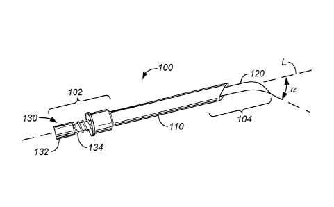
[0085] FIGS. 4A-E illustrate perspective and cross-sectional views of a
steerable device 100 capable of accessing a
target sample of material from a target site. Components of the device 100
include an optional outer sheath 110,
which can be in the form of a cannula, flexible tube or hypotube, to name a
few, and a steerable member 120. The
sheath can be made from suitable biocompatible polymers and metals such as
titanium and nickel-titanium alloys
(Nitinol), stainless steel, fluoropolymers, polyetheretherketone (PEEK),
polytetrafluoroethylene (PTFE), expanded
polytetrafluoroethylene (ePTFE), polyurethane, nylons, polyimide films
(Kapton0), and the like. Reference to
suitable polymers that can be used in the invention can be made found in: PCT
Publication WO 02/02158 Al, dated
Jan. 10, 2002, entitled Bio-Compatible Polymeric Materials; PCT Publication WO
02/00275 Al, dated Jan. 3, 2002,
entitled Bio-Compatible Polymeric Materials; and, PCT Publication WO 02/00270
Al, dated Jan. 3, 2002, entitled
Bio-Compatible Polymeric Materials.
[0086] A control mechanism 130 adapted to be controlled by a user, such as a
technician, is provided at a proximal
end 102 to enable steering. The distal end 104, which is positioned away from
the proximal end typically positioned
outside the patient's body (or nearest a user), can be adapted and configured
in a variety of ways to achieve the
diagnostic or therapeutic objective of the device. For example, the distal end
104 can be configured as a trocar,
lancet, stylet, needle, therapeutic delivery device, marker, or diagnostic
delivery device, to name a few. In the
embodiment depicted, the control mechanism 130 at the proximal end 102
includes a knob 132 and a spring 134 or
coil. Proximal and distal are, however, relative terms, which do not limit the
scope of the description.
[0087] The spring 134 may be a coiled wire formed of suitable material capable
of maintaining a desired spring
tension. A plurality of coils of the coiled body form a lumen sized and
adapted to fit around the exterior of the
control mechanism 130. Some embodiments include a second coiled body along
with a first coiled body. As
illustrated in the cross-sectional longitudinal views of FIGS. 4B and 4c, the
steerable member 120 is located within
the outer sheath 110 such that the steerable member 120 is capable of
longitudinal movement 106 within a
lumen 112 in the sheath 110. The spring 134 at the proximal end 102 fits
within an section of the sheath 110 that has
a lumen 112' at its proximal end 102 having a diameter large enough to
accommodate the steerable member 120 and
the spring 134. Thus, the sheath cups the spring at its proximal end. Although
the spring 134 can be formed from,
for example, a compression sleeve, it is anticipated that typically a spring
134 is provided that is formed from a
material capable of forming a spring with optimal spring force, such as
stainless steel. However any structure
-11-
CA 02587857 2007-05-15
WO 2006/058195
PCT/US2005/042705
;Fir i?, - ff
11- t?õ,11 õõ,p=
capable of providing spring force to the 'device for controlling the movement
of the steerable member 120, as
discussed herein, will be suitable, as will be appreciated by those of skill
in the art.
[0088] In addition to the longitudinal movement along a longitudinal axis L
that is achievable by pulling and
pushing the knob 132 proximally and distally, rotational movement 108 is also
achievable by turning the knob 132
clockwise and counterclockwise, as desired. Thus, the distal end of the
steerable device is capable of 3600
movement about at least one axis.
[0089] From the cross-sectional view shown in FIG. 4B, which is taken along
the lines D-D in FIG. 4B and
perpendicular to the longitudinal axis L of the device 100, the outer sheath
110 has a lumen 112 sized to receive a
steerable member 120 such that the steerable member 120 can move within the
lumen 112 of the outer sheath 110.
As illustrated in the cross-sectional view of FIG. 4E, taken along the lines
of E-E of FIG. 413, the diameter of the
outer sheath 110 is larger relative to the diameter of the steerable member
120 such that at least a portion of spring
134 can be positioned between the two components. Thus, the steerable member
120 has been illustrated with a
central lumen 122. Such a configuration would be useful where the steerable
member 120 is adapted, for example,
to deliver therapy (e.g. materials to a target site), or remove target tissue,
cells or fluid, to name a few. The proximal
end of the outer sheath 120 has a larger diameter and forms a cup for
retaining or engaging at least a portion of the
spring. The steerable member 120 can be formed of any suitable material
including shape memory nickel-titanium
alloys (Nitinol); the outer sheath can be formed from any suitable material
such as stainless steel, titanium tubing or
biocompatible polymers. The steerable member 120 can be configured to lock or
engage the outer sheath 110 to
control the relative movement of the steerable member 120 to the outer sheath
110.
[0090] In at least one embodiment, the device 100 is radiopaque at least at
its distal tip. The outer sheath 110 can
also be formed from plastic with a metal tip or a polymer that has been loaded
with bismuth, tantalum, platinum, or
other dense metal. The sheath can also be formed from nickel-titanium super
elastic shape memory alloys (Nitinol),
including normalized, austentitic or martensitic forms. The outer diameter of
the sheath, or exterior profile, can be
from 10-28 gauge, more typically around 23 gauge. The overall length of the
device 100 can be anywhere from 1
inch to, for example, 17 inches, or any suitable length.
[0091] In operation of the steering features, as the steerable member 120 is
advanced in a distal direction and exits
the distal end 104 of the sheath 110, the distal end of the steerable member
120 assumes a curved shape that deviates
away (angle a) from a longitudinal axis L of the device 100. The outer sheath
110 or steerable member 120 can act
as a dilator. The amount of deviation away from the central axis L is
controlled by the user and the amount of
distance the distal end 104 of the steerable member 120 extends out of the
sheath 110. As the steerable member 120
is drawn back into the sheath 110 (i.e., pulled proximally toward the user
and/or controls), the angle a is decreased.
The reduction of angle a can be caused by pressure applied to the steerable
member 120 by the interior surface 113
of the sheath 110 which causes the steerable member 120 to straighten out.
Thus, when advancing toward a target
site, the entire mechanism (sheath 110 and steerable member 120) is advanced
toward the tissue. As the location of
the device 100 relative to the target site is assessed (using, for example, an
image capture machine 60 discussed with
respect to FIG. 3) and it is determined that the trajectory of the device 100
has deviated from the desired target site
(see, for example, FIGS. 2B-C), the steerable member 120 can be advanced
distally toward the tissue while
maintaining the sheath 110 in a fixed, or largely fixed location, to enable
the device 100 to reach the target site. As
will be appreciated by those of skill in the art, the step of advancing the
device 100, and advancing only the
steerable member 120 can be alternated as required to optimize accessing the
target site. In addition to controlling
the location of the device 100 by advancing the device 100 and/or the
steerable member 120, further control can be
achieved by rotating the knob 132 clockwise and counterclockwise. The position
of any or all components of the
-12-
CA 02587857 2007-05-15
WO 2006/058195
PCT/US2005/042705
.t" ..........................
ir" lbõ), ,µ' 11.4, 0*. -11-36õ, IP ;ha .-11
device can be locked into place, e.g. by engaging the steerable member 120 and
the outer sheath 110, to prevent
further movement of the device 100 or device components, as desired.
[0092] The device 100 can achieve, for example, up to 360 movement about at
least one axis, such as longitudinal
axis L, and up to 1800, or more, movement about any remaining axes, depending
upon the curve of the steerable
member. Greater or less steerability can be provided for by altering the
design of the device as disclosed herein.
Once the device is in place, the steerable member 120 can be withdrawn from
the outer sheath HO and replaced
with, for example, a syringe, or other suction source, and a tissue sample may
then be aspirated into the outer sheath
and withdrawn from the patient. Additionally, the two part configuration
enables the outer sheath 110 to be made
with a thinner wall which results in an overall lower profile (i.e., diameter
or circumference) making the device less
invasive. Alternatively, the steerable member 120 can be replaced with a
device or system that administers therapy
to the target site.
[0093] FIGS. 5A-F illustrate perspective and cross-sectional views of another
steerable device 200 capable of
accessing a target site. The steerable device 200 has a proximal end 202 and a
distal end 204. An optional sheath
210 is provided having an inner lumen 212 for receiving a steerable member
220. In the configuration shown in FIG.
5, the steerable member 220 has a notched tubular member 221 that houses an
inner control member 223.
[0094] The inner control member 223 is configured to have a distal end having
a diameter larger than the inner
diameter of the notched tubular member 221, such that the distal end extends
beyond the distal end of the tubular
member and is prevented from being pulled within the lumen of the notched
tubular member. Thus the distal end of
the inner control member 223 can form an end 224 such as a ball or bulbous
end, as depicted, or a flange that
catches the notched tubular member. As will be appreciated by those skilled in
the art, the distal end of the inner
control member 223 can also be removable. In one configuration, the end 224
can be removably attachable to the
end of the control member 223 by appropriate mechanisms, e.g. threaded male
end on the control member 223
engaging a threaded female end of the end 224. In another configuration, the
end 224 can be soldered to the control
member 223, if desired. Designs where the end 224 and control member 223 act
in a unified manner, including
designs where the control member 223 and end 224 are one piece, are also
within the contemplated design. The
inner control member 223 is capable of movement 206 along a longitudinal axis
L of the device 200, as well as
rotational movement 208 clockwise and counterclockwise around the longitudinal
axis L of the device 200.
[0095] The notched tubular member 221 has an inner lumen 222 that is
configured to surround the control
member 223 and engage the end 224 at the distal end of the tubular member 221.
The notched tubular member 221
can also be adapted and configured to fit within the lumen 212 of the optional
sheath 210, as illustrated. When
placed within the sheath 210, the notched tubular member 221 has at least a
portion that is capable of movement 206
along a longitudinal axis L of the device 200. Additionally, at least a
portion of the tubular member 221 is fixed
within the sheath 210. In one configuration, the notched tubular member 223 is
adapted to fixedly engage the sheath
210 at a proximal end 202. For example, the notched tubular member 223 can be
adhered to the sheath 210 at a
proximal location, or can be releasably engaged at a proximal location (e.g.,
by using threads or tongue and groove
designs).
[0096] Turning to the cross-sections taken along a plane perpendicular to the
longitudinal axis L along the length
of the device 200 shown in FIGS. 5D-E, it can be seen that the control member
223 is positioned within a lumen 222
of the notched tubular member 221. Where the notched tubular member 221 cross-
section cuts across a notched
section of the tubular member 221, the lumen 222 defined by the tubular member
221 at that cross-section
communicates with the lumen 212 defmed by the sheath 210, as shown in FIG. 5D.
Conversely, where the notched
tubular member 221 cross-section cuts across a section of the tubular member
221 that is not notched, the lumen 221
-13-
CA 02587857 2007-05-15
WO 2006/058195 PCT/US2005/042705
IL IL It II it !I ir 11..õ :7:1; =11
defined by the tubular member 221 at that cross-section may not communicate
with the lumen 212 defmed by the
sheath 210. The notches 226 can be configured such that a profile, or side
view, along a longitudinal axis of the
tubular member 221 form a semicircular shape, or u shaped (as illustrated in
FIG. 5B), a triangular shape, a square
shape, etc. Notches 226 can be in the form of cuts or ridges as well.
Regardless of the geometric profile of the
notch 226 in a dimension, from at least one view, the upper opposing edges
228, 228' of the notch are positioned
such that the opposing edges 228, 228' approach each other when the notches
are compressed by moving the control
member 223. In some configurations, when the upper opposing edges 228, 228' of
the notches 226 are compressed
at least a portion of the edge 228 of the notch 226 may appear to disappear
completely, e.g., where the sides of the
notches 229, 229' come into contact with each other and appear to form a seam.
However, as well be appreciated by
those skilled in the art, other configurations of the tubular member are
possible. For example, the cross-section at
FIG. 5E can be adapted to engage at a location along its circumference, such
as by forming a seam.
[0097] In cross-section, for example, the inner control mechanism 223 has a
solid circular cross-section and is
positioned to fit within the lumen 222 of the notched tubular member 221. As
shown in FIG. 5D, which is taken
across the lines D-D in FIG. 5B, the cross-section is taken across a notch 226
of the notched tubular member 221 and
therefore the tubular member 221 has a partial circular cross-sectional shape,
such as a "c." The tubular
member 221 and inner control mechanism 223 fit within the lumen 212 of the
sheath 210. As illustrated in FIG. 5F,
when the inner control mechanism 223 is moved axially the notches 226 are
brought together and the gap between
the edges of the notches get smaller. Thus, for example, as shown in FIG. 5F,
the partial circular cross-sectional
shape shown in FIG. 5C becomes elliptically shaped for a cross-section taken
perpendicular to the longitudinal
axis L as the device 200 assumes the curved configuration shown in FIG. 5C and
the cross-section of the notched
tubular member 221 becomes an elliptically shaped, or substantially
elliptically shaped, "c" with the edges closer to
contact.
[0098] In the cross-section illustrated in FIG. 5E, which is taken along the
lines E-E of FIG. 5B, the exterior of the '
proximal end of the notched tubular member 221 is configured to engage the
interior of the proximal end of the
sheath 210 in order to maintain a permanent or semi-permanent relationship
between the two members (thus
preventing rotational movement of the notched tubular member 220 without
rotational movement of the sheath 210).
In the cross-section of FIG. 5E, the parts are maintained by the use of one or
more tongue and groove joints 218 that
engage one component with another. Other mechanisms for engaging the sheath
210 and the notched tubular
member 221 would be apparent to those skilled in the art, including, for
example, the use of a detent on one member
and depressions on another member to provide a snap fit arrangement.
[0099] Each of the sheath 210 and the notched tubular member 220 can have a
flange 217, 227 to facilitate
manipulation by the user and, in the case of the flange 227 of the notched
tubular member 220, the flange 227 can
provide a further mechanism for preventing the notched tubular member 220 from
advancing entirely into the
lumen 212 of the sheath 210 upon manipulation of the inner control mechanism
223.
[00100] In operation of the steering component, pulling or pushing the inner
control member 223 in an axial
direction 206 results in a deformation of the steerable member 220 away from a
longitudinal axis L of the
device 200. The amount of deviation of the distal end away from the central
axis L is controlled by the user and the
amount based on the amount of push/pull of the inner control of control member
223 of the steerable member 220.
As the inner control member 223 is pulled proximally (i.e., pulled proximally
toward the user and/or device
controls), the angle a of the deviation away from the longitudinal axis L, is
increased because the inner control
member 223 pulls the sides defoithing the notches 226 of the control member
(as illustrated in FIG. 5C) which
causes the steerable member 220 to bend in a direction and achieve movement
that is, for example, 1800, or more,
-14-
CA 02587857 2007-05-15
WO 2006/058195
PCT/US2005/042705
' It'
V.I. 0. qõ,,, ,õõn frõ,R õ,õIf
off the longitudinal axis in one or more planes. Additionally, one component
can be pulled, while another
component is pushed to achieve the same result.
[00101] The action of the user engaging the control mechanisms and/or flanges
causes a bending force to be applied
which results in the device steering toward a target site. As the bending
force increases; the stress on the steerable
member increases, which induces a curvature of the device. Thus, the strain
occurs when the steerable member is
distorted by the user engaging the control mechanism. The application of a
bending force results in an active
steering of the designs described in this invention, as opposed to passive
steering resulting from deformation to a
preformed shape. Combinations of active and passive steering can be used
without departing from the scope of the
invention. Further the curvilinear length of each component of the device can
remain the same, or substantially the
same, as the longitudinal length (for an unbent device) during the steering
and advancing processes. The device is
adapted and configured to defme and create its own path to the target site.
The definition and creation of a path can
occur dynamically as the device is advanced through tissue. Thus, for example,
as the device is advanced through
tissue, the denseness, or other features, of the tissue may place a stress or
strain on the device that causes the device
to deviate away from a trajectory toward a target site. Controlling the
location and direction of the distal end of the
device by engaging the control mechanisms to place a strain, such as an
opposing strain or bending force, on the
device using the control mechanisms causes the device to steer toward the
target site.
[00102] Thus, when advancing toward a target site, the entire mechanism
(sheath 210 and steerable member 220) is
advanced toward the target site. As the location of the device 200 relative to
the target site is assessed (using, for
example, an image capture machine 60 FIG. 3) and it is determined that the
trajectory has deviated from the
trajectory required to reach the desired target site (see, for example, FIGS.
2B-C), the steerable member 220 can be
engaged to cause the distal end 204 of the device to maintain or deviate from
the original trajectory by bending the
distal end 204 of the device 200. As will be appreciated by those of skill in
the art, the step of adjusting the control
member 223 can be alternated as required to optimize accessing the target
site. Additionally, a knob, such as those
illustrated in other embodiments, can be provided at the proximal end and can
be engaged to further provide
rotational control of the device 200, providing up to 3600 movement of the
device around the longitudinal axis L.
Separate movement of the sheath 200 relative to the control mechanism 220 can
be achieved where the mechanisms
are disengaged, e.g. where the tongue and groove are uncoupled, or the male
and female threads are disengaged.
[00103] FIGS. 6A-E illustrate perspective and cross-sectional views of yet
another steerable device 300 capable of
accessing a target sample. In this embodiment, an optional sheath 310 is
provided with a steerable member 320
positioned within at least a part of the lumen 312 of the sheath 310. The
steerable member 320 has a control
wire 324, or pull wire, adapted to engage the steerable member 320 at least at
two points along its length. The
control wire 324 can be used to cause a difference in location of the distal
tip of the steerable member 300 during
actuation. Thus, the wires can be thought of as differential wires for causing
differences in the location of the tip of
the devices. The control wire 324 has a length that is less than the length of
the steerable member 320. The control
wire 324 can be formed from a material having elastic properties in at least
one direction. As the control wire 324
engages the interior surface 313 of the lumen 312 of the sheath 310, the
control wire 324 is deformed which results a
deformation of the steerable member 320. A knob 332 is provided at the
proximal end 302 which in use can, directly
or indirectly, control the axial 306 and rotational 308 movement of the
steerable member 320 within the sheath 310.
As will be appreciated by those of skill in the art, the control wire 324 can
be in the form of a wire, having a circular
cross-sectional shape (as illustrated), or can be in the form of a band or
ribbon (e.g., flat strip having a square or
rectangular cross-sectional shape), or any other shape that achieves the
operational objectives of the device design.
-15-
CA 02587857 2007-05-15
WO 2006/058195
PCT/US2005/042705
F.-' )!. =11 "MI "TP
tr- 16. 11 3 3.41 Il.dr 3 33 iõõ,33 3)3 õjr
[00104] Turning to FIG. 6B, the steerabIe member 320 is in the form of a
central beveled needle 340 with a single
control wire 324, or pull wire, in the form of a guidewire attached to the
steerable member. At portions along the
length of the steerable member 320 the control wire 324 assumes a
configuration whereby it is adjacent the interior
wall or lumen 313 of the sheath 310, as shown in the cross-section of FIG. 6C,
or bows away from the steerable
member if no outer sheath is present. At other locations, the control wire 324
can assume a configuration whereby it
is adjacent the surface of the steerable member 320. At still other locations,
the control wire 324 can assume a
configuration whereby it is positioned equidistant between the interior wall
313 of the sheath 310 and the surface
323 of the control wire 324. At yet other locations, the control wire 324 can
assume a configuration whereby it
comes in contact with both the interior lumen 313 of the sheath 310 and the
exterior surface of the steerable
member 320. As will be appreciated by those skilled in the art, the diameter
of the interior lumen of the sheath 310
can be constant along its length or can vary along its length, to provide
mechanical pressure on the control wire 324
and/or deform control wire 324.
[00105] In operation of the steering features, as the steerable member 320 is
advanced in a distal direction and exits
the distal end 304 of the sheath 310, the distal end of the steerable member
320 assumes a curved shape that deviates
away (angle a) from a longitudinal axis L of the device 300 and which is
controlled by the control wire 324. The
amount of deviation away from the central axis L is controlled by the user,
the amount of distance the distal end 304
of the steerable member 320 extends out of the sheath 310, as well as by the
material properties of the control wire
324, such as elasticity, deformability, strength, etc. As the steerable member
320 is drawn back into the sheath 310
(i.e., pulled proximally toward the user and/or controls), the angle a is
decreased because pressure is applied to the
control wire 324 by the interior walls of the sheath 310 which causes the
steerable member 320 to straighten out.
Thus, when advancing toward a target site, the entire mechanism (sheath 310
and steerable member 320) can be
advanced toward the tissue. As the location of the device 300 relative to the
target site is assessed (using, for
example, an image capture machine 60 discussed with respect to FIG. 3) and it
is determined that the trajectory has
deviated from the desired target site (see, for example, FIGS. 2B-C), the
steerable member 320 can then be advanced
distally toward the target site while maintaining the sheath 310 in a fixed,
or largely fixed location, to enable the
device 300 to reach the target site. As will be appreciated by those of skill
in the art, the step of advancing the
device 300, and advancing only the steerable member 320 can be alternated as
required to optimize accessing the
target site. In some instances, steering the device may occur actively while
forming a curvilinear shape that is
equivalent, or substantially equivalent, in length to the unbent length of the
device. In addition to controlling the
location of the device 300 by advancing the device 300 and/or the steerable
member 320, further control can be
achieved by rotating the knob 332 clockwise and counterclockwise.
[00106] In another operation, the control wire 324 is pushed or pulled as the
flange 327 of the steerable
member 320 is engaged. This action results in the steerable member 320 being
held stationary with respect to
movement of the control wire 324. A locking mechanism, as described above, can
also be incorporated.
[00107] FIG. 6E illustrates an alternative cross-sectional view wherein a
spring 334 is provided to increase the
amount of control administered to the device 300.
[00108] FIGS. 7A-E illustrate perspective and cross-sectional views of still
another steerable device 400 capable of
accessing a target site. The device 400 illustrated in Fig. 7 includes the
sheath 410, having a steerable member 420
and a control member 430. Where the design of FIG. 6 provides a single control
wire 424, the design of FIG. 7 uses
more than one control wire 424, 424', or four control wires (as illustrated).
A central steerable member 425, which
can be in the form of a wire, and four lateral control wires 424, 424' are
provided that engage a control lever 433.
Movement of a tab 435 of the control lever 433 in a direction will result in
controlled movement of the distal end of
-16-
CA 02587857 2007-05-15
WO 2006/058195
PCT/US2005/042705
LP,E,:;;;
the device 400 causing the distal end of the device to curve toward a target
area, such as target tissue, cells or fluids.
The control wire can be metal, polymers, or organic fiber, such as carbon or
aramid fibers (Kevlar ). Additionally,
the control wire can be glass or ceramic.
[00109] In operation of the steering features, for example, as the tab 435 is
moved toward the right, the control
wire 424 engaging the control lever 433 on the right will be advanced
proximally causing the distal tip 404 of the
device 400 to move toward the left (i.e., movement of the tab in a first
direction will cause a movement of the distal
end 404 of the device 400 in a direction opposing the directional movement of
the tab). As illustrated in FIG. 7C the
control wires 424, 424' can be soldered 427 to the exterior of the control
lever 433. Additionally, or in place of
soldering, the control wires 424, 424' can be crimped, glued, or combinations
and/or configured to bend at a
proximal end such that the proximal end can be placed within a lumen in the
control lever 433. The operation of the
steering features enables a one-handed, highly accurate, control of both the
steering of the distal tip of the device
and the advancement of the device along a path toward a target site.
[00110] Further, as discussed above, the action of the user engaging the
control mechanisms and/or flanges causes a
bending force to be applied which results in the device steering toward a
target site. As the bending force increases,
the strain on the steerable member which induces curvature increases. The
application of a bending force results in
an active steering of the designs described in this invention, as opposed to
passive steering resulting from
deformation to a preformed shape. Further the curvilinear length of each
component of the device remains the same,
or substantially the same, as the longitudinal length (for an unbent device)
during the steering and advancing
processes. The device is configured to create its own path to the target site.
This design improves the usability,
consistency and accuracy of operation of the device, as well as the ergonomic
interface with a user and human
factors design considerations.
[00111] FIGS. 8A-E illustrate perspective and cross-sectional views of another
steerable device 500 capable of
accessing a target sample. The device 500 illustrated in Fig. 8 includes the
optional sheath 510, having a steerable
member 520 and a control member 530. Where the design of FIG. 6 provides a
single control wire 324, the design of
FIG. 8 uses a plurality of control wires 524, 524'. The provision of
additional control wires 524, 524' enables the
control lever 533 to achieve more accurate control of the distal end 504 of
the device 500. As will be appreciated by
those skilled in the art, a knob 535, or other suitable mechanism, can be
provided at the proximal end 502 in order to
provide additional rotational movement to the device 500. The operation of the
device of FIG. 8 is similar to the
operation described with respect to the embodiment depicted in FIG. 7.
[00112] FIGS. 9A-E illustrate perspective and cross-sectional views of another
steerable device 600 capable of
accessing a target sample. In this embodiment, the interior lumen of the
distal end 604 of the sheath 610 is
configured to curve or angle away from the longitudinal axis L, or further
away from the longitudinal axis L than a
section of the sheath proximal to the distal end. Thus, the distal opening 611
of the sheath 610 forms a bend 613 that
results in the distal opening 611 being positioned such that it does not cross
all, or part, of the longitudinal axis L.
[00113] In operation of the steering features, as the steerable member 620 is
moved in a longitudinal direction 606,
the distal end of the steerable member 620 advances and causes the bend 613 in
the interior of the lumen of the
sheath 610 which, in turn, bends the steerable member 620 an amount
corresponding to the bend in the interior
lumen of the sheath 610. As the distal end of the steerable member 620
continues to advance through the lumen of
the sheath 610 and extend beyond the distal end of the sheath, the steerable
member is bent away from a central
longitudinal axis L of the device in a predetermined or determinable amount.
Further rotational movement 608 can
be achieved by turning a control mechanism, such as a sheath knob 614 in a
clockwise and/or counterclockwise
direction. As illustrated by the cross-sections shown in FIGS. 9C-D, taken
along an axis perpendicular to the
-17-
CA 02587857 2007-05-15
WO 2006/058195
PCT/US2005/042705
µõ,dt 4,4 õõ
longitudinal axis of the device 600, the lumen 612 of the sheath 610 is
positioned at a first location (e.g. a midpoint
along the length of the device) more centrally located than at a second
location (e.g., a position at the distal end).
Once the distal end of the control member 620 is advanced beyond the distal
end of the sheath 610, rotational
movement of the entire mechanism 600 can be achieved by engaging knob 632. As
illustrated in FIG. 9E, the device
600 can be configured to include the use of a spring 634. Further the device
600 can be configured such that once
the control wire 623 achieves the desired location, it can be removed, leaving
an open lumen for administering
another component (e.g. a diagnostic device or tissue biopsy device).
[00114] FIGS. 10A-C illustrate perspective and cross-sectional views of yet
another steerable device 700 capable of
accessing a target sample. The device 700 has an outer sheath 710 having a
lumen 712 into which a steerable
member 720, such as a stylet, is received. The steerable member 720 engages a
top cap or knob 732 which includes
a position indicator 736 (as depicted, position indicator 736 is an arrow
provided on a surface of the knob 732).
Additional indicator markings can be provided on a lip or flange 717 of the
outer sheath 710, such that the position
indicator 736 is relative to one or more markings 736', 736" on the flange
717, thus giving a relative direction or
movement of the stylet 720 relative to the device 700 or sheath 710. In this
embodiment, like the embodiment
shown in FIG. 4, the steerable member 710 has a flexible curved distal tip
that reforms (or returns to a curved
configuration) upon advancing the steerable member 710 beyond the distal end
of the outer sheath 710. As will be
appreciated by those skilled in the art, markings can be provided on any of
the other designs provided for herein
without departing from the scope of the invention. The style of the position
indicator(s) can also be modified, as
desired. For example, in an indication of degrees in one or more directions
could be included, as well as, or in
addition to, the arrow markings. Additional markings could be provided on the
stem of, for example, the steerable
member or outer sheath, to provide additional indications of movement in an
additional plane.
[00115] In an embodiment of the markings, an arrow position indicator is
provided that corresponds to, for
example, a pointed tip 729 of the stylet. A thicker and thinner (top-bottom)
indicator 736', 736" can be provided on
the flange 717 to enable the user to determine the location of the pointed tip
729 of the stylet relative to the
sheath 710. The pointed tip can also be configured to correlate to the
exterior curve of the stylet once the stylet is
advanced beyond the end of the distal tip. of the outer sheath. The knob 732
can further be configured to engage the
proximal end of the outer sheath 710 such that the position of the flexible
member 720 is locked in place. For
example, tongue and groove, detents and channels, or any other suitable design
or configuration can be used to
engage one component with another. The implementationand design of the
markings can be modified to incorporate
human factors considerations.
[00116] In operation, a user would use the position indicators 736 to
determine the orientation of the tip of the
device relative to the target site. Further steering could be accomplished
based on, for example, determination by
reviewing an image that the device needed to be advanced, for example, to the
right 100 in order to engage the
tissue. Using the position indicator(s), the user would steer the distal tip
of the device (located within the patient) in
a manner to achieve the desired movement and advance the device toward the
target site.
[00117] FIGS. 11A-G illustrate perspective and cross-sectional views of still
another steerable device 800 capable of
accessing a target sample. The device 800 has an outer sheath 810 and an inner
steerable member 820. The Inner
steerable member 820 has a further lumen 822 which engages a steerable central
wire 824 having two, or more,
connectable sections 824', 824". As illustrated the steerable central wire 824
has two components which are
connected near a proximal end by a flexible joint 826. The flexible joint 826
enables the components to be in
flexible relationship with each other such that when the steerable member 820
is advanced distally, bending or
rotation of a proximal end of the device 800 will result in steering of the
distal end 804. As illustrated in FIG. 11C,
-18-
CA 02587857 2007-05-15
WO 2006/058195
PCT/US2005/042705
iP` ir 1õ; G..1 !Dl
the knob 832 is moved in a first direction which causes a bending of the
distal end of the device 800. As shown in a
cross-section taken perpendicular to a longitudinal axis of the device at a
distal position, the cannula portion or
control wire 824 can be positioned within a steerable sheath 820 which, in
turn, fits within the outer sheath 810. In
an alternative embodiment, the cannula portion 824 can be fitted within one or
more steering wires 825, 825' as
shown in FIG. 11B. The wires can be used to assist in the steering motion of
the device by effecting differential
lengths of the wires. For example, the embodiments illustrated in FIGS. 7-8
also use wires in the steering
mechanisms. The lumen 822 into which the cannula 824 fits increases in size
relative to the cross-section of the
cannula 824 at a location proximal to the cross-section taken at D-D shown in
FIG. 11D. This relationship is
illustrated in FIG. 11E which is from a cross-section taken at E-E in FIG.
11B. As shown in FIG. 11F, a first section
of the cannula 824', at its proximal end, engages a flexible material or joint
826 which enables the first section of
the cannula 824' to move about an axis relative to a second section of the
cannula 824" which engages the proximal
control mechanism, or knob 832. At least a portion of the device 800 has a
flexible sheath 810 that does not
surround a cannula, as shown at the cross-section G-G depicted in FIG. 11C.
[00118] In steering the device, the operation of the device 800 is similar to
the devices described above. However,
the two piece structure of the steerable member 820 results in the proximal
end of the device being rotatable relative
to the distal end of the device about the joint. The amount of rotational
movement achievable could be controlled by
the flexibility of the material used at the joint. Additionally, flexibility
could be lowered by advancing the steerable
member into the outer sheath, thereby positioning the joint section of the
steerable member within the outer sheath
at a location where the flexibility of the joint is reduced.
[00119] As will be appreciated by those skilled in the art, the operation of
the device can be such that the steerable
member is positioned wholly or partially within tissue as the device operates
and engages in steering and
longitudinal movement as the device advances toward a target site.
Additionally, or alternatively, the steerable
member can be positioned wholly or partially within another member, such as
the optional sheath or a scope, which
itself is adapted to penetrate tissue, as well as engage in steering movement.
[00120] FIGS. 12A-C illustrate cross-sectional views of a variety of distal
tip designs 900 suitable for use with any
of the steerable devices shown in FIGS. 4-11. As will be appreciated, the tip
designs 900 can be cannulated (thus
providing a lumen within the center of the tip), or form a solid needle. The
distal tip can also form a cutting
apparatus, or be configured to provide therapeutic or diagnostic delivery
mechanisms. As shown in FIG. 12A, a two-
beveled needle dual needle or coaxial configuration is provided with a bevel
core that is constrained within a needle.
A first needle 940 fits within a second cannulated needle 942. In the
embodiment illustrated in FIG. 12B a four-
beveled core needle 944 is provided within a cannulated needle 942. FIG. 12C
illustrates a four-beveled core needle
944 within a cannulated four-beveled needle 946. Other designs and embodiments
can be used without departing
from the scope of the invention. Further it will be appreciated that the core
needle configuration can correspond to
any of the steerable member designs described above, and the cannulated needle
can correspond to the outer sheath
configurations described above. Additionally, the central core needle, in
addition to being cannulated, can be
configured to be removable, alone or in combination with the sheath, to allow
a device to be positioned within the
lumen of the outer sheath. For example, a therapeutic, diagnostic or biopsy
device could be positioned within the
lumen.
[00121] FIGS. 13A-F illustrate cross-sectional views of a variety of proximal
control mechanisms suitable for use
with any of the steerable devices shown in FIGS. 4-11, the proximal control
mechanisms can be directly controlled
by a user (such as a physician or technician), or can be engaged by another
mechanism that enables the user to
control the devices from a distance (e.g., another room). Luer fittings can
also be provided at the proximal end of the
-19-
CA 02587857 2007-05-15
WO 2006/058195
PCT/US2005/042705
Jur u it. 1!. If IL.. .t" !I IL = ..:fl= !I
device to facilitate control and engagement of various components. In the
first embodiment, the outer sheath 1010
(corresponding, for example, to 110, 210, 310, 410, 510, 610, 710, 810, 910)
engages a coiled member or
spring 1034 (corresponding, for example, to 134, 334, 634). The coiled member
1034 provides control of the
longitudinal movement 1006 along an axis of the steerable member 1020
(corresponding, for example, to 120, 220,
320, 420, 520, 620, 720, 820, 920) of the device as well as curved movement
(e.g., where the coils are compressed
on one side, and not the other and the distal tip deviates away from a
longitudinal axis by an angle). FIG. 13B
illustrates a handle 1050 which can provide additional forward and backward
1006, side to side 1007, or
rotational /008 motion of the steerable member 1020 relative to the outer
sheath 1010. FIG. 13C illustrates the
handle of FIG. 13B in combination with a knob 1034 that facilitates rotational
movement of the steerable
member 1020. FIG. 13D illustrates a rotational ball or tab 1035 in combination
with wires of a steerable
member 1020, wherein the wires are connected to the ball or tab 1035 in such a
way that swiveling the ball in any
direction off a central axis results in movement of the wires within a lumen
of the outer sheath 1010 and a
movement of the steerable distal end 1004 of the device. The device of FIG.
13E illustrates the rotational ball in
combination with a coiled member 1034 or spring that can provide additional
control of the longitudinal/axial
movement of the device. FIG. 13F illustrates the rotational ball in
combination with a knob 1034 which provides
additional rotational control of the device. The embodiments employing a ball
or tab 1035 facilitate single-handed
steering and control of the device, as discussed above.
[00122] FiGs. 14A-B illustrate mechanisms for remote control of the steerable
devices shown in FIGS. 4-11. The
configuration allows for real-time or near real-time guidance of the distal
tip. An actuation lever 1112 is connected
to a linkage within a handle 1116. The linkage within the handle 1116
communicates with an actuator that is
connected to steerable member 1110. This enables control of the devices
described above without the
interventionalist (such as x-ray technician or physician) being in contact
with the exterior of a patient's body during
the assessment of where the device is positioned to access the target site. As
a result the steering action for the
device can be performed remotely from the needle site entry. Thus, by
separating the position of the user's hand
from the needle entry site, a CT-scan can be performed while operating the
steerable device without radiation
exposure to the user's hands, as shown in FIG. 16. In another embodiment,
shown in FIG. 14B, a mechanism is
provided for full gambling control remote from the radiation. A thumb knob
1132 is provided in the handle that
allows a forward and back control of the steerable device. Additionally, a
side to side movement and rotation is
enabled as well, and combinations thereof, to optimize the movement of the
distal end of the steerable device.
[00123] FIGS. 15A-B illustrate additional mechanisms for control of the
steerable devices shown in FIGS. 4-11. In
the embodiment shown in FIG. 15A, finger rings 1202, 1202' connected to
control wires 1204, 1204' that pass
through a passage formed in the proximal end of steerable device 1200. A hinge
1206 then attaches to a pivoting
connection to a self-aligning bearing or ball gimbal 1208. The ball gimbal
provides rotational freedom about two
perpendicular axes. The second control wire extends from ring 1202' to a fixed
connection on ball gimbal 1208. Ball
gimbal /208 is connected to control wires (not shown) as part of a steering
mechanism for steerable device 1200 as
described above. The distal end of the steerable device 1200 may be moved
steered by moving rings 1202, 1202'.
Steerable device 1200 may be advanced into the patient toward a suspected
lesion by moving ring 1202 down
toward ring 1202'. The entire apparatus may be attached to the patient's body
by any suitable mechanism, including,
for example, adhesive. The system illustrated in FIG. 15B illustrates a remote
control apparatus 1240 that
communicates with the steerable device 1200 using a joy stick 1242 that
rotates around a pivot so that the distal
point moves across a surface. The joy stick 1242 is connected to a linkage
1244 that can move to the left and right,
as well as forward and back using push/pull control wires, thereby bending the
steerable needle.
-20-
CA 02587857 2007-05-15
WO 2006/058195
PCT/US2005/042705
ft ...................
tf-s It 4mIt Irmli
[00124] FIG. 16 illustrates an interventionalist 56, e.g. physician or
technician, controlling the steerable devices
shown in FIGS. 4-11 to access target site on a patient 52 using, for example,
remote access devices shown in
FIGS. 14 and 15. As will be appreciated by those skilled in the art, the
remote access and control can be from a
position that is in another room (as illustrated in FIG. 3), or from any
suitable location, such as those that minimizes
exposure to the image capture machine. Additionally, remote control wireless
devices and mechanisms can be used
by the interventionalist 56 to control the active steering and movement of the
device. See, for example, U.S. Patent
Nos. 6,768,425 entitled Medical Apparatus Remote Control and Method (Flaherty
et al.); and 6,577,893 entitled
Wireless Medical Diagnosis and Monitoring Equipment (Besson et al.).
[00125] FIG. 17 illustrates a cross-sectional view of a steerable device used
as a biopsy device to capture a target
sample at its distal end. In this embodiment, the outer sheath 1310 surrounds
a steerable member 1320 which has a
distal end configured to capture tissue 82. The steerable proximal end, uses a
spring 1334 to control axial movement
and a knob 1335 to control steering and bending of the distal end 1304 of the
device. Modifications to the capture
design of the distal end to facilitate collection of cells or fluid can also
be made without departing from the scope of
the invention.
[00126] FIGS. 18A-C illustrate a variety of systems that can be incorporated
in configuration and use of the device
described above. For example, a mechanism for controlling temperature sensing
and delivery of heat or cold, as
illustrated in FIG. 18A can be provided. An energy source 1360 is provided
that communicates with an energy
delivery source 1362. The energy delivery source 1362 can deliver heat and/or
cold. A sensor 1364 can also be
provided to provide a loop to the system that controls application of further
heat and/or cold. In another
embodiment, the energy delivery source 1362 and sensor 1364 can be provided
with a system for delivering
pressurized gas 1370 as illustrated in FIG. 18B. In yet another system, such
as that illustrated in FIG. 18C, a power
supply 1360 can be provided that delivers RF energy to the distal tip of the
device 1300, which results in cutting or
cauterization of tissue and delivery of RF for cutting and coagulation. Any of
these systems can be used in
connection with any of the steerable devices shown in FIGS. 4-11.
[00127] Suitable materials for making the devices, and any component part of
the devices, including those
discussed above and disclosed herein, would be apparent to those skilled in
the art. Suitable materials include
biocompatible materials such as inorganic materials (metals, ceramics, and
glasses) and polymeric materials
(synthetic and natural). Thus, for example, stainless steel, shape memory
alloys (such as nickel-titanium alloys)
would be suitable for use in the device. Additionally, suitable polymeric
materials can be selected from a wide
variety of known biocompatible and biodegradable polymers, such as those
classified as polystyrenes,
polyphosphoester, polyphosphazenes, aliphatic polyesters and their copolymers,
such as polycaprolactone,
hydroxybutyric acid, and butylenes succinate. Other polyesters, such as nylon,
and natural polymers, such as
modified polysaccharides, may also be appropriate, depending upon the
application. In some instances, it may be
desirable to use a shape memory polymer that has the ability to store and
record large strains. Still other polymers
include polyetheretherketone, polyetherketoneketone, polyethylene,
fluoropolymers, elastomers and the like.
[00128] Other appropriate polymers that can be used in the components or
devices are described in the following
documents, all of which are incorporated herein by reference: PCT Publication
WO 02/02158 Al, dated Jan. 10,
2002 and entitled Bio-Compatible Polymeric Materials; PCT Publication WO
02/00275 Al, dated Jan. 3, 2002 and
entitled Bio-Compatible Polymeric Materials; and PCT Publication WO 02/00270
Al, dated Jan. 3, 2002 and
entitled Bio-Compatible Polymeric Materials. Still other materials such as
Bionate , polycarbonate urethane,
available from the Polymer Technology Group, Berkeley, Calif., may also be
appropriate because of the good
-21-
CA 02587857 2007-05-15
WO 2006/058195
PCT/US2005/042705
it It II ' !F it -I,
33 3 p Zit
oxidative stability, b'iocompatibility, mechanical strength and abrasion
resistance. Combinations of any suitable
material, including the materials listed here, can be used as well, without
departing from the scope of the invention.
[00129] FIG. 19 illustrates a target lung 20, of lungs 19, having a steerable
device 1600 positioned to access a target
site 82. The device 1600 has been steered away from a first trajectory (the
device shown in phantom) 1601, using
the steering capabilities described above. As a result the steerable device
1600 engages the target site 82, whereupon
any of the diagnostic, therapeutic, and marking procedures described above can
be achieved using the steering
device. The fact that the device 1600 creates its own path, which is steerable
and correctable actively while
advancing the device toward the target site, enables the device to be advanced
to the target site without the need to
repeatedly withdraw and re-advance the device. In the context of the lung,
this simplified method decreases the
likelihood that a pneumothorax will result.
[00130] For purposes of illustrating the use of the devices disclosed herein
with a scope, FIG. 20 illustrates a
flexible bronchoscope 90 with a working channel into which a steerable device
1500 has been inserted. The
steerable device 1500 has been illustrated in a curved orientation which would
result during operation using the
devices and methods discussed above. As will be appreciated by those skilled
in the art, prior to inserting the
bronchoscope 90 into a patient, an access accessory, such as a guide wire (not
shown) can be inserted into the distal
end of the scope. The guide wire can then be bent around the scope end so that
the guidewire lies outside the scope
along the length of the scope. When the bronchoscope is inserted into a
patient's lungs, the proximal end of the
guidewire remains outside the patient. The guidewire can, however, be used to
deliver diagnostic, therapy, or biopsy
tools to the distal end of the bronchoscope without having to pass the tools
through the working channel. These tools
can be delivered either simultaneously alongside the bronchoscope or after the
bronchoscope has been placed at the
selected site within the patient's lung.
[00131] FIG. 21 illustrates the steps of a method for operating and deploying
the devices described herein. In a first
step target site is identified 1400 in a patient. Once the target site is
identified 1400, a steerable device is introduced
1402. The introduction of the steerable device 1402, is typically at a
location that provides the most direct,
unimpeded access to tissue. However, as will be appreciated by those skilled
in the art, due to anatomical obstacles,
the path may not necessarily present the shortest distance to the target site.
Movement of the access point to a
different location, which has a longer path to the target site, may be
employed if desirable. For example, where
dense tissue, or bones would impede the ability of the steerable device to
reach its projected location, steering would
be required.
[00132] Once the tissue has been identified and the steerable device has been
introduced, the device is advanced
toward the target site 1404. As discussed above, the entire device can be
advanced (for example, the outer sheath
and the steerable member) or just a component can be advanced. At some point
while advancing the to the target
site, it may be desirable to stop advancing the device, or to assess the
location of the distal tip of the device relative
to the target site 1406. The assessment can be done by obtaining a series of
images, or as technology continues to
develop by assessing the location and steering of the device relative to the
target site real time, or near real time
using available imaging techniques. Once the location of the distal tip of the
steerable device is assessed, the
configuration of the distal end of the steerable device can be configured 1408
to direct the steerable device to the
target site (e.g. where the trajectory of the steerable device no longer
intersects with the location of the target site).
The steerable device is advanced to the target 1410, either with adjustment of
the distal tip, or without, as desired.
[00133] Once the distal tip of the steerable device is positioned at the
target site, the user can remove the steerable
core (where a removable steerable core is provided) 1420 and replace the core
with a tissue removal device 1422, a
therapy delivery or location marking device 1424, or a diagnostic assessment
device 1426 or introduce or extract
-22-
CA 02587857 2007-05-15
WO 2006/058195
PCT/US2005/042705
lOt ;!:5 `;71135,
energy to heat or freeze the target site 1428 or use a device as a guide to
advance or deliver additional device to the
target site 1430. As will be appreciated by those skilled in the art, each of
the processes illustrated can be repeated
(as indicated by the circular arrow path) and combinations of the steps can be
practiced during a single session with
a patient without departing from the scope of the invention. Steering
mechanisms may be incorporated in therapy,
treatment, diagnostic or marking devices.
[00134] The steerable device can be used in combination with a number of
devices to deliver therapy and/or
diagnostics. See, for example, U.S. Patent Nos. 6,945,942 entitled Device for
Biopsy and Treatment of Breast
Tumors (Van Bladel et al.); 6,789,545 entitled Method and System for
Cryoablating Fibrodenomas (Littrup et al.);
6,551,255 entitled Device for Biopsy of Tumors (Van Bladel et al.); 5,916,212
entitled Hand Held Cryosurgical
Probe System (Baust et al.); 5,846,235 entitled Endoscopic Cryospray Device
(Pasricha et al.); 5,514,536 entitled
Solutions for Tissue Preservation and Bloodless Surgery and Methods Using Same
(Taylor); 5,978,697 entitled
System and Method for MRI-Guided Cryosurgery (Maytal et al.); 6,875,209
entitled Cryoplasty Apparatus and
Method (Zvuloni et al.); 6,962,587 entitled Method for Detecting and Treating
Tumors Using Localized Impedance
Measurement (Johnson et al.); 6,663,624 entitled RF Treatment Apparatus
(Edwards et al.); 6,652,516 entitled Cell
Necrosis Apparatus (Gough); 6,632,222 entitled Tissue Ablation Apparatus
(Edwards et al.); 5,334,183 entitled
Endoscopic Electrosurgical Apparatus (Wuchinich); 5,312,329 entitled Piezo
Ultrasonic and Electrosurgical
Handpiece (Beaty et al.); 6,752,767 entitled Localization Element with
Energized Tip (Turovskiy et al.); 6,652,520
entitled Modular Biopsy and Microwave Ablation Needle Delivery Apparatus
Adapted to In Situ Assembly and
Method of Use (Moorman et al.); 6,807,446 entitled Monopole Phased Array
Thermotherapy Applicator for Deep
Tumor Therapy (Fenn et al.); 6,690,976 entitled Thermotherapy Method for
Treatment and Prevention of Breast
Cancer and Cancer in Other Organs (Fenn et al.); 6,537,195 entitled
Combination X-Ray Radiation and Drug
Delivery Devices and Methods for Inhibiting Hyperplasia; 6,390,967 entitled
Radiation for Inhibiting Hyperplasia
After Intravascular Interfention (Forman et al.); 6,840,948 entitled Device
for Removal of Tissue Lesions (Albrecht
et al.); 6,942,627 entitled Surgical Biopsy Device Having a Flexible Cutter
(Huitema); 6,758,824 entitled Biopsy
Apparatus (Miller et al.); 6,312,428 entitled Methods and Apparatus for
Therapeutic Cauterization of Predetermined
Volumes of Biological Tissue (Eggers et al.); 6,287,304 entitled Interstitial
Cautherization of Tissue Volumes with
Electrosurgically Deployed Electrodes (Eggers et al.); 6,936,014 entitled
Devices and Methods for Performing
Procedures on a Breast (Vetter et al.); 6,863,676 entitled Excisional Biopsy
Devices and Methods (Lee et al.); and
4,479,792 entitled Peritoneal Fluid Treatment Apparatus, Package and Method
(Lazarus et al.).
[00135] Other devices that can be modified to incorporate the designs and
objectives of the invention include
steerable needle, lancet, trocar, stylet, cannula, device and/or system. See,
U.S. Patents 4,013,080 entitled Cannula
Connector and Director Indicator Means for Injection System (Froning);
4,769,017 entitled Self-Sealing Infusion
Manifold and Catheter Connector (Fath et al); 5,240,011 entitled Motorized
Biopsy Needle Positioner (Assa);
5,526,821 entitled Biopsy Needle with Sample Retaining Means (Jamshidi);
5,660,185 entitled Image-Guided
Biopsy Apparatus with Enhanced Imaging and Methods (Shmulewitz); 5,735,264
entitled Motorized
Mammographic Biopsy Apparatus (Siczek et al); 6,315,737 B1 entitled Biopsy
Needle for a Biopsy Instrument
(Skinner); 6,328,701 B1 entitled Biopsy Needle and Surgical Instrument
(Terwilliger); 6,402,701 B1 entitled Biopsy
Needle Instrument (Kaplan); 6,464,648 B1 entitled Biopsy Device and Remote
Control Device Therefor
(Nakamura); 6,485,436 B1 entitled Pressure-Assisted Biopsy Needle Apparatus
and Technique (Truckai et al);
6,558,337 B2 entitled Positioner for Medical Devices such as Biopsy Needles
(Dvorak et al); 6,709,408 B2 entitled
Dual Action Aspiration Biopsy Needle (Fisher); 6,908,440 B2 entitled Dual
Action Aspiration Biopsy Needle
(Fisher); and 6,918,881 B2 entitled Biopsy Needle with Integrated Guide Pin
(Miller et al). U.S. Patent Publications
-23-
CA 02587857 2007-05-15
WO 2006/058195
PCT/US2005/042705
2:1 -4;lp n
jr- Vs 2604'013E4 Al entlaf' teerade Needle (Salcudean et al.); as well as PCT
Publications WO 00/13592 Al
entitled Device for Receiving and Actuating a Biopsy Needle (Heinrich); WO
03/077768 Al entitled Biopsy Needle
and Biopsy Needle Module that Can be Inserted into the Biopsy Device (Heske et
al); WO 2004/062505 Al entitled
Flexible Biopsy Needle (Bates et al.); and WO 2004/086977 Al entitled Coaxial
Cannula Provided with a Sealing
Element (Heske et al.).
[00136] In addition to the use of scopes to introduce the steerable needles,
lancets, trocars, stylets, cannulas, devices
and/or systems of the invention, a variety of other techniques are also
suitable for inserting the steerable devices of
the invention into a patient. One such suitable technique is the Seldinger
technique, developed by the Swedish
radiologist Sven-Ivar Seldinger to provide a method for percutaneous puncture
and catheterization of the arterial
system. The Seldinger technique employs the use of a thin walled percutaneous
device, such as a needle, to access a
patient. A guide wire is passed through the lumen of the needle. The guide
wire is advanced into the tissue (beyond
the distal end of the needle) and the needle is withdrawn. At that point, the
puncture site (where the needle and
guidewire entered the patient) can be enlarged, if desired. An outer sheath
(such as those described above) is then
advanced over the guide wire toward the target site. After the outer sheath is
positioned, the guidewire is removed
and the steerable member is inserted. Thereafter the entire system can be
advanced toward the target site directly or
employing any of the steering mechanisms described above. This technique, and
modifications that take into
consideration the device and systems designs of this invention, can also be
employed.
[00137] An additional application of the device includes the accurate delivery
of materials to a target site. Materials
includes: therapeutic and diagnostic substances, in liquid, solid, or any
other form. For example, agents suitable for
chemical pleurodesis, including radioactive isotopes, tetracycline,
chemotherapeutic agents, and talc can be
delivered using the devices and techniques of the invention.
[00138] In another application of the device and methods, accurate delivery of
materials to a target site, includes the
delivery of adhesive materials, such as those having strength values up to 1.5
psi, or more; preferably having a
strength value between 0.2-0.6 psi. In addition, the adhesive material
suitable for any of the embodiments of the
methods of the invention have viscosity levels of 1.1 centipoise and higher.
Further, materials suitable for
performing any of the methods of the invention can be selected from the group
comprising hydrogels, proteins,
polymers and cross-linking agents. The hydrogel adhesive may include material
selected from the group consisting
of hyalurons, hyaluronic acid, alginates, chitins, chitosans, and derivatives
thereof. The protein material comprises
material that can be selected from the group consisting of albumins, porcine
albumin, collagens and gelatins. The
polymer material comprises material selected from the group consisting of
poly(lactic acid) and poly(glycolide).The
cross-linking agent material comprises material that may be selected from the
group consisting of glutaraldehyde
and stable polyaldehyde. For example, adhesive material could be delivered to
a target site and allowed to cure at
the location of a small lesion. The curing of the material would provide in
situ a lump of material having a
consistency different from natural tissue that a surgeon could then use to
determine the location of the target lesion.
Determination of the location of the material could be determined by
palpation. In another embodiment, an inner
member could surround the tissue and a polymerizing adhesive material could be
extruded from between an outer
member and the inner member, the extrusion could be performed as the device
were drawn out, thereby forming a
sheath around the target site and encapsulating it. The encapsulated tissue,
now prevented from obtaining
nourishment from blood flow, would then become necrotic and could be removed
in a subsequent procedure. For
example, where a patient is undergoing chemotherapy and has a compromised
immune system, it may be desirable
to encapsulate target lesions and then, after the patient has recovered from
the chemotherapy, remove the
encapsulated lesions.
-24-
CA 02587857 2007-05-15
WO 2006/058195
PCT/US2005/042705
It fF in L.
4"P itn'g Itõ.1t jr
[00139] Although many alternative sealant formulations may be suitable for the
purposes described herein, a
preferred sealant would consist of primarily a combination of stable
polyaldehyde, albumin and collagen with or
without additional additives. The sealant can also have agents that initiate
or accelerate the clotting cascade so the
sealant can be used as a hemostatic agent. For example, a suitable material is
described in US Patent Application
Publ. No. 2004/0081676. The glue's intrinsic viscosity can be tuned to allow
for fast or slow delivery through a
delivery system and includes glue viscosity more than 1.1 centipoise. This
glue formulation is appropriate for use
with all lung tissue and structures within the pulmonary system as well as
pulmonary vasculature. It can also be
formulated and used for any adhesive or anti-adhesion purpose including
anastomosis of blood vessels and
bronchi/bronchioles and to seal pulmonary structures from air leaks, bleeding
or fluid leaks. Ideally, the sealant will
cure within a few minutes, works well in a damp or wet environment, and blocks
air or fluid from entering the
pleural cavity. Typically, the glues are composed of a condensation product of
glutaraldehyde that consists of cross-
linked albumin, including porcine albumin. Adhesion values for the glue can be
up to 1.5 psi, more preferably
between 0.2-0.6 psi. Agents can be included in the adhesives that absorb x-
rays to enhance the ability to visualize
the target site.
[00140] Still another application of the device and methods provides for the
accurate delivery of therapeutic
materials, such as chemotherapy agents, and biologically active agents, to a
target site. Yet another application of
the device and methods provides for the accurate delivery of therapeutic
materials, such as chemotherapy agents,
and biologically active agents, to a target site using a delivery medium. For
example, therapeutic materials can be
incorporated into a material being delivered, such as glue. The therapeutic
material can be incorporated into the
material to provide time-released delivery of the therapeutic material.
[00141] Yet another application of the device and method provides for the
accurate delivery using a steerable
device of bioabsorbable materials or drug delivery materials (e.g. a drug
eluting delivery device). For example, it
may be desirable to deliver all, or a part of the material to be delivered in
bioresorbable polymers. Bioresorbable
materials are those materials made from essentially the same lactic acid
molecular building blocks that occur
naturally in the human body. Long polymer chains are created to form
polylactides (PLa). Thus for example, a
biologically and biomechanically active PLa can be delivered using the
steerable device which is then resorbed
during the healing process.
[00142] In still other embodiments, biocompatible polymers, biocompatible
foams, such thermoplastic syntactic
foam, water-insoluble derivatives of hyaluronic acid in the form of gels,
films and sponges, polyglycolic acid, low-
density reticulated vitreous carbon (RVC), and hydrogels can be delivered
using the steerable devices of the
invention. The materials delivered can be prepared in colored form by
including a dye or stain to assist in easier
handling and visualization during or after the process. The materials
delivered can also be selected for its ability to
become more or less viscous as the material approaches body temperature, or to
provide growth factors, antibiotics,
or other agents to the site. Materials may also be loaded with pharmaceutical
agents which are delivered to the site
by a permeable or semi-permeable membrane.
[00143] Kits employing the devices, components and materials of the invention
can also be employed.
[00144] While preferred embodiments of the present invention have been shown
and described herein, it will be
obvious to those skilled in the art that such embodiments are provided by way
of example only. Numerous
variations, changes, and substitutions will now occur to those skilled in the
art without departing from the invention.
It should be understood that various alternatives to the embodiments of the
invention described herein may be
employed in practicing the invention. It is intended that the following claims
define the scope of the invention and
that methods and structures within the scope of these claims and their
equivalents be covered thereby.
-25-