Note: Descriptions are shown in the official language in which they were submitted.
CA 02587954 2007-05-04
WO 2006/048905
PCT/1T2005/000348
"A device and method for the therapy of obesity"
The present invention relates to devices and
methods for the therapy of obesity in general.
Particularly, the present invention relates to devices
for drawing together tissues that are suitable to be
used in a method for carrying out anastomosis in tracts
of the digestive tube.
According to a further aspect, the present
invention relates to a circular stapler, which is also
particularly suitable to be used in a method for
carrying out anastomosis in tracts of the digestive
tube.
The present invention further relates to a method
for carrying out anastomosis in tracts of the digestive
tube.
At present, surgical anastomoses are very difficult
to carry out via endoluminal access. Most of
anastomoses, in fact, are created by using open or
laparoscopic surgical techniques.
Accordingly, no effective surgical instruments are
available which offer the guide and control required to
suitably drawing together the tissue surfaces and/or
connecting the surfaces with a passage (anastomosis)
through the body cavities.
CA 02587954 2012-12-17
The problem at the heart of the present invention is
to provide devices capable of drawing tissues together and
create a passage therebetween. A further problem at the
heart of the present invention is to provide devices
capable of being used in a method for carrying out
anastomosis in tracts of the digestive tube with
endoluminal access.
This problem is solved by means of a circular stapler
as described herein.
According to a still further aspect, a problem at the
heart of the present invention is to provide a method for
carrying out anastomoses in tracts of the digestive tube
with endoluminal access.
According to a further aspect, there is provided a
circular stapler comprising a handle, a stem and an anvil
suitable to be introduced on the free end of the stem,
wherein a channel suitable to receive a guide wire at
least partially develops along a length of the stapler,
characterized by further comprising an anchoring ring
suitable to lock the anvil on the guide wire to be drawn
in position.
According to a further aspect, there is provided use
of the circular stapler as described herein for carrying
out anastomosis in tracts of the digestive tube.
Further characteristics and advantages of the
devices, circular stapler and method according to the
invention will result from the description below of
preferred exemplary embodiments, which are given as a non-
limiting indication, with reference to the attached
figures, wherein:
2
CA 02587954 2012-12-17
Fig. 1 illustrates a perspective view of a circular
stapler;
Fig. 2 illustrates a perspective view of a device to
be associated with the circular stapler from Fig. 1;
Fig. 2a illustrates a perspective view of the stapler
from Fig. 1, guide wire and device from Fig. 2
2a
CA 02587954 2007-05-04
WO 2006/048905
PCT/1T2005/000348
during an assembly step for performing a suture;
Fig. 3 illustrates a perspective view of a possible
embodiment of a positioning device;
Fig. 4 illustrates a sectional view along a plane
containing a longitudinal axis of the device from Fig.
3;
Fig. 5 illustrates a perspective view of a detail
of the device from Fig. 3;
Fig. 6 illustrates a perspective view of a detail
of the device from Fig. 3;
Fig. 7 and 8 illustrate perspective views of the
detail from Fig. 5 from different points of view;
Fig. 9 illustrates a perspective view of a possible
embodiment of a positioning device;
Fig. 10 illustrates a sectional view along a plane
containing a longitudinal axis of the device from Fig.
9;
Fig. 11 illustrates a partially sectioned,
perspective view of a detail of the device from Fig. 9;
Fig. 12 illustrates a perspective view of a detail
of the device from Fig. 9;
Fig. 13 illustrates a perspective view of a
possible embodiment of a positioning device;
Fig. 14 illustrates a sectional view along a plane
containing a longitudinal axis of the device from Fig.
3
CA 02587954 2007-05-04
WO 2006/048905
PCT/1T2005/000348
13;
Fig. 15 illustrates a perspective view of a detail
of the device from Fig. 13;
Fig. 16 illustrates a perspective view of a detail
of the device from Fig. 13;
Fig. 17-40 illustrate several steps of a method
according to the present invention;
Fig. 41 illustrates a partial perspective view of a
circular stapler, a positioning device and a guide wire,
an anchoring ring being inserted thereon;
Fig. 42 illustrates the circular stapler,
positioning device and guide wire from Fig. 41 while the
positiOning device is being inserted in the circular
stapler;
Fig. 43 illustrates the Fig. 41 in a longitudinal
section;
Fig. 44 illustrates Fig. 41 from a different point
of view.
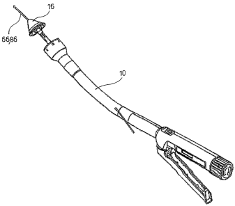
With reference to Fig. 1, with 10 has been
indicated a circular stapler portion comprising a handle
12 and a stem 14 as a whole. The structure of the
- - -
circular stapler is similar to the known circular
staplers, which are conventionally used to carry out
circular anastomosis, such as of the bowel. The
structure of the circular stapler according to the
4
CA 02587954 2007-05-04
WO 2006/048905
PCT/1T2005/000348
present invention is changed compared with the
conventional ones in that, in a preferred embodiment
thereof, it has a channel suitable to receive the guide
wire. In Fig. 2A, the circular stapler 10 has a channel
crossing the stem 14 thereof from the distal end to an
area at the proximal end from which it protrudes
outside, for example on one side. In
accordance with
different embodiments, not illustrated, the guide wire
runs all along the length of the circular stapler or
only a distal portion thereof.
The channel is suitable to receive a guide wire
(not illustrated in Fig. 1) such that the stapler can
slide therealong and be placed in the site requiring
anastomosis. An exemplary use of the circular stapler 10
will be described below with particular reference to
Fig. 29. The length of the stem 14 and the diameter
thereof are sufficient to carry out the method and reach
the desired site.
Advantageously, the stem is made of a flexible
material, such as to facilitate reaching the site
requiring anastomosis.
The stapler 10 advantageously comprises an anvil. 16
illustrated for example in Fig. 2. The anvil 16 defines
an exemplary device for drawing tissues together,
particularly a device that, besides drawing the tissues
5
CA 02587954 2007-05-04
WO 2006/048905 PCT/1T2005/000348
togeth-er, is suitable to be associated with the circular
anvil 10 such as illustrated in Fig. 1 in order to carry
out anastomosis.
The anvil 16 comprises a stem 18 and a head 20. The
stem 18 has such a longitudinal and cross size that
makes it suitable to be fit on the end of the stem 14 of
the circular stapler 10 opposite the handle 12 (Fig. 2A
and 41-44).
Advantageously, a channel 22 crosses the anvil 16
in a longitudinal direction and is suitable to receive a
guide wire, not illustrated in Fig. 2. An exemplary use
of the anvil 16 for the circular stapler 10 will be
described below with particular reference to Fig. 26-29.
An anchoring ring suitable to lock the anvil 16 on the
guide wire on which it is fitted such as to be drawn by
the guide wire in order to draw the tissues together and
create the anastomosis has been designated with 76.
=
In Fig. 3, with 24 has been generally designated a
positioning device suitable to draw tissues to4ether
which have been subjected to enterostomy, and to place
means for providing a passage (anastomosis) between the
tissues that have been drawn together.
The positioning device 24 comprises a first
component, or proximal component, designated with
numeral 26 and a second component, or distal component,
6
CA 02587954 2007-05-04
WO 2006/048905
PCT/1T2005/000348
designated with reference 28. Preferably, the
positioning device 24 extends along a longitudinal axis
30. Fig. 5, 7 and 8 illustrate perspective views of the
proximal component 26, whereas Fig. 6 illustrates a
perspective view of the distal component 28.
In accordance with a possible embodiment, the
proximal component 26 is suitably shaped to be abutted
against the edge of a first enterc)stomy in order to draw
the tissues adjacent thereto against the tissues
adjacent to a second enterostomy. The
distal component
28 is suitably shaped to be inserted through the
enterostomies.
For clarity purpose, the first enterostomy will.be
also called herein below as the proximal enterostomy,
whereas the second enterostomy will be also called the
distal enterostomy. With reference to a possible
embodiment, the first enterostomy can be a gastrostomy
and the second enterostomy can be a jejunostomy. With
reference to a different embodiment, the. first
=
enterostomy can be a proximal jejunostomy and the second
enterostomy can be a distal jejunostomy.
In accordance with a possible embodiment, the
proximal component 26 has a substantially cylindrical
outer structure. A cavity 32 being formed at one of the
bases of the cylindrical structure and longitudinally
7
CA 02587954 2007-05-04
WO 2006/048905 PC
T/IT2005/000348
thereto, preferably has a first portion defined by a
surface having the shape of a truncated cone 32a and a
second portion defined by a cylindrical surface 32b.
The cavity 32 does not run through the entire length of
the proximal component 26, leaving a base wall 34.
Furthermore, the cross size of the cavity 32 and the
proximal component 26 are preferably such as to leave an
abutment surface, for example a plane annular surface
26a contouring the cavity.
According to a possible embodiment, a lug 36,
"
preferably cylinder, shaped, extends along the
longitudinal axis 30 from the bottom of the cavity 32
towards the outside of the cavity, preferably such. that
a free end 36a of the lug 36 is completely out of the
cavity 32. In other words, the length of the lug 36 from
the base of cavity 32 to the free end 36a thereof is
preferably greater than the depth of the cavity 32. The .
lug 36 has a preferably cylindrical cavity 38 extending
along the longitudinal axis 30 and crossing the base
wall 34 leading to the opposite surface of the proximal
component 26. In other words, the cavity 38 involves the
lug 36 and base wall 34 thereby generating a duct open
at the ends thereof and suitable to receive a guide wire
not illustrated in Fig. 3-8. Preferably, the proximal
component 26 comprising the lug 36. is made as one piece.
8
CA 02587954 2007-05-04
WO 2006/048905
PCT/1T2005/000348
According to a possible embodiment, the proximal
component 26 comprises holes 40 for example for a
suture, which can be used for separating the proximal
component from the distal component, to be passed
therethrough.
In accordance with a possible embodiment, the
distal component 28 comprises a head 42 and a stem 44,
which are preferably made as one piece, that develop
along the longitudinal axis 30.
The head 42 preferably has a shape of a truncated
cone and, according to a possible embodiment, comprises
holes 46 for a suture, which can be used for example to
separate the distal component from the proximal
component, to be passed therethrough.
In accordance with a possible embodiment, the stem
44 preferably has a cylindrical structure and a free end
thereof, i.e. opposite the head 42, widens to form a
preferably annular base 48.
A channel 50 extends along the longitudinal axis 30
from the end of head 42 to the base 48 and is suitable
to receive a guide wire therein, not illustrated in Fig. .
4 or 6. The cross size of channel 50, at least in the
portion at the base 48, are such as to receive the lug
36 of the proximal component 26 therein. In other words,
the channel 50 preferably has a larger section at the
9
CA 02587954 2007-05-04
WO 2006/048905 PCT/1T2005/000348
area in which it receives the lug 36. Preferably, the
remaining part of channel 50 has the same cross size as
the cavity 38.
Fig. 3 and 4 illustrate the positioning device 24
when assembled. The proximal component 26 and the distal
component 28 are joined such that the cavity 38 and
channel 50 define a channel running all along the
assembly for introducing a guide wire, not illustrated
in Fig. 3 and 4. Particularly, Fig. 4 illustrates the
positioning device 24 sectioned along a plane comprising
the longitudinal axis 30. In the assembly position, the
proximal component 26 and the distal component 28 lock
an elastic ring 52 therebetween, which is hold in a
compressed/deformed configuration, and suitable to be
placed by the positioning device 24 in a desired
anastomotic site in which, after it has been positipned,
the elastic ring 52 takes a preset non-compressed rest
shape. (see for example Fig. 37). The elastic ring can be.
made of Nitinol, stainless steel or other satisfying
materials.
In accordance with a possible embodiment, the
elastic ring 52 in its deformed configuration, has an -
end, such as the proximal (designated with numeral 52a),
that is locked between the base 48 of the distal
component 28 and the inner diameter of the cavity 32 of
CA 02587954 2007-05-04
WO 2006/048905
PCT/1T2005/000348
the proximal component 26, i.e. inside the cylindricaa
portion 32b of the cavity 32. The opposite end of the
elastic ring 52, i.e. the distal end designated with the
numeral 52b, is preferably unfastened and abuts beneath
the head 42 of the distal component 28. In this case, it
is advantageously provided that the cross size of the
distal end 52b of the elastic ring 52 does not exceed
the cross size of the head 42 of the distal component
28.
An exemplary use of the positioning device 24 will
be described below with particular reference to the Fig.
34-37. Upon use, the outer diameter of the proximo"
component, and particularly with the flat annular
surface 26a, is intended to act as a striker against the
wall of the tissue to be drawn together, or in other
words, abut against the proximal enterostomy while it
minimizes the risk of penetration in the wall.
The distal component 28, with its head 42, is =
intended to penetrOte in the proximal and distal
enterostomies and protects the elastic ring 52 whiLe
being inserted and positioned, such as will be described
in the following.
Fig. 9-12 illustrate a possible variant embodiment
of the positioning device 24 and the proximal and distal
components thereof according to the present invention.
11
CA 02587954 2007-05-04
WO 2006/048905
PCT/1T2005/000348
The elements in common have been designated with the
same numeral used in Fig. 3-8 and will be described
below with reference to the differences from the above
embodiment.
The proximal component 26 is substantially similar
to the one illustrated in Fig. 3-5, 7 and 8. As regards
the distal component 28, the stem 44 extends straight up
to its free end opposite the head 42 that does not widen
to form a base similar to the base 48 of the distal
component described above.
Furthermore, the head 42,
preferably having the shape of a cone or truncated cone,
comprises a flange 54 extending from the outer perimeter
of the major base of the head to form a circular wall
substantially parallel to the longitudinal axis 30.
Fig. 9 and 10 illustrate the positioning device 24
when assembled, in which the channel 50 and the cavity
38 define a channel extending along the longitudinal
axis 30 through the entire length of the assembled
positioning device 24 to receive a guide wire, not
illustrated in the Fig. 9-12. In the assembled
configuration of the positioning device 24, the elastic
ring 52 is hold between the proximal component 26 and
the distal component 28 in a compressed/deformed
configuration. After the elastic ring has been
positioned, it takes a preset, uncompressed rest shape
12
CA 02587954 2007-05-04
WO 2006/048905
PCT/1T2005/000348
as described above. In the deformed configuration, the
proximal end 52a of the elastic ring 52 is fastened by
the inner diameter of the cavity 32, particularly by the
cylindrical portion 32b cif the cavity 32, whereas the
distal end 52b of the elastic ring 52 is fastened within
the circular flange 54 of the distal component 28.
The cross size of channel 50, at least in the
portion at the base 48, are such as to receive the lug
36 of the proximal component 26 therein. In other words,
the channel 50 has a larger section at the area where it
receives the lug 36. Preferably, the remaining part of
channel 50 has the same cross size as the cavity 38.
Also in this case, the elastic ring 52 can be made of
Nitinol (Ni-Ti alloy), stainless steel or other
satisfying materials.
The exemplary use of the positioning device is
similar throughout the various embodiments described.
Fig. 13-16 illustrate a possible variant embodiment
of the positioning device and the proximal and distal
components thereof according to the present invention.
The elements in common have been designated with the
same numeral used in the figures above and will be
described below with reference to the differences from
the above embodiments.
The proximal component 26 has a prismatic outer
13
CA 02587954 2007-05-04
WO 2006/048905
PCT/1T2005/000348
structure, prefeLdbly having a rectangular base. The
cavity 32 is formed at one of the bases of the structure
and does not run through the entire length of the
proximal component 26, leaving a base wall 34. The sizes
of the cavity 32 and proximal component 26 are such as
to leave a peripheral flat surface 26a.
In the base wall 34, preferably in the middle
thereof, there is provided a preferably cylindrical
cavity 38 extending along the longitudinal axis 30 and
crossing the entire thickness of the base wall.
Ribs 56, preferably on opposite parts of cavity 38
and parallel to the long sides of the rectangular base,
extend from the bottom of the cavity 32 by a height
preferably less than the depth of cavity 32.
According to a possible embodiment, the proximal
component 26 comprises holes 40 for example for a
suture, which can be used for separating the proximal
component from the . distal component, to be passed
therethrough.
The distal component 28 comprises a head 42, which
according to a possible embodiment, comprises holes (not
illustrated) for a suture, which can be used for example
to separate the distal component from the proximal
component, to be passed therethrough.
The channel 50 extends along the longitudinal axis
14
CA 02587954 2007-05-04
WO 2006/048905
PCT/1T2005/000348
30 throughout the solid thickness of the head 42,
preferably in the middle thereof, and is suitable to
receive a guide wire therein, not illustrated in Fig. 14
or 15.
The head 42_ has a tract having a substantially
pyramidal or having a truncated-pyramid shape,
preferably with a rectangular base. Two flanges 54 that
preferably involve the short sides of the rectangular
base and a limited portion of the long sides extend from
the outer periphery of the major base of the truncated-
pyramidal portion, in a direction substantially parallel
to the longitudinal axis 30.
According to a possible embodiment, there is
provided a preferably flat extension 58, arranged at a
middle portion of each long side of the rectangular base
and projecting in the direction substantially parallel
to the longitudinal axis 30 along a preferably longer
tract than the flanges 54.
Fig. 13 and 14 illustrate the positioning device 24
when assembled, in which the channel 50 and the cavity
38 are arranged alOng the longitudinal axis 30 to
receive a guide wire, not illustrated in the Fig. 13-16.
In the assembled configuration of the positioning device
24, the elastic ring 52 is hold between the proximal
component 26 and the distal component 28 in a preferably
CA 02587954 2007-05-04
WO 2006/048905
PCT/1T2005/000348
flattened, compressed/deformed configuration. After the
elastic ring has been positioned, it takes a preset,
uncompressed rest shape as described above. In
the
deformed configuration, the proximal end 52a of the
elastic ring 52 is fastened by the inner periphery of
the cavity 32. Particularly, the ribs 56 fasten the
elastic ring 52 in a flat deformed configuration, or in
other words, the elastic ring 52 is arranged between the
wall of the cavity 32 and the ribs 56. Furthermore, the
distal end 52b of the elastic ring 52 is fastened by the
flanges 54 and extensions 58, when the latter are
provided.
On the one hand, the assembled configuration of the.
positioning device 24 is ensured by the interference
between the proximal end 52a of the elastic ring 52 and
the walls of the proximal component 26 defining the
cavity 32, and on the other hand by the interference
between the distal end 52b of the elastic ring 52 and
the flanges 54 and the extensions 58, when the latter
are provided.
Also in this case, the elastic ring 52 can be made
of Nitinol, stainless steel or other satisfying
materials.
The exemplary use of the positioning device is
similar throughout the various embodiments described. In
16
CA 02587954 2007-05-04
WO 2006/048905
PCT/1T2005/000348
this latter case, the peripheral wall 26a of the
proximal component 26 is the one intended to abut
against the wall of the tissue to be drawn together
while minimizing the risk that the wall may be
penetrated. Furthermore, the angled head 42 of the
distal component 28 is intended to penetrate the
proximal and distal enterostomies and protects the
elastic ring 52 when being introduced and positioned by
fastening the ring within the flanges 54 and the
extensions 58, such as will be described in the
following.
The channel 50 and the cavity 38 are intended to
house a guide wire for transporting the positioning,
device.
The present invention further relates to a method
for the therapy of obesity and particularly a method for,
carrying out anastomosis in tracts of the digestive
tube. Fig. 17-40 illustrate several steps of a possible
embodiment of the method according to the present
invention. The examples illustrated particularly relate
to a method for carrying out an endoluminal/transluminal
gastrojejunostomy (G-J) and a jejunojejunostomy (J-J)
via transoral access.
=
In general terms, the method according to the
present invention advantageously provides to draw
17
CA 02587954 2007-05-04
WO 2006/048905
PCT/1T2005/000348
tissues together and carry out anastomosis via
endoluminal access by introducing, through a natural
orifice (such as nose, mouth, ears, anus) or other
luminal structures, guide or rail means within the
tissues to be drawn together. Suitable components or
devices can be thereby carried to the anastomotic site
such that the surfaces of the tissues are suitably drawn
together and connected with a channel (anastomosis).
Advantageously, the guide or rail means,
particularly a main guide wire or first guide wire, are
introduced such as to generate an open ring that can
begin and end in natural orifices, such as the mouth,
nose, anus or other natural orifices, such as colostomy,
trocar, abdomen incisions, wounds, fistulae. The
components or devices provided to draw the tissues
together are advantageously moved by locking the device
on the guide wire and pulling one of the guide wire
ends.
In accordance with a possible embodiment, the ends
of the open ring and accordingly of the main guide wire
are different from each other, and hence
distinguishable. Advantageously, the guide wire is
internally hollow, i.e. it has a tubular structure
suitable to receive needles for perforating the tissues
and carrying out proximal and distal enterostomies.
18
CA 02587954 2007-05-04
WO 2006/048905
PCT/1T2005/000348
PerfordLion can take place for example either by pushing
the needle through the tissues, or applying a
radiofrequency through the needle.
The open ring then crosses the proximal enterostomy
and then the distal enterostomy, for example by using a
gripping device.
The positioning device 24 is locked on the guide
wire by means of an anchoring ring 76 and drawn by the
guide wire until it is partially inserted in the
proximal enterostomy and abutted against a first tissue
portion to be joined. The positioning device 24 is
further drawn until it is partially inserted in a distal
enterostomy by drawing together the tissue portions to
be joined. Finally, the positioning device 24 partially
inserted in the proximal and distal enterostomy releases
an elastic ring 52 riding the proximal and distal
enterostomies to hold the tissue portions joined to each
other thereby generating a passage or anastomosis.
With reference to the above example, Fig. 17-29
illustrate a gastrojejunostomy (G-J) step that is
advantageously carried out by introducing guide or rail
means through a natural orifice (such as the nose or
mouth). Subsequently, the guide means, a guide wire in
this case, form an open ring crossing the points of the
tissues to be joined.
19
CA 02587954 2007-05-04
WO 2006/048905 PCT/1T2005/000348
Fig. 17 illustrates a first step, INThich is
designated as the step 1, in which a substantially
conventional laparoscope 60 has been introduced in the
abdominal cavity to view the areas to be treated. This
step can be potentially eliminated after a certain
degree of skill in the method has been achieved, thereby
the method can be made completely endoluminal and
transluminal. The laparoscope 60 is illustrated in Fig.
17 and also in the subsequent steps, but it can be
omitted as well. Alternatively or in addition to the
laparoscopic control, a gastroscopic control can be
provided, i.e. by introducing a secondary gastroscope =
for example through the esophagus or mouth having the
function of controlling the method steps. In case a
gastroscope is required to carry out several steps of
the method, a main gastroscope carrying out the method
steps and a secondary gastroscope monitoring the
operation will be introduced.
Fig. 18 illustrates a step of the method according
to the present invention, which is also designated as
=
the step 2, in which a substantially conventional main
gastroscope 62 is introduced through the esophagus,
stomach, passing through the pylorus and subsequently
the duodenum to reach the jejunum. Particularly, the
gastroscope 62 is advanced by approximatively 20-40 cm
CA 02587954 2007-05-04
WO 2006/048905
PCT/1T2005/000348
beyond the pylorus.
Fig. 19 illustrates a detail of the jejunum and the
end of the gastroscope 62. The latter conventionally
comprises several channels 64 crossing the entire length
thereof and that can be used for tools or the like to be
passed therethrough. The step from Fig. 19, also
designated as the step 3, provides that a first guide
wire 66 or main guide wire being suitable to provide the
open ring is advanced along one of the channels 64 of
the gastroscope 62. The guide wire is advanced until a
pointed end 66a thereof or a needle sliding along the
tubular structure of the guide wire protrudes from the
gastroscope. The end 66a of the guide wire 66 perforates
the jejunum wall from the inside and creates a
jejunostomy (proximal enterostomy). The laparoscope 60
is optionally provided. When this is provided, the guide
wire 66 is advanced and the jejunostomy is created under
the laparoscope visual control.
The jejunostomy can be carried out by pushing the
guide wire directly through the jejunum wall.
Alternatively, or in addition thereto, radiofrequency
energy may be applied to perforate the jejunum wall and
then advance the guide wire 66.
In other words, a first guide wire 66 being past of
guide or rail means which will be subsequently indicated =
21
CA 02587954 2007-05-04
WO 2006/048905
PCT/1T2005/000348
in greater detail, is positioned within the tissue to be
joined and passed through one of the tissue portions to
be joined. The jejunum tissue portion to be drawn near
and joined to the stomach thereby forming an anastomosis
has been designated with A.
Fig. 20 illustrates a step which has been
designated as the step 4 in which the gastroscope 62 is
removed and the guide wire 66 is left in the abdomen
within the stomach and along a jejunum tract with the
end 66a protruding 'from the jejunum at the tissue
portion A to be joined. The step 4 can be carried out
under laparoscopic control (laparoscope 60), when
provided.
Fig. 21 illustrates a step which is designated as
the step 5, in which a main gastroscope 62, of a
substantially conventional type, has been introduced
again in the stomach through the esophagus, in order to
create a gastrostomy .(distal enterostomy). Also in this
case, one may directly push either a secondary guide
wire 77 with a pointed end 77a or a needle sliding
within the tubular structure of the guide wire.
Alternatively, or in addition thereto, radiofrequency
energy can be applied in order to perforate the stomach
wall and advance the guide wire.
The gastrostomy is carried out in a stomach portion
22
CA 02587954 2007-05-04
WO 2006/048905
PCT/1T2005/000348
corresponding to the area to be joined. This portion has
been designated with A'.
Step 5 may also be carried out under laparoscopic
control.
Fig. 22 illustrates a step of the method according
to the present invention, which has been designated as
the step 6, in which the gastrostomy is enlarged by
means of a balloon catheter 72. The catheter is inserted
in the gastroscope 62 until a balloon-end 72a thereof
reaches the gastrostomy which is enlarged by inflating
the balloon.
Step 6 may also be carried out under laparoscopic
control.
Fig. 23 illustrates a step of the method according
to the present invention, which has been also designated
as the step. 7, in which the gastroscope 62 is advanced
through the gastrostomy enlarged by the balloon, within
the abdominal cavity. Step 7 can be carried out either
under gastroscopic (secondary gastroscope) and/or
laparoscopic (laparoscope 60) control. As stated above,
by gastroscopic control is meant a control carried out
by means of a secondary gastroscope, not illustrated in
Fig. 23, which is introduced through the esophagus
having only a control function. This secondary
gastroscope can be provided in every step whenever a
23
CA 02587954 2007-05-04
WO 2006/048905
PCT/1T2005/000348
gastroscopic control as an alternative or in addition to
the laparoscopic control is required.
Fig. 24 illustrates a step which has been
designated as the step 8, in which a gripping device 74
(forceps or the like) is advanced through the
gastroscope 62 and the end 66a of the guide wire 66
protruding from the jejunostomy is coupled therethrough.
Gripping the guide wire point is not required.
The gripping device 74 can be for example a loop-
shaped endoscopic instrument for polypectomies.
Fig. 25 illustrates a step which has been
designated as the step 29, in which the guide wire 66 of
the jejunum is pulled through the gastrostomy in order
to provide a first ring 80 open at the ends thereof, or
gastro-jejunum ring (ring 1), the ends thereof
protruding from the orifice used (the mouth, esophagus,
= In Fig. 25, the end of ring 80 corresponding to the
jejunum (jejunum end), i.e. the end passing through the
stomach and jejunum and protruding from portion A has
been designated with 80a, whereas the end of the ring 80
corresponding to the stomach (stomach end), i.e. the end
passing through the stomach and protruding therefrom at
portion A' has been designated with 80b. Both ends are
advantageously different from each other in order to be
distinguished.
24
CA 02587954 2007-05-04
WO 2006/048905
PCT/1T2005/000348
The ring 80 can be now used as a guide means or
rail system in order to introduce and carry suitable
anastomotic devices suitable to draw the tissues
together and carry out the anastomosis in the site of
interest. The anastomotic devices are advantageously
locked on the guide wire for example by means of an
anchoring ring 76 and one of the ends of the ring is
pulled until the anastomotic device partially enters the
proximal enterostomy, draws the tissues thereof near the
distal enterostomy and partially enters the same.
Fig. 26 illustrates a step which has been
designated as the step 10, wherein the selected
anastomotic device (anvil 16, positioning device 24,
etc.) is inserted on the guide wire from the jejunum end
80a and pulled along the ring 80 of guide wire through
the esophagus, stomach, duodenum and jejunum. Drawing is
allowed by an anchoring ring 76 which is made integral
with the guide wire such as to push against the proximal
part of the selected anastomotic device.
Though an anvil 16 has been illustrated in Fig. 26,
a positioning device 24 or other similar devices can be
used as well.
By pulling the guide wire from the end of stomach
80b, the anastomotic device can be pulled .until the
portion A of the jejunum (proximal enterostomy).
CA 02587954 2007-05-04
WO 2006/048905
PCT/1T2005/000348
As illustrated in Fig. 26, the end of the jejunum
part of the guide wire 80a is inserted in the channel 22
of the anvil 16 from the side of the stem 18. In the
case of the positioning device 24, the end of the
jejunum part of the guide wire 80a would be inserted in
the channel 50 from the side of the distal component 28.
Fig. 27 illustrates a step which has been also
designated as the step 11, in which the anastomotic
device and particularly the anvil 16 is pulled until it
has partially passed the jejunostomy (proximal
enterostomy). As already stated above, one can act under
laparoscopic control. The stem 18 of the anvil 16
crosses the jejunostomy and protrudes in the abdominal
cavity, whereas the head 20 contacts the tissue to be
drawn together. When a positioning device 24 is used,
the head would enter the jejunostomy whereas the flat
peripheral surface 26a would abut against the contouring
tissues.
Fig. 28 illustrates a step which has been also
designated as the step 12, in which by keeping on
pulling the end of the guide wire 80b from the side of
the stomach, the anvil 16 and particularly the head 20
acts as a striker against the inner wall of the portion
A of the jejunum and draws the jejunum until the portion
A is drawn near the stomach and particularly portion A'.
26
CA 02587954 2007-05-04
WO 2006/048905
PCT/1T2005/000348
The stem 18 of the anvil 16 (anastomotic device) also
partially enters the gastrostomy (distal enterostomy).
The operation can be carried out under laparoscopic
control (laparoscope 60). When a positioning device 24
is used, the head would 42 enter the gastrostomy whereas
the flat peripheral surface 26a would draw the relative
contouring tissues together.
Fig. 29 illustrates a step which has been indicated
as the step 13, in which the traction on the stomach end
80b of the guide wire is maintained in order to maintain
the portions A and A' near each other. Furthermore, a
circular stapler 10 is caused to slide on the guide wire
from the stomach end 80b until it reaches the inside of
the stomach, and until the stem 18 of the anvil 16
connects to the end of the stem 14 of the stapler 10
(such as illustrated in greater detail in Fig. 2a and
41-44). The stapler 10 carries out the anastomosis
between the portion A and the portion A' by cutting and
suturing the tissue in a circular manner. A passage 84
is thereby formed (Fig. 30) which directly communicates
the stomach and jejunum. When a positioning device 24 is
used, the passage 84 is obtained by detaching the
proximal component from the distal component and
releasing the elastic ring 52 riding both enterostomies.
When the gastrojejunostomy (G-J) has been
27
CA 02587954 2007-05-04
WO 2006/048905
PCT/1T2005/000348
completed, the guide wire 66 is removed by drawing one
end thereof.
With reference to the above example, Fig. 30-40
illustrate a jejunojejunostomy (J-J) step that is
advantageously carried out by introducing guide or rail
means through a natural orifice (such as the esophagus
or mouth). Subsequently, the guide means, a guide wire
in this case, form an open ring crossing the points of
the tissues to be joined.
Fig. 30 illustrates a step designated as the step
14, in which a substantially conventional gastroscope 62
is introduced through the esophagus, stomach, passing
through the pylorus and subsequently the duodenum to
reach the jejunum. Particularly, the gastroscope 62 is
advanced to a portion to be joined that is designated
with B in order to carry out a proximal jejunostomy,
which portion is proximally arranged relative to channel
84 (anastomosis) that has already been created.
A guide wire 86, or main guide wire, intended to
form the open ring is advanced along a channel 64. The
guide wire is advanced until a pointed end 86a thereof
or a needle sliding within the guide wire protrudes from
the gastroscope. The end 86a of the guide wire 66
perforates the jejunum wall from the inside and creates
a jejunostomy (proximal enterostomy).
28
CA 02587954 2007-05-04
WO 2006/048905
PCT/1T2005/000348
The laparoscope 60 is optionally provided. When
this is provided, the guide wire 86 is advanced and the
= jejunostomy is created under the laparoscope visual
control.
The jejunostomy can be carried out by pushing the
guide wire directly through the jejunum wall.
Alternatively, or in addition thereto, radiofrequency
energy may be applied to perforate the jejunum wall and
then advance the guide wire 86.
In other words, a guide wire 86 being part of guide
or rail means which will be subsequently indicated in
greater detail, is positioned within the tissue to be
joined and passed through one of the tissue portions B
to be joined (proximal jejunostomy).
Fig. 31 illustrates a step which has been indicated
as the step 15, in which the gastroscope 62 is removed
and the guide wire 86 is left within the stomach and.
jejunum, with the end 86a protruding from the walls of
the jejunum (proximal jejunostomy). The gastroscope 62
is then advanced through the stomach, the previously
accomplished gastrojejunostomy (step 84) and a tract of
the distal jejunum until a sufficient distance to create
the jejunumjejunum (J-J) anastomosis. The
latter
portion has been indicated with the reference B'.
Analogously to Fig. 21 and 22 (steps 5 and 6) a
29
CA 02587954 2007-05-04
WO 2006/048905
PCT/1T2005/000348
secondary guide wire is advanced along the gastroscope
62 until a pointed end thereof protrudes from the end of
the gastroscope 62. This
pointed end is then passed
through the tissue walls at the portion B' in order to
create a distal jejunostomy. The distal jejunostomy can
be also carried out either by directly pushing the
pointed end through the jejunum wall or applying
radiofrequency energy in order to perforate the wall,
subsequently advancing the guide wire.
The creation of the distal jejunostomy can be
monitored through the laparoscope.
A catheter with balloon-end may be optionally
inserted along the gastroscope. When the balloon end is
near the distal jejunostomy, the balloon is inflated in
order to dilate the distal jejunostomy and the
gastroscope is pushed in the abdominal cavity. This
dilation may be required when the laparoscope is not
ysed and monitoring is carried out through the
gastroscope such that the latter can view the end 86a of
the guide wire 86.
Fig. 32 illustrates a step which has been
designated as the step 16, in which a gripping device 74
(endoscopic forceps or the like) similar to the one used
in step 8, is advanted through the gastroscope to lock
the end 86a of the main guide wire 86 protruding from
CA 02587954 2007-05-04
WO 2006/048905
PCT/1T2005/000348
the proximal jejunostomy site. Gripping the guide wire
end is not required.
Fig. 33 illustrates a step which has been indicated
as the step 17, in which the gastrcpscope has been
removed and the main guide wire 86 Ylas been pulled
through the distal jejunostomy to form a ring 88 open at
the ends thereof, or jejunumjejunum ring (the ring 2),
the ends thereof protruding from the orifice used. In
Fig. 33 the end of the jejunum, i.e. the end passing
through the stomach and the jejunum and protruding
therefrom at portion B has been designated with 88a,
whereas the end of the stomach, i.e. the end passing
through the stomach, protruding from the
gastrojejunostomy (passage 84) and protruding from the
jejunum at portion B' has been designated with 88b.
The ring 88 can be now used as a guide means or
rail system in order to introduce and carry suitable
anastomotic devices suitable to draw the tissues
together and carry out the anastomosis in the site of
interest (J-J). As described above, the anastomotic
device is locked on the guide wire, an end thereof being
pulled in order to advance the anastomotic device.
Fig. 34 illustrates a step -which has been
designated as the step 18, wherein an allastomotic device
such as a positioning device 24 is inserted from the
31
CA 02587954 2007-05-04
WO 2006/048905 PC
T/IT2005/000348
jejunum end 88a and pulled along the ring 88 of guide
wire through the esophagus, stomach, duodenum and
jejunum. The operation may be carried out under
laparoscopic and/or gastroscopic control in order to
view the movement.
The anastomotic device may be caused to slide on
the guide wire from the jejunum end 88a. Traction is
permitted due to an anchoring ring similar to that
described above, which is made integral with the guide
wire pushing against the proximal part of the selected
anastomotic device. By pulling the guide wire from the
end of stomach 88b, the anastomotic device can be pulled
until the portion B of the jejunum.
As illustrated in Fig. 34, the positioning device
24 (channel 50 and cavity 38) is inserted on the end 88a
of the guide wire from the side of the distal component
28.
The anastomotic device and particularly the
positioning device 24 is pulled until it has partially
passed the proximal jejunostomy. The head 42 of the
distal component 28 and a part of the elastic ring 52
cross the proximal jejunostomy and protrude in the
abdominal cavity, whereas the other part of the elastic
ring 52 remains within the jejunum. The proximal
component also remains within the jejunum and abuts, for
32
CA 02587954 2007-05-04
WO 2006/048905
PCT/1T2005/000348
example with the surface 26a, against the tissu.e wall to
act as a striker.
Fig. 35 illustrates a step which has been
designated as the step 19, in which the two jejunum
branches are drawn together, optionally under
gastroscopic and/or laparoscopic vision by keeping on
pulling the anastomotic device (positioning device 24).
Particularly, the head 42 of the distal component
28 with a part of the elastic ring 52 penetrate in the
distal jejunostomy.
Fig. 36 illustrates a step which has been
designated as the step 20, in which the elastic ring 52
that takes its uncompressed configuration, such as
illustrated enlarged for example in Fig. 37 (step 21) is
pos tioned. The ends of the elastic ring 52 foad between
the proximal jejunostomy and the distal jejunostomy
thereby maintaining the portion B and portion B' joined
to each other thereby creating a circular an_astomosis.
The elastic ring 52 can be released from the positioning
device 24 by simultaneously uncoupling the distal and
proximal components 26 and 28. Alternatively, one of the
two components can be uncoupled by pulling the same
while keeping unchanged the position of the elastic ring
52 relative to the anastomotic site.
To uncouple the distal component and th_e proximal
33
CA 02587954 2007-05-04
WO 2006/048905 PC
T/IT2005/000348
component, one can use the suture threads protruding
from the holes 40 of the proximal component 26 and the
holes 46 of the distal component 28 by coupling them by
means of a suitable tool inserted in a gastroscope 62.
The suture stitches are thus gripping points for
uncoupling the proximal and distal components of the
positioning device 24 from each other.
During positioning, the enterostomy can be viewed
by means of a gastroscope or laparoscope.
When the jejunojejunostomy (J-J) has been
completed, the guide wire 86 is removed by drawing one
end thereof.
Fig. 38 illustrates a step which has been
designated as the step 22, in which the
gastrojejunostomy (G-J) and the jejunojejunostomy (J-J)
have been completed and in which there is illustrated
the route followed by the food along the digestive tract
after it has been changed.
To complete the method discussed above, either a
gastric partition obtained with a gastric bandage such
as illustrated in Fig. 39 (step 23.1) or a gastric
partition obtained with an endoscopic stapler such as
illustrated in Fig. 40 (step 23.2) can be provided.
From what has been described above, one may
appreciate how the provision of guide means carrying
34
CA 02587954 2007-05-04
WO 2006/048905
PCT/1T2005/000348
components or devices to the desired anastomotic site
through natural orifices (such as the nose, mouth, ear,
anus) or other luminal structures in order to carry out
anastomosis greatly simplifies the procedure, shortens
the patient's convalescence and eliminates the drawbacks
of traditional surgery.
The provision of components and devices that
suitably draw the tissue surfaces together and/or
connect the surfaces by means of a passage is
particularly advantageous and allows to carry out a
completely endoluminal method.
It should be understood that variations and/or
additions to what has been described and illustrated
above may be provided.
In addition to the method described above, there
may provided alternative procedures (ERCP, Chole duct,
colo-proctostomy, jejunum-colostomy).
The order of the steps of ,the method illustrated in
the annexed drawings and described above,
(gastrojejunostomy or G-J, jejunojejunostomy or J-J,
sectioning) can be readapted. For
example, with
patients that have already been subjected to gastric
bandage, the steps G-J and J-J can be completed as
described above. Subsequently, the gastric bandage can
be completely restricted by carrying out a gastric
CA 02587954 2012-12-17
partition thereby completing the procedure.
Alternatively to the use of the circular stapler 10
and anvil 16, the gastrojejunostomy G-J according to the
steps described above (Fig. 17-Fig. 29) can be carried
out by means of a positioning device 24 as described
above (similarly to the jejunojejunostomy J-J steps
corresponding to Fig. 31-38).
To the preferred embodiment of the device, stapler
or method described above, those skilled in the art,
aiming at satisfying contingent and specific
requirements, may carry out a number of modifications,
adaptations and replacement of elements with others
functionally equivalent.
*** * ***
36