Note: Descriptions are shown in the official language in which they were submitted.
CA 02588391 2007-05-17
WO 2006/059961 PCT/TR2005/000011
DESCRIPTION
Mastoid Antral Ventilation Tube
The invention relates to a mastoid antral ventilation tube that aerates and
drains
the middle ear by the route of mastoid antrum. On the contrary to the
ventilation tubes
placed on the eardrum, this tube is a ventilation and drainage tube placed
between the
mastoid antrum and the external ear canal.
The middle ear cleft comprises the air cells of the eustachian tube, middle
ear and
the mastoid. Einbryologically, this cleft develops from the first branchial
pouch, first
eustachian tube, then the middle ear and finally the mastoid antrum and the
air cells form.
The aeration of the middle ear is almost complete at birth. However, the
aeration of the
mastoid cells continue until 9 years of age. These three aerated cavities that
make up the
middle ear cleft are connected to one another. Eustachian tube connects the
middle ear to
the nasopharynx. Rather small and narrow canals called Istmus anterior and
Istmus
posterior connect middle ear to the mastoid air cells. The mucosa events
occurring in the
middle ear are not limited only to the middle ear, they extend also to mastoid
antrum and
cells through aforesaid connections. Even though the function of the mastoid
air cells is
not known exactly, they are commonly accepted as the air reservoir for the
middle ear.
Hence, especially in children, mastoid air cells contribute to the ventilation
of the middle
ear in case of obstruction or dysfunction of eustachian tube, in order to
prevent the
formation of the negative pressure. However, prolonged dysfunction of
eusthacian tube,
increase of negative pressure in the middle ear and the obstruction of istmus
anterior and
posterior cause effusion to form in both middle ear and the mastoid cells.
Initially, the
medical treatment is applied to eliminate this effusion. In patients who are
refractory to
medical treatment, a ventilation tube (myringotomy tube, tympanostomy tube,
grommet
tube) is placed on the eardrum by means of a surgical operation called
myringotomy
(paracentesis), in order to ventilate the middle ear and drain the effusion.
In this way, a
cavity is provided to equate the pressure between the middle ear and the
external medium
and to permit the drainage of effussion. When the eardrum heals spontaneously
afterwards, the tube is extruded, leaving a usually healthy eardrum. This
operation has
found a rather widespread use during the recent years. Although these tubes
have
provided important contributions to the treatment of the middle ear effusion,
they have
failed to provide the permanent and desired achievement in an non-neglectable
number of
patients. It was even reported that the long term outcomes in the patients
with and without
1
CA 02588391 2009-02-09
tube placed was similar. Moreover, many complications and sequelae to these
tubes
have been reported until present time. The object of the mastoidectomy, which
is a
surgical operation applied to open the mastoid antrum and air cells as the
last resort
for the patients who exhibit no improvement despite the repetitive
applications of
myringotomy tube, is to open the connections between the middle ear and the
mastoid cells.
Myringotomy tubes have important disadvantages. As these tubes have a
fairly small size, surgical skills and experience are required to place them
on the
eardrum. These tubes may frequently become obstructed, they are hard to aspire
and they are not suitable for medical administration for the children. They do
not
provide information as to the potency of connection between the middle ear and
the
mastoid air cells. In connection with the myringotomy tube application, the
complications and sequels such as chalk patches (myringosclerosis) on eardrum,
perforation, discharge, retraction etc. may exist. The duration of these tubes
is quite
variable based on the reaction of the body, course of the middle ear disease
and
healing of the wound. Particularly in children, it is very difficult to open
the
myringotomy tube in case it becomes obscured during or immediately after the
operation. As the air buffer in the middle ear is canceled by myringotomy
tubes, the
transport of mucus and bacteria from the nasopharynx to the middle ear through
the
eustachian tube is facilitated. Therefore, these tubes disable the functions
of the
eustachian tube during their period of stay in the patient. In addition,
placement of
these on the graft during the frequent practices of tympanoplasty brings about
disadvantages both in wound healing and surgical intervention.
US3982545A is considered to represent the most relevant state of the art,
discloses a mastoid antral ventilation tube comprising a cannula made up of a
tab
and a shaft providing a drainage-aspiration of mastoid antrum with one end
capable
of extending up to mastoid antrum via cannula. From this, the subject-matter
of
independent claim 1 differs in that the tube comprises an inner cannula made
up of a
thin and narrow canal and a drenage canal. The subject-matter of claim 1 is
therefore
2
CA 02588391 2009-02-09
novel. The problem to be solved by the present invention may be regarded as
reducing the risc of blockage of the tube.
According to the present invention, there is provided a mastoid antral
ventilation tube, which treats diseases associated with insufficiencies of
ventilation in
middle ear of a person, helps to eliminate pressure imbalances for inner ear
fluids,
enables a drug treatment for diseases of middle and inner ear, makes it
possible to
aerate and drain the middle ear cleft of the person via mastoid antral path
without
disrupting integrity of eardrum and improves a success of middle ear
operations, said
mastoid antral ventilation tube being for placement into mastoid antrum,
characterized in that the ventilation tube comprises an outer cannula
consisting of
tab, neck, bend and shaft sections, and an inner cannula consisting of a first
canal
having an end suitable for a syringe for drug administration, and a second
canal
which is a drainage canal that provides for drainage or aspiration of the
mastoid
antrum, one end of said inner cannula being capable of extending via the outer
cannula up to the mastoid antrum.
This invention aerates and drains the middle ear cleft by way of mastoid
antrum. It re-established the connection between the middle ear and the
mastoid air
cells, and equates the pressure of both middle ear and the mastoid air cells
with the
external medium. The mucosal healing of the aerated cavities of the middle ear
cleft
is provided in a natural way, without the need for any intervention to the
eardrum. A
more permanent healing of disease will be provided, and unwanted complications
and sequels are avoided. The mastoid antral ventilation tube can be used in
children
and the adult for all the diseases where the middle ear needs aeration and
drainage.
The advantage of the mastoid antral ventilation tube over the myringotomy
tubes placed on the eardrum is that it enables the eardrum to preserve its
natural
form, since no intervention to eardrum is involved. Via inner cannula, drug
administration is possible to antrum, and therefore to the middle ear cleft.
By this
application, it is possible to control the patency of the passages from the
mastoid
antrum up to nasopharynx (mastoid antrum-
2a
CA 02588391 2007-05-17
WO 2006/059961 PCT/TR2005/000011
middle ear and middle ear-nasopharynx) with sweet serums, which provides a
very
advantageous test for evaluation of the treatment. Since this tube has a
rather larger inner
diameter as compared to that of a myringotomy tube, the extent of mastoid and
middle ear
aeration will be greater, thereby it will contribute to a treatment concluding
in a short
time. In case of the middle ear aeration being considered to be insufficient
in
tympanoplasties, this tube may be easily applied. This tube covers all the
indications
where myringotomy tubes are used, and it may have wider field of use as
compared to
those.
The description of the figures which would help better understand the mastoid
antral ventilation tube according to the invention:
Figure-l: Perspective view of the external cannula of mastoid antral
ventilation
tube
Figure-2: Lateral view of the external cannula of mastoid antral ventilation
tube
Figure-3: Vertical cross-section showing the canals of the inner cannula of
mastoid antral ventilation tube
Figure-4: Perspective view of the inner and external cannula of mastoid antral
ventilation tube together
Figure-5: Perspective view of the external cannula of the mastoid antral
ventilation
tube placed in the ear
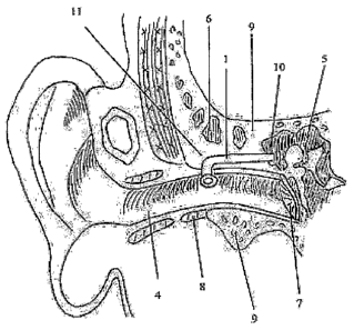
The description of the part numbers mentioned in the figures to help better
understand
the invention
1- External cannula of ventilation tube
1.1- Tab of external cannula
1.2- Lumen
1.3- Neck of external cannula
1.4- Bend of external cannula
1.5- Shaft of extenlal cannula
2- Inner cannula of ventilation tube
2.1- Thin and narrow canal of inner cannula
2.2- Drainage canal of inner cannula
2.3- External tip of the thin and narrow canal of inner cannula
2.4- External tip of the drainage canal of the inner cannula
3- Syringe
4- External ear canal
3
CA 02588391 2007-05-17
WO 2006/059961 PCT/TR2005/000011
5- Middle ear
6- Mastoid air cells
7- Eardrum
8- Cartilaginous of external ear canal
9- Bony portion of external ear canal
10- Mastoid antrum
The ventilation tube according to the invention designed for placement into
mastoid antrum (10) comprises the following elements: Mastoid antral
ventilation tube is
composed of two main elements, external (1) and inner cannula (2). External
cannula (1)
comprises the sections of tab (1.1), neck (1.3), bend (1.4) and shaft (1.5).
Lumen (1.2) of
ventilation tube starts in the middle portion of the tab (1.1). The neck
(1.3), bend (1.4) and
shaft (1.5) sections of this cannula have tubular lumen. The first section of
the tube after
the tab (1.1) is the neck (1.3). The neck (1.3) is connected to the tab (1.1)
at a certain
angle.
After the neck (1.3), comes a bend (1.4) that forms an angle of preferably 80-
90
degrees. The shaft (1.5) constitutes the final section of the external cannula
(1). The
length of the mastoid antral ventilation tube may be varied.
The inner cannula must have two canals. One of these canals must be a narrow
and thin canal (2.1) placed on one side of the imier cannula. This canal is
used for drug
administration. The luinen of the inner cannula remaining from this canal
constitutes the
drainage canal (2.2). External tip (2.4) of this canal is used for aspiration.
The two canals
of the inner cannula (2) must end at the same level on mastoid antrum (10)
without being
separated from each other. Inner cannula (2) is longer than the external
cannula (1), it
leaves the external ear canal (4) and ends pre- or retroauricularly. The
canals of the inner
cannula (2) which remain outside the external ear canal (4) must be separated
from one
another and the external tip (2.3) of the thin-narrow canal must be terminated
with a
syringe adapter (3). Inner cannula (2) must be placed into the external
cannula (1) neither
loosely nor tightly, in such a way that it gets in and out easily. Canals
(2.1, 2.2) of the
inner cannula may be also in the form of completely independent tubes.
Moreover, the
inner cannula (2) may comprise a single tube. The inner and external cannulas
(1, 2) that
make up mastoid antral tube must be made of flexible and composite materials
and they
must not collapse.
In order to apply the mastoid antral ventilation tube being disclosed, which
aerates
and drains the middle ear (5) via mastoid antrum (10), a 1-2 cm postauricular
skin
4
CA 02588391 2007-05-17
WO 2006/059961 PCT/TR2005/000011
incision is performed in consistency with the curvature of the auricle. Then
mastoidotomy
(antrotomy) is applied by a drill having a diameter of 2-3 mm from the
location defined as
Mc Evan triangle. Mastoid antrum is aspirated. Following the proper bleeding
control, the
mastoid antral ventilation tube along with its iimer and external cannulas (1,
2) is placed
by a mini-incision through the intersection of the external ear canal (4) with
the bone (9)
and the cartilage (8), and pushed into the site of mastoidotomy up to antrum
(10). The tab
(1.1) of the tube extends in the external ear canal (4) fiom the neck (1.3) to
the site of
mastoidotomy (11), and the shaft (1.5) extends up to the mastoid antrum (10).
The shaft
(1.5) length may be reduced on the condition that the tab (1.1) of the
external cannula
must remain in the external ear canal (4). The serum at body temperature is
administered
via the inner cannula (2) and aspirated. Inner cannula (2) is removed within
few days
following the disappearance of the aspirated mastoid effusion. External
cannula (1) is
maintained until the completion of the treatment. As the mastoid antral
ventilation tube
will not be spontaneously extruded, it is removed with the aid of a forceps.
As a sample application of the mastoid antral ventilation tube according to
the
invention, for a 5-year-old child, the tube must have an external cannula tab
(1.1) of 3
mm, tube lumen (1.2) diameter of 1,5 mm, tube neck (1.3) of 2 mm and shaft
(1.5) length
of 1,5 cm. The inner cannula is longer than the external cannula, up to 10 cm.
The mastoid antral ventilation tube according to the invention is used in the
treatment of the middle and inner ear diseases.
5