Note: Descriptions are shown in the official language in which they were submitted.
CA 02588449 2007-05-22
WO 2006/057859 PCT/US2005/041330
AN IMPLANT FOR INTRAOCULAR DRUG DELIVERY
This application is being filed as PCT International Patent application in the
name of Therakine Corporation, an Irish corporation, Applicant for all
countries
except the U.S., and Andreas Reiff, Scott M. Hampton, and Richard Payne, each
a
U.S. resident, Applicants for the designation of the U.S. only, on November
16, 2005.
CROSS-REFERENCE TO RELATED PATENT APPLICATION
This application claims the benefit, pursuant to 35 U.S.C. 119(e), of U.S.
provisional patent application Serial No. 60/630,75 1, filed November 24,
2004,
entitled "EYE IMPLANT WITH MEDICINE RELEASE," by Scott M. Hampton and
Andreas Reiff, which is incorporated herein by reference in its entirety.
Some references, if any, which may include patents, patent applications and
various publications, are cited and discussed in the description of this
invention. The
citation and/or discussion of such references is provided merely to clarify
the
description of the present invention and is not an admission that any such
reference is
"prior art" to the invention described herein. All references, if any, cited
and
discussed in this specification are incorporated herein by reference in their
entireties
and to the same extent as if each reference individually incorporated by
reference. In
terms of notation, hereinafter, "[n]" represents the nth reference cited in
the reference
list. For example, [ 10] represents the 10th reference cited in the reference
list,
namely, Franks WA. Limb GA. Stanford MR. Ogilvie J. Wolstencroft RA. Chignell
AH. Dumonde DC., Cytokines in human intraocular inflammation, Current Eye
Research. 11 Suppl:187-91, 1992.
FIELD OF THE INVENTION
The present invention is generally related to an ocular implant, and more
particularly, is related to an implant having at least one compound or agent
releasable
for the treatment of intraocular diseases therein.
1
CA 02588449 2007-05-22
WO 2006/057859 PCT/US2005/041330
BACKGROUND OF THE INVENTION
Many chronic disorders of the eye may and can cause long-term damage
including vision loss or blindness. Two main categories of diseases may be
differentiated: the non-infectious chronic inflammatory eye diseases and the
degenerative vasculopathies such as age related macular degeneration or
diabetic
retinopathy. Recent research suggests that inflammatory mechanisms contribute
to
degenerative diseases of the eye [19, 20, 21, 22, 23], so the categories may
be more
descriptive than casual and may have overlapping features.
In the first category, inflammatory eye diseases, the barrier that shields the
eye
from an invasion of auto aggressive white blood cells is disrupted by an
autoimmune
process allowing "eye foreign" white blood cells to invade the eye and attack
its inner
layers. The term uveitis refers to intraocular inflammations, which accounts
for
approximately 50 different entities with either infectious or autoimmune
origin. The
intraocular inflammation generally originates from the middle layer of an eye
of a
living subject, called a uvea. The uveal tract of the eye includes an iris, a
ciliary
body, and a choroid. Inflammation of the overlying retina, called retinitis,
or of the
optic nerve, called optic neuritis, may occur with or without accompanying
uveitis.
Primary uveitis ("idiopathic") is referred to the intraocular inflammation of
unknown
cause (roughly 40% of cases seen in tertiary referral centers). Secondary
uveitis (all
cases with some explanation for the uveitis) accounts for inflammatory ocular
conditions that are either associated with a systemic disease (e.g. ankylosing
spondylitis or sarcoidosis) of known infectious cause (e.g. toxoplasmosis or
CMV-
retinitis) or defined as ocular syndromes (e.g. Fuchs uveitis syndrome,
Birdshot
syndrome or serpiginous choroiditis). Masquerade syndromes, like intraocular
lymphoma, are different from primary or secondary uveitis.
The etiology and pathogenesis of uveitis is not yet fully understood. Uveitis
can be caused by infections, malignancy, exposure to toxins and autoimmune
disorders. Disturbances of immune mechanisms have long been suspected of
playing
a central role in intraocular inflammation. In the majority of cases of
endogenous
uveitis in which no link with an infectious agent can be identified,
autoimmunity has
been believed as the cause.
2
CA 02588449 2007-05-22
WO 2006/057859 PCT/US2005/041330
Clinic data collected from animals suggest that susceptibility to autoimmune
uveitis is caused by a predominant Thl response of autoreactive T cells
against retinal
antigens. Thl cells mainly produce cytokines such as INF gamma, IL2, 12, 18
while
TNF is mainly associated with cell-mediated autoimmunity. The significantly
elevated ocular and systemic levels of IL-1 beta and TNF suggest that there is
not
only a localized ocular response but a systemic response as well. The presence
of IL-
1 beta and TNF may play a role in the pathogenesis of ocular inflammation once
the
blood ocular barrier has been breached and ocular antigens have been exposed
to the
systemic immune system. Particularly, IL-6 and IL-1 may act as local
amplification
signals in pathological processes associated with a chronic eye inflammation.
Additionally, other proinflammatory cytokines such as IL2, IL4, IL6, IL8,
IL12, IL15,
IL17, ILlg and chemokines such as Matrix Metallo Proteinases (MMPs) play an
important role in the chronic inflammation of the eye.
The incidence of uveitis appears to be increasing over the last decade and is
approximately 52.4/100,000 person-years with a period prevalence of
115.3/100,000
persons. Uveitis afflicts approximately 420,000 Americans annually. The rate
of the
incidence and prevalence of uveitis is lowest in pediatric age groups,
increases with
age and is highest in patients 65 years old and older.
Ocular complications of uveitis produce profound and irreversible loss of
vision, especially when such ocular complications are unrecognized and/or
treated
improperly. Some of the most frequent complications include cataract,
glaucoma,
retinal detachment, cystoid macular edema, neovascularization of the retina,
optic
nerve and iris.
The long-term outcome of uveitis in adults is unknown because no prospective
studies are available. In the pediatric population with autoimmune conditions
(such
as juvenile rheumatoid arthritis), the risk of permanent blindness after 5
years has
remained unchanged at about 10%, despite aggressive treatment with topical
steroids
and systemic immunosuppressive therapy. About 30% have significant loss of
vision,
requiring lifelong assistance. Because uveitis causes pain and light
sensitivity, the
impact on quality of life is much more severe than the figures above indicate,
even for
"mild" cases.
3
CA 02588449 2007-05-22
WO 2006/057859 PCT/US2005/041330
In the second category of chronic eye diseases, degenerative vasculopathies,
age related or metabolic factors cause blood vessels to obliterate and no
longer supply
vital parts of the eye with blood. As a result, the eye rapidly starts to form
new blood
vessels around the occluded old vessel in order to compensate for the lack of
blood
supply. Unfortunately these repair mechanisms are frequently insufficient and
the
newly formed blood vessels often burst resulting into bleeding into the eye
and
detachment of the retina.
The most important diseases in the degenerative category include age related
macula degeneration and diabetic retinopathy, as well as cystoid macular
edema.
Macular degeneration is the most common cause of blindness in the senior
population of the developed world. In macular degeneration, the light-sensing
cells of
the macula malfunction and cease to work over time. Macular degeneration
occurs
most often in people over 60 years old, in which case it is called Age Related
Macular
Degeneration (AMD or ARMD) but can occur at all ages including children. The
most common early sign of AMD is blurred vision, straight lines appearing
wavy, and
finally leading to loss of visual acuity and color sensitivity. The macula is
the part of
the retina that provides central vision, and as it degenerates it can lead to
partial or
complete loss of vision. About 85 - 90% of AMD cases are the dry, or atrophic,
form,
in which yellowish spots of fatty deposits called drusen appear on the macula.
The
rest of AMD cases are the wet form, so called because of leakage into the
retina from
newly forming blood vessels in the choroid, a part of the eye behind the
retina.
Normally, blood vessels in the choroid bring nutrients to, and carry waste
products
away from, the retina. Sometimes the fine blood vessels in the choroid
underlying the
macula begin to proliferate, a process called choroidal neovascularization, or
CNV.
The cause is unknown. When those blood vessels proliferate, they leak, and
cells in
the macula may be damaged and may die. Laser photocoagulation is a technique
used
by ophthalmic surgeons to treat leakage from submacular neovascularizations.
Unfortunately only about half of patients with wet AMD are candidates for
laser
photocoagulation and laser photocoagulation is only effective about half the
time it is
done as a treatment for wet macular degeneration. When effective, the benefit
lasts
on the average about one year.
4
CA 02588449 2007-05-22
WO 2006/057859 PCT/US2005/041330
Diabetic retinopathy is the leading cause of acquired blindness among
Americans under the age of 65. Diabetic retinopathy may occur at any point in
time
after the onset of diabetes. Blood vessels damaged from diabetic retinopathy
can
cause vision loss in two ways: Fragile and abnormal blood vessels can develop
and
leak blood into the center of the eye, blurring vision. This is proliferative
retinopathy
and is the fourth and most advanced stage of the disease. Fluid can leak into
the
center of the macula, the part of the eye where sharp, straight-ahead vision
occurs.
The fluid makes the macula swell, blurring vision. This condition is called
macular
edema. It can occur at any stage of diabetic retinopathy, although it is more
likely to
occur as the disease progresses. About half of the people with proliferative
retinopathy also have macular edema.
Findings in the retina include dot and blot hemorrhages (tiny hemorrhages in
the retina itself), microaneurysms (out-pouchings of capillaries), and
exudates (retinal
deposits occurring as a result of leaky vessels). The development of this
condition in
type I(juvenile-onset) diabetics is rarely present prior to three or four
years following
the onset of diabetes. In type II (adult-onset) diabetics, background diabetic
retinopathy may be present at the time of diagnosis of the condition. The
great
majority of this blindness can be prevented with proper examination and
treatment by
ophthalmologists. Unfortunately, patients who are not properly referred for
evaluation and management or those who, for any reason, fail to get proper
care from
an ophthalmologist, are at the greatest risk of vision loss.
Various treatment options have been developed for patients who are affected
by these 2 categories of disorders.
In case of the inflammatory eye diseases, the treatments of noninfectious
and/or autoimmune uveitis include administering topical steroid eyedrops
and/or
corticosteroids, combined with antimicrobials and cycloplegic drops. Even
though
most patients will have a mild form of uveitis, the disease can linger for
months
(many cases continue for years), and residual damage to the iris or the lens
is not
uncommon. Glaucoma (increased pressure in the eye) is an additional side
effect of
steroid eyedrops and can further limit the patient's vision. For certain
cases, it may
require injection of steroids into the tissue around the eye. If this is not
effective,
corticosteroids can be given orally, with well known side effects such as
weight gain
5
CA 02588449 2007-05-22
WO 2006/057859 PCT/US2005/041330
(including fat deposits developing on the face) increased risk of infections,
osteoporosis, weakness, diabetes, slow wound healing with easy bruising, acne,
salt
retention, and hypertension. Additional risks in the eye include cataract and
glaucoma.
Clinical research has shown that the use of antibodies designed to modulate
elements of the immune system lead to positive outcomes in inflammatory and
degenerative conditions of the eye. However, the antibody compounds must be
administered systemically either by intravenous (IV) or sub-cutaneous
injection. The
problem with this systemic application is the risk of systemic infections,
reactivation
of tuberculosis and demyelination in the brain in patients with multiple
sclerosis.
Furthermore, since the eye is a well-shielded organ with natural barriers to
the blood,
treatments with antibodies require much higher doses than those requires in
rheumatoid arthritis. Thus the cost of such a treatment can be prohibitively
expensive.
In the case of inflammatory eye diseases, treatment is facilitated by using
anti-
cytokines or anti-chemokines that modulate chronic inflammatory eye disease,
and a
number of such drugs are being used systemically with good success. However
the
systemic use, such as an intravenous injection, is expensive, and is
associated with
side effects and not always effective. By giving these drugs directly into the
eye
through the device(s) and method(s) according to several embodiments of the
present
invention, systemic side effects can be avoided and better local control of
the
inflammation can be achieved. In addition the patients' immune system remains
substantially unchanged since the present invention allows the modulation of
local
inflammation only.
For the patients with degenerative vasculopathies, among other unique
features, the present invention allows direct drug delivery into the eye but
instead of
using anti-cytokines or anti-chemokines, protein inhibitors, so called MAP-
Kinase
inhibitors, will be used to precisely block intracellular signals that would
lead to the
formation of new blood vessels. The protein inhibitors are delivered directly
into the
eye over an extended time period. This in turn can prevent catastrophic
bleeding from
or into the eye and avoid costly laser surgeries to reattach the retina. These
drugs
have already been successfully used in the treatment of solid tumors where
they
6
CA 02588449 2007-05-22
WO 2006/057859 PCT/US2005/041330
prevent the formation of new blood vessels thereby shutting off the blood
supply to
the growing tumor leading to its death. Inflammation is implicated as a
contributing
factor in degenerative eye diseases, such as macular degeneration, and
effective
treatment of these diseases may require the use of multiple agents to modulate
inflammation and new vessel formation.
The intracellular signal transduction pathways involved in inflammation and
cell transformation and their relationship to autoimmune diseases are only
beginning
to be explored. The identification of enzymes involved in signaling from the
plasma
membrane to the nucleus in lymphocytes and the cells involved in autoimmune
diseases will likely contribute significantly to future understanding of
mechanisms
responsible for lymphocyte differentiation and for the discrimination of self
from non-
self in developing and mature cells.
Chemical manipulations of the enzymes involved in these pathways known as
selective kinases or downstream transcription factors provide a unique
opportunity for
novel therapeutic interventions. It is feasible that inhibition of specific
signal
transduction or transcription factor targets might interrupt the perpetuation
mechanisms involved in many autoimmune diseases. The blockade of the
appropriate
pathway could provide an opportunity to reestablish homeostasis by inhibition
of
cellular responses, such as lymphokine gene expression and cellular release of
proinflammatory cytokines such as TNF and others.
Despite the differences in the antigens that they recognize and in the
effector
functions they carry out, B and T lymphocytes utilize remarkably similar
signal
transduction components to initiate responses. Even though the signaling
pathways
are highly diverse, they display an extraordinary degree of specificity for a
given
transcription factor or transcription factor family. A number of transcription
factor
families, including those for activator protein 1 (AP-1)/activating
transcription factor
2 (ATF2), nuclear factor [kappa] B (NF- [kappa] B), nuclear factor of
activated T
cells (NF-AT), signal transducer and activator of transcription (STAT), p53,
and
nuclear hormone receptors, have been implicated as critical regulators of gene
expression in the setting of inflammation
7
CA 02588449 2007-05-22
WO 2006/057859 PCT/US2005/041330
In animal models of uveitis such as endotoxin-induced uveitis (EIU), a
signaling pathway known as the extracellular signal-regulated kinase (ERK)
pathway
plays an important role in the inflammation of the retina.
Furthermore another Mitogen-activated protein kinase (MAPK) cascade, one
of the major protein kinase families involved in intracellular signaling has
been
implicated in the activation of Anti-endothelial cell antibodies (AECA) in the
sera of
patients with Behcet's disease (BD) and uveitis. AECA of the IgM subtype can
play a
pathogenic role in induction of vasculitis and inflammatory lesions of BD by
directly
activating endothelial cells (HDMEC), independent from the help of
proinflammatory
cytokines such as TNF alpha or IL-1 alpha. These antibodies facilitate the
perpetuation of a chronic inflammatory response by attracting lymphocytes to
leave
the bloodstream and infiltrate the eye. Inhibition of the enzymes of the MAPK
cascade pathways stopped the antibody production.
In summary, even though the evidence of the role of small molecule inhibitors
in the treatment of uveitis is still largely unexplored, preliminary evidence
suggests
that small molecule inhibitors may play an important role in the treatment of
uveitis in
the near future.
Since multiple signaling pathways are known to be involved in all of the
diseases discussed, it is very likely that the most effective local treatment
for these
diseases will be to use multiple compounds that are selective to the disease-
specific
pathways that cause the inflammation and/or the degeneration. The current
treatment
paradigm for degenerative eye diseases has been to administer a single
compound,
usually systemically, even though it has been shown that the separate
processes of
inflammation and neovascularization occur simultaneously. Targeting multiple
pathways, by using combinations of anti-cytokines, anti-chemokines, kinase
inhibitors, and other signal modulating agents, delivered locally, will allow
the
treatment of these eye diseases with superior outcomes and safety, and
represent a
new approach to the treatment of the leading causes of blindness. Because of
the
complexity of these diseases, it is not yet clear whether the best treatment
option
would be a single implanted delivery device that releases multiple compounds
or a
collection of implanted delivery devices that each releases only a single
compound,
8
CA 02588449 2007-05-22
WO 2006/057859 PCT/US2005/041330
each of which would allow a physician to tailor the treatment to achieve
specific
treatment profiles.
Therefore, a heretofore unaddressed need exists in the art to address the
aforementioned deficiencies and inadequacies.
SUMMARY OF THE INVENTION
In one aspect, the present invention relates to an implant for intraocular
drug
delivery for the treatment of intraocular inflammatory or degenerative
diseases. In
one embodiment, the implant includes a body portion. The body portion has a
first
end portion, a second, opposite end portion, an outer surface, an interior
surface, and a
length L defined between the first end portion end and the second end portion.
The
body portion defines a cavity with a first opening at the first end portion,
and a
second, opposite opening at the second end portion. In one embodiment, the
body
portion has a cross-section of a circle, a square, an oval, or a polygon. The
implant
further includes a solid material received in the cavity, where the solid
material
comprises a depot material and an effective amount of at least one therapeutic
compound or agent.
The implant may also include a first membrane covering the first opening of
the body portion, through which the at least one therapeutic compound or agent
is
controllably released to the environment of the implant, and a second membrane
covering the second opening of the body portion, through which the at least
one
therapeutic compound or agent is controllably released to the environment of
the
implant. The first membrane and the second membrane each is made from a
biodegradable material.
In one embodiment, the implant is implanted in or around the vitreous or other
parts of the posterior chamber of the eye of a living subject so that the
cavity of the
implant is in fluid communication with the vitreous or other parts of the
posterior
chamber of the eye through at least one of the first opening and the second,
opposite
opening. When the implant is implanted in an eye of a living subject, the
effective
amount of at least one therapeutic compound or agent is released to the
environment
of the implant through at least one of the first opening and the second,
opposite
opening over an extended period of time. In one embodiment, the effective
amount
9
CA 02588449 2007-05-22
WO 2006/057859 PCT/US2005/041330
of at least one therapeutic compound or agent is released to the environment
of the
implant by diffusion through and dissolution of the depot material that
comprises a
soluble binder material.
The body portion of the implant, in one embodiment, is made from an inert
polymeric material selected from polysulfone, polyetherimide, polyimide,
polymethylmethacrylate, siloxanes, other acrylates, polyetheretherketone,
copolymers
of any of these compounds, and biocompatible implantable polymers.
In another embodiment, the body portion of the implant is made from a
biodegradable material such that when the effective amount of at least one
therapeutic
compound is released to the environment of the implant, the body portion
gradually
resorbs or degrades in situ. The biodegradable material includes a
biodegradable
polymeric material selected from modified poly(saccharides), including starch,
cellulose, and chitosan, fibrin, fibronectin, gelatin, collagen, collagenoids,
tartrates,
gellan gum, dextran, maltodextrin, poly(ethylene glycol), poly(propylene
oxide),
poly(butylene oxide), Pluoronics, modified polyesters, poly(lactic acid),
poly(glycolic
acid), poly(lactic-co-glycolic acid), modified alginates, carbopol, poly(N-
isopropylacrylamide), poly(lysine), triglyceride, polyanhydrides,
poly(ortho)esters,
poly(epsilon-caprolactone), poly(butylene terephthalate), polycarbonates,
triglyceride, copolymers of glutamic acid and leucine, poly(hydroxyalkanoates)
of
the PHB-PHV class, proteins, polypeptides, proteoglycans, polyelectolytes, and
any
copolymer or combination of them.
The soluble binder material, in one embodiment, comprises at least one of
modified poly(saccharides), including starch, cellulose, and chitosan, sugars
and
modified sugars, including trehalose, sucrose, sucrose esters, polyalcohols,
poly(vinyl
alcohol), glycerol, fibrin, fibronectin, gelatin, collagen, collagenoids,
tartrates, gellan
gum, heparin, carrageenan, pectin, xanthan, dextran, maltodextrin,
poly(ethylene
glycol), poly(propylene oxide), poly(butylene oxide), Pluoronics, modified
alginate
hydrogels, carbopol, poly(lysine), proteins, polypeptides, polyelectolytes,
proteoglycans, and any copolymer or combination of them.
The at least one therapeutic compound or agent, in one embodiment,
comprises at least one biologic immunomodulator or anti-inflammatory agent
that
specifically or functionally oppose the action of Tumor Necrosis Factor alpha
CA 02588449 2007-05-22
WO 2006/057859 PCT/US2005/041330
(TNFa); the Interleukines including Interleukine-1, Interleukine-2,
Interleukine-4,
Interleukine-6, Interleukine-8, Interleukine-12, Interleukine-15, Interleukine-
17, and
Interleukine-18; Anti-chemokines and anti-metalloproteases that specifically
or
functionally oppose the action of MCP-1 (9-76), Gro-alpha (8-73), V MIPII,
CXCR4,
Met-CCL5, Met-RANTES, CCR1, RANTES (CCL5), MIP 1 alpha (CCL3), IP 10
(CXCL10), VEGF, MCP 1-4 (CCL1, CCL8, CCL7, CCL13), CINC, Cognate
receptor, GRO, CXCR4, Stromal-derived factor-1, CCR4, CCR5, and CXCR3;
Chemokines or synthetic molecules that are structurally or functionally
equivalent to
Interleukine-10 and Interleukine-12; and Tumor Growth Factors (TGF) and
related
anti-inflammatory growth factors. Co-stimulatory molecule inhibitor including
CTLA4 Ig, anti CD11, anti CD2, fusion protein of LFA3e and IgGFc; inhibitors
of
nitric oxide (NO) or inducible nitric oxide synthase (iNOS), adhesion molecule
inhibitors including alpha4-integrin inhibitor, inhibitors of P selectin or E
selectin or
ICAMl or VCAM, alpha-melanocyte stimulating hormone (alpha-MSH), anti HSP 60
or Heme Oxygenase (HO)-1, and heat shock proteins.
The at least one therapeutic compound or agent may also comprise at least one
of the following signal pathway modulators or involve in the signaling
pathways to
reduce or inhibit inflammation and angiogenesis, including NF-kappa B
inhibitors
such as Pyrrolidine dithiocarbamate (PTDC), Proteasome inhibitor, MG-132,
Rolipram, an inhibitor of type 4 phosphodiesterase, CM101, for example;
inhibitors of
other transcription factors such as activator protein 1 (AP 1), activating
transcription
factor 2 (ATF2), nuclear factor of activated T cells (NF-AT), signal
transducer and
activator of transcription (STAT), p53, Ets family of transcription factors
(Elk-1 and
SAP-1), nuclear hormone receptors; small molecule inhibitors that inhibit or
block the
following intracellular signaling pathways, or regulatory enzymes/kinases, for
example: PTEN, P13 Kinases, P38 MAP Kinase and other MAP Kinases, all stress
activated protein kinases (SAPKs), the ERK signaling pathways, the JNK
signaling
pathways (JNK1, JNK2), all RAS activated pathways, all Rho mediated pathways,
and all NIK, MEKK-1, IKK-1, IKK-2 pathways; and other intracellular and
extracellular signaling pathways.
In another embodiment, the at least one therapeutic compound or agent
comprises any combination of the agents mentioned above.
11
CA 02588449 2007-05-22
WO 2006/057859 PCT/US2005/041330
In an alternative embodiment, the at least one therapeutic compound or agent
comprises at least one of antibodies, nanobodies, antibody fragments,
signaling
pathway inhibitors, transcription factor inhibitors, receptor antagonists,
small
molecule inhibitors, oligonucleotides, fusion proteins, peptides, protein
fragments,
allosteric modulators of cell surface receptors such as G-protein coupled
receptors
(GPCR), cell surface receptor internalization inducers, and GPCR inverse
agonists.
In another aspect, the present invention relates to an implant for intraocular
drug delivery. In one embodiment, the implant has a body portion having an
outer
surface and an interior surface, where the interior surface defines a cavity
with at least
one opening. In one embodiment, the outer surface of the body portion has a
geometric shape of a hemisphere. The implant also has an effective amount of
at least
one therapeutic compound or agent received in the cavity, where when the
implant is
implanted in the eye of a living subject, the effective amount of at least one
therapeutic compound or agent is released to the environment of the implant
through
the at least one opening over an extended period of time.
The implant further has a soluble binder material, where at least one
therapeutic compound or agent is stabilized with the soluble binder material
to form a
compound that is received in the cavity. The soluble binder material comprises
at
least one of modified poly(saccharides), including starch, cellulose, and
chitosan,
sugars and modified sugars, including trehalose, sucrose, sucrose esters,
polyalcohols,
poly(vinyl alcohol), glycerol, fibrin, fibronectin, gelatin, collagen,
collagenoids,
tartrates, gellan gum, heparin, carrageenan, pectin, xanthan, dextran,
maltodextrin,
poly(ethylene glycol), poly(propylene oxide), poly(butylene oxide),
Pluoronics,
modified alginate hydrogels, carbopol, poly(lysine), proteins, polypeptides,
polyelectolytes, proteoglycans, and any copolymer or combination of them.
In one embodiment, the implant may comprises a membrane covering the at
least one opening of the body portion, through which the at least one
therapeutic
compound or agent is controllably released to the environment of the implant,
where
the membrane is made from a biodegradable material.
The body portion of the implant in one embodiment is made from an inert
polymeric material selected from the group of polysulfone, polyetherimide,
polyimide, polymethylmethacrylate, siloxanes, other acrylates,
polyetheretherketone,
12
CA 02588449 2007-05-22
WO 2006/057859 PCT/US2005/041330
copolymers of any of the these compounds, and similar engineered biocompatible
implantable polymers.
In another embodiment the body portion is made from a biodegradable
material such that when the effective amount of at least one therapeutic
compound is
released to the environment of the implant, the body portion gradually resorbs
or
degrades in situ. The biodegradable material comprises a biodegradable
polymeric
material selected from modified poly(saccharides), including starch,
cellulose, and
chitosan, fibrin, fibronectin, gelatin, collagen, collagenoids, tartrates,
gellan gum,
dextran, maltodextrin, poly(ethylene glycol), poly(propylene oxide),
poly(butylene
oxide), Pluoronics, modified polyesters, poly(lactic actid), poly(glycolic
acid),
poly(lactic-co-glycolic acid), modified alginates, carbopol, poly(N-
isopropylacrylamide), poly(lysine), triglyceride, polyanhydrides,
poly(ortho)esters,
poly(epsilon-caprolactone), poly(butylene terephthalate), polycarbonates,
triglyceride, copolymers of glutamic acid and leucine, poly(hydroxyalkanoates)
of
the PHB-PHV class, proteins, polypeptides, proteoglycans, polyelectolytes, and
any
copolymer or combination of them.
In one embodiment, the at least one therapeutic compound or agent comprises
at least one immunomodulator or anti-inflammatory agent that specifically or
functionally opposes the action of Tumor Necrosis Factor alpha (TNFa); the
Interleukines including Interleukine-1, Interleukine-2, Interleukine-4,
Interleukine-6,
Interleukine-8, Interleukine- 12, Interleukine- 15, Interleukine- 17, and
Interleukine- 18;
Anti-chemokines and anti-metalloproteases that specifically or functionally
oppose
the action of MCP-1 (9-76), Gro-alpha (8-73), V MIPII, CXCR4, Met-CCL5, Met-
RANTES, CCR1, RANTES (CCL5), MIP 1 alpha (CCL3), IP 10 (CXCL10), VEGF,
MCP 1-4 (CCL1, CCL8, CCL7, CCL13), CINC, Cognate receptor, GRO, CXCR4,
Stromal-derived factor-1, CCR4, CCR5, and CXCR3; Chemokines or synthetic
molecules that are structurally or functionally equivalent to Interleukine-10
and
Interleukine-12; and Tumor Growth Factors (TGF) and related anti-inflammatory
growth factors; co-stimulatory molecule inhibitor including CTLA4 Ig, anti
CD11,
anti CD2, fusion protein of LFA3e and IgGFc; inhibitors of nitric oxide (NO)
or
inducible nitric oxide synthase (iNOS); adhesion molecule inhibitors including
alpha4-integrin inhibitor; inhibitors of P selectin or E selectin or ICAM1 or
VCAM;
13
CA 02588449 2007-05-22
WO 2006/057859 PCT/US2005/041330
alpha-melanocyte stimulating hormone (alpha-MSH); anti HSP 60 or Heme
Oxygenase (HO)-1; and heat shock proteins.
The at least one therapeutic compound or agent may also comprise at least one
of the following signal pathway modulators or involve in the following
pathways to
reduce or inhibit inflammation and angiogenesis, including NF-kappa B
inhibitors
such as Pyrrolidine dithiocarbamate (PTDC), Proteasome inhibitor, MG-132,
Rolipram, an inhibitor of type 4 phosphodiesterase, CM101, for example;
inhibitors of
other transcription factors such as activator protein 1(AP1), activating
transcription
factor 2 (ATF2), nuclear factor of activated T cells (NF-AT), signal
transducer and
activator of transcription (STAT), p53, Ets family of transcription factors
(Elk-1 and
SAP-1), nuclear hormone receptors; small molecule inhibitors that inhibit or
block the
following intracellular signaling pathways, or regulatory enzymes/kinases, for
example: PTEN, P13 Kinases, P38 MAP Kinase and other MAP Kinases, all stress
activated protein kinases (SAPKs), the ERK signaling pathways, the JNK
signaling
pathways (JNK1, JNK2), all RAS activated pathways, all Rho mediated pathways,
and all NIK, MEKK-1, IKK-1, IKK-2 pathways; and other intracellular and
extracellular signaling pathways.
In another embodiment, the at least one therapeutic compound or agent
comprises at least two therapeutic compounds, at least one of which is an anti-
cytokine or anti-chemokine for the treatment of inflammatory diseases by
simultaneously and synergistically blocking signal transduction pathways
involved in
the inflammatory and/or autoimmune disorders related to the eye of a living
subject.
In yet another embodiment, the at least one therapeutic compound or agent
comprises at least one of antibodies, nanobodies, antibody fragments,
signaling
pathway inhibitors, transcription factor inhibitors, receptor antagonists,
small
molecule inhibitors, oligonucleotides, fusion proteins, peptides, protein
fragments,
interference RNA, allosteric modulators of cell surface receptors such as G-
protein
coupled receptors (GPCR), cell surface receptor internalization inducers, and
GPCR
inverse agonists.
In one embodiment, the at least one therapeutic compound or agent is in the
form of a plurality of particles, which are releasable to the environment of
the
implant.
14
CA 02588449 2007-05-22
WO 2006/057859 PCT/US2005/041330
The effective amount of at least one therapeutic compound or agent, in one
embodiment, is released to the environment of the implant by diffusion through
and
dissolution of the soluble binder material.
In one embodiment, when the implant is implanted in the eye of a living
subject, the implant is placed in or around the vitreous or other parts of the
posterior
chamber of the eye of a living subject so that the cavity of the implant is in
fluid
communication with the vitreous or other parts of the posterior chamber of the
eye
through the at least one opening.
In yet another aspect, the present invention relates to an eye implant. In one
embodiment, the eye implant includes a first material, and a second material
containing an effective amount of at least one therapeutic compound or agent,
where
the first material and the second material are arranged to form a solid, and
when the
eye implant is implanted in an eye of a living subject, the effective amount
of at least
one therapeutic compound or agent is releasable to the environment of the
implant
over an extended period of time. The eye implant may comprise a third material
containing an effective amount of at least one therapeutic compound or agent.
In one embodiment, the first material and the second material are formed in a
layer structure. In another embodiment, the first material, the second
material and the
third material are formed in a layer structure. When the eye implant is
implanted in
the eye of a living subject, materials in different layers are released to the
environment of the eye implant at different rates, respectively or one after
another.
Alternatively, the first material and the second material are formed in a
wafer-
like structure. The first material and the second material may be also formed
to a
solid such that at any given position, the density of the material is
substantially one of
the densities of the first material and the density of the second material.
In one embodiment, the first material comprises an inert polymeric material
selected from the group of polysulfone, polyetherimide, polyimide,
polymethylmethacrylate, siloxanes, other acrylates, polyetheretherketone,
copolymers
of any of the these compounds, and similar engineered biocompatible
implantable
polymers.
The first material in another embodiment comprises a biodegradable material
such that when the effective amount of at least one therapeutic compound or
agent is
CA 02588449 2007-05-22
WO 2006/057859 PCT/US2005/041330
released to the environment of the eye implant, the first material gradually
degrades or
dissolves in situ. The biodegradable material comprises a biodegradable
polymeric
material selected from modified poly(saccharides), including starch,
cellulose, and
chitosan, fibrin, fibronectin, gelatin, collagen, collagenoids, tartrates,
gellan gum,
dextran, maltodextrin, poly(ethylene glycol), poly(propylene oxide),
poly(butylene
oxide), Pluoronics, modified polyesters, poly(lactic actid), poly(glycolic
acid),
poly(lactic-co-glycolic acid), modified alginates, carbopol, poly(N-
isopropylacrylamide), poly(lysine), triglyceride, polyanhydrides,
poly(ortho)esters,
poly(epsilon-caprolactone), poly(butylene terephthalate), polycarbonates,
triglyceride, copolymers of glutamic acid and leucine, poly(hydroxyalkanoates)
of
the PHB-PHV class, proteins, polypeptides, proteoglycans, polyelectolytes, and
any
copolymer or combination of them.
The second material fu.rther comprises a soluble binder material. The at least
one therapeutic compound or agent is stabilized with the soluble binder
material. The
soluble binder material in one embodiment comprises at least one of modified
poly(saccharides), including starch, cellulose, and chitosan, sugars and
modified
sugars, including trehalose, sucrose, sucrose esters, polyalcohols, poly(vinyl
alcohol),
glycerol, fibrin, fibronectin, gelatin, collagen, collagenoids, tartrates,
gellan gum,
heparin, carrageenan, pectin, xanthan, dextran, maltodextrin, poly(ethylene
glycol),
poly(propylene oxide), poly(butylene oxide), Pluoronics, modified alginate
hydrogels,
carbopol, poly(lysine), proteins, polypeptides, polyelectolytes,
proteoglycans, and any
copolymer or combination of them.
The effective amount of at least one therapeutic compound or agent is released
to the environment of the eye implant by diffusion through and dissolution of
the
soluble binder material.
In one embodiment, when the eye implant is implanted in the eye of a living
subject, the eye implant is placed in or around the vitreous or other parts of
the
posterior chamber of the eye of a living subject.
In a fiuther aspect, the present invention relates to a method of treating
inflammatory and degenerative diseases in or around the eye. In one
embodiment, the
method includes the step of providing an eye implant having a first material,
and a
second material containing an effective amount of at least one therapeutic
compound
16
CA 02588449 2007-05-22
WO 2006/057859 PCT/US2005/041330
or agent, where the first material and the second material are arranged to
form a solid.
Furthermore, the method includes the step of implanting the eye implant in an
eye of a
living subject. The effective amount of at least one therapeutic compound is
releasable to the environment of the eye implant over an extended period of
time. The
method also includes the step of leaving the eye implant in the eye.
In one embodiment, the first material comprises an inert polymeric material
selected from the group of polysulfone, polyetherimide, polyimide,
polymethylmethacrylate, siloxanes, other acrylates, polyetheretherketone,
copolymers
of any of the these compounds, and similar engineered biocompatible
implantable
polymers. In another embodiment, the first material comprises a biodegradable
material such that when the effective amount of at least one therapeutic
compound or
agent is released to the environment of the eye implant, the first material
gradually
degrades or dissolves in situ.
The second material further comprises a soluble binder material, and wherein
at least one therapeutic compound or agent is stabilized with the soluble
binder
material. The effective amount of at least one therapeutic compound or agent
is
released to the environment of the eye implant by diffusion through and
dissolution of
the soluble binder material.
These and other aspects of the present invention will become apparent from
the following description of the preferred embodiment taken in conjunction
with the
following drawings, although variations and modifications therein may be
affected
without departing from the spirit and scope of the novel concepts of the
disclosure.
BRIEF DESCRIPTION OF THE DRAWINGS
The accompanying drawings illustrate one or more embodiments of the
invention and, together with the written description, serve to explain the
principles of
the invention. Wherever possible, the same reference numbers are used
throughout
the drawings to refer to the same or like elements of an embodiment, and
wherein:
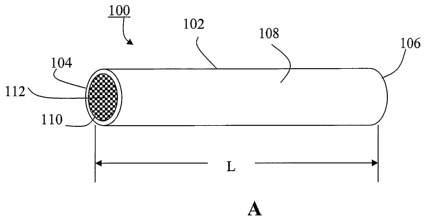
Fig. 1 shows schematically an implant according to one embodiment of the
present invention: (a) a perspective view, and (b) a cross sectional view.
Fig. 2 shows schematically an implant according to another embodiment of the
present invention: (a) a perspective view, and (b) a cross sectional view.
17
CA 02588449 2007-05-22
WO 2006/057859 PCT/US2005/041330
Fig. 3 shows schematically an implant according to yet another embodiment of
the present invention: (a) a perspective view, and (b) a cross sectional view.
Fig. 4 shows schematically an implant according to an alternative embodiment
of the present invention: (a) in a first state, (b) a second state, and (c) a
third state.
Fig. 5 shows schematically an implant according to one embodiment of the
present invention: (a) a perspective view, and (b) a sectional view.
Fig. 6 shows schematically an implant according to another embodiment of the
present invention: (a) a perspective view, (b) a partially cross sectional
view, and (c)
compounds and/or agents in the implant releasing to the environment.
Fig. 7 shows schematically an implant according to an alternative embodiment
of the present invention: (a) a perspective view, and (b) a cross sectional
view.
Fig. 8 shows schematically an implant according to a further embodiment of
the present invention.
Fig. 9 shows schematically an implant according to yet a further embodiment
of the present invention: (a) in a first state, and (b) in a second state.
Fig. 10 shows schematically an implant according to one embodiment of the
present invention: (a) a cross sectional view, and (b) compounds and/or agents
in the
implant.
DETAILED DESCRIPTION OF THE INVENTION
The present invention is more particularly described in the following examples
that are intended as illustrative only since numerous modifications and
variations
therein will be apparent to those skilled in the art. Various embodiments of
the
invention are now described in detail. Referring to the drawings of Figs. 1-
10, like
numbers indicate like components throughout the views. As used in the
description
herein and throughout the claims that follow, the meaning of "a", "an", and
"the"
includes plural reference unless the context clearly dictates otherwise. Also,
as used
in the description herein and throughout the claims that follow, the meaning
of "in"
includes "in" and "on" unless the context clearly dictates otherwise.
Moreover, titles
or subtitles may be used in the specification for the convenience of a reader,
which
shall have no influence on the scope of the present invention. Additionally,
some
terms used in this specification are more specifically defmed below.
18
CA 02588449 2007-05-22
WO 2006/057859 PCT/US2005/041330
DEFINITIONS
The terms used in this specification generally have their ordinary meanings in
the art, within the context of the invention, and in the specific context
where each
term is used.
Certain terms that are used to describe the invention are discussed below, or
elsewhere in the specification, to provide additional guidance to the
practitioner in
describing the apparatus and methods of the invention and how to make and use
them.
For convenience, certain terms may be highlighted, for example using italics
and/or
quotation marks. The use of highlighting has no influence on the scope and
meaning
of a term; the scope and meaning of a term is the same, in the same context,
whether
or not it is highlighted. It will be appreciated that the same thing can be
said in more
than one way. Consequently, alternative language and synonyms may be used for
any
one or more of the terms discussed herein, nor is any special significance to
be placed
upon whether or not a term is elaborated or discussed herein. Synonyms for
certain
terms are provided. A recital of one or more synonyms does not exclude the use
of
other synonyms. The use of examples anywhere in this specification, including
examples of any terms discussed herein, is illustrative only, and in no way
limits the
scope and meaning of the invention or of any exemplified term. Likewise, the
invention is not limited to various embodiments given in this specification.
Furthennore, subtitles may be used to help a reader of the specification to
read
through the specification, which the usage of subtitles, however, has no
influence on
the scope of the invention.
As used herein, "around", "about" or "approximately" shall generally mean
within 20 percent, preferably within 10 percent, and more preferably within 5
percent
of a given value or range. Numerical quantities given herein are approximate,
meaning that the term "around", "about" or "approximately" can be inferred if
not
expressly stated.
As used, the term "uveitis" is referred generally to intraocular
inflammations,
which account for at least 50 different entities with either infectious or
autoimmune
origin, Prirnar,y uveitis ("idiopathic") is referred to the intraocular
inflammation of
unknown cause (roughly 40% of cases seen in tertiary referral centers).
Secondary
uveitis (all cases with some explanation for the uveitis) accounts for
inflammatory
19
CA 02588449 2007-05-22
WO 2006/057859 PCT/US2005/041330
ocular conditions that are either associated with a systemic disease (e.g.
ankylosing
spondylitis or sarcoidosis) of known infectious cause (e.g. toxoplasmosis or
CMV-
retinitis) or defined as ocular syndromes (e.g. Fuchs uveitis syndrome,
Birdshot
syndrome or serpiginous choroiditis). Masquerade syndromes, like intraocular
lymphoma, are different from primary or secondary uveitis.
The term "compound" is referred to a chemical combination of two or more
elements that may have an impact on any living system such as a cell, nerve or
tissue.
Examples of compounds that may be related to practicing the present invention
include those in the following exemplary list:
Anti-inflammatory compounds:
a) Anti-cytokines
= Anti-Tumor Necrosis Factor alpha (TNFa) such as
(1) Etanercept (p75 TNFr fusion protein)
(2) Infliximab (chimeric Anti TNF Mab)
(3) Adalimumab (human Anti TNF Mab)
(4) Onercept (soluble p55 TNFr)
Or other compounds, such as antibodies, nanobodies, antibody fragments,
and receptor antagonists.
= Anti-Interleukin-1 such as
(1) Anakinra (IL-1 type 1 receptor antagonist)
(2) IL1 Trap (Regeneron, an IL-1 type 1 receptor plus IL-1 fusion protein)
or other compounds
= Anti-Interleukin-2 such as
(1) Daclizumab or other compounds
= Anti-Interleukin-4 such as
(1) Human Anti-IL-4 antibody, E coli derived goat IgG (R&D systems)
(2) Human Anti-IL-4 antibody, E coli derived murine IgG (R&D systems)
Or other compounds
= Anti-Interleukin-6 such as
(1) MRA (Chugai Pharmaceuticals/Roche) or other compounds
= Anti-Interleukin-8 such as
CA 02588449 2007-05-22
WO 2006/057859 PCT/US2005/041330
(1) Anti-EGF-R antibody (C225) or other compounds
= Anti-Interleukin-12 such as
(1) Human Anti-IL-12 antibody, E coli derived goat IgG (R&D systems)
(2) Human Anti-IL-12 antibody, E coli derived murine IgG (R&D
systems)
Or other compounds
= Anti-Interleukin- 15 such as
(1) Human Anti-IL-15 antibody, E coli derived goat IgG (R&D systems)
(2) Human Anti-IL- 15 antibody, E coli derived murine IgG (R&D
systems)
Or other compounds
= Anti-Interleukin-17 such as
(1) Human Anti-IL-17 antibody, E coli derived goat IgG (R&D systems)
(2) Human Anti-IL-17 antibody, E coli derived murine IgG (R&D
systems)
Or other compounds
= Anti-Interleukin- 18 such as
(1) Human Anti-IL-18 antibody, E coli derived goat IgG (R&D systems)
(2) Human Anti-IL- 18 antibody, E coli derived murine IgG (R&D
systems)
Or other compounds
b) Cytokines
= Interleukin 10 and 12
c) TGF beta and related anti-inflammatory growth factors
d) Anti-chemokines/Anti-Metalloproteases
= MCP-1 (9-76),
= Gro-alpha (8-73),
= V MIPII
= CXCR4
= Met-CCL5
= Met-RANTES
21
CA 02588449 2007-05-22
WO 2006/057859 PCT/US2005/041330
= oral CCR1 antagonist and others
And all other potential compounds which antagonize the following
chemokines and metalloproteases or its receptors:
= RANTES (CCL5)
= MIP 1 alpha (CCL3)
= IP 10 (CXCL10)
= VEGF
= MCP 1-4 (CCL1, CCL8, CCL7, CCL13)
= CINC
= Cognate receptor
= GRO
= CXCR4
= Stromal-derived factor-1
= CCR4, CCR5, and CXCR3 and others
e) Co stimulatory molecule inhibitors:
= CTLA4Ig
= Efalizumab (anti CD 11 a) binds to unique CD 11 a chain of LFA1
= Alefacept (anti CD2) fusion protein of LFA3e and IgGFc and others
f) Inhibitors of nitric oxide (NO) or inducible nitric oxide synthase (iNOS)
g) Other
= Adhesion molecule inhibitors: such as alpha4-integrin inhibitor, inhibitors
of P selectin or E selectin, ICAM1, VCAM and others
= Alpha-melanocyte stimulating hormone (alpha-MSH)
= Anti HSP 60 or Heme oxygenase (HO)-1, heat shock proteins
Anti-angiogenic/Anti-degenerative compounds:
a) NF-kappa B inhibitors such as
= Pyrrolidine dithiocarbamate (PTDC)
= Proteasome inhibitor, MG- 132
= Rolipram, an inhibitor of type 4 phosphodiesterase
= CM101
And others
22
CA 02588449 2007-05-22
WO 2006/057859 PCT/US2005/041330
b) Inhibitors of other transcription factors such as
= Activator protein 1 (AP 1)
= Activating transcription factor 2 (ATF2)
= Nuclear factor of activated T cells (NF-AT)
= Signal transducer and activator of transcription (STAT)
= p53
= Ets family of transcription factors (Elk-1 and SAP-1)
= Nuclear hormone receptors
c) Small molecule inhibitors that inhibit or block the following intracellular
signaling pathways, or regulatory enzymes/kinases, for examples:
= PTEN
= P13 Kinases
= P38 MAP Kinase and other MAP Kinases
= All stress activated protein kinases (SAPKs)
= The ERK signaling pathways
= The JNK signaling pathways (JNK1, JNK2)
= All RAS activated pathways
= All Rho mediated pathways
= NIK, MEKK-1, IKK-1, IKK-2.
Tumor Necrosis Factor alpha (TNFa) plays a pivotal role in most animal
models of uveitis. In addition it regulates most cytokines and chemokines and
indirectly influences the inflammatory process. Multiple clinical trials have
demonstrated that TNF inhibition is beneficial in treating uveitis and other
inflammatory eye conditions such as Behcet's disease (BD) [13,16]. Currently
available TNF inhibitors include Etanercept (p75 TNFr fusion protein),
Infliximab
(chimeric Anti TNF Mab), Adalimumab (human Anti TNF Mab), and Onercept
(soluble p55 TNFr). Currently applied doses for various autoimmune diseases:
Etanercept: 50 mg once a week SQ or 0.8 mg/kg/wk for a child; Adalimumab: 40
mg
EOW SQ or app. lmg/kg/wk for a child; and Infliximab: 3 -10 mg/kg at 0, 2, 6
weeks
and then every other month IV. Infliximab has been shown to improve vision in
23
CA 02588449 2007-05-22
WO 2006/057859 PCT/US2005/041330
patients with degenerative diseases such as choroidal neovascularization [19],
macular
edema [20, 23], macular degeneration [21], and branch retinal vein occlusion
[22].
Interleukin-1 (IL-1) appears to have a more pivotal role in endotoxin induced
uveitis than TNF-alpha, and IL-1 beta is one of the principal mediators of LPS-
induced uveitis. IL-1 may act as local amplification signal in pathological
processes
associated with chronic eye inflammation [10]. IL-Ibeta causes blood brain
barrier
(BRB) breakdown by opening tight junctions between RVE cells and possibly by
increasing transendothelial vesicular transport. Currently available IL-1
inhibitors
include [1] Anakinra (IL-1 type 1 receptor antagonist) and ILl Trap
(Regeneron, an
IL-1 type 1 receptor plus IL-1 fusion protein). In addition synthetic IL-1
blockers
(CK-138, 139) are effective in treatment of IL-1 alpha induced uveitis in the
rat.
Currently applied doses for various autoimmune diseases: Anakinra: 100 mg/d SQ
or
app. lmg/kg/d for a child.
IL-2 is initially identified as a T cell growth factor that is produced by T
cells
following activation by mitogens or antigens. Since then, it has also been
shown to
stimulate the growth and differentiation of B cells, natural killer (NK)
cells,
lymphocyte activated killer (LAK) cells, monocytes/macrophages and
oligodendrocytes. At the amino acid sequence level, there is approximately 72%
similarity between mature porcine and human IL-2 and approximately 80%
similarity
between rat and mouse IL-2. IL-2 is expressed upon stimulation of T-cells and
is a
commonly used marker for T-cell activation. The primary, known physiologic
effect
of IL-2 is to act as a T lymphocyte growth factor. Elevated aqueous and serum
levels
of IL-2 have been observed in patients with uveitis, especially with acute
anterior
uveitis and BD [2, 9, 11]. Suppression of serum IL2levels has been shown to be
beneficial in animals and humans with various forms of uveitis [1]. Currently
available IL-2 inhibitors include Daclizumab, a monoclonal antibody, that
exerts its
effect by binding to the alpha subunit (CD25) of the human interleukin (IL)-2
receptor
on the surface of activated lymphocytes, thus preventing the binding of IL-2.
Currently applied doses for transplant rejection: 1 mg/kg/dose for a total of
5 doses
for children and adults.
IL-4 is a pleiotropic cytokine produced by activated T cells, mast cells, and
basophiles. It was initially identified as a B cell differentiation factor
(BCDF), as well
24
CA 02588449 2007-05-22
WO 2006/057859 PCT/US2005/041330
as a B cell stimulatory factor (BSFl). IL-4 has since been shown to have
multiple
biological effects on hematopoietic and non-hematopoietic cells, including B
and T
cells, monocytes, macrophages, mast cells, myeloid and erythroid progenitors,
fibroblasts, and endothelial cells. Rat, mouse and human IL-4 are species-
specific in
their activities. IL-4 can induce the production of IFN-gamma and other
inflammatory cytokines under certain conditions. IL-4 can exert a dose-
dependent
differential effect on the induction of immune responses and on autoimmunity.
IL4 is
an important cytokine in the regulation of IL6 and perhaps other cytokine
production
by endothelium in vivo. IL-4 secreting cells are significantly increased in
active BD.
Active and in remission BD patients have increased serum levels of IL-4. PBMC
from patients with BD produced higher levels of IL-4. In addition IL-4 plays
an
important role in the late phase of EAU. Similarly, treatment with IL-4
significantly
decreased the development of uveitis from 68 % to 30.4 % in rats with HSP
induced
uveitis. Furthermore there are significantly elevated IL-4 levels in aqueous
humors of
patients with complicated cataracts. Anti-Interleukin-4 (IL-4) includes human
anti-
IL-4 antibody, E coli derived goat IgG (R&D systems), human anti-IL-4
antibody, E
coli derived murine IgG (R&D systems), or other compounds.
IL-6 is also known as interferon-b2, 26-kDa protein, B cell stimulatory factor-
2 (BSF-2), hybridoma/plasmacytoma growth factor, hepatocyte stimulating
factor,
cytotoxic T cell differentiation factor, and macrophage-granulocyte inducing
factor
2A (MGI-2A). IL-6 is a multi-functional protein that plays important roles in
host
defense, acute phase reactions, immune responses, and hematopoiesis [4, 8, 14,
18].
IL-6 is expressed by a variety of normal and transformed cells including T
cells, B
cells, monocytes/macrophages, fibroblasts, hepatocytes, keratinocytes,
astrocytes,
vascular endothelial cells, and various tumor cells. It plays an important
role as an
inflammatory mediator in VKH [ 15]. In addition especially IL-6 levels
increase
significantly following laser photocoagulation and IL-6 is one of the dominant
contributing factors in the occurrence of postoperative inflammation.
Currently
applied doses for arthritis: 8 mg/kg/dose for children and adults. Anti-
Interleukin-6
(IL-6) includes MRA (Chugai Pharmaceuticals) or other compounds. IL-6 is one
of
several elevated pro-inflammatory signaling molecules found in both macular
degeneration and branch vein occlusion [21, 22].
CA 02588449 2007-05-22
WO 2006/057859 PCT/US2005/041330
IL-8 is also referred to as neutrophil chemotactic factor (NCF), neutrophil
activating protein (NAP), monocyte-derived neutrophil chemotactic factor
(MDNCF),
T cell chemotactic factor (TCF), granulocyte chemotactic protein (GCP) and
leukocyte adhesion inhibitor (LAI). Many cell types, including monocyte
/macrophages, T cells, neutrophils, fibroblasts, endothelial cells,
keratinocytes,
hepatocytes, chondrocytes, and various tumor cell lines, can produce IL-8 in
response
to a wide variety of pro-inflammatory stimuli such as exposure to IL-1, TNF,
LPS,
and viruses. IL-8 is a member of the CXC subfamily of chemokines. IL-8 plays a
role in the progression of intraocular inflammation, and granulocytes are
thought to be
a possible source of IL-8 in endophthalmitis [7]. IL-8 contributes to the
chemotactic
signal for the recruitment of leukocytes in EIU. Anti-IL-8 antibody treatment
partially blocks EIU in rabbits. IL-8 is one of the dominant contributing
factors in the
occurrence of postoperative inflammation. IL-8 mediated mechanisms are
responsible
for ocular lesions in BD and there is a close relationship between the cell-
associated
IL-8 and the disease activity. Anti-Interleukin-8 (IL-8) has anti-EGF-R
antibody
(C225) or other compounds.
IL-12 is also known as natural killer cell stimulatory factor (NKSF) or
cytotoxic lymphocyte maturation factor (CLMF), and it is a hetero-dimeric
pleiotropic
cytokine made up of a 40 kDa (p40) subunit and a 35 kDa (p35) subunit. The IL-
12
p40 subunit is shared by IL-23, another heterodimeric cytokine that has
biological
activities similar to, as well as distinct from, IL-12. IL-12 is produced by
macrophages and B cells and has been shown to have multiple effects on T cells
and
natural killer (NK) cells. While mouse IL-12 is active on both human and mouse
cells, human IL-12 is not active on mouse cells. IL-12 is a cytokine that
facilitates
cytolytic T-cell responses, enhances the lytic activity of NK cells and
induces the
secretion of interferon-gamma by both T and NK cells. IL-12 plays a pivotal
role in
the initiation and maintenance of the intraocular inflammation. IL-12 has an
inhibitory effect on endotoxin-induced inflammation in the eye suggesting that
IL-12
can have an immunoregulatory function in some forms of inflammatory eye
disease.
High levels of IL-12 in the vitreous and/or aqueous humor in patients with
uveitis of
non-neoplastic etiology have been observed [5, 6]. Serum IL-12 levels are
associated
with a general clinical improvement during treatment. In addition IL-12 plays
a
26
CA 02588449 2007-05-22
WO 2006/057859 PCT/US2005/041330
substantial part in the pathogenesis of BD and there is a correlation of IL-12
plasma
levels with disease activity, so that anti-IL- 12 or pro-IL- 12 or IL- 12
itself may be of
use depending on specific clinical symptoms. Anti-Interleukin-12 (IL-12)
includes
human anti-IL-12 antibody, E coli derived goat IgG (R&D systems), human anti-
IL-
12 antibody, E coli derived murine IgG (R&D systems), or other compounds.
IL- 15 shares many biological properties with IL-2, including T, B and natural
killer cell-stimulatory activities. Human IL-15 shares approximately 97% and
73%
sequence identity with simian and mouse IL-15, respectively. Both human and
simian
IL-15 are active on mouse cells. IL-15 mRNA is expressed by a wide variety of
cells
and tissues and is most abundantly expressed by adherent peripheral blood
mononuclear cells, fibroblasts and epithelial cells. IL- 15 is a novel
cytokine that
induces T cell proliferation, B cell maturation, natural killer cell
cytotoxicity, and may
have a pivotal role in the pathogenesis of inflammatory disease, acting
upstream from
tumour necrosis factor alpha (TNF alpha). IL-15 is elevated in RA patients,
especially in those with long-term disease and is involved in the perpetuation
of RA
synovitis. IL-15 and interleukin 18 (IL18) are cytokines produced principally
by
macrophages during innate immune response and subsequently profoundly
influence
adaptive immunity. In addition this cytokine plays an important role in the
biology of
pathologic scar formation and is involved in the regulation of apoptosis. Its
exact role
in uveitis is still unclear. Anti-Interleukin- 15 (IL-15) includes humans anti-
IL- 15
antibody, E coli derived goat IgG (R&D systems), humans anti-IL-15 antibody, E
coli
derived murine IgG (R&D systems), or other compounds.
IL-17 is also known as CTLA-8, is a T cell-expressed pleiotropic cytokine that
exhibits a high degree of homology to a protein encoded by the ORF13 gene of
herpes
virus Saimiri. Both recombinant and natural IL-17 have been shown to exist as
disulfide linked homo-dimers. At the amino acid level, human IL-17 shows 72%
and
63% sequence identity with herpes virus and rat IL-17, respectively. The IL-17
family comprises at least six members, including IL-17, IL-17B, IL-17C, IL-
17D, IL-
17E (IL-25) and IL-17F. All IL-17 family members share a set of spatially
conserved
cysteine residues, which suggest that IL- 17 family members may be related to
the
cysteine knot superfamily. IL-17 upregulates the expression of several pro-
inflammatory cytokines and it modulates the immune response during viral
infections.
27
CA 02588449 2007-05-22
WO 2006/057859 PCT/US2005/041330
IL17 may act as a potent upstream mediator of cartilage collagen breakdown in
inflammatory joint diseases but its exact role in uveitis is still unclear.
Active BD was
characterized by a higher increase of IL-17 compared to remission BD. Anti-
Interleukin-17 (IL-17) includes human anti-IL-17 antibody, E coli derived goat
IgG
(R&D systems), human anti-IL-17 antibody, E coli derived murine IgG (R&D
systems), or other compounds.
IL- 18 is also known as interferon-gamma-inducing factor (IGIF) and IL-1 g,
and it is a cytokine which shares biologic activities with IL-12 and
structural
similarities with the IL-1 family of proteins. Porcine IL- 18 cDNA encodes a
precursor molecule (pro-IL-18) that shares 77% sequence identity with human
pro-IL-
18. Pro-IL-18 lacks a hydrophobic signal peptide but contains a leader
sequence that
is analogous to the IL-lb pro-domain. IL-18 is expressed in the epithelial
cells in iris,
ciliary body, and retina in the eyes, but its role in the eye remains
undetermined. IL-
18 up-regulation is a feature of BD and suggests that IL- 18 may contribute to
the local
inflammatory response. Active BD was characterized by a higher increase of IL-
18
and IFN-gamma, compared to remission BD. Anti-Interleukin-18 (IL-18) includes
human anti-IL-18 antibody, E coli derived goat IgG (R&D systems), human anti-
IL-
18 antibody, E coli derived murine IgG (R&D systems), or other compounds.
Tumor growth factor beta two, TGF(3-2, is reduced below normal in ocular
inflammation such as Fuch's heterochromic cyclitis [12]. The etiology is
unknown,
but restoration of normal levels in the vitreous could help to reduce severity
as the
compound is known to be neuroprotective in some animals. Interferon gamma,
IFNy,
may be one of the mediators for induced expression of HLA antigens on iris
cells
which may play a role in the pathogenesis of anterior uveitis and iritis [17].
Anti-Chemkines and Anti-Metalloproteases (ACM): Anti-chemokines and
anti-metalloproteases which specifically or functionally oppose the action of
MCP-1
(9-76), Gro-alpha (8-73), V MIPII, CXCR4, Met-CCL5, Met-RANTES, CCR1,
RANTES (CCL5), MIP 1 alpha (CCL3), IP 10 (CXCL10), VEGF, MCP 1-4 (CCL1,
CCL8, CCL7, CCL13), CINC, Cognate receptor, GRO, CXCR4, Stromal-derived
factor-1, CCR4, CCR5, CXCR3 and the like.
Chemokines [chemoattractant cytokines and Matrix Metallo Proteinases
(MIlVIPs)] comprises a complex super family of at least 40-50 low molecular
weight
28
CA 02588449 2007-05-22
WO 2006/057859 PCT/US2005/041330
proteins (usually between 6-14 KD). They have varying cellular targets and
biological responses. High levels of M1VIPs are found in patients with chronic
uveitis
and contribute to the damage often seen in these eyes. Since MMPs are capable
of
releasing proinflammatory cytokines bound to components of the extracellular
matrix,
and facilitate the secretion of active TNF-alpha by cleavage of the membrane
bound
form, it is conceivable that MMPs contribute to the chronicity of some uveitis
cases.
The amounts of IL-lbeta, IL-12 and IL-lra correlate with levels of MMP-2 and
MMP-9. CXC chemokine GRO is essential for neutrophil infiltration in LPS-
induced
uveitis in rabbits. Most of GRO production is mediated by TNF alpha and IL-1.
GRO and IL-8 act in concert to mediate neutrophil infiltration.
Some representative examples of chemokines include: RANTES (CCL5), MIP
1 alpha (CCL3), IP 10 (CXCL10), VEGF, MCP 1-4 (CCL1, CCL8, CCL7, CCL13),
CINC, Cognate receptor, GRO, CXCR4, and Stromal-derived factor-1.
Chemokine antagonists are available in the form of MCP-1(9-76), Gro-
alpha(8-73), vMIPII, CXCR4, Met-CCL5, Met-RANTES and have been shown to be
beneficial in rat models of arthritis and glomerulonephritis as well as murine
models
of atherosclerosis, spinal cord injury, and tumor.
Cytokines (CK): IL-10 is an anti-inflammatory or inflammation modulating
cytokine which has been found to reduce the effects of many of the cytokines
listed
above [3]. IL-12 is usually pro-inflammatory but there are some indications
that it
also has a regulatory role in the supression of specific immune responses.
Treatment
using molecules which are structurally or functionally equivalent to
Interleukine-10
and Interleukine-12 may help to reduce inflammation in some disease states.
Other signal pathway modulators: Other signal pathway molecules are well
known to those versed in the art, the following list is not exclusive or
complete but
contains those factors whose modulation could prove useful in the control of
inflammation andlor degeneration of ocular tissue: co-stimulatory molecule
inhibitor
including CTLA4 Ig, anti CD11, anti CD2, fusion protein of LFA3e and IgGFc;
inhibitors of nitric oxide (NO) or inducible nitric oxide synthase (iNOS);
adhesion
molecule inhibitors including alpha4-integrin inhibitor, inhibitors of P
selectin or E
selectin or ICAM1 or VCAM, alpha-melanocyte stimulating hormone (alpha-MSH),
anti HSP 60 or Heme Oxygenase (HO)-1, heat shock proteins; NF-kappa B
inhibitors
29
CA 02588449 2007-05-22
WO 2006/057859 PCT/US2005/041330
such as Pyrrolidine dithiocarbamate (PTDC), Proteasome inhibitor, MG-132,
Rolipram, an inhibitor of type 4 phosphodiesterase, CM101, for example;
inhibitors of
other transcription factors such as activator protein 1 (AP 1), activating
transcription
factor 2 (ATF2), nuclear factor of activated T cells (NF-AT), signal
transducer and
activator of transcription (STAT), p53, Ets family of transcription factors
(Elk-1 and
SAP- 1), nuclear hormone receptors; small molecule inhibitors that inhibit or
block the
following intracellular signaling pathways, or regulatory enzymes/kinases, for
example: PTEN, P13 Kinases, P381VIAP Kinase and other MAP Kinases, all stress
activated protein kinases (SAPKs), the ERK signaling pathways, the ]NK
signaling
pathways (JNK1, JNK2), all RAS activated pathways, all Rho mediated pathways,
and all NIK, MEKK-1, IKK-1, IKK-2 pathways; and other intracellular and
extracellular signaling pathways.
The term "agent" is broadly defined as anything that may have an impact on
any living system such as a cell, nerve or tissue. For examples, the agent can
be a
chemical agent. The agent can also be a biological agent. The agent may
comprise at
least one known component. The agent can also be a physical agent. Other
examples
of agent include biological warfare agents, chemical warfare agents, bacterial
agents,
viral agents, other pathogenic microorganisms, emerging or engineered threat
agents,
acutely toxic industrial chemicals (TICS), toxic industrial materials (TIMS)
and the
like. Preferably, biological or pharmacological agents are employed to
practice the
present invention. Examples of agent types that may be related to practicing
the
present invention include antibodies, nanobodies, antibody fragments,
signaling
pathway inhibitors, transcription factor inhibitors, receptor antagonists,
small
molecule inhibitors, oligonucleotides, fusion proteins, peptides, protein
fragments,
allosteric modulators of cell surface receptors such as G-protein coupled
receptors
(GPCR), cell surface receptor internalization inducers, and GPCR inverse
agonists.
The term "inert polymeric material" is referred to a biocompatible non-
degrading polymer that includes but is not limited to one of polysulfone,
polyetherimide, polyimide, polymethylmethacrylate, siloxanes, other acrylates,
polyetheretherketone, copolymers of any of the these compounds, and similar
engineered biocompatible implantable polymers.
CA 02588449 2007-05-22
WO 2006/057859 PCT/US2005/041330
The term "biodegradable material" is referred to a material that may be
selected from modified poly(saccharides), including starch, cellulose, and
chitosan,
fibrin, fibronectin, gelatin, collagen, collagenoids, tartrates, gellan gum,
dextran,
maltodextrin, poly(ethylene glycol), poly(propylene oxide), poly(butylene
oxide),
Pluoronics, modified polyesters, poly(lactic actid), poly(glycolic acid),
poly(lactic-co-
glycolic acid), modified alginates, carbopol, poly(N-isopropylacrylamide),
poly(lysine), triglyceride, polyanhydrides, poly(ortho)esters, poly(epsilon-
caprolactone), poly(butylene terephthalate), polycarbonates, triglyceride,
copolymers of glutamic acid and leucine, poly(hydroxyalkanoates) of the PIHB-
PHV
class, proteins, polypeptides, proteoglycans, polyelectolytes, and any
copolymer or
combination of them, in addition to other materials well known to those versed
in the
art and which appear in the scientific and technical literature.
The term "soluble binder" is referred to a material that is selected from the
following list, which is not a complete enumeration of the many choices
available to
those skilled in the art: modified poly(saccharides), including starch,
cellulose, and
chitosan, sugars and modified sugars, including trehalose, sucrose, sucrose
esters,
polyalcohols, poly(vinyl alcohol), glycerol, fibrin, fibronectin, gelatin,
collagen,
collagenoids, tartrates, gellan gum, heparin, carrageenan, pectin, xanthan,
dextran,
maltodextrin, poly(ethylene glycol), poly(propylene oxide), poly(butylene
oxide),
Pluoronics, modified alginate hydrogels, carbopol, poly(lysine), proteins,
polypeptides, polyelectolytes, proteoglycans, and any copolymer or combination
of
them.
The term "depot material" is referred to a material that includes at least one
of
a biodegradable material, a soluble binder or any combinations of them.
DESCRIPTION OF THE PREFERRED EMBODIMENTS
Among other things, the present invention relates to the treatment of chronic
disorders of the eye that may and can cause long-term damage including vision
loss or
blindness.
Various treatment options have been developed for patients who are affected
by these disorders. In case of the inflammatory eye diseases, for examples,
patients
are treated with a combination of immunosuppressive medications in addition to
31
CA 02588449 2007-05-22
WO 2006/057859 PCT/US2005/041330
topical steroid eye drops. This has three major disadvantages: it may leave
the
patients vulnerable to infections; it could cause damage to their inner
organs,
especially liver and kidney; and it may cause cataracts and increase
intraoccular
pressure (glaucoma) in the eye. In case of the degenerative vasculopathies,
moreover,
existing treatments are not generally effectual.
The present invention provides a different approach and offers a viable and
superior treatment solution for inflammatory and/or degenerative eye diseases.
By
delivering signal pathway modulating drugs directly into the eye in situ
through the
device(s) and method(s) according to several embodiments of the present
invention,
systemic side effects can be avoided and precise treatment of the disease at
the site is
enabled.
Thus, among other things, the present invention allows delivery of compounds
or agents, such as monoclonal antibodies or kinase inhibitors, directly into
an eye of a
living subject such as a patient or a animal, which may allow one to
dramatically
reduce chronic eye diseases by modulating the signal pathways to suppress
inflammation without supression of the immune system and allow dramatic
reduction
in the formation of new blood vessels thus preventing bleeding and retinal
detachment.
Without intent to limit the scope of the invention, various embodiments of the
present invention are described below.
The present invention discloses an implant having a first material, and a
second material containing an effective amount of at least one therapeutic
compound
or agent. When the implant is implanted in an eye of a living subject, the
effective
amount of at least one therapeutic compound or agent is releasable to the
environment
of the implant over an extended period of time for the treatment of
intraocular
inflammatory and/or degenerative eye diseases therein.
Referring to Fig. 1, an implant 100 is shown according to one embodiment of
the present invention. In this embodiment, the implant 100 includes a body
portion
102. The body portion 102 has a first end portion 104, a second, opposite end
portion
106, an outer surface 108, an interior surface 110, and a length L defined
between the
first end portion end 104 and the second end portion 106. The body portion 102
defines a cavity 112 with a first opening 1 12a at the first end portionl04,
and a
32
CA 02588449 2007-05-22
WO 2006/057859 PCT/US2005/041330
second, opposite opening 112b at the second end portion 106. In this
embodiment,
the body portion 102 has a cross-section of a circle. The body portion 102 can
also
has other cross-section shapes such as a square, an oval, or a polygon.
The implant 100 further includes a solid material 120 received in the cavity
112. The solid material 120 includes a depot material and an effective amount
of at
least one therapeutic compound or agent 122, where the effective amount of at
least
one therapeutic compound or agent is released to the environment of the
implant 100
by diffusion through and dissolution of the depot material. The depot material
has a
soluble binder material.
The implant may also include a first membrane covering the first opening
112a of the body portion 102, through which the at least one therapeutic
compound or
agent is controllably released to the environment of the implant 100, and a
second
membrane covering the second opening 112b of the body portion 102, through
which
the at least one therapeutic compound or agent is controllably released to the
environment of the implant 100. The first membrane and the second membrane
each
is made from a biodegradable material.
The body portion 102 of the implant 100, in one embodiment, is made from an
inert polymeric material selected from polysulfone, polyetherimide, polyimide,
polymethylmethacrylate, siloxanes, other acrylates, polyetheretherketone,
copolymers
of any of these compounds, and biocompatible implantable polymers. For this
embodiment, the body portion 102 still exists and substantially keeps its
physical
form when and after the effective amount of at least one therapeutic compound
is
released to the environment of the implant 100.
In another embodiment, the body portion 102 of the implant 100 is made from
a biodegradable material such that when the effective amount of at least one
therapeutic compound is released to the environment of the implant 100, the
body
portion 102 gradually resorbs or degrades in situ. In other words, for this
embodiment, the body portion 102 gradually disappears and no longer exists in
its
physical form when and after the effective amount of at least one therapeutic
compound is released to the environment of the implant 100. The biodegradable
material includes a biodegradable polymeric material selected from modified
poly(saccharides), fibrin, fibronectin, gelatin, collagen, collagenoids,
tartrates, gellan
33
CA 02588449 2007-05-22
WO 2006/057859 PCT/US2005/041330
gum, dextran, maltodextrin, poly(ethylene glycol), poly(propylene oxide),
poly(butylene oxide), Pluoronics, modified polyesters, poly(lactic actid),
poly(glycolic acid), poly(lactic-co-glycolic acid), modified alginates,
carbopol,
poly(N-isopropylacrylamide), poly(lysine), triglyceride, polyanhydrides,
poly(ortho)esters, poly(epsilon-caprolactone), poly(butylene terephthalate),
polycarbonates, triglyceride, copolymers of glutamic acid and leucine,
poly(hydroxyalkanoates) of the PHB-PHV class, proteins, polypeptides,
proteoglycans, polyelectolytes, and any copolymer or combination of them. The
modified poly(saccharides) includes starch, cellulose, and chitosan.
The soluble binder material comprises at least one of modified
poly(saccharides), sugars and modified sugars, including trehalose, sucrose,
sucrose
esters, polyalcohols, poly(vinyl alcohol), glycerol, fibrin, fibronectin,
gelatin,
collagen, collagenoids, tartrates, gellan gum, heparin, carrageenan, pectin,
xanthan,
dextran, maltodextrin, poly(ethylene glycol), poly(propylene oxide),
poly(butylene
oxide), Pluoronics, modified alginate hydrogels, carbopol, poly(lysine),
proteins,
polypeptides, polyelectolytes, proteoglycans, and any copolymer or combination
of
them. The modified poly(saccharides) includes starch, cellulose, and chitosan.
The at least one therapeutic compound or agent, in one embodiment, includes
at least one of the following signal pathway modulators or involves in the
following
signaling pathways that specifically or functionally oppose the action of
Tumor
Necrosis Factor alpha (TNFa); the Interleukines including Interleukine-1,
Interleukine-2, Interleukine-4, Interleukine-6, Interleukine-8, Interleukine-
12,
Interleukine- 15, Interleukine- 17, and Interleukine- 18; Anti-chemokines and
anti-
metalloproteases that specifically or functionally oppose the action of MCP-1
(9-76),
Gro-alpha (8-73), V MIPII, CXCR4, Met-CCL5, Met-RANTES, CCR1, RANTES
(CCL5), MIP 1 alpha (CCL3), IP 10 (CXCL10), VEGF, MCP 1-4 (CCL1, CCL8,
CCL7, CCL13), CINC, Cognate receptor, GRO, CXCR4, Stromal-derived factor-1,
CCR4, CCR5, and CXCR3; Chemokines or synthetic molecules that are structurally
or functionally equivalent to Interleukine-10 and Interleukine-12; and Tumor
Growth
Factors (TGF) and related anti-inflammatory growth factors, co-stimulatory
molecule
inhibitor including CTLA4 Ig, anti CD11, anti CD2, fusion protein of LFA3e and
IgGFc; inhibitors of nitric oxide (NO) or inducible nitric oxide synthase
(iNOS),
34
CA 02588449 2007-05-22
WO 2006/057859 PCT/US2005/041330
adhesion molecule inhibitors including alpha4-integrin inhibitor, inhibitors
of P
selectin or E selectin or ICAM1 or VCAM, alpha-melanocyte stimulating hormone
(alpha-MSH), anti HSP 60 or Heme Oxygenase (HO)-1, heat shock proteins; NF-
kappa B inhibitors such as Pyrrolidine dithiocarbamate (PTDC), Proteasome
inhibitor, MG-132, Rolipram, an inhibitor of type 4 phosphodiesterase, CM101,
for
example; inhibitors of other transcription factors such as activator protein 1
(AP 1),
activating transcription factor 2 (ATF2), nuclear factor of activated T cells
(NF-AT),
signal transducer and activator of transcription (STAT), p53, Ets family of
transcription factors (Elk-1 and SAP-1), nuclear hormone receptors; small
molecule
inhibitors that inhibit or block the following intracellular signaling
pathways, or
regulatory enzymes/kinases, for example: PTEN, P13 Kinases, P38 MAP Kinase and
other MAP Kinases, all stress activated protein kinases (SAPKs), the ERK
signaling
pathways, the JNK signaling pathways (JNK1, JNK2), all RAS activated pathways,
all Rho mediated pathways, and all related NIK, MEKK-1, IKK-1, IKK-2 pathways;
and other intracellular and extracellular signaling pathways.
In one embodiment, the implant 100 is implanted in or around the vitreous or
other parts of the posterior chamber of the eye of a living subject so that
the cavity
112 of the implant 100 is in fluid communication with the vitreous or other
parts of
the posterior chamber of the eye through at least one of the first opening
112a and the
second, opposite opening 112b.
Other implantation sites for place the implant 100 includes the Canal of
Petit,
the retrozonular space, the uvea, the choroid of the posterior chamber of the
eye, the
ciliary body, the zonules, pars plana, the ciliary process, the ciliary
muscles, the
trabecular meshwork, within the sclera or the conjunctiva or at the boundary
of the
sclera and the conjunctiva, within the anterior chamber of the eye in the
anterior
chamber in the anatomical angle, Schlemm's Canal, in the cornea at or near the
limbus.
When the implant 100 is implanted in an eye of a living subject, the effective
amount of at least one therapeutic compound or agent is released to the
environment
of the implant 100 through at least one of the first opening 112a and the
second,
opposite opening 112b over an extended period of time, by diffusion through
and
dissolution of the soluble binder. The releasing rate of the at least one
therapeutic
CA 02588449 2007-05-22
WO 2006/057859 PCT/US2005/041330
compound or agent, for example, 1x10⁴ U per day, is controllable by varying
the
interior diameter of the cavity 112 of the implant 100, the density of the at
least one
therapeutic compound or agent, and the binder dissolution rate. The total
amount of
the at least one therapeutic compound or agent delivered is controllable by
adjusting
the length of the body portion 102 of the implant 100. The implant 100 may be
left in
the eye, removed, or may degrade in situ.
Referring to Fig. 2, another embodiment of an implant 200 of present
invention is shown. The implant 200 has a body portion 210 containing a depot
material. The body portion 210 has an outer surface 220 and an interior
surface 230,
where the interior surface 230 defines a cavity 260 with at least one opening
240. In
one embodiment, the outer surface 220 of the body portion 210 has a geometric
shape
of a hemisphere. The outer surface 220 of the body portion 210 can take other
geometric shapes. The implant 200 also has an effective amount of at least one
therapeutic compound or agent received in the cavity 260. The at least one
therapeutic compound or agent is stabilized with the depot material to form a
compound 250 that is received in the cavity 260. When the implant 200 is
implanted
in the eye of a living subject, the effective amount of at least one
therapeutic
compound or agent is released to the environment of the implant 200 through
the at
least one opening 240 over an extended period of time.
Optionally, the implant 200 includes a membrane for covering the at least one
opening 240 of the body portion 210, through which the at least one
therapeutic
compound or agent is controllably released to the environment of the implant
200.
The membrane can be made from a biodegradable material.
The body portion 210 of the implant in one embodiment can be made from a
non-biodegradable material including an inert polymeric material.
Preferably, the hemisphere implant 200 is formed with a biodegradable gel
material such as alginate, in which the at least one therapeutic compound or
agent
(active agent) have been dispersed. The hemisphere implant 200 is covered with
a
coating that is impermeable to the active agent. The opening 240 in the
coating is
located near the center of the flat side of the hemisphere implant 200. The
active
agent, such as Etanercept, an anti-TNFa compound, MCP-1(9-76), or a chemokine
antagonist, is released from the opening 240 by diffusion through of the
36
CA 02588449 2007-05-22
WO 2006/057859 PCT/US2005/041330
biodegradable material. The rate and total amount of the active agent release
is
controlled by varying the size of the opening 240, the size of the implant
200, the
density of the active agent, and diffusion coefficient of the alginate. After
the
conclusion of the treatment, (for example, 90 days) the entire implant 200
including
coating gradually resorbs or degrades in situ.
Fig. 3 shows an alternative embodiment of an implant 300 of the present
invention. In the embodiment, the implant 300 is formed in the form of a
biocompatible polyimide tube 302 having a first end 304, an opposite, second
end
306, an interior surface 308 and an exterior surface 310. The interior surface
308
defines a cavity 312 therein. The tube 302 has a cross-section of polygon. The
tube
302 may have other types of cross-section or be formed of some other
biocompatible
material. The cavity 312 of the tube 302 is filled with an active agent, such
as
Adalimumab, an anti-TNFa antibody and an anti-IL-1 or anti IL-6, compound in
an
appropriate stabilizing solution 314. The first and second ends 304 and 306 of
the
tube 302 are sealed with membranes 312a and 312b, respectively, which control
the
release of the active agent 322 into the surrounding tissue at therapeutic
levels for an
extended duration, for example, 2 months. The implant 300 may be left in the
eye,
removed, or may resorb in situ by using degrading materials instead of non-
degrading
materials.
Fig. 4 shows another embodiment of an implant 400 of the present invention.
In the embodiment, the implant 400 is formed in the form of a solid,
multisided prism
430 with a biodegradable material, such as a polyanhydride, and active agents,
for
example, monoclonal antibodies. The active agents are dispersed and stabilized
within the solid, multisided prism 430. The active agents of the implant 400
are
released by diffusion through and degradation of the prism 430 over time. As
the
treatment proceeds over time, the implant 400 is gently degraded so that the
size of
the implant 400 is reduced, as shown in Figs. 4A-4C. For example, Fig. 4A
represents the initial size of the implant 400 (in a first state), while Fig.
4B represents
the size of the implant 400 at a later time (in a second state), and Fig. 4C
represents
the size of the implant 400 at a time that is later than the time of Fig. 4B
(in a third
state). In one embodiment, the rate and total amount of the active agent
release is
37
CA 02588449 2007-05-22
WO 2006/057859 PCT/US2005/041330
controllable by varying the size of the implant 400, the density of the active
agents,
and degradation rate of the biodegradable material, individually or in
combination.
Referring to Fig. 5, an implant 500 is shown according to one embodiment of
the present invention. The implant 500 is formed in the form of a cylindrical
porous
wafer 510 with a biodegradable material, such as poly(lactic-co-glycolic)
acid, with a
number of collections 520 of active agents 530 dispersed and stabilized within
the
cylindrical porous wafer 510. The cylindrical porous wafer 510 has a height,
H, and a
diameter, D. The active agents 530, which include antagonists to TNFa, IL2,
and IL4
in a ratio of 350:20:1, are released by diffusion through and degradation of
the
implant 500. The rate and total amount of the active agent release is
controlled by
varying the porosity, the size of the implant 500 by having different H and/or
D, the
density of the active agents, and the degradation rate of the biodegradable
material.
After implanted, the implant 500 is gradually degraded and eventually
dispersed in
situ.
Fig. 6 shows another embodiment of an implant 600 of the present invention.
The implant 600 is formed in a hollow multifaceted polyhedron 620 with a
biodegradable material, for example, a modified chitosan. The implant 600 has
a
number of openings 640 formed on surfaces of the hollow multifaceted
polyhedron
620. Active agents, e.g., RNA aptamers, are encapsulated in vacuoles 660 of
poly(L)lysine and filled in the hollow multifaceted polyhedron 620. After the
implant
600 is implanted in a pre-selected implantation site of an eye of a living
subject such
as a patient or a lab animal, the active agents are released from the interior
of the
hollow multifaceted polyhedron 620 through the number of openings 640.
Following
release of the active agents from the vacuoles 660, the implant 600 is
gradually
degraded and eventually resorbed in situ.
Referring to Fig. 7, an alternative embodiment of an implant 700 is shown. In
this embodiment, the implant 700 includes active agents, for example,
synthetic
antibody fragments, contained by a combination of materials, where each
material has
a different release profile. For example, the agents are dispersed within a
porous
biodegradable poly(ortho)ester 710, which releases them over a 6 month period.
The
pores are filled with agents dispersed in gelatin 720, which releases them
over, for
example, a 2 week period. In an alternative embodiment, the agents are
dispersed in
38
CA 02588449 2007-05-22
WO 2006/057859 PCT/US2005/041330
layers of different materials 730 which dissolve at different rates, allowing
stepwise
control of the release rates as each layer dissolves. The layers can be
dissolved one
after another, or respectively at same or different rates.
Referring to Fig. 8, an implant 800 is shown according to one embodiment of
the present invention. In this embodiment, the implant 800 includes active
agents,
such as peptides, entrapped in a layer-by-layer structure using compounds of
controlled permeability and/or degradation in alternate layers of, for
example,
polyelectrolytes with opposite charges 810 and 820, like
carboxymethylcellulose and
protamine sulfate. When the implant 800 is implanted in an implantation site,
materials in different layers are released to the environment of the implant
800 at
different rates, respectively or one after another.
Fig. 9 shows an implant 900 including active agents that are stabilized in
layer-by-layer coated particles 910 of pure compound(s) or compound(s) in a
depot
material, which are entrapped in a degradable matrix 920, such as starch
carbonate.
The particles 910 degrade and release the active agents at a faster rate than
the matrix
degrades, leaving behind a sponge-like structure 930 that completely resorbs
after the
duration of the treatment.
Fig. 10 shows another embodiment of an implant 1000 of the present
invention. The implant 1000 comprises active agents that are stabilized in
layer-by-
layer coated particles 1002 of pure compound(s) or compound(s) in a depot
material.
The active agents are entrapped in a degradable matrix 1004, such as a starch
carbonate. The matrix 1004 degrades and releases the particles 1002, which
then
begin to release the active agents at a rate depending on both the particle
depot
material and the coating type and thickness.
Another aspect of the present invention provides a method of treating
inflammatory and degenerative diseases in or around the eye. In one
embodiment, the
method includes the step of providing an eye implant having a first material,
and a
second material containing an effective amount of at least one therapeutic
compound
or agent, where the first material and the second material are arranged to
form a solid;
and when the eye implant is implanted in the eye of a living subject, the
effective
amount of at least one therapeutic compound or agent is releasable to the
environment
of the implant over an extended period of time. Furthermore, the method
includes the
39
CA 02588449 2007-05-22
WO 2006/057859 PCT/US2005/041330
step of implanting the eye implant in an eye of a living subject. The
effective amount
of at least one therapeutic compound is releasable to the environment of the
eye
implant over an extended period of time. The method also includes the step of
leaving the eye implant in the eye.
The first material includes an inert polymeric material or a biodegradable
material such that when the effective amount of at least one therapeutic
compound or
agent is released to the environment of the eye implant, the first material
gradually
degrades or dissolves in situ.
The second material further includes a soluble binder material with which the
at least one therapeutic compound or agent is stabilized. The effective amount
of at
least one therapeutic compound or agent is released to the environment of the
eye
implant by diffusion through and dissolution of the soluble binder material.
The foregoing description of the exemplary embodiments of the invention has
been presented only for the purposes of illustration and description and is
not intended
to be exhaustive or to limit the invention to the precise forms disclosed.
Many
modifications and variations are possible in light of the above teaching.
The embodiments were chosen and described in order to explain the principles
of the invention and their practical application so as to enable others
skilled in the art
to utilize the invention and various embodiments and with various
modifications as
are suited to the particular use contemplated. Alternative embodiments will
become
apparent to those skilled in the art to which the present invention pertains
without
departing from its spirit and scope. Accordingly, the scope of the present
invention is
defined by the appended claims rather than the foregoing description and the
exemplary embodiments described therein.
REFERENCES
1. Murray PI. Clay CD. Mappin C. Salmon M. Molecular analysis of resolving
immune responses in uveitis. Clinical & Experimental Immunology.
117(3):455-61, 1999 Sept.
2. Lacomba MS. Martin CM. Chamond RR. Galera JM. Omar M. Estevez EC.
Aqueous and serum interferon gamma, interleukin (IL) 2, IL-4, and IL-10 in
patients with uveitis. Archives of Ophthahnology. 118(6):768-72, 2000 Jun.
CA 02588449 2007-05-22
WO 2006/057859 PCT/US2005/041330
3. Calder VL. Shaer B. Muhaya M. Mclauchlan M. Pearson RV. Jolly G. Towler
HM. Lightman S. Increased CD4+ expression and decreased IL-10 in the
anterior chamber in idiopathic uveitis. Investigative Ophthalmology & Visual
Science. 40(9):2019-24, 1999 Aug.
4. de Boer JH. van Haren MA. de Vries-Knoppert WA. Baarsma GS. de Jong
PV. Postema FJ. Rademakers AJ. Kijlstra A. Analysis of IL-6 levels in human
vitreous fluid obtained from uveitis patients, patients with proliferative
intraocular disorders and eye bank eyes. [Journal Article] Current Eye
Research. 11 Supp1:181-6, 1992.
5. Muhaya M. Calder V. Towler HM. Shaer B. McLauchlan M. Lightman S.
Characterization of T cells and cytokines in the aqueous humour (AH) in
patients with Fuchs' heterochromic cyclitis (FHC) and idiopathic anterior
uveitis (IAU). [Journal Article] Clinical & Experimental Immunology.
111(1):123-8, 1998 Jan.
6. el-Shabrawi Y. Livir-Rallatos C. Christen W. Baltatzis S. Foster CS. High
levels of interleukin-12 in the aqueous humor and vitreous of patients with
uveitis. [Journal Article] Ophthalmology. 105(9):1659-63, 1998 Sept.
7. de Boer JH. Hack CE. Verhoeven AJ. Baarsma GS. de Jong PT. Rademakers
AJ. de Vries-Knoppert WA. Rothova A. Kijlstra A. Chemoattractant and
neutrophil degranulation activities related to interleukin-8 in vitreous fluid
in
uveitis and vitreoretinal disorders. [Journal Article] Investigative
Ophthalmology & Visual Science. 34(12):3376-85, 1993 Nov.
8. Perez VL. Papaliodis GN. Chu D. Anzaar F. Christen W. Foster CS. Elevated
levels of interleukin 6 in the vitreous fluid of patients with pars planitis
and
posterior uveitis: the Massachusetts eye & ear experience and review of
previous studies. [Review] [20 refs] [Journal Article. Review. Review,
Tutorial] Ocular Immunology & Inflammation. 12(3):193-201, 2004 Sept.
9. Muhaya M., Calder VL., Towler H., Jolly G., Mclauchlan M., Lightman S.
Characterization of phenotype and cytokine profiles of T cell lines derived
from the vitreous humor in ocular inflammation in man. Clinical and
Experimental Immunology. 116(3): 410-414, June 1999.
41
CA 02588449 2007-05-22
WO 2006/057859 PCT/US2005/041330
10. Franks WA. Limb GA. Stanford MR. Ogilvie J. Wolstencroft RA. Chignell
AH. Dumonde DC. Cytokines in human intraocular inflammation. Current
Eye Research. 11 Suppl:157-91, 1992.
11. Hooks JJ. Chan CC. Detrick B. Identification of the lymphokines,
interferon-
gamma and interleukin-2, in inflammatory eye diseases. Investigative
Ophthalmology & Visual Science. 29(9):1444-51, 1988 Sept.
12. de Boer JH. Limpens J. Orengo-Nania S. de Jong PT. La Heij E. Kijlstra A.
Low mature TGF-beta 2 levels in aqueous humor during uveitis. [Journal
Article] Investigative Ophthalmology & Visual Science. 35(10):3702-10, 1994
Sept.
13. Perez-Guijo V. Santos-Lacomba M. Sanchez-Hernandez M. Castro-Villegas
Mdel C. Gallardo-Galera JM. Collantes-Estevez E. Tumour necrosis factor-
alpha levels in aqueous humour and serum from patients with uveitis: the
involvement of HLA-B27. Current Medical Research & Opinion. 20(2):155-
7, 2004.
14. Murray PI. Hoekzema R. van Haren MA. de Hon FD. Kijlstra A. Aqueous
humor interleukin-6 levels in uveitis. Investigative Ophthalmology & Visual
Science. 31(5 ): 917-20, 1990 May.
15. Norose K. Yano A. Wang XC. Tokushima T. Umihira J. Seki A. Nohara M.
Segawa K. Dominance of activated T cells and interleukin-6 in aqueous humor
in Vogt-Koyanagi-Harada disease. Investigative Ophthalmology & Visual
Science. 35(1):33-9, 1994 Jan.
16. Lacomba MS., Martin CM., Gallardo Galera JM, Gomez Vidal MA, Esetevez
EC, Chamond RR, Omar M. Aqueous humor and serum tumor necrosis factor-
alpha in clinical uveitis. Opthalmic Res 33:251-255, 2001.
17. Abi-Hanna D. McCluskey P. Wakefield D. HLA antigens in the iris and
aqueous humor gamrna interferon levels in anterior uveitis. [Journal Article]
Investigative Ophthalmology & Visual Science. 30(5):990-4, 1989 May.
18. Feys J. Emond JP. Salvanet-Bouccara A. Dublanchet A. [Interleukin-6 and
other cytokines in the aqueous humor in uveitis and endophthalmitis].
[French] Journal Francais d Opthalmologie. 17(11):634-9, 1994.
42
CA 02588449 2007-05-22
WO 2006/057859 PCT/US2005/041330
19. Markomichelakis NN, Theodossiadis PG, Pantelia E, Papaefthimiou S,
Theodossiadis GP, Sfikakis PP. Infliximab for chronic cystoid macular edema
associated with uveitis. Am J Ophthalmol. 2004 Oct;138(4):648-650.
20. Sfikakis PP, Markomichelakis N, Theodossiadis GP, Grigoropoulos V,
Katsilambros N, Theodossiadis PG. Regression of sight-threatening macular
edema in type 2 diabetes following treatment with the anti-tumor necrosis
factor monoclonal antibody infliximab. Diabetes Care. 2005 Feb;28(2):445-
447.
21. Markomichelakis NN, Theodossiadis PG, Pantelia E, Papaefthimiou S,
Theodossiadis GP, Sfikakis PP. Infliximab for chronic cystoid macular edema
associated with uveitis. Am J Ophthalmol. 2004 Oct;138(4):648-650.
22. Seddon JM, George S, Rosner B, Rifai N. Progression of age-related macular
degeneration: prospective assessment of C-reactive protein, interleukin 6, and
other cardiovascular biomarkers. Arch Ophthalmol. 2005 Jun;123(6):774-782.
23. Noma H, Funatsu H, Yamasaki M, Tsukamoto H, Mimura T, Sone T, Jian K,
Sakamoto I, Nakano K, Yamashita H, Minamoto A, Mishima HK.
Pathogenesis of macular edema with branch retinal vein occlusion and
intraocular levels of vascular endothelial growth factor and interleukin-6. Am
J Ophthalmol. 2005 Aug;140(2):256-261.
43