Note: Descriptions are shown in the official language in which they were submitted.
CA 02588692 2007-05-22
WO 2006/068921 PCT/US2005/045459
DEVICE FOR OPHTHALMIC DRUG DELIVERY
This application claims the priority of U.S. Provisional Application No.
60/638,775 filed December 22, 2004.
Field of the Invention
The present invention generally pertains to a device for ophthalmic drug
delivery. More particularly, but not by way of limitation, the present
invention
pertains to such a device for posterior segment ophthalmic drug delivery.
Description of the Related Art
Several diseases and conditions of the posterior segment of the eye threaten
vision. Age related macular degeneration (ARMD), choroidal neovascularization
(CNV), retinopathies (e.g., diabetic retinopathy, vitreoretinopathy),
retinitis (e.g.,
cytomegalovirus (CMV) retinitis), uveitis, macular edema, glaucoma, and
neuropathies are several examples.
AR.MD is the leading cause of blindness in the elderly of developed countries.
ARMD attacks the center of vision and blurs it, making reading, driving, and
other
detailed tasks difficult or impossible. About 200,000 new cases of ARMD occur
each
year in the United States alone. Current estimates reveal that approximately
forty
percent of the population over age 75, and approximately twenty percent of the
population over age 60, suffer from some degree of macular degeneration. "Wet"
ARMD is the type of ARMD that most often causes blindness. In wet ARMD, newly
formed choroidal blood vessels (CNV) leak fluid and cause progressive damage
to the
retina.
I
CA 02588692 2007-05-22
WO 2006/068921 PCT/US2005/045459
In the particular case of CNV in ARMD, three main methods of treatment are
currently being developed, (a) photocoagulation, (b) photodynamic therapy, and
(c)
the use of angiogenesis inhibitors. Photocoagulation is the most common
treatment
modality for CNV. However, photocoagulation can be harmful to the retina and
is
impractical when the CNV is near the fovea. Furthermore, over time,
photocoagulation often results in recurrent CNV. Photodynamic therapy is a
relatively new technology. The long-term efficacy of photodynamic therapy to
treat
ARMD is still largely unknown. Oral or parenteral (non-ocular) administration
of
anti-angiogenic compounds is also being tested as a systemic treatment for
ARMD.
However, due to drug-specific metabolic restrictions, systemic administration
usually
provides sub-therapeutic drug levels to the eye. Therefore, to achieve
effective
intraocular drug concentrations, either an unacceptably high dose or
repetitive
conventional doses are required.
Various needles and cannulae have been used to deliver drugs to the back of
the eye, external to the globe. Examples of such needles and cannulae are
disclosed in
U.S. Patent No. 6,413,245 and the references cited therein. U.S. Patent No.
6,413,245
discloses preferred cannulae for sub-Tenon, juxtascleral delivery of a drug
depot to
the posterior segment of a human eye and is incorporated herein by reference.
These
preferred cannulae have a distal portion with a radius of curvature
substantially equal
to the radius of curvature of the globe of the human eye. When these cannulae
are
used to create such a drug depot, drug reflux may sometimes occur during or
immediately after administration.
A need remains in the field of ophthalmology for improved devices for the
administration of an ophthalmic drug, especially to the posterior segment of
the eye.
Improved devices are also needed to minimize or prevent drug reflux as
described
2
CA 02588692 2007-05-22
WO 2006/068921 PCT/US2005/045459
above, and to facilitate drug depot placement. These improved devices should
be safe
for the patient, should be easy for the physician to use, and should improve
the
efficacy of drug administration.
Summary of the Invention
The present invention is an ophthalmic drug delivery device including a body
having a plunger chamber, a first actuation chamber, and a second actuation
chamber.
A plunger assembly having a first sealing member is slidably disposed within
the
plunger chamber. The device includes a first actuation assembly having a first
contact
member disposed in the plunger chamber, a second sealing member slidably
disposed
in the first actuation chamber, and a spring member disposed between the first
sealing
member and the first contact member. The device also includes a second
actuation
assembly having a second contact member disposed in the plunger chamber and a
third sealing member slidably disposed in the second actuation chamber. A
cannula is
fluidly coupled to the first actuation chamber and the second actuation
chamber.
Brief Description of the Drawings
For a more complete understanding of the present invention, and for further
objects and advantages thereof, reference is made to the following description
taken in
conjunction with the accompanying drawings in which:
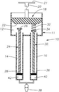
Fig. 1 is a front, sectional, schematic view of a drug delivery device
according
to a preferred embodiment of the present invention with the plunger assembly
in a
fully undepressed position;
Fig. 2 is a fragmentary, front, sectional, schematic view of the device of
Fig. I
with the plunger assembly in a partially depressed positioin;
3
CA 02588692 2007-05-22
WO 2006/068921 PCT/US2005/045459
Fig. 3 is a fragmentary, front, sectional, schematic view of the device of
Fig. 1
with the plunger assembly in a fully depressed position; and
Fig. 4 is a front, sectional, schematic view of a drug delivery device
according
to a second preferred embodiment of the present invention with the plunger
assembly
in a fully undepressed position.
Detailed Description of the Preferred Embodiments
The preferred embodiments of the present invention and their advantages are
best understood by referring to Figures 1-4 of the drawings, like numerals
being used
for like and corresponding parts of the various drawings.
As shown in Fig. 1, drug delivery device 10 preferably includes a body 11
having a plunger chamber 12, an actuation chamber 14, and an actuation chamber
16;
a plunger assembly 18 having a handle 20 and a sealing member 22; an actuation
assembly 24 having a contact member 26 and a sealing member 28; an actuation
assembly 30 having a spring member 32, a contact member 34, and a sealing
member
36; and a cannula 38 fluidly coupled to both actuation chamber 14 and
actuation
chamber 16. Device 10 is preferably sized so as to comfortably fit within a
physician's hand.
Sealing member 22 is in slidable, fluid tight engagement with the interior
surface of plunger chamber 12. Spring member 32 is preferably coupled to
sealing
member 22 on a first end and contact member 36 on a second end. Sealing member
28 is in slidable, fluid tight engagement with the interior surface of
actuation chamber
14. Sealing member 36 is in slidable, fluid tight engagement with the interior
surface
of actuation chamber 16. Cannula 38 may be any conventional blunt-tip cannula
or
sharp-tip needle suitable for ophthalmic drug delivery. Preferred cannulae for
cannula
4
CA 02588692 2007-05-22
WO 2006/068921 PCT/US2005/045459
38 for use in sub-Tenon, juxtascleral delivery of a drug depot to the
posterior segment
of a human eye are disclosed in U.S. Patent No. 6,413,245.
A dosage form 40 is disposed within actuation chamber 16 between sealing
member 36 and cannula 38. A dosage form 42 is disposed within actuation
chamber
14 between sealing member 28 and cannula 38. Device 10 is preferably packaged
with dosage forms 40 and 42 preloaded. Alternatively, dosage forms 40 and 42
may
be loaded by the user prior to administration.
Dosage forms 40 and 42 may be any dosage form containing a drug or
pharmaceutically active agent. Dosage forms 40 and 42 may be in liquid, semi-
solid,
or solid form. For example, dosage forms 40 and 42 may be a solution, a
suspension,
an emulsion, an ointment, a gel forming solution, a gel, a bioerodable
polymer, a non-
bioerodable polymer, or a powder. Preferably, dosage forms 40 and 42 include
any
ophthalmically acceptable pharmaceutically active agent. Examples of
pharmaceutically active agents suitable for dosage forms 40 and 42 are
disclosed in
U.S. Patent No. 6,416,777, which is incorporated herein by reference. One
preferred
pharmaceutically active agent is angiostatic steroids for the prevention or
treatment of
diseases or conditions of the posterior segment of the eye, including, without
limitation, ARMD, CNV, retinopathies, retinitis, uveitis, macular edema, and
glaucoma. Such angiostatic steroids are more fully disclosed in U.S. Patent
Nos.
5,679,666 and 5,770,592, which are incorporated herein by reference. Preferred
ones
of such angiostatic steroids include 4,9(11)-Pregnadien-170~21-diol-3,20-dione
and
4,9(11)-Pregnadien-17c~21-diol-3,20-dione-2l-acetate. In addition, dosage
forms 40
and 42 may include a combination of a glucocorticoid and an angiostatic
steroid as
pharmaceutically active agents. For this. combination, preferred
glucocorticoids
include dexamethasone, fluoromethalone, medrysone, betamethasone,
triamcinolone,
5
CA 02588692 2007-05-22
WO 2006/068921 PCT/US2005/045459
triamcinolone acetonide, prednisone, prednisolone, hydrocortisone, rimexolone,
and
pharmaceuitcally acceptable salts thereof, and preferred angiostatic steroids
include
4,9(11)-Pregnadien-17c~21-diol-3,20-dione and 4,9(11)-Pregnadien-17c~21-diol-
3,20-
dione-21-acetate. Dosage forms 40 and 42 may also comprise conventional non-
active excipients to enhance the stability, solubility, penetrability, or
other properties
of the active agent.
Device 10 is especially suitable for the delivery of a dosage fonn 40 and a
dosage form 42 that exhibit some kind of mutual incompatibility and are best
kept
separate until just before delivery. In addition, dosage form 40 may include
one of the
ophthalmically acceptable pharmaceutically active agents suitable for
localized
delivery to the posterior segment of the eye mentioned hereinabove, and dosage
form
42 may include a biocompatible polymer for preventing drug reflux during sub-
Tenon,
juxtascleral delivery of a drug depot to the posterior segment of the eye. A
preferred
polymer is a biocompatible, bioerodable polymer.
The following describes a preferred procedure by which a physician may use
drug delivery device 10 for sub-Tenon, juxtascleral delivery of a drug depot
to the
posterior segment of an eye. Preferred cannulae for cannula 38 for such drug
delivery
are disclosed in U.S. Patent No. 6,413,245. In the superior temporal quadrant
of the
eye, the physician uses fine scissors to create a small incision in the
conjuctiva and
Tenon's capsule to bare sclera at a point about 8 mm to about 9 mm posterior
to the
limbus. Cannula 38 of device 10 is then inserted through the incision. The
distal tip
of cannula 38 is advanced along the curvature of the sclera until the tip is
located in
the desired position. The physician then slowly depresses head 21 of handle 20
so that
sealing member 22 of plunger assembly 18 cooperates with spring niember 32 and
contact member 34 of actuation assembly 30 to slide sealing member 36 toward
6
CA 02588692 2007-05-22
WO 2006/068921 PCT/US2005/045459
cannula 38. As sealing member 36 is moved toward cannula 38, dosage form 40,
which contains an appropriate pharmaceutically active agent, is slowly
dispensed from
cannula 38 to create a drug depot on the outer surface of the sclera below the
Tenon's
capsule. When sealing member 36 reaches the position shown in Fig. 2, spring
member 32 is partially compressed, substantially all of dosage form 40 has
been
dispensed from cannula 38, and all of dosage form 42 remains in actuation
chamber
14. The spring force of spring member 32 may be optimized for different
volumes,
forms, viscosities, and delivery rates of dosage form 40. As the physician
continues to
slowly depress head 21 of handle 20, sealing member 22 then cooperates with
contact
member 26 of actuation assembly 24 to slide sealing member 28 toward cannula
38.
As sealing member 28 is moved toward cannula 38, dosage form 42, which
contains a
biocompatible, bioerodable polymer, is slowly dispensed from cannula 38 to
seal the
sub-Tenons space anterior to the drug depot and prevent reflux of dosage form
40.
When sealing member 28 reaches the position shown in Fig. 3, spring member 32
is
fully compressed, and substantially all of dosage form 42 has been dispensed
from
cannula 38. The physician slowly withdraws cannula 38 from the incision. The
physician then applies an antibiotic ointment, and optionally applies a
pressure patch
to the incision.
As shown in Fig. 4, drug delivery device l0a has a substantially identical
structure to device 10 with the exception that actuation chambers 14 and 16
are
formed adjacent to one another instead of with a space therebetween like in
device 10.
The operation of device l 0a is substantially identical to the operation of
device 10.
From the above, it may be appreciated that the present invention provides an
improved device for the administration of an ophthalmic drug, especially to
the
posterior segment of the eye. The device of the present invention also
minimizes or
7
CA 02588692 2007-05-22
WO 2006/068921 PCT/US2005/045459
prevents drug reflux during ophthalmic drug delivery. The device is safe for
the
patient, easy for the physician to use, and improves the efficacy of drug
administration.
The present invention is illustrated herein by example, and various
modifications may be made by a person of ordinary skill in the art. For
example,
although the use of the device of the present invention is described above in
connection with sub-Tenon, juxtascleral delivery of a drug depot to the
posterior
segment, it can also be utilized in connection with other ophthalmic or non-
ophthalmic drug delivery. As another example, handle 20 may be replaced with
an
automated assembly for displacing sealing member 22, if desired.
8