Note: Descriptions are shown in the official language in which they were submitted.
CA 02588866 2007-05-22
WO 2006/058018 PCT/US2005/042349
1
IMPLANT DEVICE USED IN MINIMALLY
INVASIVE FACET JOINT HEMI-ARTHROPLASTY
Technical Field
The present invention relates generally to a device for
use in minimally invasive spine surgery. More particularly, it
refers to a pre-made, pre-shaped metallic implant implanted
using an arthroscopic type portal or classic open surgical
method to achieve a spinal facet joint hemi-arthroplasty to
resurface any or all of the forty-eight superior facets of the
inferior Occiput-Cl through L5-S1 vertebrae.
Background Art
In the United States alone, about 10% of the entire
population will suffer from back pain sometime in the next
twelve months. More people will contract back pain in the next
year than any other injury or disease except the common cold
and flu. About one-third will not recover and have to live
with persistent, disabling symptoms. The number is cumulative
year after year.
One of the root causes of back pain, particularly
persistent and disabling back pain, are facet joints, small
joints located behind adjacent vertebrae in the spine that
allow for spinal motion.
Present surgical solutions available for the millions of
people with facet joint dysfunctions are complex, invasive,
high-risk operations requiring pedicle screws for fixation and
CA 02588866 2007-05-22
WO 2006/058018 PCT/US2005/042349
2
significant reduction or elimination of natural joints and
replacement with prosthetic apparatus such as those described
in United States Patents 6,610,091, 6,579,319, 6,132,464,
6,113,637 and U.S. Patent Application 2003/0028250. In
general, the present art requires prolonged recovery times,
from six to twenty-four months, and offers uncertain outcomes.
High risk equates to frequent litigation, which forces non-
surgical symptomatic treatment while the disease or
consequences of injury progressively worsen.
With the advent of new, safer and less invasive surgical
techniques and technology, the growth of spine surgery now
outpaces every other orthopedic surgery segment. Its growth is
further fueled by an enormous demand. Improvements in devises
used in spinal joints is needed.
Disclosure of Invention
The present invention provides a pre-made pre-shaped
metallic implant for use in minimally invasive spine surgery.
The use of a pre-shaped metallic overlay of this invention for
facet joint resurfacing of diseased, painful, deteriorated or
overstressed joints offers three distinct advantages over
larger prosthetic implants; namely, (1) using a thin metallic
overlay allows for minimally invasive insertion that is safer,
less traumatic and requires far less recovery time compared to
a prosthetic; (2) the overlay does not require the use of
cements, pedicle screws or other fixation methods that can work
CA 02588866 2007-05-22
WO 2006/058018 PCT/US2005/042349
3
their way loose over time; and, (3) the implant has two fins or
blades to provide lateral stability and two teeth to provide
temporary fixation and a rough or porous inner surface amenable
to bone in growth providing permanent natural fixation. The
implant also has a polished outside that allows for smooth,
natural, pain free articulation of the joint.
The implant is specifically designed for use in an
arthroscopic type portal for standalone procedures, but also
may be used in classic open surgery. This implant provides a
unique, stronger and superior resurfacing and may be used for,
but not limited to: (1) an adjunct to instrumented vertebral
fusion when implanted in the two facet joints immediately above
and below the two joints adjoining the instrumentation thereby
eliminating the risk of collateral post-operative facet joint
pain resulting from additional stress placed on facet joints,
(2)when used to resurface adjoining facet joints directly above
and below a disk replacement by eliminating the risk of
collateral post-operative facet joint pain resulting from
additional stress placed on facet joints by the disk
replacement, and, (3) as a stand along treatment for diseased,
painful or deteriorated facet joints.
The invention accomplishes its goal of resurfacing a
painful, diseased or deteriorated spinal facet joints by
providing a resurfacing implant to replace the joint surface
with a small metal on bone overlay. The overlay, constructed
CA 02588866 2007-05-22
WO 2006/058018 PCT/US2005/042349
4
of cobalt chrome or such other biocompatible metal or metallic
alloy appropriate for joint hemi-arthroplasties, is one of
several sizes for various segments of the spine, similarly
sized for different facet joints or groups of joints in the
spine and are attached to the joint using a straightforward
process without the need for screws or cements. The facet
joints may be accessed using an arthroscopic type portal
eliminating the need for open surgery, hospitalization and long
recovery periods. The procedure also may be performed as an
adjunct to other procedures such as instrumented fusion and
disc replacement in a traditional open surgery. Because the
side of the implant that attaches to bone is porous, the bone
heals onto it, permanently fixing it into place. A uniquely
designed set of blades and teeth provides temporary fixation to
the joint and prevents migration. A crimping system allows the
implant to be fixed into place, holding it firmly until bone in
growth is complete. The side making contact with the joint is
highly polished providing a smooth, virtually frictionless
surface that undergoes virtually no wear and tear. The inside
is rough or porous providing an amenable surface-for bone in
growth.
According to one broad aspect of the invention, a unique
metallic prosthetic overlay is provided. The metallic overlay
is generally shaped to the naturally shaped contour of the bone
it resurfaces and is highly polished on the outside to provide
CA 02588866 2007-05-22
WO 2006/058018 PCT/US2005/042349
frictionless articulation of the joint and rough or porous on
the inside to promote and provide a surface to allow the
natural bone to grow into the overlay, providing a permanent
fixation. In the interim between implantation and bone in-
growth, the overlay is mechanically crimped into place using
two teeth opposed to each other and one to two blades on the
inside of the implant that bite into the bone to prevent
lateral migration. The overlay is further held into place by
the natural pressure of the inferior and superior sides of the
joint as they come together in their natural position.
The system to insert the prosthetic overlay includes any
number of instruments allowing preparation of the joint and the
implant to be placed using a minimally invasive surgical
arthroscopic technique to access to the joint that include a
director probe to determine the correct facet joint angle, a
separator to assist with separation of the vertebrae to improve
access to the joint, an osteotome to make a small cut in the
bone to prepare the surface for the implant, a broach to
prepare the bone to match the implant shape, an impactor to
impact the implant into place and a crimp to fix the implant to
prevent migration prior to healing and a unique implant. By
way of example only, the director may include a planer blade or
rasp to remove any bone spurs or overgrowth and to flatten the
facet joint surface in preparation for implant placement.
CA 02588866 2007-05-22
WO 2006/058018 PCT/US2005/042349
6
Brief Description of Drawings
Many advantages of the present invention will be apparent
to those skilled in the art with a reading of this
specification in conjunction with the attached drawings,
wherein like reference numerals are applied to like elements
and wherein:
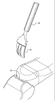
Figure 1 shows a top right isometric view of the implant;
Figure 2 shows a bottom left isometric view of the
implant;
Figure 3 shows a sectional view along line 3-3 of FIG. 1;
Figure 4 shows a perspective view of a tool used to
prepare the facet joint for receipt of the implant;
Figure 5 shows the tool inserted in the facet joint;
Figure 6 shows the insert about to be installed in the
facet joint;
Figure 7 shows the insert embedded in the facet joint;
Figure 8 shows a sectional view along line 8-8 in FIG. 7.
Best Mode for Carrying Out the Invention
Referring to FIG. 1, the prosthetic implant 10 of this
invention has a highly polished concave outside back portion 12
and a convex highly polished surface on a top portion 14. The
highly polished surface is a cobalt-chrome alloy, a titanium
alloy or other biologically acceptable material capable of
forming a smooth highly polished surface. Referring to FIG. 2,
an inside convex surface is textured 18 to encourage new bone
CA 02588866 2007-05-22
WO 2006/058018 PCT/US2005/042349
7
growth and adhesion. Blades 20 and 22 attached to inside
surface 16 bite into bone to promote adhesion. The bottom
surface 24 of the top portion of implant 10 is concave to fit
tightly over a bone as shown in FIGS. 7 and 8. Teeth 26 and 28
are used for imbedding into adjacent bone to prevent movement
of implant 10.
Prior to imbedding the implant in the facet joint 32, a
preparation tool 30 is used to slightly spread the joint 32.
Tool end 34 is inserted into the joint 32 to provide sufficient
space for inserting implant 10 into joint 32 as seen in FIGS. 7
and 8.
The use of the prosthetic implant 10 has two advantages
over the prior art:
(1) It is minimally invasive, low risk, fast (about
20 minutes per joint in an outpatient setting compared to
about three hours in a hospital followed by a three day
stay), and has a recovery time measured in a few weeks
(compared to six to twenty-four months); and
(2) It has a high success rate, does not preclude
other surgical options, and is non-limiting and permanent.
The present invention is directed at overcoming, or at
least improving upon, the disadvantages of the prior art by
achieving the following:
= Reversal of the risk/benefit ratio of the present
procedures versus the invention;
CA 02588866 2007-05-22
WO 2006/058018 PCT/US2005/042349
8
= A stand-alone minimally invasive procedure versus
major open surgery;
= Employed as an adjunct to major open surgery in
concert with long fusion and with disc replacement
surgery to strengthen adjacent facet joints;
= Outpatient versus inpatient surgery (about 20 minutes
per joint versus hours);
= Reduced morbidity;
= Reduced blood loss;
= Reduced time under anesthesia;
= Reduced risk;
= Recovery time dramatically reduced;
= Minimal scarring that decreases the risk of failed
back syndrome and improves revisions surgery outcome;
= Reduced risk of post operative infection by
significantly reducing operating room time and soft
tissue destruction;
= Prolonging the functional life of long segment
fusions and disc replacement;
= No preclusion of other surgical or non-invasive
treatment options; and,
= Projected high success rate by utilizing accepted
procedures facilitated through an arthroscopic
technique and resurfacing implant.
CA 02588866 2007-05-22
WO 2006/058018 PCT/US2005/042349
9
It is anticipated that the availability of this method,
instrumentation and implant will increase the number of
surgeries performed because they offer the first safe
outpatient solution to a predominant cause of joint pain. The
inventor also expects that virtually all patients receiving
this procedure will be able to walk the same day as surgery and
be fully functional within a few weeks. Present surgical
solutions require hospitalization of about three days and six
to twenty-four months' recovery.
Aside from the obvious positive clinical outcome, the
significant favorable financial impact on disability, worker's
compensation and health care insurers is considerable.
Spinal facet implant units are calculated per joint. Each
patient has two joints per spinal segment and six segments (T12
to L1 through L5-Sl) in the lumbar spine, or twelve lumbar,
fourteen cervical and twenty-eight thoracic joints. Each
surgery is likely to involve multiple joints, with a probable
average of four per patient.
The invention accomplishes its goal of reducing,
preventing or eliminating spinal facet joint pain by providing
a resurfacing implant to replace the joint surface with a small
metal on bone overlay. The overlay, constructed of cobalt
chrome, a material previously approved by the FDA for other
joint hemi-arthroplasty, or such other metallic construction as
may be safely used, is one of several sizes for various
CA 02588866 2007-05-22
WO 2006/058018 PCT/US2005/042349
segments of the spine, may be similarly sized for different
joints and is attached to the joint using a straightforward
process without the need for screws or cements with the aid of
custom designed instruments. The joint is accessed using an
arthroscopic type portal eliminating the need for open surgery,
hospitalization and long recovery periods (unless the procedure
is performed as an adjunct to other procedures such as
instrumented fusion and disc replacement in a traditional open
surgery). Because the side that attaches to bone is porous,
the bone heals onto it, permanently fixing it into place. A
uniquely designed set of blades and teeth prepares the joint
and a unique crimping system allows the implant to be fixed
into place, holding it firmly until bone in growth is complete.
The side making contact with the jointlis highly polished
providing a smooth, virtually frictionless surface that
undergoes virtually no wear and tear. The resurfacing implant
is a securely fixed porous hemi-arthroplasty of the facet
joints of the spine.
The metallic overlay is generally shaped to the natural
contour of the bone it resurfaces and is highly polished on the
outside to provide frictionless articulation of the joint and
rough and porous on the inside to promote and provide a surface
to allow the natural bone to grow into the overlay, providing a
permanent fixation. In the interim between implantation and
bone in-growth, the overlay is mechanically crimpled into place
CA 02588866 2007-05-22
WO 2006/058018 PCT/US2005/042349
11
using two teeth opposed to each other that bite into the bone
to prevent migration. The overlay is further held into place
by the natural pressure of the inferior and superior sides of
the joint as they come together in their natural position.
The system includes any number of instruments 30 allowing
preparation of the joint 32 and the implant 10 to be placed
using a minimally invasive surgical arthroscopic technique to
access to the joint that include a director probe to determine
the correct facet joint angle, a separator to assist with
separation of the vertebrae to improve access to the joint, an
osteotome to make a small cut in the bone to prepare the
surface for the implant, a broach to prepare the bone to match
the implant shape, an impactor to impact the implant into place
and a crimp to fix the implant to prevent migration prior to
healing and a unique implant. By way of example only, the
director may include a planer blade or rasp to remove any bone
spurs or overgrowth and to flatten the facet joint surface in
preparation for implant placement.
Equivalent elements can be substituted for the elements of
the implant of this invention to provide substantially the same
function in substantially the same way to achieve substantially
the same result.