Note: Descriptions are shown in the official language in which they were submitted.
CA 02589253 2013-04-12
WO 2006/069709 PCT/EP2005/013802
METHOD AND DEVICE FOR SHAPING .AN ENERGY INPUT BEAM
10
Technical field
The present invention relates to a method for shaping an en-
ergy input beam for imaging purposes, in particular to an ir-
radiation method for imaging a region of investigation of an
object with at least one energy input beam. Furthermore, the
present invention relates to a method and devices for imaging
the region of investigation on the basis of the irradiation
method, in particular for imaging tomography.
Technical Background
The non-destructive investigation of samples is an important
object in various technical fields like material sciences,
medical examinations, archaeology, construction technique,
techniques concerning security matters etc.. One approach for
obtaining an image of a sample e.g. by computer tomography
CA 02589253 2007-05-28
WO 2006/069709 PCT/EP2005/013802
2
(CT) is based on an irradiation trough a object plane from
different projection directions with X-rays, followed by a
reconstruction of the object plane on the basis of attenua-
tion data measured at different directions. The irradiation
of a region of investigation 2' with a conventional fan beam
5' created by an X-ray source 210' is schematically illus-
= trated in Figure 9. The fan beam 5' comprises a continuous
distribution of electro-magnetic fields shaped according to
an emission characteristic of the X-ray source 210'. The en-
tirety of the attenuation data measured with a detector 310'
can be described in terms of so-called Radon data in a Radon
space.
The most relevant conventional reconstruction methods known
today can be summarized as methods based on the iterative re-
construction or those based on the so-called filtered back-
projection. The iterative reconstruction methods have essen-
tial disadvantages in terms of extremely long calculation
times. On the other hand, the filtered back-projection method
has a general disadvantage as an interpolation step included
in the reconstruction results in errors and artifacts which
have a tendency even to increase with increasing space fre-
quency. Another problem of the filtered back-projection
method is related to the discretization of the Radon data
from which the image data have to be reconstructed. To get an
optimal filtered back-projection reconstruction it would be
necessary to exactly match the projected irradiation rays
with detector elements of a detector. This is in general not
the case. For this reason, uncertainties or smoothing effects
from the reconstruction of Radon data by means of filtered
back-projection algorithms are introduced.
T. Bortfeld et al. have described a so-called Chebyshev do-
main filtered back projection (CD-FBP) algorithm for the re-
CA 02589253 2007-05-28
WO 2006/069709
PCT/EP2005/013802
3
construction of two-dimensional images from a plurality of
projections along the projection directions ("Phys. Med.
Biol.", Vol. 44, 1999, p. 1105-1120). With this CD-FBP algo-
rithm, the projections are represented as decompositions,
which are subjected to the above filtered back-projection re-
construction. The projections are measured e.g. with a fan
beam geometry, wherein attenuation values according to single
projection lines with even angular intervals relative to each
other are measured. The single projection lines measured with
different projection directions of the fan beam can be re-
sorted for providing parallel projections to be used for the
image reconstruction. The CD-FBP algorithm has not yielded a
practical implementation. The algorithm assumes an ideal fan
beam geometry, which is not available in practice. Therefore,
the T. Bortfeld et al. algorithm requires an interpolation
step like the conventional filtered back-projection. Further-
more, as the CD-FBP algorithm is inherently discrete, there
is a lack of adaptation to the continuous radiation charac-
teristic of conventional radiation sources. Finally, the CD-
FBP algorithm has an essential disadvantage in terms of arti-
facts occurring in the reconstructed images.
The above disadvantages are associated not only with the con-
ventional CT imaging, but also with all available reconstruc-
tion methods related to Radon data.
Objective of the invention
The objective of the invention is to provide improved methods
for irradiating an object, which avoid the disadvantages of
the above conventional techniques and which in particular can
be used for improved imaging. In particular, the objective of
the invention is to provide an irradiation method with an im-
proved adaptation of a radiation source to the image recon-
CA 02589253 2013-04-12
WO 2006/069709
PCT/EP2005/013802
4
struction algorithm to be used. Furthermore, the objective of
the invention is to provide an improved imaging device, which
is capable of improved imaging a region of investigation in
particular with reduced artifacts.
10
Summary of the invention
According to a first general aspect of the invention, an ir-
radiation method is provided wherein at least one energy in-
put beam is shaped with at least one beam mask for providing
a plurality of individual energy input beam components. The
beam mask is made of an energy input shielding material with
through holes providing the number of beam components and
forming the distribution thereof. The individual energy input
beam components formed by the shielding effect of the mask
(lattice) are directed through an object along a plurality of
projection lines for imaging a region of investigation in the
object.
The energy input beam with a primary continuous radiation
characteristic is generated as a fan beam or a cone beam by
using an energy input beam source. Subsequently, the energy
input beam is shaped with the beam mask. The essential advan-
tage of shaping the energy input beam for forming the energy
input beam components is given by the fact that a discrete
irradiation characteristic is provided inherently. The irra-
diation characteristic can be adapted to an image reconstruc-
tion algorithm used for imaging the region of investigation.
This adaptation is obtained simply by selecting a predeter-
CA 02589253 2007-05-28
WO 2006/069709 PCT/EP2005/013802
mined beam mask and/or adjusting the beam mask relative to
the energy input beam source. The beam mask has a predeter-
mined geometry with a fixed distribution and fixed size(s) of
the through holes. Furthermore, the energy input (e. g. dose)
5 can be reduced essentially.
The fan or cone beams are adjusted such that the projection
directions are set in at least one common plane crossing the
region of investigation or, alternatively, in varying in-
clined planes crossing the region of investigation for ob-
taining helical projection data.
The term "region of investigation" (ROI) used herein gener-
ally refers to an object under investigation or a part
thereof. The ROI can be described as a 2- or 3-dimensional
entity. The term "projection direction" used herein generally
refers to the linear course of an energy input through the
ROI. The projection direction can be defined by angles rela-
tive to a coordinate system used. If fan or cone beams are
considered, the term "projection direction" indicates the
orientation of a central (or: main) beam component in the fan
or cone beam.
According to a preferred embodiment of the invention, the en-
ergy input beam source is movable relative to the object,
wherein the beam mask and the energy input beam source are
connected with each other. In this case, the beam mask is
called source mask. The projection directions can be selected
by moving the energy input beam source and the source mask
relative to the region of investigation. Particularly pre-
ferred is an embodiment, wherein the projection directions
are set subsequently by rotating the energy input beam source
with the beam mask around the object.
CA 02589253 2007-05-28
WO 2006/069709
PCT/EP2005/013802
6
According to an alternative embodiment of the invention, a
plurality of beam masks is provided at predetermined posi-
tions relative to the ROI. In this case, the beam masks are
called frame masks. The beam masks are distributed around the
ROI. In this case, the projection directions can be selected
by driving at least one energy input beam source at each of
the frame masks. As an example, a plurality of energy input
beam sources can be distributed according to the distribution
of the beam masks. Alternatively, one energy input beam
source is serially moved to each of the beam masks.
For an adaptation to discrete reconstruction algorithms, the
energy input beam components are formed with an equal angle
spacing. The projection lines passing the through holes of
the mask cross a spherical surface around the source with
equal arc length spacing. To this end, the beam mask can have
a planar shape or a curved shape (e.g. cylindrical shape). If
according to a particularly preferred embodiment of the in-
vention a planar beam mask is used with through holes all
having an equal size or a curved beam mask is used with
through holes having different sizes, a complete irradiation
of the region of investigation without overlap of the energy
input beam components being parallel to a particular projec-
tion direction can be obtained. In this case, artefacts can
be reduced in the image reconstruction. Alternatively, a pla-
nar beam mask with through holes all having an equal size or
a curved beam mask can be used with through holes having dif-
ferent sizes.
According to a further advantageous embodiment of the inven-
tion, the energy input beam is shaped not only with regard to
forming the energy input beam components, but also with re-
gard to the outer boundary of the energy input beam. To this
end, the imaging method of the invention comprises the fur-
CA 02589253 2007-05-28
WO 2006/069709 PCT/EP2005/013802
7
ther step of setting the beam angle of the energy input beam.
This embodiment has particular advantages with regard to an
adaptation of the beam source (possibly in combination with
the beam mask) to a particular object to be investigated. For
irradiating a small object, the beam angle can be decreased
so that the overall irradiation dose can be reduced.
Preferably, the beam angle is set with an aperture serving as
a diaphragm or shutter. Advantageously, the aperture has a
simple construction. Furthermore, it can be simply mounted on
a conventional imaging device, like e.g. a conventional CT
device for an adaptation to the present invention. Further
advantages of the aperture derive from the availability of
two degrees of freedom for adjusting the beam angle. With a
first alternative, the beam angle is adjusted by setting a
diameter of the aperture. This allows a flexible adaptation
of the aperture to the object under investigation. According
to a second alternative, the beam angle is adjusted by set-
ting a distance between the aperture and the beam source.
With the decreasing distance, the beam angle is increased.
Both alternatives can be combined.
According to a second general aspect of the invention, an im-
aging method for imaging the ROI is provided wherein the ROI
is irradiated using a method according to the above first
general aspect and a plurality of projection functions corre-
sponding to the plurality of projection directions is deter-
mined. Each of the projection functions comprises attenuation
values measured with energy input beam components being par-
allel to a current projection direction. The measured at-
tenuation values are subjected to an image reconstruction
procedure which is known as such.
CA 02589253 2007-05-28
WO 2006/069709
PCT/EP2005/013802
8
The measured attenuation values provide discrete projection
profiles representing the projection functions, wherein the
projection values of each discrete projection profile com-
prise the attenuation values corresponding to predetermined
energy input beam components with the same projection direc-
tion.
, The present invention provides another essential advantage in
terms of data handling. Due to an adjustment of the cross-
sections of the energy input beam components with the beam
mask, attenuation values can be measured with predetermined
groups of detector elements of the detector device. The
groups of detector elements have predetermined, e.g. equal
sizes for all beam components. Only these predetermined
groups are to be read out without resizing so that the amount
of data to be processed is reduced. Preferably, the attenua-
tion values are measured with at least one 1-dimensional
straight detector or with at least one 2-dimensional planar
detector.
Advantageously, the imaging method of the invention can be
implemented with various image reconstruction procedures.
Preferably, an image reconstruction procedure is used as de-
scribed in EP 04031043.5, the priority of which is claimed
with the present specification. With this method, the image
function is determined from Radon data comprising a plurality
of projection functions measured corresponding to the plural-
ity of predetermined projection directions. The image func-
tion is determined as a sum of polynomials multiplied with
values of the projection functions. In practical implementa-
tions, this image reconstruction is based on the measurement
of attenuation values corresponding to the discrete irradia-
tion beam components having equal angles relative to each
other. Alternatively, the image reconstruction procedure can
CA 02589253 2007-05-28
WO 2006/069709 PCT/EP2005/013802
9
comprise a decomposition of the attenuation values as de-
scribed with further details by T. Bortfeld et al. (see
above).
It is an essential advantage of the invention, that the imag-
ing can be used in various applications like many applica-
tions in medical imaging, for example CT, PET, SPECT, etc..
However, there are a lot more possible applications like
light tomography, any multidimensional imaging for industrial
testing or biological research and so on. Preferably, the im-
age function is determined from Radon data measured in an X-
ray computer tomography (CT) device, a PET imaging device, a
SPECT imaging device, or a neutron based transmission detec-
tion system. The object under investigation comprises e.g. a
biological organism or a part thereof, a fluid composition, a
solid material, a work-piece, and/or an object to be investi-
gated for security reasons.
According to a third general aspect of the invention, an im-
aging device for imaging a region of investigation of an ob-
ject is provided, wherein the imaging device includes at
least one beam mask made of an energy input shielding mate-
rial with through holes. The beam mask is adapted for forming
individual, discrete energy input beam components. Further-
more, the imaging device comprises a measuring device for
measuring projection functions corresponding to a plurality
of projection directions. The measuring device includes at
least one energy input beam source and at least one detector
device for measuring the projection functions. The energy in-
put beam source is arranged for creating at least one energy
input beam to be shaped with the beam mask. Furthermore, the
imaging device includes a reconstruction circuit for recon-
structing an image function on the basis of measured projec-
tion functions.
CA 02589253 2007-05-28
WO 2006/069709 PCT/EP2005/013802
Preferably, the energy input beam source being a adapted for
creating a fan beam or a cone beam source is movably arranged
on a source carrier. Particularly preferred is a source car-
5 rier which has a ring shape so that the energy input beam
source can be rotated e.g. on a circle or along a helical
path around the object.
The detector device comprises at least one detector array of
10 detector elements for detecting attenuation values represent-
ing the attenuation of the energy input corresponding to the
plurality of predetermined projection directions.
According to further preferred embodiments of the invention,
the imaging device comprises at least one of a first adjust-
ment device for adjusting a distance between the source mask
and the energy input beam source and a second adjustment de-
vice for adjusting a diameter of an beam angle aperture
and/or a distance between the aperture and the energy input
beam source.
If the at least one beam mask comprises one source mask being
movable with the energy input beam source, in particular with
the fan or cone beam source, advantages with regard to the
adjustment of the source relative to the mask can be ob-
tained. Preferably, the source mask is detachably connected
with the energy input beam source, so that the imaging device
can be adapted to a particular application simply by changing
the source mask.
If a plurality of frame masks for shaping the energy distri-
bution function of the energy input beam source is provided
as beam mask(s), advantages with regard to setting the pro-
jections directions can be obtained. Preferably, the frame
CA 02589253 2007-05-28
WO 2006/069709 PCT/EP2005/013802
11
masks are fixed at equal arc lengths on a common source car-
rier. As an example, the source carrier is a ring-shaped
shield containing the frame masks. Advantageously, the energy
input beam source can be shielded with the ring-shaped shield
at positions other than the positions of the frame masks. Ac-
cordingly, an essential dose reduction can be obtained.
According to a further modification of the invention using
the frame masks, the detector device comprises a plurality of
fixed frame detectors for detecting attenuation values repre-
senting the attenuation of energy input corresponding to the
plurality of predetermined projection directions. In this
case, the frame detectors can be fixed on the source carrier
at predetermined positions. Preferably, the frame detectors
are positioned adjacent to the frame masks.
According to various preferred applications of the invention,
the measuring device comprises an X-ray computer tomography
(CT) device, an ultrasound tomography device, a PET imaging
device, a light tomography device, a Gamma-ray imaging de-
vice, a SPECT imaging device, or a neutron based transmis-
sion detection system.
According to a fourth general aspect of the invention, a beam
mask is provided, which is made of an energy input shielding
material with through holes. The energy input shielding mate-
rial comprises e. g. tungsten, lead or copper. Tungsten is
preferred in terms of a high absorption (shielding effect)
and high mechanical stability. The beam mask is capable of
forming energy input beam components for irradiating a region
of investigation of an object.
CA 02589253 2007-05-28
WO 2006/069709
PCT/EP2005/013802
12
Brief description of the drawings
Further details and advantages of the invention are described
in the following with reference to the attached drawings,
which show:
Figure 1 a schematic illustration of an embodiment of
beam shaping according to the invention;
Figures 2 to 5 schematic illustrations of embodiments of beam
masks used according to the invention;
Figure 6 a schematic illustration of the combination of
a beam source with a beam angle aperture and a
source mask according to the invention;
Figure 7 a further illustration of directing discre-
tized fan beams through an object under inves-
tigation;
Figure 8 a schematic representation of an embodiment of
an imaging device according to the invention;
and
25 Figure 9 a schematic illustration of directing a con-
ventional fan beam through a region of inves-
tigation (prior art).
Embodiments of the invention
The invention is described in the following with reference to
the application in computer tomography. In this case, the im-
aging device according to the invention includes the main
components of a current medical CT-system, with the energy
CA 02589253 2007-05-28
WO 2006/069709
PCT/EP2005/013802
13
input beam being an X-ray beam. It is emphasized that the in-
vention can be implemented in an analogous way with the other
applications mentioned above. Furthermore, the following de-
scription of the preferred embodiments mainly refers to the
step of energy beam shaping with a beam mask. Details of CT
or other imaging devices as well as details of the image re-
construction used for implementing the invention are not de-
scribed as they are known from conventional techniques or
from EP 04031043.5:
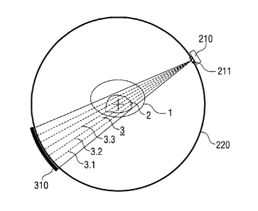
Directing a discretized fan or cone beam 3 through an object
1 with ROI 2 in a CT system for collecting projection data is
schematically illustrated in Figure 1. The CT-system (not
completely illustrated, for further components see Figure 8)
includes a ring-shaped source carrier 220 (CT ring) in which
the radiation source 210 (X-ray tube) and a detector device
310 are rotating in a way that the whole system can finish a
complete turn within e.g. 0.3 to 0.5 s.
As a beam mask, the source mask 211 is used, which is detach-
ably fixed to the radiation source 210. The source mask 211
is adapted for shaping the energy distribution function of
the radiation source 210. To this end, the source mask 211 is
made of an energy input shielding material with through
holes. Discrete, individual X-ray beam components 3.1, 3.2,
3.3, ... passing the through holes are formed for the above
image reconstruction methods, which do not require all possi-
ble ray components resulting from the geometry of the tube
and the detector geometry for the reconstruction.
The fan beam 3 is directed with varying projection directions
through the ROT 2. The projection directions are adjusted in
a way that a predetermined number of parallel X-ray beam com-
ponents is achieved for the imaging conditions. Each fan beam
CA 02589253 2007-05-28
WO 2006/069709 PCT/EP2005/013802
14
3 represents a bundle of fan beam components 3.1. 3.2, 3.3
.... Each of the fan beam components 3.1. 3.2, 3.3 ... can be
considered as a straight pencil beam. While these pencil
beams of one fan beam do not have the same individual projec-
tion directions, the determination of discrete projection
profiles for image reconstruction is obtained =by resorting
beam components, which belong to different projections direc-
tions of the beam 3. As the result, groups of parallel beam
components are obtained.
The detector device 310 is a linear or 2-dimensional array of
detector elements being shaped according to a spherical ref-
erence surface adapted to the radius of the CT ring. Alterna-
tively, a detector device with a straight (1-dimensional) or
a planar (2-dimensional) arrangement of detector elements can
be used. The detector device 310 consists of e. g. 1 to 64
rows of detector elements (if it is more than one row it
would be called a multi-slice-CT) and approximately 700 and
1000 detector elements per row. Current detector elements
have a size of e.g. 0.5 to 1.0 mm. Within each single turn
the data are read about 1000 times. The object 1, e.g. a pa-
tient, is moving through the CT-ring by using a patient ta-
ble, that is moving continuously. By this method a so-called
helical or spiral CT data set can be gathered, because the
data that are collected are located on a spiral net.
Further details of various source masks used according to the
invention are shown in Figures 2 to 5. The source mask 211
comprises a shielding plate 212 for example made by tungsten
with through holes 213. The source mask is fixed to the ra-
diation source (e.g. X-ray tube), in particular to a frame
214 of an output window 215 of the radiation source 210 by a
detachable fixing element, like e.g. a clip element or a snap
connection.
CA 02589253 2007-05-28
WO 2006/069709 PCT/EP2005/013802
The shielding plate 212 can have a cylindrical shape (Figures
2, 4) or a planar shape (Figures 3, 5) or any other appropri-
ate shape adapted to the geometric conditions of the imaging
5 device. A cylindrical shielding plate 212 is oriented with
the cylinder axis parallel to the axis of a CT ring. The
thickness of the shielding plate 212 is e.g. 100 pm to 5 mm.
The through holes 213 are arranged such that the projection
lines starting at the radiation source 210 cross the circle
10 in line with the detector elements at predetermined posi-
tions, in particular, they can be arranged with an equal arc
length spacing. Depending on the imaging conditions, the
through holes 213 are arranged with a line or an area distri-
bution.
The number and size(s) of the through holes 213 is selected
in dependence on the particular application of the imaging
method. For CT imaging, e. g. 200 through holes 213 are pro-
vided on an area in the range of about 1 mm2 to 100 mm2. Ac-
cordingly, the discrete fan beam 3 comprises e.g. 200
straight fan beam components. By increasing the number of
rays and projections, a higher resolution can be achieved,
that means the number of pixels which can be reconstructed
free of artifacts can be increased.
According to Figures 2 and 3, the sizes of the through holes
213 are selected such, that all beam components have the same
cross-section, i.e. all through holes of the cylindrical mask
have the same size (e. g. 10 to 200 pm) while through holes
in the centre of the planar source mask beam 211 are smaller
than through holes at the outer boundary of the planar source
mask beam 211. On the other hand, according to Figures 4 and
5, the sizes of the through holes 213 are selected such, that
the beam components have varying cross-sections in the detec-
CA 02589253 2007-05-28
WO 2006/069709
PCT/EP2005/013802
16
tor domain. Preferably, the cross-sections are selected in
the range of e.g. 100 pm to 4 mm. In particular, the cross-
sections are set in dependence on the detector resolution,
which can be obtained with a particular detector. The largest
cross-sections are provided in the centre of beam 3, while
the smallest cross-sections are provided at the boundary of
beam 3.
Figure 6 schematically shows an embodiment of combining the
radiation source 210 with the source mask 211 and a beam an-
gle aperture 216. An adjustable carrier 217 is arranged on
the frame 214 of the output window 215 for holding at least
one of the components 211 and 216. Generally, the adjustable
carrier 217 serves as at least one adjustment device. The
beam angle aperture 216 is made of a shielding plate with a
central hole. The diameter and/or the perpendicular distance
between the beam angle aperture 216 and the radiation emitter
210.1 of the radiation source 210 can be adjusted with the
adjustable carrier 217 for defining a beam angle a of the
fan beam 3. The source mask 211 is shaped as outlined above
for defining the beam components. The distance between the
source mask 211 and a radiation emitter 210.1 can be varied
for obtaining a required imaging resolution. The adjustable
carrier 217 can be operated manually or electrically, e. g.
with piezo-electric drive units.
With the discrete fan beam 3 generated by the mask illus-
trated above, the signals from the detector elements of the
detector device detecting the attenuation along the corre-
sponding projection lines are read-out at certain positions
of the radiation source and the detector device only. The
read out positions are those arc length positions on the
ring-shaped source carrier, which fulfil the condition of se-
CA 02589253 2007-05-28
WO 2006/069709 PCT/EP2005/013802
17
lecting parallel fan beam components with the same projection
directions.
For reducing the radiation or particle exposure of the object
under investigation, it is preferred to direct the energy in-
put (e.g. radiation) into the object under investigation only
at the above read-out positions, namely the detector element
signals are read out only when the combination of a radiation
source and the detector device is oriented to the suitable
positions. During the movement of the radiation source, this
condition is fulfilled for certain times and/or for certain
arc length positions of the radiation source. As long as the
read-out condition is not fulfilled, the radiation source can
be shut off or shielded. Shielding the radiation source is
preferred for keeping radiation conditions stable.
The shielding function can be fulfilled by a ring-shaped
shield 222 which is schematically illustrated in Figure 7
with a plurality of radiation windows 223. The ring-shaped
shield 222 can be detachably fixed to the source carrier 220
for adapting the geometric properties of the shield 222 to
the practical application and in particular to the mask used.
As an example, the ring-shaped shield 222 comprises 201 ra-
diation windows 223 each having a diameter of 6 mm (with a
diameter of the CT-ring: 80 cm).
The source masks 211 described above can be omitted if each
radiation window 223 of the ring-shaped shield 222 is pro-
vided with a frame mask 224 which is illustrated in Figure 7
as an example only. In fact, the source and frame masks 211,
224 need not be provided simultaneously. The frame masks 224
can be designed as the source masks shown in Figures 2 to 5.
CA 02589253 2007-05-28
WO 2006/069709 PCT/EP2005/013802
18
Figure 8 schematically illustrates an embodiment of the imag-
ing device 100. The imaging device 100 comprises the measur-
ing device with an energy generator 200 and a detector device
300 and the reconstruction device 400 being connected with
the measuring device 200, 300. Furthermore, a holding device
500 is provided, which is e.g. a carrier table as it is known
from CT systems or any other carrier or substrate holder for
arranging an object under investigation in the measuring de-
vice and for adjusting the geometry of the object relative to
the energy generator 200 and the detector device 300. Further
components like a control device, a display device etc. (not
shown) are provided for as they are known per se from prior
art devices.
The energy generator 200 comprises the radiation source 210,
like e.g. a movable X-ray tube with a source mask arranged on
the source carrier 220 (e.g. a guide rail or gantry). The de-
tector device comprises a detector array 310 which is movably
arranged on the source carrier 220 in opposite relationship
relative to the radiation source 210. With this structure,
the projection direction through the ROI (parallel to the
plane of drawing) can be set by rotating the combination of
components 210, 310 around the holding device 500.
The source carrier 220 is illustrated as a circle allowing a
rotation of the energy generator 200 and the detector device
300 around an object. According to a modification, the source
carrier can have an ellipse shape or another shape. This can
represent an advantage in terms of an adaptation to the ge-
ometry of the object to be investigated.