Note: Descriptions are shown in the official language in which they were submitted.
CA 02589362 2007-06-01
WO 2006/060586 PCT/US2005/043489
METHODS AND SYSTEMS FOR ACCESSING THE PERICARDIAL SPACE
FIELD OF THE INVENTION
This invention relates generally to methods and systems for accessing the
pericardial space via the vascular system and atrial wall, particularly
through the superior
vena cava and right atrial wall to deliver treatment in the pericardial space.
BACKGROUND OF THE INVENTION
The human heart wall consists of an inner layer of simple squamous epithelium,
referred to as the endocardium, overlying a variably thick heart muscle or
myocardium and
is enveloped within a multi-layer tissue structure referred to as the
pericardium. The
innermost layer of the pericardium, referred to as the visceral pericardium or
epicardium,
clothes the myocardium. The epicardium reflects outward at the origin of the
aortic arch
to form an outer tissue layer, referred to as the parietal pericardium, which
is spaced from
and forms an enclosed sac extending around the visceral pericardium of the
ventricles and
atria. An outermost layer of the pericardium, referred to as the fibrous
pericardium,
attaches the parietal pericardium to the sternum, the great vessels and the
diaphragm so
that the heart is confined within the middle mediastinum. Normally, the
visceral
pericardium and parietal pericardium lie in close contact with each other and
are separated
only by a thin layer of a serous pericardial fluid that enables friction free
movement of the
heart within the sac. The space (really more of a potential space) between the
visceral and
parietal pericardia is referred to as the pericardial space. In common
parlance, the visceral
pericardium is usually referred to as the epicardium, and epicardium will be
used hereafter.
Similarly, the parietal pericardium is usually referred to as the pericardium,
and
pericardium will be used hereafter in reference to parietal peiricardium.
Access to the pericardial space is desirable in order to provide a variety of
cardiac
therapies, including delivery of therapeutic agents (defined herein as
including genetic
agents, biologic agents, and pharmacologic agents), placement of electrical
medical leads
for pacing, cardioversion, defibrillation or EGM monitoring, removal of
pericardial fluid
for diagnostic analysis, or other purposes. A variety of mechanisms have been
developed
for accessing the pericardial space, ranging from a simple puncture by means
of a large
CA 02589362 2007-06-01
WO 2006/060586 PCT/US2005/043489
2
bore needle to intricate catheter or cannula based systems provided with
sealing and
anchoring mechanisms.
Access to the pericardial space may be accomplished from outside the body by
making a thoracic or sub-xiphoid incision to access and cut or pierce the
pericardial sac.
Access to the pericardial space from the exterior of the body, accomplished by
passing a
cannula or catheter type device through the chest wall and thereafter passing
the cannula or
catheter or a further instrument through the pericardium into the pericardial
space, is
disclosed in U.S. Patent Nos. 5,827,216, 5,900,433, and 6,162,195 issued to
Igo, U.S.
Patent No. 5,336,252 issued to Cohen, and U.S. Patent Nos. 5,972,013,
6,206,004,
6,592,552 by Schmidt, for example. In certain cases the pericardial sac is cut
by a cutting
instrument as disclosed in U.S. Patent Nos. 5,931,810, 6,156,009, and
6,231,518 issued to
Grabek et al.
Alternatively, an elongated perforating instrument device is introduced from a
skin
incision by a transvenous or transarterial approach into the right or left
heart chambers,
respectively, and a cutting or piercing or penetrating mechanism at the distal
end of the
elongated perforating instrument is operated to penetrate through the atrial
or ventricular
wall of the right or left heart chamber into the surrounding pericardial space
without
perforating the pericardial sac. For example, a transvenous catheter provided
with a
hollow helical needle adapted to rotated and pierce through the wall of a
right or left heart
chamber to access the pericardial space to deliver pharmacologic agents is
disclosed in
U.S. Patent Nos. 5,797,870 issued to March et al. A transvenous catheter
introduced into
the right ventricular chamber to provide access through the right ventricular
wall to enable
passage of an electrical medical lead into the pericardial space is disclosed
in, U.S. Patent
No. 4,991,578 issued to Cohen, and U.S. Pat. No. 5,330,496 issued to
Alferness, for
example.
It is preferable to effect transvenous access into the pericardial space from
the right
atrial heart chamber through the atrial wall due to the relatively low blood
pressure of right
atrial blood during systole to lessen the possibility of leakage of blood into
the pericardial
space. Consequently, it has been proposed to transvenously introduce an
elongated
electrical medical device through the venous system and either the inferior
vena cava or
the superior vena cava into the right atrial chamber and perforating through
the right atrial
wall into the pericardial space. In U.S. Patent No. 4,946,457 issued to Elliot
it is proposed
CA 02589362 2007-06-01
WO 2006/060586 PCT/US2005/043489
3
to transvenously introduce an elongated electrical medical lead through the
venous system
and superior vena cava into the right atrial chamber and perforating through
the right atrial
wall to advance and dispose the distal electrode of the lead into the
pericardial space. It
has also been proposed that a preferred site of penetration of catheters or
electrical medical
leads through the atrial wall into the pericardial space is within the right
atrial appendage
as disclosed in U.S. Patent Nos. 5,269,326 issued to Verrier, 6,200,303 issued
to Verrier et
al and 5,968,010 issued to Waxman et al. Transvenous approaches through either
of the
inferior vena cava or the superior vena cava are disclosed in these patents.
It is customary in the implantation of transvenous cardiac pacing leads and
cardioversion/defibrillation leads to access the venous system that drains
into the superior
vena cava and to lodge and fix a pace/sense or cardioversion/defibrillation
electrode within
the right ventricle, the right atrium or a cardiac vein accessed through the
coronary sinus.
The proximal connector ends of such leads are coupled to implantable pulse
generators (IPGs) that are implanted subcutaneously in the thoracic region. It
is preferable
to implant the IPGs in the thoracic region, rather than the groin or abdominal
region,
because the thoracic region is more stable than the abdominal or groin region
during
ambulation and other normal body movement and the IPG is 'less likely to
migrate from the
subcutaneous implantation site. Consequently, the transvenous access into the
right atrium
is made through the superior vena cava.
A distal pace/sense electrode of an atrial pacing lead is typically lodged
into the
atrial appendage and various active and passive fixation mechanisms are
employed to hold
the electrode in place. Atrial pacing leads have been designed in a variety of
ways to
overcome the inherent difficulty of routing the distal end of an atrial lead
or any other
elongated medical device through the superior vena cava into the right atrial
heart chamber
and then into the atrial appendage. However, care is taken in the design of
such leads and
delivery mechanisms and techniques to avoid perforating the atrial wall
It is proposed in the above-referenced '326 patent to alternatively route a
pacing
lead or cardioversion/defibrillation lead through a perforation'of the atrial
wall in the atrial
appendage to lodge a pace/sense electrode and/or cardioversion/defibrillation
electrode
within the pericardial space and to subcutaneously implant an IPG or
implantable monitor
or drug dispenser in the thoracic region. The suggested routing of the
electrical medical
CA 02589362 2007-06-01
WO 2006/060586 PCT/US2005/043489
4
lead or catheter is through the thoracic venous system, through the superior
vena cava, and
through the atrial wall of the atrial appendage into the pericardial space.
It is a relatively simple matter to route a perforating instrument through the
venous
system draining into the right atrium through the inferior vena cava since the
instrument
body is relatively straight within the right atrium and axial force can be
applied to
perforate the atrial wall while observing the advancement under fluoroscopy.
It is not a
simple matter to advance the distal end perforating mechanisms of the
perforating
instruments disclosed in the above-identified patents through the superior
vena cava into
the right atrial heart chamber and then into the atrial appendage. The
physiology and
shape of the atrial appendage requires that the direction of advancement of
the distal end
be reversed or abruptly changed after it is disposed in the right atrial heart
chamber.
Moreover, the atrial wall in the atrial appendage tends to yield somewhat if
blunt
force is applied against its endocardial surface. Consequently, the precise
application of
perforating force and advancement of the distal end perforating element must
be carefully
controlled, which is difficult to manage through the bend in the instrument
body.
It would therefore be desirable to provide a method and system for accessing
the
atrial appendage via the superior vena cava and applying force through an
elongated
perforating instrument sufficient to safely penetrate through or perforate the
atrial wall
without penetrating or perforating the pericardial sac enclosing the
pericardial space.
In addition, after the perforation is made, the transvenous advancement of an
electrical
medical lead or therapeutic catheter through the perforation made in the
atrial wall via the
superior vena cava can be difficult to accomplish. It would be desirable that
such a system
and method facilitate that advancement.
It would also be particularly desirable to facilitate access to the
pericardial space to
enable chronic delivery of pharmacologic agents to the heart as suggested in
the above-
referenced '326, '303, and '010 patents. In particular it is noted that the
pericardial fluid
provides an excellent medium for delivery of pharmacologic agents to the
cardiac muscles
and coronary vessels without distribution to other organs. Among the
clinically significant
pharmacologic agents (i.e., drugs) which could advantageously be delivered to
the heart
via the pericardial fluid are those which improve cardiac contractility (e.g.,
digitalis drugs,
adrenergic agonists, etc.), suppress arrhythmias (e.g., class 1, II, III, and
IV agents and
specialized drugs such as amiodarone, which is particularly potent but has
severe systemic
CA 02589362 2007-06-01
WO 2006/060586 PCT/US2005/043489
side effects), dilate coronary arteries (e.g., nitroglycerin, calcium channel
blockers, etc.),
and lyse clots in the coronary circulation (e.g., thrombolytic agents such as
streptokinase or
tissue-type plasminogen activator (TPA)).
Examples of other pharmacologic agents which may be administered into the
5 pericardial space include: agents to protect the heart pharmacologically
from the toxic
effects of drugs administered to the body generally for other diseases, such
as cancer;
antibiotics, steroidal and non-steroidal medications for the treatment of
certain pericardial
diseases; and growth factors to promote angiogenesis or neovascularization of
the heart.
The delivery of further pharmacologic agents into the pericardial space is
disclosed
in the above-referenced '433 patent, wherein cardio-active or cardio-vascular
active drugs
are selected from vasodilator, antiplatelet, anticoagulant, thrombolytic, anti-
inflammatory,
antiarrhythmic, inotropic, antimitotic, angiogenic, antiatherogenic and gene
therapy
bioactive agents. The approaches to the pericardial space include those
disclosed in the
above-referenced '326 patent or transthoracically, e.g., under the xiphoid
process, i.e., by a
sub-xiphoid surgical approach.
In particular, it is proposed in the '433 patent to deliver such pharmacologic
agents
into the pericardial space to treat or to prevent vascular thrombosis and
angioplasty
restenosis, particularly coronary vascular thrombosis and angioplasty
restenosis, thereby to
decrease incidence of vessel rethrombosis, unstable angina, myocardial
infarction and
sudden death. It is proposed to deliver a congener of an endothelium-derived
bioactive
agent, more particularly a nitrovasodilator, representatively the nitric oxide
donor agent
sodium nitroprusside, to the pericardial space at a therapeutically effective
dosage rate to
abolish cyclic coronary flow reductions (CFR's) while reducing or avoiding
systemic
effects such as suppression of platelet function and bleeding. Particular
congeners of an
endothelium-derived bioactive agent include prostacyclin, prostaglandin El,
and a
nitrovasodilator agent. Nitrovasodilater agents include nitric oxide (NOX) and
NOX
donor agents, including L-arginine, sodium nitroprusside and nitroglycycerine.
The so-
administered nitrovasodilators are effective to provide one or more of the
therapeutic
effects of promotion of vasodilation, inhibition of vessel spasm, inhibition
of platelet
aggregation, inhibition of vessel thrombosis, and inhibition of platelet
growth factor
release, at the treatment site, without inducing systemic hypoiension or
anticoagulation.
CA 02589362 2007-06-01
WO 2006/060586 PCT/US2005/043489
6
The above-referenced '433 patent also discloses intrapericardial injection of
drugs
for the treatment of malignant or loculated pericardial effusions in man.
Drugs that have
been injected into the pericardial space include antibiotic, antineoplastic,
radioactive and
fibrinolytic agents. This method of site-specific drug delivery has been shown
to be
effective in attaining higher, longer-lasting drug levels in the pericardial
fluid with lower
plasma concentrations and less systemic toxicity.
It is therefore desirable to provide a system and method for chronically
accessing the
pericardial space to deliver such therapeutic agents to treat cardiac
disorders or to prevent
or ameliorate a cardiac insult.
BRIEF SUMMARY OF THE INVENTION
Accordingly, the present invention provides systems and methods that access a
desired location through a tissue wall within a patient's body, e.g., the
pericardial space
through the right atrial wall in the atrial appendage via the venous system
draining through
the superior vena cava into the right atrium to enable chronic implantation of
a distal
segment of a therapeutic catheter, e.g., a drug delivery catheter coupled to a
drug pump, or
an electrical medical lead coupled to an IPG or implantable heart monitor
(IHM), in the
pericardial space.
In a preferred embodiment of the present invention, the system comprises an
elongated steering instrument, an elongated fixation catheter, and an
elongated tissue
penetration instrument. The elongated tissue penetration element may be
separate from or
may be incorporated into an elongated medical device comprising one of an
electrical
medical lead and/or a therapeutic catheter.
The electrical medical lead or therapeutic catheter and the elongated fixation
catheter may advantageously be left in place during chronic implantation after
a distal
segment of the therapeutic catheter or electrical medical lead is lodged in
the desired
location, and the proximal end of the therapeutic catheter is coupled to an
implantable
infusion pump (IIP) or the proximal end of the electrical medical lead is
coupled to an IPG
or IHM.
One form of an elongated steering instrument comprise a steerable guide
catheter
of the type having a guide catheter delivery lumen terminating in a delivery
lumen exit port
at a guide catheter distal end, a deflectable distal segment, and a deflector
operable from
the guide catheter proximal end to steer the guide catheter distal end to the
tissue wall.
CA 02589362 2007-06-01
WO 2006/060586 PCT/US2005/043489
7
The elongated fixation catheter has a fixation catheter lumen extending
between
proximal and distal fixation catheter lumen openings, a penetrable seal
closing the fixation
catheter lumen, and a distal tissue fixation mechanism, the fixation catheter
sized to be
extend through said guide catheter delivery lumen to dispose said distal
fixation
mechanism and distal fixation catheter lumen opening proximate the tissue
wall. The
elongated fixation catheter is adapted to be manipulated to extend the distal
fixation
mechanism away from the distal guide catheter delivery lumen opening into the
atrial wall
to engage and stabilize the atrial wall. The elongated penetration instrument
has a tissue-
penetrating element sized and adapted to be passed through the fixation
catheter lumen,
through the penetrable seal and through the tissue wall into the desired
location.
Alternatively, the elongated steering instrument comprises a steerable stylet
or
guidewire having an outer tubular member and an inner member wherein a distal
segment
may be selectively deflected to induce a like deflection in the fixation
catheter.
The distal fixation mechanism is preferably a fixation helix extending
distally from the
penetrable seal and adapted to be rotated and screwed into tissue wall. .
The tissue wall is preferably the right atrial wall within the atrial
appendage that is
accessed by advancing the steerable guide catheter through a venous pathway
and the
superior vena cava into the right atrial chamber and deflecting the
deflectable distal
segment of the guide catheter body to dispose the delivery lumen exit port
against the right
atrial wall. The desired location is preferably the pericardial space accessed
by a
perforation through the right atrial wall
The methods and systems of the present invention would be best used with an
implantable drug pump coupled to a therapeutic agent delivery therapeutic
catheter
extending through the fixation catheter lumen, through the penetrable seal and
through the
tissue wall into the desired location, preferably through the right atrial
wall into the
pericardial space. Similarly, the methods and systems of the present invention
would be
best used with an IPG or an IHM coupled to an electrical medical lead
extending through
the fixation catheter lumen, through the penetrable seal and through the
tissue wall into the
desired location, preferably through the right atrial wall into the
pericardial space.
Advantageously, the fixation catheter may be removed or left in place to
maintain a
seal of the right atrial wall and to protect or reinforce the electrical
medical lead or
CA 02589362 2007-06-01
WO 2006/060586 PCT/US2005/043489
8
therapeutic catheter extending through the venous pathway to the perforation
of the right
atrial wall.
BRIEF DESCRIPTION OF THE DRAWINGS
These and other advantages and features of the present invention will be more
readily understood from the following detailed description of the preferred
embodiments
thereof, when considered in conjunction with the drawings, in which like
reference
numerals indicate identical structures throughout the several views, and
wherein:
FIG. 1 is a schematic illustration of a bilumen guide catheter body;
FIG. 2 is a perspective sectional view of the bilumen guide catheter body of
FIG. 1;
FIG. 3 is plan view of a guide catheter incorporating the guide catheter body
of
FIG. 1 with a guide catheter hub and deflection wire;
FIG. 4 is a plan view of the deflection wire incorporated into the guide
catheter of
FIG. 3;
FIG. 5 is a plan view of a fixation catheter having a fixation catheter lumen
closed
by a penetrable seal at the fixation catheter body distal end and a distally
extending
fixation helix;
FIG. 6 is a partial section view of a distal segment of the fixation catheter
of FIG. 5
extending from the delivery lumen exit port of the steerable guide catheter of
FIG. 3 with
the fixation helix screwed into a tissue wall, particularly the right atrial
wall;
FIG. 7 is a partial section view of the distal segments of the fixation
catheter and
steerable guide catheter as in FIG. 6 with an elongated tissue penetration
instrument
extending through the fixation catheter lumen, the penetrable seal, and the
right atrial wall
into the pericardial space;
FIG. 8 is a plan view of a therapeutic catheter, e.g., a drug infusion or
drainage or
fluid sampling catheter, adapted to be advanced through the fixation catheter
lumen, the
penetrable seal, and the right atrial wall into the pericardial space in FIGs.
6 and 7;
FIGs. 9 and 10 are plan views of electrical medical leads adapted to be
advanced
through the fixation catheter lumen, the penetrable seal, and the right atrial
wall into the
pericardial space in FIGs. 6 and 7 to dispose one or more of a pace/sense
electrode, a
cardioversion/defibrillation electrode, and a sensor in the pericardial space
at a selected
site of the left atrium and/or left ventricle;
CA 02589362 2007-06-01
WO 2006/060586 PCT/US2005/043489
9
FIG. 11 is a schematic illustration of the advancement of the steerable guide
catheter of FIG. 3 into the right atrium through the superior vena cava and
the deflection of
the guide catheter distal end into the atrial appendage;
FIG. 12 is a schematic illustration of the advancement of the distal segment
of the
fixation catheter of FIG. 5 out of the guide catheter delivery lumen and the
rotation of the
fixation helix into the atrial wall;
FIG. 13 is a schematic illustration of the advancement of one of a tissue
penetration
instrument, a therapeutic catheter, and an electrical medical lead through the
fixation
catheter lumen, the distal seal, the atrial wall and into the pericardial
space;
FIG. 14 is a schematic illustration of the coupling of the proximal ends of a
pair of
cardioversion/defibrillation leads incorporating electrodes of FIGs. 9 and 10
with an ICD
IPG following removal of the steerable guide catheter enabling implantation of
the ICD
IPG subcutaneously in the thoracic region;
FIG. 15 is a schematic illustration of the coupling of the proximal end of an
electrical medical lead of FIG. 9 or FIG. 10 with an IPG following removal of
the steerable
guide catheter enabling implantation of the IPG subcutaneously in the thoracic
region;
FIG. 16 is a schematic illustration of the coupling of the proximal end of a
drug
infusion therapeutic catheter of FIG. 8 with an implantable drug dispenser
following
removal of the steerable guide catheter enabling implantation of the
implantable drug
dispenser subcutaneously in the thoracic region; and
FIG. 17 is a schematic illustration of the advancement of the fixation
catheter of
FIG. 5 into the right atrium through the superior vena cava and the deflection
of the
fixation probe distal end into the atrial appendage employing a steerable
stylet or
guidewire.
DETAILED DESCRIPTION OF THE INVENTION
In the following detailed description, references are made to illustrative
embodiments of methods and apparatus for carrying out the invention. It is
understood
that other embodiments can be utilized without departing from the scope of the
invention.
The present invention can be implemented employing steerable guide catheters
having a single lumen or multiple lumens extending the length of the
therapeutic catheter
body. For convenience, the illustrated preferred embodiments depict steerable
therapeutic
catheters having at least one delivery lumen and a deflection lumen that can
receive a
CA 02589362 2007-06-01
WO 2006/060586 PCT/US2005/043489
deflection mechanism to induce bends and curves in at least an intermediate
segment of
the therapeutic catheter body.
For example, one possible form of steerable guide catheter is disclosed in
commonly assigned Patent Application Publication US 2004/0116848 published
June 17,
5 2004 and depicted in FIGs. 1- 4. A bilumen therapeutic catheter body 12 is
depicted in
FIGs. 1 and 2, which forms part of a steerable guide catheter 10 depicted in
FIG. 3, with a
bend induced in the intermediate segment 52 thereof. The elongated therapeutic
catheter
body 12 has a therapeutic catheter axis 18 and extends from a therapeutic
catheter body
proximal end 14, which is adapted to be coupled with and separated from the
therapeutic
10 catheter hub 80 shown in FIG. 3, to a therapeutic catheter body distal end
16. A delivery
lumen 24 extends through the therapeutic catheter body 12 from a delivery
lumen proximal
end opening at the therapeutic catheter body proximal end 14 to a delivery
lumen distal
end opening at the therapeutic catheter body distal end 16. A deflection lumen
26 extends
alongside the delivery lumen 24 through the therapeutic catheter body 12 from
a deflection
lumen proximal end opening 32 through sheath 34 to either a deflection lumen
closed
distal end proximal to the therapeutic catheter body distal end 16 or a
deflection lumen
distal end opening at the therapeutic catheter body distal end 16, depending
upon the type
of steerable therapeutic catheter formed with the steerable therapeutic
catheter body 12.
Generally speaking, the therapeutic catheter body 12 includes a number of
segments, e.g., segments 50, 52 and 54, along its length formed of different
materials and
structural components to provide different handling characteristics. The
segments 50 and
52 are formed of respective outer sheath segments 40 and 42 of materials that
contribute to
making the most proximal segment 50 relatively stiff to impart column strength
and
torqueability and to making intermediate segment 52 more flexible and bendable
upon
manipulation of the deflection mechanism. The distal segment 54 incorporates a
soft
sheath 34 that is intended to be atraumatic at therapeutic catheter body
distal end 16 to
avoid injury to tissue. Intermediate segment 52 is axially joined to proximal
segment 50 at
junction 36, and the intermediate segment 52 is joined to distal segment 54 at
junction 38.
The present invention improves the flexibility of the bendable intermediate
segment 52 and the characteristics of the atraumatic distal segment 54 and
offers further
advantages in fabrication and handling characteristics.
CA 02589362 2007-06-01
WO 2006/060586 PCT/US2005/043489
11
The deflection lumen 26 is adapted to receive a deflection mechanism 30
extended
through in the outer sheath side opening 32 operable to selectively impart a
bend in the
intermediate segment 52 of the therapeutic catheter body 12. The deflection
mechanism
30 shown schematically in FIG. I comprises one of a permanently inserted and
distally
attached deflection wire, a removable stylet, a removable guide wire or
conductors for
applying current to and resistively heating a shape memory alloy strip
inserted into the
intermediate segment 52. The removable stylet can be a steerable stylet of the
types
described in commonly assigned U.S. Patent Nos. 5,873,842 and 6,146,338, for
example.
Referring to FIG. 2, the therapeutic catheter body 12 is formed of a proximal
outer
sheath segment 40 and an intermediate outer sheath segment 42 encasing a
tubular wire
braid 28, a delivery lumen liner 44 defining delivery lumen 24, and a
deflection lumen
liner 46 defining the deflection lumen 26. The delivery and deflection lumen
liners 44 and
46 may have a substantially uniform cross-sectional area along the lengths
thereof or may
vary along the lengths thereof. It is desirable for the therapeutic catheter
body 12 to be
constructed to assure that the delivery and defledion lumens 24 and 26
maintain their
cross-sectional shape and to provide the desired flexibility, pushability,
torqueability and
low profile of the therapeutic catheter body 12 required for its intended use
in a steerable
therapeutic catheter. It is further desirable that the inner surfaces of the
lumen liners 44
and 46 are lubricious to enable free passage or movement of devices
therethrough. It is
also desirable that the lumen liners 44 and 46 resist rupture or penetration.
The lumen
diameter and wall thickness of the deflection lumen liner 46 and its specific
properties may
depend in part upon the diameter and type of deflection mechanism 30 intended
to be
inserted into the deflection lumen 26 and the requisite clearance to assure
smooth
movement of a movable deflection mechanism. The tubular wire braid 28 may be
of a
variety of different materials and configurations designed to impart the
desired stiffness to
the therapeutic catheter shaft section and in particular ensure that the cross-
sectional shape
of the delivery and deflection lumen liners 44 and 46 to remain substantially
undistorted as
the therapeutic catheter body 12 undergoes high flexure encountered traversing
sharp
bends in the vascular pathway. Exemplary materials, material characteristics,
and methods
of fabrication of the components of the therapeutic catheter body 12 are
described in detail
in the above referenced Patent Application Publication 2004/0116848.
CA 02589362 2007-06-01
WO 2006/060586 PCT/US2005/043489
12
The steerable guide catheter 100 illustrated in FIG. 3 comprises the
therapeutic
catheter body 12 modified at the therapeutic catheter body distal end 16 and
attached at the
therapeutic catheter body proximal end 14 to a universal hub 120. Universal
hub 120 is
formed of hub body 60, modified by the depicted elongated side port extension
122 and
incorporating a hemostasis valve. Preferably, the universal handle 120 is
separable from
the therapeutic catheter body proximal end 14 or the therapeutic catheter body
12 and the
universal handle 120 are splittable to enable removal from any elongated
medical device
delivered through the guide catheter delivery lumen.
The deflection mechanism 30 of the steerable therapeutic catheter 100
illustrated
in FIG. 3 preferably comprises a deflection wire 110 that is also depicted in
FIG. 4. The
deflection wire 110 is inserted through the hub deflection lumen 64 and the
therapeutic
catheter body deflection lumen 26 that collectively comprise a deflection wire
lumen and
is affixed at the deflection wire lumen distal end to the therapeutic catheter
body 12. The
therapeutic catheter body distal segment 54 only comprises a distal segment of
the delivery
lumen liner 44 and the distal outer sheath 34 (shown in broken lines to
illustrate interior
components). The deflection lumen liner 46 is truncated proximal to the
therapeutic
catheter body distal end 16. The distal outer sheath 34 is reflow molded in
the distal
segment 54 to encase the depicted components to either provide the therapeutic
catheter
body distal end 16 having the same diameter as the therapeutic catheter body
12 along its
length as depicted or having a taper to a reduced diameter surrounding the
distal end of the
delivery lumen liner 44.
The deflection wire 110 comprises a length of stainless steel wire 112
extending
from a proximal knob 114 coupled to the proximal end of stainless steel wire
112 to a ring
118 welded to the deflection wire distal end 116. The wire 112 can have a
diameter of
about 0.008 inches tapered down to 0.006 inches. In this illustrated
fabrication of
steerable therapeutic catheter 100, the stainless steel wire 112 extends from
the distal point
of attachment proximally through the deflection wire lumen 26 extending
through the
intermediate segment 52 and the non-deflectable proximal segment 50 of the
therapeutic
catheter body 12 and then through the hub deflection wire lumen 64 within side
branch or
port 122. The deflection wire knob 114 can be pulled away from the side branch
or port to
induce the bend in the intermediate outer sheath segment 42 depicted in broken
lines.
CA 02589362 2007-06-01
WO 2006/060586 PCT/US2005/043489
13
The guide catheter body 12 can be between about 50 cm and 300 cm in length,
but
is typically and more preferably between about 65 cm and 80 cm in length. The
guide
catheter body 12 is preferably circular or slightly oval or triangular in
cross-section and
having a maximal outer diameter in the range of 7 French (2.3 mm) to 14 French
(4.7
mm). Typically, proximal segment 50 constitutes about 70-90% of the total
length of
therapeutic catheter body 12, and relatively more flexible intermediate
segment 52 and
distal segment 54 constitute the remaining 10% - 30% of the length of
therapeutic catheter
body 12. The delivery lumen diameter is preferably about 0.120 inches (3.0
mm), and the
deflection lumen diameter is preferably about 0.020 inches (0.5 mm).
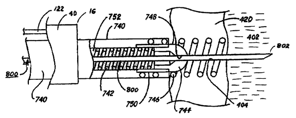
An exemplary elongated fixation catheter 700 is depicted in FIG. 5 that
comprises
a fixation catheter body 740 that extends from a fixation catheter body
proximal end 754
to a fixation catheter distal end 756. The elongated fixation catheter 700 has
a fixation
catheter lumen 752 extending between proximal and distal fixation catheter
lumen
openings at the respective fixation catheter proximal and distal ends 754 and
756. A distal
tissue fixation mechanism, in this instance a fixation helix 746 is mounted to
extend
distally from the fixation catheter distal end 756, terminating in a sharp
tissue penetrating
helix point 758.
The fixation catheter length FCL exceeds the length of the steerable guide
catheter
100. The diameter of the fixation catheter 700 is preferably uniform through
length FCL
and the helix length HL and fits into the delivery lumen 24 of steerable guide
catheter 100
as the steerable guide catheter distal end is advanced to a tissue wall,
particularly the right
atrial wall 420 in the atrial appendage as shown in FIGs. 6 and 7.
In the preferred use of the embodiments of the invention, the steerable
therapeutic
catheter 100 is advanced through a tortuous venous pathway through the
superior vena
cava and into the right atrium, and the guide catheter body distal end 16 is
deflected into
the atrial appendage. The distal fixation helix 746 and a distal segment or
segment of the
fixation catheter body 740 are advanced from the guide catheter delivery lumen
24 to
extend from the guide catheter distal end 16 and screwed into the tissue wall,
particularly
the right atrial wall 420.
To accomplish this, the fixation helix 746 is adapted to be advanced from the
guide
catheter delivery lumen exit port so that helix point 758 penetrates the
endocardium. The
fixation catheter body 740 extends proximally from the guide catheter hub 80
and is
CA 02589362 2007-06-01
WO 2006/060586 PCT/US2005/043489
14
adapted to be grasped near proximal end 754 and rotated to screw the distal
fixation helix
746 into the right atrial wall 420 as shown in FIG. 6. The number of helix
turns and the
helix length HL are selected to provide adequate fixation to the right atrial
wall 420
without perforating through it when the helix 746 is screwed into the right
atrial wall 420.
The distal fixation helix 746 is typically formed of an electrically
conductive
platinum-iridium alloy that is typically employed for chronic fixation of
electrical medical
leads to cardiac tissue. Therefore, fixation catheter 700 can optionally be
formed to be
used as a pacing lead to deliver pacing pulses and sense atrial electrical
activity through
the fixation helix 746. In this variation, a coiled wire conductor 742 shown
in FIG. 6
extends from a distal crimp sleeve 750 attached to the fixation helix 746
proximally within
the fixation catheter lumen 752 to a connector element 760 adapted to inserted
into a
connector bore of an IPG or monitor in a manner well known in the art.
As shown in FIGs. 6 and 7, the delivery lumen exit port at the fixation
catheter
distal end 756 is closed by a molded polymer (e.g., silicone rubber) seal 744.
having a pre-
formed slit 748. Seal 744 is flexible and penetrable by an elongated tissue-
penetrating
instrument 800 that passes through slit 748 as depicted in FIG. 7. The
elongated tissue-
penetrating instrument 800 has a sharp penetrating tip or element 802 that can
be advanced
through the penetrable seat 744 (by widening lit 748), and then advanced
axially, within
the turns of the fixation helix 746, through the right atrial wall 420, and
into the pericardial
space 402. The elongated tissue penetrating instrument 800 is advanced through
the
fixation catheter lumen 752 after the fixation helix is 746 is screwed into
the right atrial
wa11420.
The elongated penetration instrument 800 may simply function to create a
perforation 404 through right atrial wal1420 axially aligned with the fixation
catheter
lumen 752 whereupon the elongated penetration instrument 800 is withdrawn from
the
fixation catheter lumen 752. In this case, the penetration instrument may
comprise a stylet
or a guidewire having a relatively stiff sharp distal tip penetrating element
802 that passes
through the penetrable seal 748 and penetrates or perforates the right atrial
wal1420 as it is
advanced and is then withdrawn. In this instance, one of a therapeutic
catheter, e.g., a gene
delivery catheter or a drug delivery catheter, a drainage or fluid sampling
catheter, and an
electrical medical lead bearing one or more of a sense electrode, a
stimulation electrode,
e.g., a pace/sense electrode or cardioversion/defibrillation electrode, and a
physiologic
CA 02589362 2007-06-01
WO 2006/060586 PCT/US2005/043489
sensor, e.g. a pressure sensor, a temperature sensor or a cardiac motion or
acceleration
sensor, may be advanced through fixation catheter lumen 752, through
penetrable seal 744,
through the perforation 404 and to a desired site or location in the
pericardial space 402
adjacent the left atrium or left ventricle.
5 If penetration instrument 800 is a guidewire, the therapeutic catheter may
be
advanced over the guidewire through fixation catheter lumen 752, through
penetrable seal
748, through the perforation 404 and to a desired site or location in the
pericardial space
402 adjacent the left atrium or left ventricle, and the guidewire may then be
withdrawn.
An electrical medical lead having a through lumen may be advanced over the
guidewire
10 through fixation catheter lumen 752, through penetrable seal 744, through
the perforation
404 and to a desired site or location in the pericardial space 402 adjacent
the left atrium or
left ventricle, and the guidewire may then be withdrawn.
Alternatively, the elongated penetration instrument 800 may comprise one of a
therapeutic catheter, e.g., a gene delivery catheter or a drug delivery
catheter, a drainage or
15 fluid sampling catheter, and an electrical medical lead bearing one or more
of a sense
electrode, a stimulation electrode, e.g., a pace/sense electrode or
cardioversion/defibrillation electrode, and a physiologic sensor, e.g. a
pressure sensor, a
temperature sensor or a cardiac motion or acceleration sensor, all adapted to
be advanced
through fixation catheter lumen 752, through penetrable seal 744, through the
perforation
404 and to a desired site or location in the pericardial space 402 adjacent
the left atrium or
left ventricle.
FIGs. 8 - 10 illustrate various types of elongated medical devices that may be
introduced through the fixation catheter lumen 752 into the pericardial space
402 as
illustrated in FIG. 7 for temporary or chronic use. In each case, the
elongated medical
device includes a device body that is sized to fit into the fixation catheter
lumen and pass
through the penetrable seal 744 and may include a through lumen that enables
advancement over a guidewire that may be employed as the tissue penetrating
instrument
800.
A therapeutic catheter 500 is illustrated in FIG. 8 for delivery of drugs or
withdrawal of fluids from the pericardial space 402. The therapeutic catheter
500 may be
employed as a therapeutic catheter, e.g., a gene delivery catheter or a drug
delivery catheter
or a drainage or fluid sampling catheter. The therapeutic catheter 500
comprises an
CA 02589362 2007-06-01
WO 2006/060586 PCT/US2005/043489
16
elongated therapeutic catheter body 504 extending between a proximal fluid
connector 502
and a therapeutic catheter body distal end 506. A fluid transmitting lumen 508
extends
from a proximal lumen end opening at the fluid connector 502 and one or more
delivery
lumen exit ports at or near the therapeutic catheter body distal end 506.
Fluid transmitting
lumen 508 may function as a through lumen for over the wire advancement of the
therapeutic catheter body 504 over a guidewire if a delivery lumen exit port
is axially
aligned with fluid transmitting lumen.
The fluid connector 502 is shaped and adapted to be coupled to an implantable
drug dispenser for chronic dispensation of drugs or agents from a reservoir of
an IIP into
the pericardial space 402. Alternatively, the fluid connector can be located
outside the
patient's body and attached to an external drug dispenser temporary delivery
of drugs or
therapeutic agents or fluid evacuation device for temporarily sampling or
draining
pericardial fluid from the pericardial space 402
An electrical medical lead 510 is depicted in FIG. 9 for transmission of
electrical
signals from the heart or a physiologic sensor or delivery of electrical
stimulating pulses,
e.g., pacing pulses, to one or more of the left atrium and left ventricle. The
electrical
medical lead 510 may be adapted for chronic implantation to be coupled to a
subcutaneously implanted IPG or IHM or may be extended through the patient's
skin to an
external pulse generator or monitor for temporary use. The electrical medical
lead 510
bears one or more of a sense electrode and a stimulation electrode, e.g., one
or more
pace/sense electrode and/or a physiologic sensor, e.g. a pressure sensor, a
temperature
sensor or a cardiac motion or acceleration sensor.
For example, the electrical medical lead 510 is formed of an elongated lead
body
516 extending between a proximal lead connector comprising a connector ring
512 and a
connector pin 514 and a distal tip pace/sense electrode 524. The proximal lead
connector is
shaped and adapted to be coupled to a subcutaneously implanted IPG or monitor
or can be
located outside the patient's body and attached to an external pulse generator
or monitor
for temporarily pacing or monitoring the heart from the pericardial space 402.
A proximal
ring pace/sense electrode 522 and a physiologic sensor 520, e.g., a pressure
sensor, are
disposed along the elongated lead body 516 proximal to the distal tip
pace/sense electrode
524. Lead conductors extend within lead body between the proximal connector
ring 512
and pin 514 and the pace/sense electrodes 522 and 524 and the physiologic
sensor 520.
CA 02589362 2007-06-01
WO 2006/060586 PCT/US2005/043489
17
The physiologic sensor 520 and the pace/sense electrode 522 may be combined so
that electrical medical lead 510 functions in the manner of the combined
pacing and
pressure sensing lead disclosed in commonly assigned U.S. Patent No.
5,564,434. A lead
lumen 528 extends from a proximal lumen end opening axially through connector
pin 514
through the length of the lead body 516 and either terminates at extends
axially through tip
pace/sense electrode 524 to function as a stylet lumen or a through lumen for
over the wire
advancement of the lead body 516 over a guidewire.
A further electrical medical lead 530 comprising a
cardioversion/defibrillation lead
is depicted in FIG. 10 that would typically be employed with at least one more
cardioversion/defibrillation lead adapted to be disposed about the heart and
coupled to a
cardioversion/defibrillation IPG. The electrical medical lead 530 and the
other
cardioversion/defibrillation lead would typically also include pace/sense
electrodes and
connector elements as described above with respect to electrical medical lead
510 to
enable sensing and processing of heart signals to trigger delivery of
cardioversion/defibrillation shocks or pacing therapies as necessary.
The electrical medical lead 530 is formed of an elongated lead body 536
extending
between a proximal lead connector comprising a connector pin 534 and a lead
body distal
tip 532. The proximal lead connector is shaped and adapted to be coupled to a
subcutaneously implanted cardioversion/defibrillation IPG. An elongated,
relatively large
surface area cardioversion/defibrillation electrode 540 extends along a distal
segment of
the lead body 536 that would be disposed within the pericardial space 402
alongside the
left ventricle to deliver cardioversion/defibrillation shocks through the mass
of the left
ventricle. A lead conductor extends within lead body 536 between the proximal
connector
pin 534 and the cardioversion/defibrillation electrode 540. A lead lumen 538
extends from
a proximal lumen end opening axially through connector pin 534 through the
length of the
lead body 536 and either terminates at extends axially through distal tip 532
to function as
a stylet lumen or a through lumen for over the wire advancement of the lead
body 536 over
a guidewire.
The heart 400 and the surrounding pericardial sac 406 depicted in FIGs. 11 -
14 are
cut away in part to expose the epicardium and the right heart chambers of the
right atrium
(RA) and the right ventricle (RV), which are separated by the tricuspid
valve). Venous
blood drains into the RA through the superior vena cava (SVC) and the inferior
vena cava
CA 02589362 2007-06-01
WO 2006/060586 PCT/US2005/043489
18
(not shown). The RA appendage 408 extends somewhat laterally of the axis of
the RA
between the SVC and tricuspid valve.
In FIG. 11, the steerable guide catheter body 40 of FIG. 3 is advanced into
the RA
through the SVC, and the guide catheter distal end 16 is directed toward the
right atrial
wall 420 by selectively operating the deflection wire 30/110 to induce a bend
in segment
52 as shown in FIG. 1. The fixation catheter 700 is advanced through the guide
catheter
delivery lumen to dispose the distal fixation helix 746 toward the right
atrial wall 420.
As shown in FIG. 12, the distal segment of the fixation catheter 700 is
extended out
of the guide catheter delivery lumen, and the fixation helix 746 is rotated to
screw it into
the atrial wall 420 as shown in greater detail in FIG. 6. In FIG. 13, one of a
tissue
penetration instrument 800, a therapeutic catheter 500, and an electrical
medical lead
510/530 are extended through the fixation catheter lumen, the distal seal, the
atrial wall,
and into the pericardial space 402 in the manner shown in detail in FIG. 7.
The tissue penetration instrument 800 can be removed if it is not necessary to
employ it in the advancement of a distal segment of the therapeutic catheter
500 or the
electrical medical lead 510/530 into the pericardial space 402. If the tissue
penetration
instrument 800 is employed, and if it is or functions as a guidewire, then it
is left in place
so that the one of the therapeutic catheter 500 or the electrical medical lead
510/530 can be
advanced over it. Then, the penetration instrument 800 is retracted leaving
the distal
segment of one of the therapeutic catheter 500 or the electrical medical lead
510/530
within the pericardial space 402.
The steerable guide catheter 100 is removed either before or after the
advancement
of a distal segment of the therapeutic catheter 500 or the electrical medical
lead 510/530
into the pericardial space 402 by retracting it over the fixation catheter
700. The steerable
guide catheter 100 is retracted and removed from over the fixation catheter
700, leaving it
in place with the fixation helix 746 screwed into the right atrial wall 420 as
shown in FIG.
13.
FIG. 14 is a schematic illustration of the coupling of the proximal end of an
electrical medical lead 510 of FIG. 9 or 530 of FIG. 10 with a implantable
cardioverter/defibrillator (ICD) IPG 550 following removal of the steerable
guide catheter
100 enabling implantation of the IPG 550 subcutaneously in the thoracic
region. The
depicted electrical medical lead 510/530 incorporates both an elongated
CA 02589362 2007-06-01
WO 2006/060586 PCT/US2005/043489
19
cardioversion/defibrillation electrode 540 and a distal tip pace/sense
electrode 524. The
cardioversion/defibrillation electrode 540 and a distal tip pace/sense
electrode 524 are
advanced through the lumen and penetrable seal of the fixation catheter 700
and through
the atrial wall 420 and disposed in the pericardial space 402 in the manner
described
above. The ICD IPG 550 is also coupled to a second electrical medical lead 560
that is
transvenously advanced through the SVC and RA to dispose an elongated
cardioversion/defibrillation electrode 562 and a distal tip pace/sense
electrode 564 in the
RV.
The ICD IPG 550 comprises a hermetically sealed enclosure or housing 554 that
encloses electrical circuitry and a battery power source and a connector
header 552 having
connector bores that the lead connector assemblies fit into to couple the lead
electrodes to
the electrical circuitry in a manner well known in the art. The electrical
circuitry may be
coupled to the exterior electrically conductive surface of the housing 554 to
form an
indifferent electrode for pacing and/or cardioversion/defibrillation. The ICD
IPG may
comprise the MEDTRONIC Marquis VR or DR ICD IPG, for example, that senses
electrical heart activity and delivers pacing pulses between the pace/sense
electrodes 524
and 564 and delivers cardioversion/defibrillation shocks between the elongated
cardioversion/defibrillation electrodes 540 and 562 in response to detection
of a
ventricular tachycardias. The ICD IPG 550 may further comprise the MEDTRONIC
InSync ICD Cardiac Resynchronization and ICD System IPG that delivers
synchronized
pacing pulses to the right and left ventricles through the pace/sense
electrodes 564 and
524, respectively.
It will be understood that a bi-ventricular pacemaker IPG may be substituted
for the
ICD IPG 550 and employed with pacing leads 510 of FIG. 9 with pace/sense
electrodes
522 and 524 disposed in pericardial space 402 and one of the RA and RV to
provide multi-
chamber pacing therapies. It will also be understood that the implantable
medical lead
510/530 may further support a physiologic sensor 520 that is coupled with
circuitry within
the ICD IPG 550 or a multi-chamber pacemaker IPG and'employed in the
determination of
a pacing rate or the detection of a tachyarrhythmia.
Elaborate implantable hemodynamic monitors (IHMs) for recording the EGM from
electrodes placed in or about the heart and other physiologic sensor derived
signals, e.g.,
one or more of blood pressure, blood gases, temperature, electrical impedance
of the heart
CA 02589362 2007-06-01
WO 2006/060586 PCT/US2005/043489
and/or chest, and patient activity have been proposed in the prior art. FIG.
15 is a
schematic illustration of the coupling of the proximal end of an electrical
medical lead 510
of FIG. 9 with an IHM 570 following removal of the steerable guide catheter
100 enabling
implantation of the IHM 570 subcutaneously in the thoracic region. The
depicted
5 electrical medical lead 510 supports the physiologic sensor 520 and/or one
or both of the
sense electrodes 522 and 524 on the lead body 528 disposed in the pericardial
space 402.
The physiologic sensor 520 and/or one or both of the sense electrodes 522 and
524
are advanced through the lumen and penetrable seal of the fixation catheter
700 and
through the atrial wall 420 and disposed in the pericardial space 402 in the
manner
10 described above. The IHM 570 comprises a hermetically sealed enclosure or
housing 574
that encloses electrical circuitry and a battery power source and a connector
header 572
having connector bores that the lead connector assemblies fit into to couple
the lead
electrodes and sensor 520 to the electrical circuitry in a manner well known
in the art. The
electrical circuitry may be coupled to the exterior electrically conductive
surface of the
15 housing 554 to form an indifferent electrode for far field EGM sensing.
FIG. 16 is a schematic illustration of the coupling of the proximal end of a
drug
infusion catheter 500 of FIG. 8 with an implantable drug dispenser following
removal of
the steerable guide catheter 100 enabling implantation of the implantable drug
dispenser
subcutaneously in the thoracic region. The therapeutic catheter 500 is
depicted in FIG. 14
20 extending between such an IIP 200 implanted subcutaneously in the thoracic
region of the
body through a venous pathway and through the superior vena cava SVC into the
RA and
through the right atrial wall 420 into the pericardial space 402. The IIP 200
is coupled to
the proximal end of the drug delivery catheter 500 and implanted
subcutaneously in a
thoracic region of the patient's body. The IIP 200 and therapeutic catheter
500 may take
the form of the Medtronic SynchroMed Infusion System. The battery powered
IIP 200
can be advantageously programmed to frequently or continuously deliver drug
boluses of
drugs that have a short duration of activity directly to an efficacious site.
The IIP 200 is
surgically implanted subcutaneously under the skin such that the refill port
210 is directed
outward. The IIP reservoir can be refilled through port 210 accessed
transcutaneously as
necessary. Adverse side effects are reduced and the mental and physical states
of many
patients are improved by the automatically administered drug therapy. It is
not necessary
to rely upon the patient to comply with the prescribed regimen.
CA 02589362 2007-06-01
WO 2006/060586 PCT/US2005/043489
21
It will be understood that structure and functions of both the lIP 200 and an
IPG or
IHM of the types described in reference to FIGs. 14 - 16 may be combined into
a single
implantable medical device.
A variety of deflectable or steerable stylets and guidewires have been
proposed,
and in some cases clinically introduced, to, aid in direct implantation of an
endocardial
cardiac lead having a lead body lumen. One approach has been to employ
deflectable
stylets wherein the stylet distal segment can be deflected or curved while
within the lead
body lumen from the proximal end thereof. Thus, it is conceived that such
steerable
stylets or guidewires can be employed instead of the steerable guide catheter
100 to
advance the fixation catheter 700 through the venous pathway, the SVC the RA
and into
the atrial appendage and to deflect the fixation catheter distal end and
fixation helix 746
toward the atrial wall 420 in the atrial appendage 412.
Use of a two-piece steerable stylet (or guidewire) 900 to accomplish the
advancement and to deflect the fixation catheter distal end and fixation helix
746 toward
the atrial wall 420 is depicted in FIG. 17. The two-piece steerable stylet 900
comprises a
straight, tubular, outer sleeve or member 902 and a curved inner wire 904 or
member
received within the outer member lumen enabling relative movement of the inner
and
outer members as disclosed in U.S. Patent No. 5,728,148 to Bostrom et al, for
example.
The outer member of the '148 patent is relatively straight when unrestrained,
and a
curve can be induced in the inner member. The curvature of the inner member
904
induces a like curvature in the outer member 902 when the inner member 904 is
advanced
distally through the lumen of the outer member 902. Alternatively, a two-piece
stylet 900
comprising a curved outer member 902 and a relatively straight inner member
904 are also
known to the art. In such a two-piece stylet 900, the relative position of the
inner member
904 with respect to the outer member 902 determines the degree to which the
curved
member (inner or outer) is allowed to display its preset curvature. The inner
member 904
can be completely withdrawn from the lumen of the outer member 902 in such a
two-piece
steerable stylet 900.
Further steerable stylets (or guidewires) have been developed or proposed
where
the inner member distal end is attached to the outer member 902 at or near the
outer
member distal end, and the outer member 902 is fabricated to enable selective
deflection
of a distal segment thereof. Such a steerable stylet 900 typically employs an
outer member
CA 02589362 2007-06-01
WO 2006/060586 PCT/US2005/043489
22
902 that is generally straight when unrestrained and the inner member 904
functions as a
deflection wire (also referred to as a traction wire, a pull wire, a push wire
or a push-pull
wire) extending through a lumen of the outer member to an attachment point at
or near the
outer member distal end. The inner member 904 or deflection wire is pushed or
pulled on
at its proximal end typically through a handle that is permanently or
removably attached to
the outer member proximal end. The proximal retraction or distal advancement
of the
inner member 904 or deflection wire causes at least a distal segment of the
outer member
902 to bend or deflect. An example of such a deflection mechanism can be found
in U.S.
Patent Nos. 4,815,478, and 6,146,338, for example, which disclose the use of a
push-pull wire extending through a guidewire lumen for deflecting a guidewire
distal end
by manipulating the push-pull wire at the guidewire proximal end. The '338
patent
discloses a steerable stylet handle at the stylet body proximal end that is
manipulated by
one hand operation to induce a bend in a distal segment of the stylet outer
member 902.
In FIG. 17, the curvature of the steerable stylet outer member 902 of either
of the above-
described types induces a like curvature in the fixation catheter 700 that
deflects the distal
fixation helix 746 into the atrial appendage 412 and toward the atrial wall
420. The
implanting physician can rotate the fixation catheter proximal end to screw
the fixation
helix 746 into the right atrial wall 420 as described above.
Typically, such a two-piece steerable stylet 900 does not have an enlarged
proximal
handle or end or the handle can be removed, and other instruments having
through lumens
can be advanced over the outer member 902 or the inner member 904 if the outer
member
can be removed. Advantageously, referring again to FIG. 7, the inner member
904 or the
outer member 902 (or both) may function as a penetration instrument 800 that
is advanced
more distally within the turns of the fixation helix 746 through the atrial
wall 420. The
inner member 904 or outer member 902 (or both) may function as a guidewire
enabling
advancement of a further electrical medical lead 500/510 or catheter 530 over
the
stylet/guidewire 900 to dispose the distal segment thereof in the pericardial
space 402.
All patents and publications referenced herein are hereby incorporated by
reference in their
entireties.
It will be understood that certain of the above-described structures,
functions and
operations of the above-described preferred embodiments are not necessary to
practice the
CA 02589362 2007-06-01
WO 2006/060586 PCT/US2005/043489
23
present invention and are included in the description simply for completeness
of an
exemplary embodiment or embodiments.
In addition, it will be understood that specifically described structures,
functions
and operations set forth in the above-referenced patents can be practiced in
conjunction
with the present invention, but they are not essential to its practice.
It is to be understood, that within the scope of the appended claims, the
invention may be
practiced otherwise than as specifically described without actually departing
from the spirit
and scope of the present invention. The disclosed embodiments are presented
for purposes
of illustration and not limitation, and the present invention is limited only
by the claims
that follow.