Note: Descriptions are shown in the official language in which they were submitted.
CA 02589569 2014-07-08
MRI BIOPSY DEVICE
Field of the Invention
The present invention relates, in general, to a method of imaging assisted
tissue sampling and,
more particularly, to an improved method for positioning a biopsy probe with
respect to a
magnetic resonance imaging (MRI) breast coil for acquiring subcutaneous
biopsies and
for removing lesions.
Background of the Invention
Core biopsy devices have been combined with imaging technology to better
target a lesion in
breast tissue. One such commercially available product is marketed under the
trademark
name MAMMOTOMETm, by Ethicon Endo-Surgery, Inc. An embodiment of such a
device is described in U.S. Patent No. 5,526,822 issued to Burbank, et al., on
June 18,
1996. Its handle receives mechanical and electrical power as well as vacuum
assist from a
remotely positioned control module that is spaced away from the high magnetic
field of a
Magnetic Resonance Imaging (MRI) machine.
As seen from that reference, the instrument is a type of image-guided,
percutaneous coring,
breast biopsy instrument. It is vacuum-assisted, and some of the steps for
retrieving the
tissue samples have been automated. The physician uses this device to capture
"actively"
(using the vacuum) the tissue prior to severing it from the body. This allows
the sampling
of tissues of varying hardness. In addition, a side opening aperture is used,
avoiding
having to thrust into a lesion, which may tend to push the mass away, cause a
track
metastasis, or cause a hematoma that, with residual contrast agent circulating
therein, may
mimic enhancement in a suspicious lesion. The side aperture may be rotated
about a
longitudinal axis of the probe, thereby allowing multiple tissue samples
without having to
otherwise reposition the probe. These features allow for substantial sampling
of large
lesions and complete removal of small ones.
1
CA 02589569 2014-07-08
Vacuum assisted core biopsy devices have been adapted to be safe and
compatible with
various imaging modalities, including Magnetic Resonance Imaging (MRI). In
particular,
portions of a biopsy system placed near the magnet core of an MRI machine need
to be
nonresponsive to the strong magnetic field to prevent becoming drawn toward
the magnet
core or to malfunction. Further, the MRI machine depends upon sensing
extremely weak
radio frequency (RF) signals emanated by tissue after being excited by a
strong change in
the magnetic field. Components placed in the RF shielded MRI suite need to
avoid
producing electromagnetic interference (EMI) and need to avoid having
materials that
would distort RF signals sufficient to create artifacts in the MRI scan data.
A successful approach has been to segregate motive power generation, graphical
user
interface, vacuum assist, and closed loop control in a control module that has
typically
been placed about 6 feet away from the magnet core to mitigate detrimental
interaction
with its strong magnetic field and/or sensitive radio frequency (RF) signal
detection
antennas. An intuitive graphical user interface (GUI) provides a range of
preprogrammed
functionality incorporated into a control module to efficiently use time in an
MRI suite to
take tissue samples.
As an example, in U.S. Pat. No. 6,752,768, a control button may be depressed
to change a
mode of operation of a core biopsy device with this mode displayed remotely on
a display.
While a full function GUI has numerous clinical benefits, the clinician may
find the control
module inconveniently remote during hands-on portions of the procedure. In
addition,
some MRI machines have such increased sensitivity and/or increased magnet
field
strength that it is desirable to increase the distance of the control monitor
(e.g., 30 feet)
from the MRI machine. Further, even if the control monitor is sufficiently
close, some
clinicians prefer a simplified user interface to simplify training
familiarity.
Consequently, a significant need exists for a biopsy system compatible for use
in an MRI
suite with biopsy controls with enhanced convenience and intuitiveness.
Brief Summary of the Invention
In one aspect, the invention overcomes the above-noted and other deficiencies
of the prior art
by providing a handpiece of a magnetic resonance imaging (MRI) compatible core
biopsy
2
CA 02589569 2015-09-18
,
system that includes a graphical user interface that facilitates user control
even with
vacuum, power generation, and control processing components remotely
positioned away
from the MRI magnet and sensitive radio frequency (RF) receiving components.
Thereby,
a clinician may have the full functionality of vacuum assisted core biopsy
systems yet not
be inconvenienced by the distance from a remotely positioned control module.
In one embodiment there is disclosed a biopsy device comprising: an outer
cannula having a
lateral tissue receiving aperture; an inner tubular cutter disposed for
translation within the
cannula; a sensor responsive to a translation position of the inner tubular
cutter; a body,
wherein the cannula extends distally from the body, wherein the body further
includes a
graphical display attached to the body proximally to the outer cannula
operably
configured to depict the translation position in response to the sensor.
In one embodiment, there is provided a biopsy device comprising: an outer
cannula having a
lateral tissue receiving aperture; an inner tubular cutter disposed for
translation within the
cannula; a sensor responsive to a translation position of the inner tubular
cutter; a body,
wherein the cannula extends distally from the body, wherein the body includes
a graphical
display attached to the body proximally to the outer cannula, wherein the
graphical
display includes a cutter indicator, wherein the cutter indicator comprises a
plurality of
indicators, wherein the plurality of indicators are together operably
configured to depict
the translation position in response to the sensor.
There is also described a surgical biopsy system comprising: a handpiece
including an
elongated, hollow cannula and a cutter rotatably and axially positionable
relative to the
hollow cannula, the hollow cannula having a lateral port for receiving the
tissue sample
into the hollow cannula; a power source remotely positioned from the handpiece
and
operatively coupled via a mechanical drive cable to the cutter for rotating
and translating
the cutter; a control unit operatively associated with the handpiece; and a
display attached
to the handpiece and operatively configured with the control unit wherein the
display
provides a graphical display of an operational mode of the surgical biopsy
system.
These and other objects and advantages of the present invention shall be made
apparent from
the accompanying drawings and the description thereof.
3
CA 02589569 2015-09-18
Brief Description of the Figures
The accompanying drawings, which are incorporated in and constitute a part of
this
specification, illustrate embodiments of the invention, and, together with the
general
description of the invention given above, and the detailed description of the
embodiments
given below, serve to explain the principles of the present invention.
FIGURE 1 is a perspective disassembled view of a Magnetic Resonance Imaging
(MRI)
biopsy system including a handpiece ("biopsy device") having intuitive
graphical controls
consistent with aspects of the invention.
3a
CA 02589569 2007-05-22
FIGURE 2 is an isometric view of a lateral fence and pedestal of a
localization fixture of the
MRI biopsy system of FIG. 1.
FIGURE 3 is an isometric view of a guidance assembly mounted on a right
primary targeting
rail of FIG. 2.
FIGURE 4 is an exploded isometric view of the guidance assembly of FIG. 3 and
the sleeve
trocar and introducer obturator of FIG. 1.
FIGURE 5 is an isometric view of the introducer obturator inserted into the
sleeve trocar of
FIGS. 1 and 4.
FIGURE 6 is an aft right isometric view of the MRI biopsy device of FIG. 1
with a
disposable probe assembly and keypad control disengaged from a reusable
holster
portion.
FIGURE 7 is a fore left isometric view of the MRI biopsy device of FIG. 1 with
the
disposable probe assembly and keypad control disengaged from the reusable
holster
portion.
FIGURE 8 is a fore left exploded isometric view of the reusable holster
portion of FIG. 7.
FIGURE 9 is a top view of the disposable probe assembly of FIG. 7 with an
upper cover
removed to expose interior components of a carriage cavity.
FIGURE 10 is a fore left exploded isometric view of the disposable probe
assembly of FIG.
7.
FIGURE 11 is an aft left isometric view of the localization fixture and
guidance assembly
installed into a breast coil of FIG. 1.
FIGURE 12 is an aft isometric view of the MRI biopsy device of FIG. 7 into the
guidance
assembly of FIG. 11.
FIGURE 13 is a top detail view of a display portion of the MRI biopsy device
of FIG. 7.
FIGURE 14 is an aft right isometric view of the MRI biopsy device,
localization fixture and
breast coil of FIG. 12 with insertion of a marker deploying instrument through
a probe of
the disposable probe assembly.
4
CA 02589569 2014-07-08
Detailed Description of the Invention
An MRI biopsy device advantageously includes is partially disposable for
sterility purposes
with a reusable portion for economy. Inconvenience of mechanical, electrical,
and
pneumatical coupling to a remotely placed control portion, necessitated by a
strong
magnetic field and sensitive RF receiving components of an MRI machine, is
mitigated.
First, proximal detachable intuitive controls and displays on the MRI biopsy
device give
interactive control even after insertion into localizing and guiding
structures. Second,
binding of mechanical coupling to the MRI biopsy device is sensed prior to
equipment
damage or malfunction. Third, mechanical coupling is moved closer to
engagement
points between the MRI biopsy device and guiding structures to reduce torque
loads,
especially those transferred through its distal probe. Fourth, a single
mechanical drive
cable drives a fixed ratio transmission that translates and rotates a cutter
of the distal
probe to realize an effective fixed ratio translation/rotation sampling cut
without the
encumbrance of two mechanical drive cables.
Turning to the Drawings, wherein like numerals denote like components
throughout the
several views, in FIGS. 1-3, a Magnetic Resonance Imaging (MRI) compatible
biopsy
system 10 has a control module 12 that typically is placed outside of a
shielded room
containing an MRI machine (not shown) or at least spaced away to mitigate
detrimental
interaction with its strong magnetic field and/or sensitive radio frequency
(RF) signal
detection antennas. As described in U.S. Pat. No. 6,752,768, a range of
preprogrammed
functionality is incorporated into the control module 12 to assist in taking
these tissue
samples. The control module 12 controls and powers an MRI biopsy device
("handpiece") 14 that is positioned and guided by a localization fixture 16
attached to a
breast coil 18 that is placed upon a gantry (not shown) of the MRI machine.
A cable management spool 20 is placed upon a cable management attachment
saddle 22 that
projects from a side of the control module 12. Wound upon the cable management
spool
20 is a paired electrical cable 24 and mechanical cable 26 which are bundled
into
sheathed cable 27 for communicating control signals and cutter
rotation/advancement
motions respectively. In particular, electrical and mechanical cables 24, 26
each have one
end connected to respective electrical and mechanical ports 28, 30 in the
control module
CA 02589569 2007-05-22
12 and another end connected to a reusable holster portion 32 of the MRI
biopsy device
14. An MRI docking cup 34, which may hold the holster portion 32 when not in
use, is
hooked to the control module 12 by a docking station mounting bracket 36.
An interface lock box 38 mounted to a wall provides a tether 40 to a lockout
port 42 on the
control module 12. The tether 40 is advantageously uniquely terminated and of
short
length to preclude inadvertent positioning of the control module 12 too close
to the MRI
machine. An in-line enclosure 44 may advantageously register the tether 40,
electrical
cable 24 and mechanical cable 26 to their respective ports 42, 28, 30 on the
control
module 12.
Vacuum assist is provided by a first vacuum line 46 that connects between the
control
module 12 and an outlet port 48 of a vacuum canister 50 that catches liquid
and solid
debris. A tubing kit 52 completes the pneumatic communication between the
control
module 12 and the MRI biopsy device 14. In particular, a second vacuum line 54
is
connected to an inlet port 56 of the vacuum canister 50. The second vacuum
line 54
divides into two vacuum lines 58, 60 that are attached to the MRI biopsy
device 14. With
the MRI biopsy device 14 installed in the holster portion 32, the control
module 12
performs a functional check. Saline is manually injected into biopsy device 14
to serve as
a lubricant and to assist in achieving a vacuum seal. The control module 12
actuates a
cutter mechanism (not shown) in the MRI biopsy device 14, monitoring full
travel.
Binding in the mechanical cable 26 or within the biopsy device 14 is monitored
with
reference to motor force exerted to turn the mechanical cable 26 and/or an
amount of
twist in the mechanical cable 26 sensed in comparing rotary speed or position
at each end
of the mechanical cable 26.
Just proximal to a display area 61 on the reusable holster portion 32, a
remote keypad 62,
which is detachable from the reusable holster portion 32, communicates via the
electrical
cable 24 to the control module 12 to enhance clinician control of the MRI
biopsy device
14, especially when controls that would otherwise be on the MRI biopsy device
14 itself
are not readily accessible after insertion into the localization fixture 16
and/or placement
of the control module 12 is inconveniently remote (e.g., 30 feet away). An aft
end
thumbwheel 63 on the reusable holster portion 32 is also readily accessible
after insertion
to rotate the side from which a tissue sample is to be taken.
6
CA 02589569 2007-05-22
Left and right parallel upper guides 64, 66 of a localization framework 68 are
laterally
adjustably received respectively within left and right parallel upper tracks
70, 72 attached
to an under side 74 and to each side of a selected breast aperture 76 formed
in a patient
support platform 78 of the breast coil 18. A base 80 of the breast coil 18 is
connected by
centerline pillars 82 that are attached to the patient support platform 78
between the breast
apertures 76. Also, a pair of outer vertical support pillars 84, 86 on each
side spaced about
a respective breast aperture 76 respectively define a lateral recess 88 within
which the
localization fixture 16 resides.
In FIGS. 1-2, a selected breast is compressed along an inner (medial) side by
a medial plate
90 downwardly received into a medial three-sided frame 92 of the localization
framework
68. The breast is compressed from an outside (lateral) side of the breast by a
lateral fence
94 downwardly received into a lateral three-sided frame 96 of the localization
framework
68, defining an X-Y plane. The X-axis is vertical (sagittal) with respect to a
standing
patient and corresponds to a left to right axis as viewed by a clinician
facing the
externally exposed portion of the localization fixture 16.
Perpendicular to this X-Y plane extending toward the medial side of the breast
is the Z-axis,
which typically corresponds to the orientation and depth of insertion of a
probe 98 of a
disposable probe assembly 100 of the MRI biopsy device 14 or of a sleeve
trocar 102
with inserted introducer obturator 104. For clarity, the term Z-axis may be
used
interchangeably with "axis of penetration", although the latter may or may not
be
orthogonal to the spatial coordinates used to locate an insertion point on the
patient.
Versions of the localization fixture 16 described herein allow a nonorthogonal
axis of
penetration to the X-Y axis to a lesion at a convenient or clinically
beneficial angle. An
origin of the spatial coordinates may be imaging the dents imparted to the
tissue by the
lateral fence 94. Alternatively, a disposable fiducial pointer 106 held by a
fiducial holder
108 is filled with an MRI imagable material (e.g., KY jelly, saline,
gadolinium) and
sealed with a cap 110.
The probe 98, sleeve trocar 102 and fiducial pointer 106 are guided by the
localization fixture
16. With particular reference to FIG. 2, a lateral fence supported pedestal
120 spatially
positions left and right primary targeting rails 121, 122 that in turn guide
the fiducial
pointer 106, the sleeve / trocar 102, or the probe 98 of the biopsy device 14
(FIG. 1). The
7
CA 02589569 2007-05-22
primary targeting rails 121, 122 each include an attachment axle 124 that
receives in
either a left or right side axle hub 125 of a (Y-axis) height yoke 126 that is
vertically
adjustable upon a pedestal main body 128, that in turn is laterally adjustable
upon the
lateral fence 94. Alternatively, a breast coil may enable mounting the
pedestal main body
on the medial plate 90 for accessing medially. The pedestal main body 128
includes a
proximal upright rectangular column 132 with a thinner wall 134 projecting
from its
distal side that flares laterally outward (defining left and right vertical
rectangular slots
136, 138) as part of a bracket 140 with top and bottom hanger arms 144, 146
that slide
laterally respectively on a top track 148 and a proximally open lower track
150 formed in
the lateral fence 94. A lateral (X-axis) adjustment lever 151 may be raised to
lift its distal
end 149 out of engagement with a bottom track 147 formed in the lateral fence
94 as the
lateral adjustment lever 151 is repositioned to the left or right to a desired
location with
reference to a lateral measurement guide 145.
The height yoke 126 is a rectangular cuff interrupted in a mid-portion of a
distal side to form
locking left and right hands 152 respectively which ride vertically in the
left and right
vertical rectangular slots 136, 138. The locking left and right hands 152 have
respective
ridged proximal surfaces (not shown) that are selectively drawn proximally
into locking
engagement by a height locking lever 156 with a ridged surface 158 on a
proximal side of
each vertical rectangular slot 136, 138. Lifting the height locking lever 156
takes the
height yoke 126 out of locking engagement to the pedestal main body 128 as the
height
yoke 126 is vertically repositioned. For height adjustment, the proximal top
surface of the
height yoke 126 serves as a sight 160 to read a height measurement scale 162
presented
on a proximal surface of the height locking lever 156.
The attachment axle 124 allows rotation so that an axis of penetration may
include an upward
or downward trajectory. In the illustrative version, proximal comers of the
height yoke
126 include angle detents 164 (e.g., -15 , 00, +15 ) that are selectable by an
angle lock
lever 166. The primary targeting rail 122 includes a distal detent 167 that
serves as a
home reference for the fiducial holder 108 (FIG. 1).
In FIGS. 3-4, a guidance assembly 200, that may be attached to the lateral
fence supported
pedestal 120 of FIG. 2, includes a cradle 202 whose upper lateral side 202a
flares
upwardly to engage a bottom channel 203 of the primary targeting rail 122. A
lower
8
CA 02589569 2007-05-22
lateral side 202b flares horizontally to provide a holster guide track 204
that underlies the
axis of penetration. To provide additional guidance to the MRI biopsy device
14 (FIG. 1),
a secondary targeting rail 206 includes a lateral channel 208 that is guided
along a
longitudinal guide tab 210 of the primary targeting rail 122. When fully
engaged thereon,
a pawl 212 pivoting under urging of a pawl spring 214 about a vertical pawl
pin 216 in a
lateral window 218 proximally positioned in the secondary targeting rail 206
drops into a
proximal detent 220 proximally positioned on the primary targeting rail 122.
The pawl
spring 214 may maintain the pawl 212 in a neutral position that serves in both
assembly
and later removal of the secondary targeting rail 206 or comprises a pair of
opposing pawl
springs (not shown) for that purpose.
In FIGS. 4-5, the sleeve trocar 102 includes a hollow shaft (or cannula) 223
that is proximally
attached to a cylindrical hub 224 and has a lateral aperture 226 proximate to
an open
distal end 228. The cylindrical hub 224 has an exteriorly presented thumbwheel
230 for
rotating the lateral aperture 226. The cylindrical hub 224 has an interior
recess 232 that
encompasses a duckbill seal 234, wiper seal 236 and a seal retainer 238 to
provide a fluid
seal when the shaft 223 is empty and for sealing to the inserted introducer
obturator 104.
The introducer obturator 104 advantageously incorporates a number of
components with
corresponding features. A hollow shaft 242 includes a fluid lumen 244 that
communicates
between an imageable side notch 246 and a proximal port 248. The hollow shaft
242 is
longitudinally sized to extend when fully engaging a piercing tip 249 out of
the distal end
228 of the sleeve trocar 102. An obturator handle 250 encompasses the proximal
port 248
and includes a locking feature 252, which includes a visible angle indicator
254, that
engages the sleeve thumbwheel 230 to ensure that the imageable side notch 246
is
registered to the lateral aperture 226 in the sleeve trocar 102. An obturator
seal cap 256
may be engaged proximally into the obturator handle 250 to close the fluid
lumen 244.
The obturator seal cap 256 includes a locking or locating feature 258 that
includes a
visible angle indicator 259 that corresponds with the visible angle indicator
254 on the
obturator thumbwheel cap 230. The obturator seal cap 256 may be fashioned from
either
a rigid, soft, or elastomeric material.
Returning to FIGS. 3, 4, the sleeve trocar 102 is guided, during penetration
of tissue, by a
sleeve mount 260 having a sleeve hub 262 that receives the cylindrical hub 224
of the
9
CA 02589569 2007-05-22
sleeve trocar 102. The sleeve mount 260 has a lateral sleeve hub channel 264
that slides
along top and bottom guide flanges 266, 268 of the secondary targeting rail
206, each
having an aligned and recess ridged, ratcheting surface 270 that interacts
with a
respective top and bottom ratcheting feature 272, 274 on respective top and
bottom rail
lock rocker latches 276, 278 that are engaged by respective top and bottom
latch pins
280, 282 in respective sides of the sleeve mount 260. The ratcheting features
272, 274
are proximally ramped such as to allow distal movement. Distal portions of
each rail
lock rocker latches 276, 278 are biased away from the sleeve mount 260 by
respective
rail lock compression springs 284, 286 to bias the ratcheting features 272,
274 into
contact with the ridges surfaces 270 of the guide flanges 266, 268.
Simultaneous
depression of the rail lock rocker latches 276, 278 allow the sleeve mount 260
to be
drawn proximally, withdrawing any sleeve trocar 102 supported therein, until
the sleeve
mount 260 reaches a proximal end of the secondary targeting rail 206,
whereupon the
sleeve mount 260 rotates the pawl 212 clockwise (as viewed from the top) and
is thus
engaged to the secondary targeting rail 206 as the secondary targeting rail
206 is
unlocked from the primary targeting rail 122, causing removal therefrom with
continued
proximal movement.
Before mounting the secondary targeting rail 206 onto the primary targeting
rail 122 in the
first place, the sleeve mount 260 is advantageously adjustably positioned on
the
secondary targeting rail 206 to set a desired depth of penetration. In
particular, a depth
guide 290 is formed by a crescent-shaped depth indicator 292 having a lateral
channel
296 shaped to engage the top and bottom guide flanges 266, 268. Forward ramped
surfaces 298 on the top and bottom of the lateral channel 296 are positioned
to engage
the ridged ratcheting surfaces 270 on the secondary targeting rail 206,
allowing assembly
by inserting the depth indicator 292 from a distal end of the secondary
targeting rail 206.
Frictional engagement thereafter resists further proximal movement and
strongly
opposes any distal movement, especially from a depth lead screw 300 of the
depth guide
290, whose distal end 302 rotates within an outboard hole 304 in the depth
indicator 292
and whose proximal end deflects laterally as a depth actuator lever 305 is
used to rotate
and longitudinally position the depth lead screw 300 therein. A mid portion of
the depth
lead screw 300 is received in a longitudinal through hole 306 formed in the
sleeve mount
260 outboard of its lateral channel 208. For coarse depth adjustment, outer
lead threads
CA 02589569 2007-05-22
307 on the depth lead screw 300 selectively engage the sleeve mount 260 until
top and
bottom coarse adjust buttons 308, 310 are inwardly depressed into the sleeve
mount 260,
compressing respective top and bottom coarse adjust compression springs 312,
314.
Each coarse adjust button 308, 310 includes a respective vertically elongate
aperture
316, 318 whose inward surface presents a worm gear segment 320, 322 to engage
the
outer lead threads 307 on the depth lead screw 300 when urged into engagement
by
relaxed coarse adjust compression screws 312, 314.
Returning to FIG. 3, the thumbwheel 230 is depicted as engaged to the sleeve
hug 262 of the
sleeve mount 260 with other portions of the sleeve trocar 102 omitted.
Application s
consistent with the present invention may include a probe of an MRI biopsy
device that
includes a piercing tip or that otherwise is used without passing through a
hollow shaft
(cannula) 223. As such, the thumbwheel with similar sealing members may be
incorporated into the sleeve mount 260.
In FIGS. 6-7, the MRI biopsy device 14 has the disposable probe assembly 100
depicted
detached from the reusable holster portion 32 and with the remote keypad 62
released
from the reusable holster portion 32. The sheathed cable 27 is joined to an
underside of
the reusable holster portion 32 distal to the aft end thumbwheel 63 to enhance
balance and
support of the reusable holster portion, which in turn may be engaged to the
holster guide
track 204 (FIG. 4) by an I-beam shaped holster rail 324 whose upper surface
326 is
engaged within a bottom channel 328 of a holster base plate 330. A ridged
member 331
upon the holster base plate 330 guides the disposable probe assembly 100
during
engagement. A narrowed upper distal surface 332 of the holster rail 324 also
engages
downward gripping flanges 334 extending downward just proximal to a distal
thumbwheel 336 of the disposable probe assembly 100. An under slung shell 337
is
fastened to the proximal undersurface portion of the holster base plate 330.
The disposable probe assembly 100 also has an undersurface that backwardly
slides into
engagement with the reusable holster portion 32. In particular, a narrowed
proximal end
338 is formed into an upper cover 340 with a distal locking arm 342 separated
from the
upper cover 340 on each side except proximally to present an unlocking button
344 on an
exposed surface 346 of the upper cover 340 that is depressed to disengage a
locking
11
CA 02589569 2007-05-22
surface 348 (FIG. 6) from a distal lip 350 of a distally open receiving
aperture 352 in the
reusable holster portion 32 of the holster plate 330.
A recessed deck 354 in an upper proximal surface of a proximal top cover 356
of the reusable
holster portion 32 is shaped to receive the remote keypad 62. A lower shell
358 mates to
the proximal top cover 356. The proximal top cover 356 also defines the upper
portion of
the receiving aperture 352. The recessed deck 354 has a front guide hole 360
and a back
locking aperture 362 registered to respectively receive a front tooth 363 and
a flexing
unlock tab 364 at an aft end of the remote keypad 62 to selectively engage and
disengage
the keypad 62 from the reusable holster portion 32. The keypad 62 also
includes a
translation rocker button 366 that has a distal advance, a default neutral,
and an aft retr' act
command position. An aft button 368 may be programmed for mode functions such
as
saline flush.
With particular reference to FIG. 6, the disposable probe assembly 100 has a
plurality of
interconnections presented on an aft docking end 370. A rightward canted
vacuum hose
nib 372 is positioned to receive a vacuum conduit (not shown) that would be
gripped by a
friction clip 373 extending under and aft thereof to prevent inadvertent
release. A right
side slot 374 is distally open and formed between the holster base plate 330
and proximal
top cover 356 to receive such a vacuum conduit as the disposable probe
assembly 100 is
engaged to the reusable holster portion 32. A center splined driveshaft 375
engages the aft
end thumbwheel 63 and communicates with the distal thumbwheel 336 to rotate a
side
aperture 376 in probe 98 to a desired side, as visually confirmed by an arrow
indicator
378 on the distal thumbwheel 336. A right splined driveshaft 380 effects
cutter translation
and a left splined driveshaft 382 effects cutter rotation.
The distal thumbwheel 336 and probe 98 are mounted to a cylindrical hub 384,
which is a
distal portion of the lower shell 358 that extends beyond the mating with the
upper cover
340. A sample through hole 386 communicates through the cylindrical hub 384
for
receiving a rotating and translating cutter tube 388 (FIG. 9) that enters the
probe 98 and
for receiving tissue samples (not shown) deposited by a retracting cutter tube
388. As the
cutter tube 388 fully retracts into a carriage cavity 390 formed between the
upper cover
340 and proximal portion of the lower shell 358, a distally extending tip 392
from a
vacuum tube 394 encompassed by the cutter tube 388 dislodges the retracted
tissue
12
CA 02589569 2007-05-22
sample onto a sample retrieval platform 396, which is a relieved area between
the upper
cover 340 and the cylindrical hub 384.
In FIG. 8, it should be appreciated that the sheathed cable 27 connects to the
holster base
plate 330 and communicates a single mechanical drive rotation to a fixed ratio
transmission 398 mounted to the holster base plate 330 and electrically
communicates
with an encoder 400 coupled to the fixed ratio transmission 398 aft of the
receiving
aperture 352. The sheathed cable 27 also communicates electrically with the
display area
61 via a wire bundle (not shown) and with the keypad 62 via a cable assembly
402, the
latter including a strain relief bracket 404 that grips a keypad cable 406 and
is fastened
proximate to the sheathed cable 27. The fixed ratio transmission 398 has a
pass-through
port 408 that receives a distal end the center splined driveshaft 375 (FIG. 6)
to rotatingly
engage a proximally received beveled shaft 410 distally presented by the aft
end
thumbwheel 63 and sealed by an 0-ring 412. A right port 414 distally presented
by the
fixed ratio transmission 398 engages for rotation the right splined driveshaft
380 from the
disposable probe assembly 100 for advancing and retracting ("translation") the
cutter tube
388. A left port 416 distally presented by the fixed ratio transmission 398
engages for
rotation the left splined driveshaft 382 from the disposable probe assembly
100 for
rotating the cutter tube 388 when a distal cutting edge of the cutter tube 388
slides past
the side aperture 376 of the probe 98.
In FIGS. 9-10, the carriage cavity 390 of the disposable probe assembly 100
includes a cutter
carriage 418 having a threaded longitudinal bore 420 that encompasses an
elongate
translation shaft 422 whose proximal termination is the right splined
driveshaft 380
supported by an aft right cylindrical bearing 424 received in an aft wall 425
of the lower
shell 358. A race about the outer circumference of the cylindrical bearing 424
receives an
0-ring 426. A distal end 428 of the threaded translation shaft 422 rotates
within a distal
right cylindrical bearing 430 engaged to a forward wall 432 of the lower shell
358. A race
about the outer circumference of the cylindrical bearing 430 receives an 0-
ring 434. A
threaded central portion 436 of the elongate translation shaft 422 resides
between an
unthreaded distal over-run portion 438 and an unthreaded proximal over-run
portion 440,
both sized to allow the threaded longitudinal bore 420 of the cutter carriage
418 to
disengage from the threaded central portion 436.
13
CA 02589569 2007-05-22
A distal compression spring 442 and a proximal compression spring 444
respectively reside
on the unthreaded distal and proximal over-run portions 438, 440 to urge the
threaded
longitudinal bore 420 of the cutter carriage 418 back into engagement with the
threaded
central portion 436 upon reversal of rotation of the elongate translation
shaft 422. In
particular, the cutter carriage 418 includes a top longitudinal channel 446
that slidingly
engages an undersurface of the upper cover 340 (not shown) and a bottom
longitudinal
guide 448 that engages a longitudinal track 450 on a top surface of the lower
shell 358.
Thus rotationally constrained, rotation of the elongate translation shaft 422
causes
corresponding longitudinal translation of the cutter carriage 418 with distal
and aft pairs
of gripping flanges 452, 454 maintained laterally to the left to engage
respectively distal
and proximal races 456, 458 formed on each side of a toothed portion 460 of a
cutter spur
gear 462, which has a longitudinal bore for applying vacuum.
To that end, the vacuum hose nib 372 is attached to a mounting structure 464
that is gripped
between the upper cover 340 and the lower shell 358 to present an orifice 466
within the
carriage cavity 390 that is aligned with the longitudinal bore of the cutter
gear 462 and
that is in fluid communication with the vacuum hose nib 372.
With particular reference to FIG. 10, the proximal end of the vacuum tube 394
is received in
the orifice 466. A rectangular guide 467 supports the distally extending tip
392 of the
vacuum tube 394 and is engaged between the upper cover 340 and the lower shell
358.
The cutter tube 388 encompasses and translates relative to the vacuum tube
394. A seal
cap 468 attached to a proximal end of the cutter gear 462 dynamically seals to
the outer
circumference of the vacuum tube 394 so that vacuum pressure supplied
proximate to the
distally extending tip 392 is not released within the carriage cavity 390. The
cutter tube
388 is advanced around the open distal end of the vacuum tube 394, across the
sample
retrieval platform 396 to seal against a back seal 470 that substantially
closes a proximal
opening 472 into a sleeve union 474 that rotates within the cylindrical hub
384. The
sleeve union 474 has a distal end 476 engaged for rotation with the distal
thumbwheel
336. Distal and proximal 0-rings 478, 480 reside respectively within distal
and proximal
races 482, 484 that straddle a lateral passage 486 of the sleeve union 474 to
provide a
degree of frictional resistance against inadvertent rotation and
advantageously seal the
lateral passage 486 for vacuum assistance to prolapse tissue and to retract
samples. A
noncircular opening 488 is centered in a distal face of the distal thumbwheel
336. A
14
CA 02589569 2007-05-22
proximal end of a probe tube 490 of the probe 98 extends through the
noncircular opening
488 to receive a distal end of the cutter tube 388. A lateral tube 492
attached along its
length to the probe tube 490 communicates with the lateral passage 486 of the
union
sleeve 474. The lateral tube 492 defines a lateral lumen that communicates
with the a
cutter lumen defined by the probe tube 490 / cutter tube 388 below the side
aperture 376
through lumen holes 494 (FIG. 9).
The center splined driveshaft 375 that is turned by the aft end thumbwheel 63
rotates in turn a
shaft 496 whose keyed distal end 498 in turn is engaged to and rotates a
pinion gear 500
that is in gear engagement to a proximal spur gear 502 that forms an outer
proximal
circumference of the sleeve union 474. A cylindrical distal tip 504 of the
keyed distal end
498 rotates within an axle hole (not shown) in the lower shell 358. Rotation
of the aft end
thumbwheel 63 thus rotates the probe 98.
A distal elbow pneumatic fitting 506 is supported in the lower shell 358 to
have an upper end
508 communicating with the lateral passage 486 of the sleeve union 474 and an
aft end
510 attached to a vent pneumatic conduit 512 supported by the lower shell 358.
The other
end of the vent pneumatic conduit 512 is attached to a distal end 514 of a
proximal elbow
pneumatic fitting 516 whose lateral end 518 is open to atmosphere. Sizing of
various
components that vent atmospheric pressure through the lumen holes 494 from the
lateral
end 518 are such that a tissue sample may be withdrawn through the probe tube
490. Yet
a greater pneumatic draw of air through the vacuum hose nib 372 prior to
severing a
tissue sample results in a sufficient low pressure at the side aperture 376 to
prolapse tissue
for severing.
An elongate rotation shaft 520 proximally terminates in the left splined
driveshaft 382 that is
supported for rotation by a left aft cylindrical bearing 522 having a race
about an outer
circumference that receives an 0-ring 524 and is received in the aft wall 425
of the lower
shell 358. A distal end 526 of the elongate rotation shaft 520 is received for
rotation in a
left distal cylindrical bearing 528 having a race about an outer circumference
that receives
an 0-ring 530 and that is received within the front wall 425 of the lower
shell 358. As the
cutter carriage 418 advances to position the cutter tube 388 to slide past the
side aperture
376, the cutter spur gear 460 engages a spur gear portion 532 of the elongate
rotation
shaft 520. Rotating the cutter tube 388 in proportion to an amount of rotation
advantages
CA 02589569 2007-05-22
secures an effective severing of tissue. Eliminating rotation when not
severing
advantageously enhances retraction of tissue sample retraction.
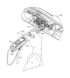
In use, in FIG. 11, the localization fixture 16 has been installed into the
breast coil 18. The
guidance assembly 200 has been preset for a desired insertion point, a desired
axis of
penetration, and a depth of penetration. After the sleeve trocar 102 /
introducer obturator
104 have been inserted and imaged to confirm placement, the introducer
obturator 104 is
removed and the probe 98 of the biopsy device 14 is inserted, as depicted in
FIG. 12. The
shape of the sleeve trocar 102 aligns the probe 98, visually assisted by
lining up the arrow
indicator 378 on the distal thumbwheel 336 with the visible angle indicator on
the
thumbwheel 230 of the sleeve trocar 102. The surgeon may effect operation of
the biopsy
device 14 by depressing the translation rocker button 366 and aft button 368
on the
keypad 62 while referencing status information about the biopsy device 14 on
the display
area 61. In FIG. 13, the display area 61 advantageously includes a cutter
position bar
graph 534 having distal and proximal indications 536, 538 that may be compared
with
how many light segments 540 have been illuminated to indicate progress of the
cutter
tube 388 relative to the side aperture 376. The aft button 368 may be toggled
to cycle the
biopsy device 14 through three modes, indicated by a position LED indicator
542, a
sample LED indicator 544, and a clear LED indicator 546 with a corresponding
label that
graphically depicts operation of the biopsy device in that mode. In
particular, a position
mode depiction 548 illustrates that the cutter tube 388 may be advanced and
retracted, for
instance, closing the side aperture 376 prior to insertion of the probe 98
into the sleeve
trocar 102. In a sample mode depiction 550, vacuum assistance is implemented,
drawing
sufficient air through the cutter tube 388 to prolapse tissue into the open
side aperture 376
that is maintained while translating the cutter tube 388. In a clear mode
depiction 552,
vacuum is maintained while fully retracting the cutter tube 388 to retract a
tissue sample.
In FIG. 14, a marker device 548 is deployed through the sample through hole
386 in the
cylindrical hub 388.
While the present invention has been illustrated by description of several
embodiments and
while the illustrative embodiments have been described in considerable detail,
it is not the
intention of the applicant to restrict or in any way limit the scope of the
appended claims
to such detail. Additional advantages and modifications may readily appear to
those
skilled in the art.
16
CA 02589569 2007-05-22
For example, while closed loop feedback sensing of a component that is related
to cutter tube
position has various advantages, determination of cutter position may be
achieved in
other ways consistent with the present invention. For instance, loading on
drive
components may be sensed at either full advancement and/or full retraction
which are
used to calibrate an estimate cutter position based on duration of a
translation command.
As another example, rather than discrete LED indicators and labeled
depictions, applications
consistent with aspects of the invention may include a graphical display
(e.g., organic
liquid crystal display) that is capable of interactive presentations of
intuitive instrument
status information. Alternatively or in addition, a touch screen capability
may be
incorporated to allow instrument control input as well as display.
For another example, applications consistent with aspects of the present
invention may be
used in conjunction with different diagnostic imaging modalities (e.g.,
ultrasonic,
computed tomography (CT).
17