Note: Descriptions are shown in the official language in which they were submitted.
CA 02590175 2007-06-08
WO 2006/063206 PCT/US2005/044564
1
SYSTEM AND METHOD FOR ANCHORING SUTURE TO BONE
BACKGROUND OF THE INVENTION
1. The Field of the Invention
[0001] The present invention relates generally to devices for anchoring soft
tissue to
bone, and more precisely, to devices that secure suture to the bone and soft
tissue.
2. The Relevant Technology
[0002] There are a number of surgical procedures in which it is necessary to
draw soft
tissue and bone together. One such procedure is rotator cuff repair, in which
the connective
tissue of the displaced rotator cuff is to be drawn against the bone of the
shoulder until the
joint is able to heal properly.
[0003] A variety of anchoring systems are presently used to carry out such
procedures.
Unfortunately, many known systems are somewhat unwieldy, unreliable, or
difficult to
implant in the patient. Some such systems require the surgeon to take some
type of action to
lock the suture at its desired length. Thus, the surgeon must keep the suture
at the desired
length while carrying out the locking operation. If the suture length changes
during locking,
the surgeon may need to perform additional steps to adjust the length of the
suture.
Furthermore, many such systems have excessive components that must be
implanted into the
body.
[0004] Accordingly, a need exists for systems and methods for attaching soft
tissue to
bone that remedy the shortcomings of the prior art. More particularly, there
is a need for a
system that is compact, has relatively few components, and is easily lockable
during surgery.
BRIEF DESCRIPTION OF THE DR.AWINGS
[0005] Various embodiments of the present invention will now be discussed with
reference to the appended drawings. It is appreciated that these drawings
depict only typical
embodiments of the invention and are therefore not to be considered limiting
of its scope.
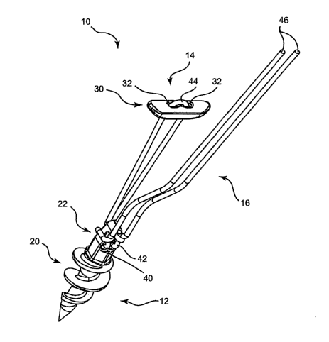
[0006] Figure 1 is a perspective view of an attachment system according to one
embodiment of the invention, with an anchor, a tissue retainer, and a suture
threaded through
the anchor and the tissue retainer.
[0007]. Figure 2 is a perspective view of the anchor of Figure 1, in
isolation.
CA 02590175 2007-06-08
WO 2006/063206 PCT/US2005/044564
2
[0008] Figure 2 is a perspective view of the anchor of Figure 1, in isolation.
[0009] Figure 3 is a perspective view of the anchor of Figure 1, with the
suture threaded
loosely through the passageways of the suture retention portion of the anchor.
[0010] Figure 4 is a perspective view of the anchor of Figure 1, with the
suture threaded
tightly through the passageways of the suture retention portion of the anchor.
[0011] Figure 5 is a perspective view of an anchor of an attachment system of
one
alternative embodiment of the invention.
[0012] Figure 6 is a perspective view of the anchor of Figure 5 with the
suture threaded
loosely through the passageways of the suture retention portion of the anchor.
[0013] Figure 7 is a perspective view of the anchor of Figure 5 with the
suture threaded
tightly through the passageways of the suture retention portion of the anchor.
[0014] Figure 8 is a perspective view of an anchor of an attachment system of
another
alternative embodiment of the invention.
[0015] Figure 9 is a perspective view of the anchor of Figure 8 with the
suture threaded
loosely through the passageways of the suture retention portion of the anchor.
[0016] Figure 10 is a perspective view of the anchor of Figure 8 with the
suture threaded
tightly through the passageways of the suture retention portion of the anchor.
[0017] Figure 11 is a perspective view of an attachment system according to
another
embodiment of the invention.
[0018] Figure 12 is a front elevation, section view of the attachment system
of claim 1,
with the anchor in the process of being implanted into the bone of a shoulder.
[0019] Figure 13 is a front elevation, section view of the attachment system
of claim 1,
with the anchor implanted in the bone of the shoulder and the tissue retainer
in the process of
being inserted through an opening in the rotator cuff.
[0020] Figure 14 is a front elevation, section view of the attachment system
of claim 1,
with the anchor and the tissue retainer in place.
[0021] Figure 15 is a front elevation, section view of the attachment system
of claim 1,
with the anchor and the tissue retainer in place, and with the suture drawn
taught to attach the
rotator cuff to the bone.
DETAILED DESCRIPTION OF THE PREFERRED EMBODIMENTS
[0022] The present invention relates to systems that can be used to draw a
bone and a
portion of soft tissue together. By increasing the size of the line locks, it
is also appreciated
that the line locks can be used outside of surgical procedures for any use
where it is desired to
CA 02590175 2007-06-08
WO 2006/063206 PCT/US2005/044564
3
selectively tighten a line such as a rope, cord, string, or other conventional
type of line that
extends between two objects, or to bring the two objects closer together.
[0023] In this application, the term "attach" is broadly interpreted to
include securement
of separate elements to each other, and the integral formation of separate
elements with each
other. Thus, two portions of an object that are unitarily formed in a single
operation may be
said to be "attached" together. The term "symmetry," without modification,
includes any
known type of symmetry, including mirror symmetry across a plane and radial
symmetry
about an axis.
[0024] The term "direction," when used in connection with motion of a flexible
member
such as a line, does not necessarily refer to a static vector. Rather,
a"direction" may refer to
motion of the line along a pathway, toward one specified end of the pathway.
Thus, stating
that a line is only able to move along a pathway in one direction means that
the line can only
be advanced toward one end of the pathway. The line moves along the pathway in
one
direction even though in the course of advancement along the pathway, segments
of the line
will simultaneously be moving along a variety of differently-oriented vectors.
[0025] A "long axis" refers to an axis of symmetry or extension along which an
object
has a length that is substantially its largest dimension. The term "retain"
refers to limiting
relative motion between two objects in some manner. The term "locking" refers
to fixation
of the relative positions of two objects in such a manner that relative
translation or rotation
along or about at least one axis is substantially prevented along at least one
direction until the
objects have been unlocked.
[0026] Referring to Figure 1, a perspective view illustrates a system 10
according to one
embodiment of the invention. The system 10 may be used to attach soft tissue
(not shown in
Figure 1) to bone (also not shown in Figure 1). According to one example, the
system 10
may be used to attach a torn rotator cuff to the bone of the shoulder to
promote proper healing
of the shoulder joint.
[0027] In the embodiment of Figure 1, the system 10 includes an anchor 12, a
tissue
retainer 14, and a suture 16 that couples the tissue retainer 14 to the anchor
12. The anchor
12 is designed to be implanted in bone, and the tissue retainer 14 is designed
to be inserted
through an opening in the tissue and then drawn toward the anchor 12 by the
suture 16 to
draw the soft tissue toward the bone.
[0028] As shown, the anchor 12 includes a bone retention portion 20 and a
suture
retention portion 22. The bone retention portion 20 is designed to be embedded
into the bone
in such a manner that the bone retention portion 20 securely fastens the
anchor 12 to the
CA 02590175 2007-06-08
WO 2006/063206 PCT/US2005/044564
4
bone. The suture retention portion 22 is designed to retain a portion of the
suture 16 in such a
manner that, when the suture 16 is under tension, the suture 16 can only be
drawn through the
suture retention portion 22 in a manner that brings the tissue retainer 14
closer to the anchor
12. The configuration and operation of the bone retention portion 20 and the
suture retention
portion 22 will be shown and described in greater detail subsequently.
[0029] The tissue retainer 14 has a body 30 with a relatively elongated shape
designed to
be insertable through a relatively small opening in the soft tissue. The
elongated shape is
further designed to abut the tissue on the opposite side of the small opening
in such a manner
that, in response to tension on the suture 16, the tissue retainer 14 is able
to draw the soft
tissue toward the anchor 12. The body 30 bounds two passageways 32, which may
be sized
and arranged to permit relatively free passage of the suture 16 therethrough
along either
direction. The present invention is not limited to the tissue retainer
configuration illustrated
in Figure 1; rather, a wide variety of shapes and sizes may be used.
[0030] The suture 16 has a first anchor portion 40 and a second anchor portion
42, both
of which pass through the suture retention portion 22 of the anchor 12. The
suture 16 further
has a retainer portion 44 that passes through the passageways 32 of the tissue
retainer 14.
Additionally, the suture 16 has two working ends 46 that are available to be
drawn by the
surgeon to induce motion of the first and second anchor portions 40, 42
through the suture
retention portion 22, thereby drawing the tissue retainer 14 toward the anchor
12.
[0031] Referring to Figure 2, a perspective view illustrates the anchor 12 of
the system 10
of Figure 1 in isolation. As illustrated, the bone retention portion 20 has a
plurality of threads
50 that extend outward to engage the bone. The bone retention portion 20 also
has a
sharpened end 52. The anchor 12 has a long axis 54 about which the threads 50
extend along
a generally helical, tapered path. The threads 50 may be "self-tapping," or
shaped to form
their own canal in the bone in response to torque and pressure of the
sharpened end 52
against the surface of the bone. Alternatively, the threads 50 may be shaped
to rotate into
engagement with a pre-formed and/or pre-tapped aperture formed in the bone.
[0032] As yet another alternative, an anchor according to the invention may be
retained in
bone via other mechanisms. For example, an anchor according to the invention
may be
configured as a "tack" with a sharpened end that penetrates the bone
sufficiently for
retention. Such an anchor may optionally be driven into the bone at an angle,
or driven into
the bone and subsequently rotated, to enhance retention. Alternatively, such
an anchor may
be barbed or may have fold-out wings or other structures designed to block
withdrawal of the
CA 02590175 2007-06-08
WO 2006/063206 PCT/US2005/044564
anchor from the bone. Any bone retention structure known in the art may be
used in
combination with the other inventive features disclosed herein.
[0033] The anchor 12 is substantially rigid, and therefore does not depend
upon flexion of
any part of the anchor 12 to enable retention of the anchor 12 in the bone.
The anchor 12
may be formed of a biocompatible metal such as titanium. Alternatively, a
bioabsorbable
material or a nonbioabsorbable polymer may be used to form the anchor 12.
[0034] In the embodiment of Figure 2, the suture retention portion 22 has a
hexagonal
collar 60 designed to be insertable into a hexagonal bore of a driver (not
shown) such that the
driver is able to impart torque as well as axial pressure to the anchor 12.
The suture retention
portion 22 bounds a primary passageway 62, a first secondary passageway 64,
and a second
secondary passageway 66. A first notch 68 and a second notch 70 extend outward
from
opposite sides of the primary passageway 62. The first and second notches 68,
70 extend at
right angles to the primary passageway 62, and are therefore oriented
generally perpendicular
to the primary passageway 62. The notches 68, 70 cooperate with the
passageways 62, 64, 66
to lock the suture 16 against motion through the suture retention portion 22
along one
direction, as will be further explained in connection with Figures 3 and 4.
[0035] Referring to Figure 3, a perspective view illustrates the anchor 12
with a portion
of the suture 16. As shown, the first and second anchor portions 40, 42 of the
suture 16 are
inserted relatively loosely through the passageways 62, 64, 66 of the suture
retention portion
22. From the tissue retainer 14 (not shown in Figure 3), the first and second
anchor portions
40, 42 pass through the primary passageway 62, and then extend outward
generally parallel to
the first and second notches 68, 70, respectively. The first and second anchor
portions 40, 42
define first and second compression sections 80, 82 as they pass over the
first and second
notches 68, 70, respectively.
[0036] The first and second anchor portions 40, 42 then pass around the sides
of the
suture retention portion 22, and then through the first and second secondary
passageways 64,
66, respectively. From the first and second secondary passageways 64, 66, the
first and
second anchor portions 40, 42 are routed toward the primary passageway 62. The
first and
second anchor portions 40, 42 then extend between the first and second
compression sections
80, 82 and the first and second notches 68, 70, respectively. First and second
compressed
sections 84, 86 are thereby defined in the first and second anchor portions
40, 42, at the
locations where the first and second anchor portions 40, 42 extend underneath
the first and
second compression sections 80, 82, respectively.
CA 02590175 2007-06-08
WO 2006/063206 PCT/US2005/044564
6
[0037] Referring to Figure 4, a perspective view illustrates the anchor 12
with a portion
of the suture 16 threaded relatively tightly through the passageways 62, 64,
66 of the suture
retention portion 22. Substantially all of the slack has been removed from the
first and
second anchor portions 40, 42. Consequently, the first and second compressed
sections 84,
86 extend through the first and second notches 68, 70, respectively, and are
held against the
notches 68, 70 by the first and second compression sections 80, 82,
respectively.
[0038] As the compressed sections 84, 86 conform to the shapes of the notches
68, 70,
respectively, the notches 68, 70 serve to create additional bends in the
corresponding
compressed sections 84, 86 to enhance retention of the anchor portions 40, 42
by the anchor
12. Such bends enhance locking of the compressed sections 84, 86 because there
is greater
friction keeping the compressed sections 84, 86 in place. Furthermore, there
is no direct path
along which tension on the working portions 46 can act to draw the compressed
sections 84,
86 through the space between the compression sections 80, 82 and the notches
68, 70,
respectively.
[0039] The result of the manner in which the suture 16 is routed through the
passageways
62, 64, 66 is that the first and second anchor portions 40, 42 can be drawn
through the
passageways 62, 64, 66 in a manner that brings the tissue retainer 14 closer
to the anchor 12,
but not in a manner that permits the tissue retainer 14 and the anchor 12 to
move apart.
Tension tending to pull the tissue retainer 14 away from the anchor 12
increases the
magnitude of the force by which the compressed sections 84, 86 are pressed
into the notches
68, 70 by the compression sections 80, 82, respectively.
[0040] Conversely, tension on the working portions 46 of the suture 16 tends
to pull the
compressed sections 84, 86 free of the notches 68, 70, respectively, to permit
motion of the
anchor portions 40, 42 through the passageways 62, 64, 66 to draw the tissue
retainer 14
closer to the anchor 12. When the tension on the working portions 46 abates,
the compressed
sections 80, 82 are again pressed into the notches 68, 70 due to tension in
the portion of the
suture 16 between the anchor 12 and the tissue retainer 14.
[0041] The first and second anchor portions 40, 42 can also be independently
drawn
through the passageways 62, 64, 66. More precisely, a surgeon can draw only
the working
portion 46 adjacent to the first anchor portion 40 to pull the first
compressed section 84 free
of the first notch 68, thereby permitting the first anchor portion 40 to
advance through the
primary passageway 62 and the first secondary passageway 64 along a direction
that draws
the tissue retainer 14 closer to the anchor 12. Similarly, a surgeon can draw
only the working
portion 46 adjacent to the second anchor portion 42 to pull the second
compressed section 86
CA 02590175 2007-06-08
WO 2006/063206 PCT/US2005/044564
7
free of the second notch 70, thereby permitting the second anchor portion 42
to advance
through the primary passageway 62 and the second secondary passageway 66 along
a
direction that draws the tissue retainer 14 closer to the anchor 12.
[0042] The two portions of the suture 16 that extend between the tissue
retainer 14
(shown in Figure 1) and the anchor 12 remain substantially the same length
because suture is
able to pass relatively freely from one portion to the other through the
passageways 32 of the
tissue retainer 14. Drawing only one of the working portions 46 provides a
mechanical
advantage that moves the tissue retainer 14 only half as fast as drawing both
working portions
46, thereby facilitating fine-tuning of the position of the soft tissue and
the level of tension in
the suture 16. Furthermore, the ability to draw only one of the working
portions 46 provides
the surgeon with additional operating flexibility.
[0043] Referring to Figure 5, a perspective view illustrates an anchor 112
according to
one alternative embodiment of the invention. As shown, the anchor 112 has a
bone retention
portion 20 and a suture retention portion 122. The bone retention portion 20
may be
substantially identical to that of the anchor 12 of Figures 1 through 4.
However, the tissue
retention portion 122 is configured differently from the tissue retention
portion 22 of the
previous embodiment.
[0044] More precisely, the suture retention portion 122 has a hexagonal collar
60 like that
of the previous embodiment. However, in place of the passageways 62, 64, 66,
the suture
retention portion 122 has a primary passageway 162 and a secondary passageway
164. The
suture retention portion 122 also has a notch 168 that extends generally
perpendicular to the
primary passageway 162 in a manner similar to that of the second notch 70 of
the previous
embodiment. The anchor 112 is thus designed to receive and lock only one
suture portion in
a manner that will be shown and described in connection with Figure 6.
[0045] Referring to Figure 6, a perspective view illustrates a system 110 that
includes the
anchor 112 as well as a tissue retainer 114 and a suture 116. Like the tissue
retainer 14, the
tissue retainer 114 has a body 130 that bounds a passageway (not shown)
through which the
suture 116 passes. The suture 116 may be similar to that of the suture 16 of
the previous
embodiment.
[0046] In the embodiment of Figure 6, the suture 116 has an anchor portion 140
that is
received by the suture retention portion 122 of the anchor 112. Additionally,
the suture 116
has a retainer portion 144 that is secured to the tissue retainer 114 and a
working end 46 that
can be manipulated by the surgeon to draw the tissue retainer 114 toward the
anchor 112.
The retainer portion 144 may be secured to the tissue retainer 114 via a knot
148. According
CA 02590175 2007-06-08
WO 2006/063206 PCT/US2005/044564
8
to alternative embodiments, bonding, insert molding, application of rigid
fasteners, or the like
may be used in place of the knot 148 to secure the retainer portion 144 to the
tissue retainer
114.
[0047] From the tissue retainer 114, the anchor portion 140 of the suture 116
passes
through the primary passageway 162, and then extends along the notch 168 to
define a'
compression section 180 of the anchor portion 140. The anchor portion 140 then
extends
around the suture retention portion 122 and through the secondary passageway
164. From
the secondary passageway 164, the anchor portion 140 extends between the
compression
section 180 and the notch 168 to define a compressed section 184 that will be
pressed into the
notch 168 by the compression section 180 when the suture 116 is tensioned.
[0048] Referring to Figure 7, a perspective view illustrates the system 110 of
Figure 6,
with the suture 116 routed relatively tightly through the suture retention
portion 122 of the
anchor 112. As in the previous embodiment, the compressed section 184 conforms
to the
shape of the notch 168, and the notch 168 thereby serves to create additional
bends in the
compressed section 184 to enhance retention of the anchor portion 140 by the
anchor 112.
[0049] Like the suture retention portion 22 of the previous embodiment, the
suture
retention portion 122 of the anchor 112 retains the anchor portion 140 in a
manner that
permits motion of the anchor portion 140 through the passageways 162, 164
along only one
direction. More precisely, tension tending to pull the tissue retainer 114
away from the
anchor 112 increases the magnitude of the force by which the compressed
section 184 is
pressed into the notch 168 by the compression section 180. Conversely, tension
on the
working portion 146 of the suture 116 tends to pull the compressed section 184
free of the
notch 168 to permit motion of the anchor portion 140 through the passageways
162, 164 to
draw the tissue retainer 114 closer to the anchor 112. When the tension on the
working
portion 146 abates, the compressed section 180 is again pressed into the notch
168 due to
tension in the portion of the suture 116 between the anchor 112 and the tissue
retainer 114.
[0050] Thus, the surgeon is able to draw the tissue retainer 114 closer to the
anchor 112
by simply pulling on the working portion 146. The one-way locking provided by
the suture
retention portion 122 keeps the tissue retainer 114 from moving apart from the
anchor 112.
The system 110 may be particularly useful in applications in which a more
compact anchor is
desired, and in which the strength of double-suturing is not needed.
[0051] Referring to Figure 8, a perspective view illustrates an anchor 212
according to
another alternative embodiment of the invention. As shown, the anchor 212 has
a bone
retention portion 20, which may be substantially identical to those of the
previous two
CA 02590175 2007-06-08
WO 2006/063206 PCT/US2005/044564
9
embodiments. Additionally, the anchor 212 has a suture retention portion 222.
The suture
retention portion 222 has a hexagonal collar 601ike those of the previous
embodiments, but is
otherwise configured differently from the suture retention portions 22, 122 of
the previous
embodiments.
[0052] Additionally, the anchor 212 has a primary passageway 262, a secondary
passageway 264, and a retention passageway 266. The suture retention portion
222 also has a
notch 268 that extends generally perpendicular to the primary passageway 262
in a manner
similar to that of the second notch 70 of the first embodiment. The anchor 212
is thus
designed to receive two suture portions, and to lock via the retention
passageway 266, and to
lock the other in a manner similar to that of the previous embodiments, as
will be shown and
described in connection with Figure 9.
[0053] Referring to Figure 9, a perspective view illustrates a system 210 that
includes the
anchor 212 as well as a tissue retainer 14 and a suture 216. The tissue
retainer 14 may be
substantially identical to that of the first embodiment, and thus has a body
30 that bounds two
passageways 32 through which the suture 216 passes. The suture 216 may be
similar to that
of the suture 16 of the first embodiment.
[0054] In the embodiment of Figure 9, the suture 216 has a first anchor
portion 240 and a
second anchor portion 242, each of which is received by the suture retention
portion 222 of
the anchor 212. Additionally, the suture 216 has a retainer portion 244 that
is secured to the
tissue retainer 14 and a working end 246 that can be manipulated by the
surgeon to draw the
tissue retainer 14 toward the anchor 212. The retainer portion 244 may pass
through the
passageways 32 of the tissue retainer 14 in a manner that permits relatively
free motion of the
retainer portion 244 therethrough.
[0055] From the tissue retainer 14, the first anchor portion 240 of the suture
216 passes
through the primary passageway 262, and then extends along the notch 268 to
define a
compression section 280 of the first anchor portion 240. The first anchor
portion 240 then
extends around the suture retention portion 222 and through the secondary
passageway 264.
From the secondary passageway 264, the first anchor portion 240 extends
between the
compression section 280 and the notch 268 to define a compressed section 284
that will be
pressed into the notch 268 by the compression section 280 when the suture 216
is tensioned.
[0056] The second anchor portion 242 of the suture 216 passes through the
retention
passageway 266 (not visible in Figure 9). A knot 288 is formed in the second
anchor portion
242 to keep the second anchor portion 242 from being withdrawn from the
retention
passageway 266 toward the tissue retainer 14. Thus, the second anchor portion
242 is
CA 02590175 2007-06-08
WO 2006/063206 PCT/US2005/044564
secured to the suture retention portion 222 in a manner that maintains tension
in the suture
216 between the anchor 212 and the tissue retainer 14. In alternative
embodiments, bonding,
insert molding, application of rigid fasteners, or the like may be used in
place of the knot 288
to secure the second anchor portion 242 to the retention passageway 266.
[0057] Referring to Figure 10, a perspective view illustrates the system 210
of Figure 9,
with the suture 216 routed relatively tightly through the suture retention
portion 222 of the
anchor 212. As in the previous embodiment, the compressed section 284 conforms
to the
shape of the notch 268, and the notch 268 thereby serves to create additional
bends in the
compressed section 284 to enhance retention of the first anchor portion 240 by
the anchor
212.
[0058] Like the suture retention portion 22 of the first embodiment, the
suture retention
portion 222 of the anchor 212 retains the first anchor portion 240 in a manner
that permits
motion of the anchor portion 240 through the passageways 262, 264 along only
one direction.
More precisely, tension tending to pull the tissue retainer 14 away from the
anchor 212
increases the magnitude of the force by which the compressed section 284 is
pressed into the
notch 268 by the compression section 280. The first anchor portion 240 is
therefore unable to
move through the suture retention portion 222. The second anchor portion 242
is locked in
place due to abutment of the knot 248 against the portion of the suture
retention portion 222
that surrounds the retention passageway 266.
[0059] Conversely, tension on the working portion 246 of the suture 216 tends
to pull the
compressed section 284 free of the notch 268 to permit motion of the first
anchor portion 240
through the passageways 262, 264 to draw the tissue retainer 14 closer to the
anchor 212.
When the tension on the working portion 246 abates, the compressed section 280
is again
pressed into the notch 268 due to tension in the portion of the suture 216
between the anchor
212 and the tissue retainer 14.
[0060] Thus, the surgeon is able to draw the tissue retainer 14 closer to the
anchor 212 by
simply pulling on the working portion 246. The one-way locking provided by the
suture
retention portion 222 keeps the tissue retainer 14 from moving apart from the
anchor 212.
Free motion of the retainer portion 244 through the passageways 32 of the
tissue retainer 14
enables the portions of the suture 216 between the anchor 212 and the tissue
retainer 14 to
remain at substantially the same length as the tissue retainer 14 is drawn
toward the anchor
212.
[0061] In the systems 10, 110, 210 of the preceding figures, the anchor
provides the
mechanism by which a suture can be drawn only in one direction, i.e., to bring
the anchor and
CA 02590175 2007-06-08
WO 2006/063206 PCT/US2005/044564
11
tissue retainer closer together, but not to permit them to move apart.
However, in alternative
embodiments of the invention, such functionality may instead be provided by
the tissue
retainer. Figure 11 provides one exemplary embodiment in which one-way suture
motion is
provided by the tissue retainer instead of the anchor.
[0062] Referring to Figure 11, a perspective view illustrates a system 310
according to
another alternative embodiment of the invention. As shown, the system 310
includes an
anchor 312, a tissue retainer 314, and a suture 316. The anchor 312 has a bone
retention
portion 20, which may be identical to that of the first embodiment, and a
suture retention
portion 322 that is configured differently from those of the previous
embodiments. More
precisely, the suture retention portion 322 is designed to permit relatively
free passage of the
suture 316 along either direction, while the tissue retainer 314 permits
motion of the suture
316 along substantially only one direction.
[0063] More specifically, the suture retention portion 322 has a passageway
324 through
which the suture 316 extends. The passageway 324 does not restrict motion of
the suture 316
along either direction. The tissue retainer 314 has a body 330 that bounds a
first passageway
332, a second passageway 334, and a retention passageway (not visible). The
body 330
further comprises a notch 338 that extends generally perpendicular to the
first passageway
332.
[0064] The suture 316 has an anchor portion 340 that passes through the
passageway 324
of the suture retention portion of the anchor 312. Additionally, the suture
316 has a first
retainer portion 342 and a second retainer portion 344, both of which are
retained by the
tissue retainer 314. The first and second retainer portions 342, 344 are
retained in a manner
that permits motion of the first retainer portion 342 only in a direction that
draws the tissue
retainer 314 closer to the anchor 312. The suture 316 has a worlcing portion
346 that can be
manipulated to draw the tissue retainer 314 toward the anchor 312.
[0065] The first retainer portion 342 passes through the first and second
passageways
332, 334 in a manner similar to that of the first and second anchor portions
240, 242 of the
embodiment of Figure 9 to enable motion of the first retainer portion 342
through the
passageways 332, 334 along only one direction. Thus, a compression section 380
and a
compressed section 384 are defined in the first retainer portion 342. As in
previous
embodiments, the compression section 380 extends generally parallel to the
notch 338, and
the compressed section 384 passes between the compression section 380 and the
notch 338.
In response to tension in the anchor portion 340, the compression section 380
presses the
CA 02590175 2007-06-08
WO 2006/063206 PCT/US2005/044564
12
compressed section 384 against the notch 338. Consequently, bends are formed
in the
compressed section 384 to enhance locking of the first retainer portion 342.
[0066] A knot 148 is provided in the second retainer portion 344 to keep the
second
retainer portion 344 from being withdrawn through the retention passageway
(not shown) in
response to tension on the anchor portion 340. In alternative embodiments,
bonding, insert
molding, application of rigid fasteners, or the like may be used in place of
the knot 148 to
secure the second retainer portion 344 to the retention passageway.
[0067] In order to tighten the tissue retainer 314 against the anchor 312, the
surgeon
simply pulls on the working portion 346. The one-way locking provided by the
tissue
retainer 314 keeps the tissue retainer 314 from moving apart from the anchor
312. Free
motion of the anchor portion 340 through the passageway 324 of the anchor
portion 322
enables the portions of the suture 316 between the anchor 312 and the tissue
retainer 314 to
remain at substantially the same length as the tissue retainer 314 is drawn
toward the anchor
312. Accordingly, although the tissue retainer 314 provides one-way suture
locking instead
of the anchor 312, the embodiment of Figure 11 provides easy tightening of
tissue against
bone in a manner similar to that of the previous embodiments.
[0068] Referring to Figure 12, a front elevation, partially sectioned view
illustrates an
initial step in the usage of the system 10 of Figures 1 through 4 to attach a
piece of soft tissue
410 to a bone 412. The soft tissue 410 may be a piece of connective tissue,
skin, or the like.
According to one exemplary method of use, the soft tissue 410 may be a rotator
cuff, and the
bone 412 may be part of the corresponding shoulder bone, such as the right
shoulder of a
patient, as viewed from the front. For clarity, a portion of the bone 412 and
the soft tissue
410 have been partially sectioned, and the various elements of the system 10
have been
sectioned substantially in their entirety.
[0069] Figure 12 illustrates insertion of the anchor 12 into the bone 412
through the use
of an inserter 414, which may have a hollow shape that defines a bore 416 with
a generally
hexagonal cross section. The tissue retainer 14 is positioned within the bore
416, and the
suture 16 extends out of the bore 416 at one end to leave the working portions
46 exposed.
The hexagonal collar 60 of the anchor 12 is retained in the opposite end of
the bore 416 such
that the hexagonal cross section of the bore 416 causes the anchor 12 to
rotate in response to
rotation of the inserter 414. The inserter 414 may have a handle or the like
(not shown) to
facilitate manual rotation thereof. If desired, the system 10 may be factory
assembled within
the inserter 414, as illustrated in Figure 12, so that a surgeon need not
insert the hexagonal
collar 60 into the bore 416 prior to surgical use.
CA 02590175 2007-06-08
WO 2006/063206 PCT/US2005/044564
13
[0070] In order to implant the anchor 12 in the bone 412, the sharpened end 52
of the
inserter may be pressed against the surface of the bone 412 and rotated
clockwise via the
inserter 414. The sharpened end 52 may then penetrate the bone 412, and the
threads 50 may
engage the bone such that the anchor 12 cannot be withdrawn from the bone 412
in the
absence of relative rotation in the opposite direction. In alternative
embodiments, the anchor
12 need not be self-tapping. Rather, a drill, reamer, or the like may be used
to form a channel
in the bone 412, and the channel may optionally be tapped prior to insertion
of the anchor 412
to facilitate insertion of the anchor 412.
[0071] After the anchor 12 has reached the proper position, the inserter 414
is withdrawn
to leave the anchor 12 embedded securely in the bone 412. Then, the tissue
retainer 14 may
be inserted through the soft tissue 410, as will be shown and described in
connection with
Figure 13.
[0072] Referring to Figure 13, a front elevation, partial section view
illustrates another
step of an exemplary method of using the system 10 to attach the soft tissue
410 and the bone
412 together. For clarity, the suture 16 and a portion of the anchor 12 have
not been
sectioned in Figure 13. This format will also be followed in Figures 14 and
15. As shown,
an opening 420 has been formed in the soft tissue 410. The opening may be
formed via a
needle, cannula, or the like.
[0073] According to one alternative embodiment, a tissue retainer (not shown)
may have
a generally sharpened shape selected to permit the tissue retainer to be
pressed into the soft
tissue 410 to penetrate the soft tissue 410, thereby forming the opening 420.
The tissue
retainer may be pushed into place by hand, or via an inserter designed to
retain a trailing end
of the tissue retainer so that a sharpened leading edge can be pressed through
the soft tissue
420.
[0074] Returning to the embodiment shown in Figure 13, a cannulated inserter
(not
shown) different from the inserter 414 may optionally be used to puncture the
soft tissue 410
to form the opening 420. Such a cannulated inserter may also contain the
tissue retainer 14 to
facilitate insertion of the tissue retainer 14 through the soft tissue 410.
The end of the
cannulated inserter may simply be inserted through the opening 420, and a
plunger within the
cannulated inserter may be actuated to eject the tissue retainer 14 so that
the tissue retainer 14
remains on the proper side of the soft tissue 410 after withdrawal of the end
of the cannulated
inserter from the opening 420.
[0075] According to alternative steps, the tissue retainer 14 may be inserted
through the
opening 420 by manually positioning it within the opening 420, and then
pressing it through
CA 02590175 2007-06-08
WO 2006/063206 PCT/US2005/044564
14
with a rod or other rigid insertion device. Once the tissue retainer 14 has
passed through the
opening 420, it may tend to reorient itself into an orientation parallel to
the soft tissue 410 in
response to tension on the suture 16, as will be illustrated in connection
with Figure 14.
[0076] Referring to Figure 14, a front elevation, partial section view
illustrates another
step of an exemplary method of using the system 10 to attach the soft tissue
410 and the bone
412 together. As shown, the tissue retainer 14 has passed fully through the
opening, and is
oriented generally parallel to the soft tissue 410. The working ends 46 may be
pulled slightly
to provide tension in the portion of the suture 16 between the anchor 12 and
the tissue
retainer 14 to draw the tissue retainer 14 into the orientation of Figure 14.
[0077] Due to its elongated shape, when oriented as in Figure 14, the tissue
retainer 14 is
too long to pass back through the opening 420. Accordingly, as long as tension
remains in
the portion of the suture 16 between the tissue retainer 14 and the anchor 12,
the tissue
retainer 14 will remain generally in the orientation illustrated. Further
tension may be
applied to draw the soft tissue 410 toward the bone 412, as will be shown in
Figure 15.
[0078] Referring to Figure 15, a front elevation, partial section view
illustrates the soft
tissue 410 and the bone 412, drawn and secured together via the system 10. As
shown,
further tension has been applied to the working ends 46 to cause the first and
second anchor
portions 40, 42 to move through the passageways 62, 64, 66 of the anchor 12,
thereby
shortening the portion of the suture 16 between the tissue retainer 14 and the
anchor 12.
[0079] As described previously, the passageways 62, 64, 66 and the notches 68,
70 (not
visible in Figure 15) are configured such that the anchor portions 40, 42 are
only able to
move through the passageways 62, 64, 66 along one direction, i.e., the
direction
corresponding to motion of the tissue retainer 14 toward the anchor 12. Relief
of tension on
the working portions 46 does not result in motion of the soft tissue 410 back
away from the
anchor 412. Accordingly, the working portions 46 may be drawn by degrees until
the soft
tissue 410 is sufficiently close to the bone 412, as desired by the surgeon.
Once the soft
tissue 410 has been drawn to the appropriate position, the working ends 46 may
be cut short,
and the first and second anchor portions 40, 42 will remain engaged by the
anchor 12 to keep
the soft tissue 410 in place.
[0080] The present invention has particular relevance to surgery, and more
particularly to
tissue retention through the use of sutures. However, the principles,
structures, and methods
of the present invention may also be extended to other fields, including the
use of larger
anchors for locking ropes or cables in a wide variety of applications.
CA 02590175 2007-06-08
WO 2006/063206 PCT/US2005/044564
[0081] The present invention may be embodied in other specific forms without
departing
from its spirit or essential characteristics. For example, above are described
various
alternative examples of different tissue anchoring systems. It is appreciated
that various
features of the anchoring systems can be mixed and matched to form a variety
of other
alternatives, each of which may have a different suture threading system,
tissue retainer,
and/or bone retention structure according to the invention. As such the
described
embodiments are to be considered in all respects only as illustrative and not
restrictive. The
scope of the invention is, therefore, indicated by the appended claims rather
than by the
foregoing description. All changes which come within the meaning and range of
equivalency
of the claims are to be embraced within their scope.