Note: Descriptions are shown in the official language in which they were submitted.
CA 02590545 2007-06-05
-1-
=
Method for coating membranes
The invention concerns a method for applying reactive films containing solids
to
microporous membranes, membranes produced accordingly and diagnostic
elements which contain them.
So-called carrier-bound tests are often used for the qualitative or
quantitative
analytical determination of constituents of fluids, in particular of body
fluids such
as blood. In these tests reagents and in particular specific detection
reagents and
auxiliary reagents are embedded or immobilized in appropriate layers of a
solid
carrier. These layers are referred to as detection elements. The liquid sample
is
brought into contact with these detection elements in order to determine the
corresponding analyte. The reaction of liquid sample and the reagents that are
present initially in a dry form and are redissolved by the sample usually
results in a
signal that can be detected optically or electrochemically when a target
analyte is
present and in particular a colour change which can be analysed visually or
with the
aid of an instrument usually by means of reflection photometry. Other
detection
methods are for example based on electrochemical methods and detect changes in
charge, potential or current.
Since, in contrast to conventional laboratory tests, the detection reagents
are
initially present in a dry form, carrier-bound tests are often also referred
to as "dry
chemistry tests".
Test elements or test carriers for dry chemistry tests are often in the form
of test
strips which essentially consist of an elongate support layer made of plastic
material
and detection elements mounted thereon as test fields. However, test carriers
are
also known which are designed as square or rectangular wafers.
The photometric detection of low molecular analytes in blood by means of dry
chemistry test strips usually comprises the separation of erythrocytes which
interfere with the photometric measurement.
CA 02590545 2007-06-05
-2-
The enzymes required for the analyte detection are usually located in a water-
resistant, insoluble film in which a hydrophobic matrix consisting of film
formers
contains all or at least some of the detection reagents (i.e. essentially
enzymes and
indicator system), into which the sample penetrates and in which the colour-
forming reaction takes place. These films are applied by means of various
established coating methods (e.g. knife-coating) on non-absorbent,
mechanically
stable support materials (such as e.g. Pokalon foil made of bisphenol-A
polycarbonate).
The term film former means polymers which allow mechanically stable, water-
resistant reagent layers to be coated (e.g. Propiofan a vinyl propionate
plastic
dispersion).
In addition these reactive films usually contain swelling agents. Swelling
agents are
water-soluble polymers which substantially influence the viscosity of the
coating
paste, which result in a fine dispersion of the reagents in the hydrophobic
partial
zones of the water-resistant layer and which facilitate the penetration of the
sample
into the layer (examples are alginate, Keltrol , Gantrez , Eudragit, etc.).
The "open porosity" and thus the ability of the analyte to penetrate into the
reactive
film can be positively affected by the addition of fillers (also known as film
openers)
(cf. e.g. US 4,312,834). Fillers are water-insoluble, non-swelling, readily
wettable,
fine, inorganic or organic particles which do not optically scatter light or
only to a
slight degree and enable even relatively large molecules (for example lipids
in the
form of lipoproteins) and even cells (e.g. erythrocytes) to penetrate into
water-
resistant films. Examples of fillers are chalk, cellulose, diatomaceous earth,
Celatom,
kieselguhr, silicic acid etc..
In the first generation of blood glucose test strips (e.g. "Hamoglukotest" 20-
800
from Boehringer Mannheim cf. also US 3,630,957) the reactive film only
contained
a film former (Propiofan ) and a swelling agent (alginate) in addition to the
detection chemistry. In the case of these very dense i.e. less open-pored,
wipe-
CA 02590545 2007-06-05
-3-
resistant films erythrocytes cannot penetrate into the reactive film, although
low
molecular weight constituents of the blood such as in particular glucose are
indeed
able to penetrate. Hence, a separate blood separation was not necessary. The
drop of
blood in which it was intended to determine blood glucose was simply applied
directly onto the reactive film of the test strip. After one minute incubation
of the
blood drop on the reactive film, the blood was wiped off, after a further
minute
reaction time the colour development could be read from the same side of the
strip
to which the blood was previously applied as a measure of the analyte
concentration.
Hence, it was for the first time possible to detect glucose directly in whole
blood.
Since these reactive films contained no fillers, they only allow the slow
penetration
of low molecular weight, readily water-soluble analytes such as glucose but
not the
detection of large and hydrophobic molecules (such as e.g. cholesterol (CHOL),
HDL (high density lipoprotein, i.e. lipoproteins of higher density),
triglycerides
(TG), creatine kinase (CK) etc.).
The use of glass fibre fleeces to separate erythrocytes (see among others US
4,816,224) especially in combination with open-pored reactive films containing
fillers (e.g. the test strips of the Reflotron product line from Roche
Diagnostics and
later the so-called "non-wipe tests" of the Accutrend line from Roche
Diagnostics)
was a milestone in the development of dry chemistry tests for detecting
analytes in
whole blood. In addition to considerably more rapid kinetics, especially with
regard
to the penetration of the analyte into the detection film, enzymatic reaction
and
colour reaction, these test superstructures also enable the detection of
relatively
large, hydrophobic molecules (e.g. CHOL, HDL, TG, etc.).
However, a disadvantage of the glass fibre fleece technology is the relatively
unfavourable ratio of the volume of usable plasma to the blood volume used
(also
referred to as blood/plasma yield in the following). Furthermore, the supply
of
oxygen to the reactive film proved to be a limitation in the case of an
oxidative
analyte detection (i.e. in an analyte detection using analyte oxidase and
reaction of
the hydrogen peroxide formed with peroxidase in the presence of an indicator
CA 02590545 2007-06-05
-4-
which is converted in this process from a (usually colourless) reduced form
into a
(usually coloured) oxidized form) especially in so-called stacked structures
(glass
fibre fleece for separating the erythrocytes and the reactive film form a
stacked
composite; the blood sample is applied to the glass fibre fleece, it
penetrates the
glass fibre fleece while separating the red blood cells and the serum or
plasma
formed in this manner penetrates into the underlying reactive film layer where
the
actual detection and indicator reaction takes place which can then be observed
from
the side of the stacked composite that is opposite to the blood application
site) so
that it is only possible to achieve a measuring range that is limited at the
top end.
Thus, in order to reduce the blood volume, the most recent generation of test
strips
uses blood-separating membranes (cf. e.g. EP-A 0 654 659) or very thin one-
layer or
two-layer films (cf. US 5,536,470 and US 6,036,919). The blood/plasma yield of
such
membrane-based systems is usually considerably more advantageous than is the
case with glass fibre technology. Both membrane-based systems are elucidated
in
the following.
US 5,536,470 discloses test fields which consist of a thin film layer. A
sample of
whole blood is applied to one side of the film layer. A colour reaction can be
detected from the opposite side without the erythrocytes being able to
penetrate
from the sample application side to the detection side. The film layer can be
coated
on a transparent support (e.g. foil) or on a membrane. Hence, the film
disclosed in
US 5,536,470 acts as a combined blood (coloured substance) separation and
detection layer. A high proportion of pigment is necessary to fulfil the
former
function (blood (coloured substance) separation) i.e. the pigment content is
at least
30 % by weight in this case based on the solids content of the film-forming
paste. A
high content of film former is also necessary to ensure the mechanical
stability of
such film layers containing a high proportion of pigment. The pigment and film
former should be present in approximately the same weight ratio. Inert fillers
(i.e.
so-called film openers) should if possible not be present in these film layers
or, if
they are present, then they should only be present in the film forming paste
in very
small amounts (less than 10 % of the total solids content) because otherwise
the
blood-separating property of the film layer is no longer ensured. However, due
to
CA 02590545 2007-06-05
-5-
the low filler content in the film-forming paste of at most 10 %, the films
disclosed
in US 5,536,470 are not sufficiently open-pored to be permeable to large,
hydrophobic analytes (e.g. lipids).
In the case of a glucose detection using thin two-layer films on transparent
foil, the
first layer (i.e. the layer which rests directly on the foil) is a reactive
film which
contains film formers, swelling agents and an optically transparent filler
(e.g.
Transpafill , a sodium aluminium silicate from Degussa) in addition to the
enzyme-indicator system. In analogy to a wet chemical photometer test, the
transparent first layer forms quasi the cuvette in which the photometric
analyte
detection occurs. The second layer applied to the first layer contains a high
proportion of a highly refractive pigment (e.g. titanium dioxide) while
dispensing
with film openers or fillers. Blood is applied directly to the second layer,
the
photometric detection takes place from the opposite side of the test strip
through
the transparent support foil in the first layer.
The optically opaque, less open-pored second layer fulfils in this case a
double
function. On the one hand, as a blood-separating film it prevents erythrocytes
from
penetrating into the reactive first layer, and on the other hand, it reflects
the light
falling through the first layer and prevents the red erythrocyte colour from
shining
through to the detection side.
The advantage of such a system compared to erythrocyte separation by means of
a
glass fibre fleece is the lower sample volume that is required and the rapid
kinetics
when detecting low molecular analytes.
The disadvantage of this two-layer structure is that large hydrophobic
molecules
(e.g. lipoproteins, cholesterol, triglycerides, HDL etc.) cannot diffuse
through the
blood-separating second layer and can thus not be detected in the first layer.
Hence, an alternative is to use blood-separating membranes. Blood-separating
membranes (i.e. membranes generating plasma or serum from whole blood) are
CA 02590545 2007-06-05
-6-
very asymmetric membranes (usually polyether or polyether sulfone e.g. BTS-SP-
300 from the Pall Co., PrimeCareX or SG from Spectral Diagnostics) i.e.
membranes whose pore diameter is not uniform, but rather have an open-pored
and a narrow-pored side. Blood is usually applied to the more open-pored side
of
the membrane. The erythrocytes are held back in the tapering pores as the
sample
material passes through the membrane (cf. EP 0 654 659).
Blood-separating membranes are basically used in two forms in dry chemistry
test
strips. In the so-called one layer structure the blood-separating membrane in
addition to blood separation also fulfils the function of a support for the
detection
chemistry. For this purpose the membrane is impregnated with a system
comprising
an aqueous indicator and detection system (e.g. by means of bath impregnation
or
slot nozzle metering).
In order to ensure a rapid dissolution of the impregnated and dried enzymes
and a
rapid wetting of the membrane by the sample material, wetting agents are
usually
added to the impregnation solution.
A disadvantage of the one-layer membrane structure is that the membrane is
optically non-transparent in the dry state (the refractive index of air is
about 1.00;
the refractive index of the membrane is about 1.35 - 1.38 i.e. the difference
between
the refractive indices is about 0.35 - 0.38 so that the membrane appears to be
non-
transparent), however, it becomes optically considerably more transparent in
the
wet state (the refractive index of water is about 1.33 so that the difference
between
the refractive indices is only about 0.02 - 0.05) and thus the intrinsic
colour of
blood of the erythrocytes separated in the lower membrane zones shines through
and influences the photometric measurement.
This can be reduced or prevented by adding white pigments (e.g. titanium
dioxide,
refractive index about 2.55) to the impregnation solution. Since optically
opaque
white pigments have particle sizes in the range of half the wavelength of the
light to
be reflected (0.2 to 0.4 m), they can enter the pores of the membrane (which
typically have a diameter of 0.2 to 10 m) during the impregnation and thus
narrow
CA 02590545 2007-06-05
-7-
and block them and hence make it impossible or more difficult for large
hydrophobic molecules to enter and pass through the membrane.
Consequently one-layer structures with blood-separating membranes are used
exclusively to detect small, readily water-soluble analytes (e.g. glucose).
A two-layer membrane structure allows many problems of the one-layer structure
to be circumvented. In this case another, more narrow-pored detection membrane
which absorbs the plasma from the blood-separating membrane (e.g. Biodyne A or
Loprodyne = 0.2 / 0.45 pm nylon membrane from the Pall Company) is adjacent to
the blood-separating membrane. In this case optically opaque white pigments
are
not necessary. Furthermore, the detection system present in the second
membrane
does not come into direct contact with the blood-separating system which,
especially in the field of lipid tests, enables the use of wetting agents that
readily
dissolve lipids and also have a haemolytic effect.
However, disadvantages of the two-layer structure are a complicated, expensive
test
configuration. The manufacturing process makes high demands on the mechanical
test strip assembly because a close contact without gaps if possible has to be
ensured
so that serum or plasma can pass from the blood-separating membrane into the
detection membrane. Inherent disadvantages of the system are the slow wetting
of
the test structure, an unfavourable blood/plasma yield compared to the one-
layer
structure and slower kinetics due to the narrower pores of the detection
membrane.
Thus, in summary the disadvantages of the methods of the prior art are that
open
detection films having a high proportion of fillers are necessary especially
to detect
large hydrophobic molecules, but such open detection films alone do not ensure
a
separation of interfering blood components (above all erythrocytes,
haemoglobin)
for test strips that are analysed optically. In contrast suitable blood
separation
systems (films, membranes) allow the penetration of large hydrophobic
molecules,
if at all, then only to an inadequate extent. Systems that are basically
suitable for
detecting large hydrophobic molecules (such as the combination of glass fibre
fleece
and an open detection film or two-layer membrane structures) only inadequately
CA 02590545 2010-04-27
-8-
solve the problem because they are complicated to manufacture and prone to
interference and are suboptimal in their test performance (large volumes of
blood
required, limited upper measuring range or slow reaction kinetics).
The present invention seeks to eliminate these disadvantages. The present
invention
seeks to provides a dry chemistry test device (and a corresponding production
process
therefor) which enables the detection of large hydrophobic molecules and, in
particular, lipids, which are present in biological samples as lipoprotein
complexes, in
very small amounts of whole blood, wherein a separation of red blood cells is
integrated into the device and rapid kinetics of the detection reaction is
achieved.
In accordance with one embodiment of the present invention, there is provided
a
method for applying a reactive film containing solids to a microporous,
absorbent,
blood-separating membrane, wherein the reactive film containing solids is a
water-
resistant, water-insoluble film which contains at least portions of the
detection
reagents for an analyte to be detected in a hydrophobic matrix of film formers
and has
a high proportion of film openers based on the amount of film formers, said
film
openers being water-insoluble, non-swelling, readily wettable, fine, inorganic
or
organic particles which do not optically scatter light or only to a slight
degree and
enable even relatively large molecules and cells to penetrate into water-
resistant films
and are present in a mass ratio of film opener to film former of 10:1 to 1:1;
and
wherein the membrane is first moistened with water or with an aqueous solution
and
the reactive film containing solids is applied to the membrane while the
membrane is
still moist.
In accordance with another embodiment of the present invention, a microporous,
absorbent, blood-separating membrane is provided, which membrane is formed by
the
method described herein.
DOCSMTL: 3862212\1
CA 02590545 2010-04-27
- 8a-
In accordance with another aspect of the present invention, there is provided
a
microporous absorbent, blood-separating membrane coated with a reactive film
containing solids wherein the reactive film containing solids is a water-
resistant,
water-insoluble film which contains all or at least portions of the detection
reagents
for an analyte to be detected in a hydrophobic matrix of film formers and
which
contains film openers, said film openers being water-insoluble, non-swelling,
readily
wettable, fine, inorganic or organic filler particles which do not optically
scatter light
or only to a slight degree and enable even relatively large molecules and
cells to
penetrate into water-resistant films and are present in a mass ratio of film
opener to
film former of 10:1 to 1:1.
In accordance with yet another embodiment of the present invention, a
diagnostic
element for detecting constituents of body fluids is provided containing a
membrane
coated with a reactive film as described herein.
Reactive films containing solids are water-resistant, water-insoluble films
which
contain all or at least some of the detection reagents in a hydrophobic matrix
of film
formers. In addition to the actual detection chemistry which typically
comprises
enzymes, co-enzymes, mediators, indicators or indicator systems, etc.,
reactive films
can contain water-resistant film formers, film openers and optionally
optically
DOCSMTL: 3862212\1
CA 02590545 2007-06-05
-9-
blocking pigments (the latter being used to reduce the optical transparency)
and
other components known to a person skilled in the art (wetting agents,
swelling
agents etc.).
According to the invention inorganic or organic and in particular particulate
materials come into consideration as film openers (also referred to as
fillers). Such
film openers are known to a person skilled in the art. For example as already
mentioned above water-insoluble, non-swelling, readily-wettable, fine
inorganic or
organic particles which do not scatter light or only to a slight degree and
enable
even relatively large molecules (for example lipids in the form of
lipoproteins) and
even cells (e.g. erythrocytes) to rapidly penetrate into water-resistant films
are
suitable. Examples of fillers are chalk, cellulose, diatomaceous earth,
Celatom,
kieselguhr, silicic acid etc.. Celatom and kieselguhr have proven to be
particularly
suitable for the purposes of the invention.
According to the invention especially organic polymers which enable the
formation
of mechanically stable, water-resistant reagent layers come into consideration
as
film formers. Such film formers are known to a person skilled in the art. For
example as already mentioned above vinyl propionate plastic dispersions e.g.
Propiofan , Eudragit (a dispersion of an acrylic resin), Mowiol (a polyvinyl
alcohol) etc. are suitable.
According to the invention the reactive film is coated onto a microporous
support
layer which can also be referred to as a microporous membrane. Especially when
using whole blood as a sample material, it is advantageous when the membrane
has
blood-separating properties i.e. is able to retain coloured components (above
all
erythrocytes, haemoglobin) from a whole blood sample and thus to generate
plasma
or serum from whole blood. Such membranes are known to a person skilled in the
art. Examples are polyether or polyether sulfone membranes which are
preferably
asymmetric. Examples of these are BTS SP 300 (Pall), Prime Care X or SG
(Spectral
Diagnostics).
CA 02590545 2007-06-05
- 10-
According to the invention it has proven to be advantageous to moisten the
microporous membrane before coating it with the coating paste that is intended
to
form the reactive film and to carry out the coating while it is still in a
moist state. It
is especially advantageous to apply the reactive film coating directly after
the
moistening i.e. if possible in one process step.
If reactive films are applied to membranes that have not been pre-moistened
i.e. to
dry membranes, blood-separating membranes coated with reactive films are
obtained which, as expected, hold back erythrocytes. The reactive film applied
to
the membrane fills up with plasma. In principle a colour development that
depends
on the amount of analyte can be observed. However, dry membranes coated with
reactive films exhibit only suboptimal results when analysing blood samples
especially if the analyte is a large or hydrophobic molecule (CHOL, TG, HDL
etc.).
In this case the extent of colour development in the reactive film is
considerably less
than expected.
Application of plasma containing lipids directly to this reactive film i.e.
without
prior separation of blood from whole blood by means of the membrane, leads,
however, to the expected colour development. If the same samples are firstly
guided
through the dry coated blood-separating membrane, there is almost no colour
development despite the ensured wetting of the reactive film with plasma. This
experimental finding leads to the supposition that the permeability of the
membrane to large hydrophobic molecules is greatly reduced by applying the
relatively open film containing fillers.
Using the triglyceride test as an example it was possible to explicitly show
that the
permeability of small, readily soluble, temporarily formed analyte
intermediates
(e.g. glycerol, H202) through the membrane-reactive film composite was ensured
(i.e. there was no difference in colour between sample application from above
(directly on the reactive film) and from below (sample is applied to the
membrane
and penetrates this membrane before contacting the reactive film) even if the
membrane was dry coated.
CA 02590545 2007-06-05
-11-
Furthermore, it was observed that the metastable coating pastes started to
segregate
during the application of open films containing fillers and pigments to
absorbent,
non-premoistened membranes. The pigment and filler fractions of the coating
pastes concentrated during the coating process at the doctor blade gap. This
resulted in very inhomogeneous coatings.
Although it was possible to apply more stable coating pastes and thus more
homogeneous films to absorbent, non-premoistened membranes by reducing the
proportion of pigment/filler in the coating paste, the permeability to
analytes was
not promoted by this means because films with a low proportion of fillers are
rather
less open-pored and thus more impermeable to analytes and especially to large
or
hydrophobic analytes.
It was now unexpectedly found that it is possible to apply metastable reactive
films
containing solids to absorbent membranes without reducing the analyte
permeability if the membranes are coated in a moistened state.
This can advantageously be carried out technically by for example firstly
guiding the
membrane through a water bath in a process step and subsequently applying the
paste containing solids to the membrane while it is still moist by means of a
doctor
blade or slot die.
A positive side-effect apart from the increased analyte permeability, is that
considerably more homogeneous films are applied because the tendency of
metastable pastes containing solids to segregate during the coating process is
considerably reduced when using moist membranes. More homogeneous films
ultimately mean improved precisions in photometric measurements and thus lower
coefficients of variation in the concentration determination.
Furthermore, this process can be used to apply open-pored films on membranes
by
using higher proportions of filler in the coating paste.
CA 02590545 2007-06-05
-12-
Since this method apparently reduces the penetration of components of the
reactive
film into membranes during the coating process, it enables, due to the better
spatial
separation of blood separation (in the membrane) and reactive film (on the
membrane), the reactive film components which promote the detection reaction
as
a whole (e.g. special wetting agents, which readily dissolve lipoproteins and
activate
lipases and esterases) to be added to the reactive film in a one-layer
structure
without at the same time causing haemolysis due to wetting agents in the blood-
separating membranes.
The moistening of the membrane is preferably carried out by bath impregnation,
slotted nozzle impregnation or spraying. Water or aqueous solutions can be
used to
moisten the membrane which can for example contain buffers, wetting agents
(generally also referred to as surfactants or detergents) to improve the
wetting of the
membrane etc.
The method according to the invention is especially suitable for coating
reactive
films in which the ratio of the masses of film opener to film former is 10:1
to 1:1.
Especially films having a ratio of masses of film opener to film former of 5:1
to 2:1
have proven to be particularly suitable. Reactive films having such a mass
ratio are
characterized by a relatively large open porosity which facilitates or allows
for the
first time penetration of large hydrophobic molecules into the reactive film.
It was
previously not possible to produce such membranes coated with homogeneous,
open-pored films. Therefore correspondingly coated microporous membranes are
also a subject matter of the present invention.
The reactive film is applied by means of known methods such as e.g. doctor
knife
coating, roller application or slot die coating.
In addition to the coated membranes according to the invention, another
subject
matter of the present invention is a diagnostic element for detecting
constituents of
body fluids comprising a membrane coated with a reactive film. Body fluids in
this
connection are in particular blood, serum, plasma, urine, saliva, sweat etc.,
blood
being preferred. The constituents to be detected are typically analytes that
are to be
CA 02590545 2007-06-05
-13-
detected in body fluids and in particular large and/or hydrophobic molecules
such
as CHOL, TG, HDL, etc. which are present in blood in the form of lipoprotein
complexes, but they also include fructosamine, creatine kinase (CK), glutamate
oxalate transferase (GOT), glutamate pyruvate transaminase (GPT), amylase,
haemoglobin, albumin.
An advantage of the subject matter of the invention is that the method
according to
the invention enables for the first time reactive films with a high content of
fillers
(based on the amount of film former) to be coated on membranes and thus to
generate stable, homogeneous open films. These films are particularly
advantageous
for the detection of large hydrophobic analytes in whole blood. The membrane
fulfils the function of blood separation i.e. it holds back erythrocytes and
optionally
haemoglobin so that the blood colour does not interfere with the subsequent
analyte detection by means of optical methods. The intimate contact between
the
membrane and detection film is ensured so that a rapid and substantially
complete
transfer of the serum/plasma into the reactive film occurs. The analytes can
be
detected in a few l of whole blood. The coating method is excellently
suitable for
automated processes, in particular for the large-area manufacture of reactive
films
coated on membranes and also in roll or tape processes.
The invention is further elucidated by the following examples and figures.
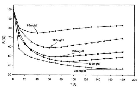
Figure 1 shows the kinetic measurement time course for test strips which
contain a
"moist-coated" membrane in the presence of blood samples containing different
triglyceride contents (65, 207, 294, 494 and 728 mg/dl) in which the relative
reflectance (R in %) is plotted versus time (t in s).
Figure 2 shows the relative reflectance (R) at different triglyceride
concentrations (c
in mg/dl) in blood samples for dry-coated (2) and moist-coated (1) membranes.
CA 02590545 2007-06-05
-14-
The numerals and abbreviations in the figures have the following meaning:
1 measurement curve for moist-coated membrane
2 measurement curve for dry-coated membrane
R relative reflectance
t time
c concentration
Example 1
Method for applying a reactive film to a blood-separating membrane and a
corresponding test device for detecting triglycerides in whole blood
1. Production of the coating paste
a.) Gantrez solution:
35.5 g water is added to 58.5 g of an 85 millimolar phosphate buffer (pH 7.5).
After adding 1.7 g MgSO4i 5.2 g Gantrez S 97 (copolymer of methyl vinyl
ether and maleic acid anhydride, GAF Corporation chemical division) is
added in small portions and stirred for 3 hours until the Gantrez is
completely
swollen. Afterwards 4.5 g of a 32 % NaOH solution is added and after a
further 30 minutes stirring 0.6 g PVP (polyvinylpyrrolidone 25,000) is
sprinkled in and stirred for a further 20 minutes until it has completely
dissolved. Subsequently the pH of the paste preparation is adjusted with 32 %
NaOHtoapHof6.7-7Ø
b.) Symperonic solution:
1.3 g Symperonic F68 (polyoxyethylene-co-oxypropylene, ICI) is dissolved in
5.3 g water while stirring for 20 minutes.
c.) 17.0 g Propiofan 70 D (50 % polymer dispersion of vinyl propionate in
water,
demonomerized, source BASF, Ludwigshafen) is added to the Gantrez
solution described in a.) and after 30 minutes stirring 26.5 g Celatom MW 25
(kieselguhr, CHEMAG) was added within 10 minutes and stirred for a further
CA 02590545 2007-06-05
-15-
20 minutes. Afterwards 6.6 g of the Symperonic solution described in b.) is
added to the preparation and stirred for a further 10 minutes.
d.) Refloblau solution
1.7 g Refloblau (4-(4-dimethylaminophenyl)-5-methyl-2-(3,5-dimethoxy-4-
hydroxyphenyl)-imidazole dihydrochloride, Roche Diagnostics) is dissolved
protected from light in 23.3 g 35 C warm water by stirring for 15 minutes on a
magnetic stirrer.
e.) Titanium dioxide/Refloblau partial preparation
While protected from light 22.5 g of an 85 mmolar phosphate buffer (pH 7.5)
is added first and 4.3 g Ti02 (RN 56, Kronos Titan) is sprinkled in within 5
minutes using a dissolver stirrer at 450 rpm and afterwards it is stirred for
a
further 5 minutes. Finally 25 g of the Refloblau solution prepared in d.) is
added within 5 minutes to the Ti02 suspension and stirred for a further 30
minutes. Afterwards the Ti02/Refloblau partial preparation is stored until use
in a refrigerator while protected from light.
f.) ATP solution
1.7 g ATP (adenosine triphosphate; di-sodium salt) is dissolved in 3.3 g
water.
g.) DONS solution
1.3 g DONS (dioctylsodium sulfosuccinate) is dissolved in 5.3 g acetone.
h.) MPSC solution
0.03 g MPSC (methylphenylsemicarbazide) is dissolved in 0.6 g 1-methoxy-2-
propanol while protected from light.
i.) Enzyme solution
The following enzymes (present as lyophilisates) are dissolved successively in
15.9 g of an 85 millimolar phosphate buffer (pH 7.5) where the respective
weighed-in amount of enzyme depends on the specific activity of the enzyme
CA 02590545 2007-06-05
- 16-
batch that is used:
40 kilo units (about 1.8 g) glycerokinase (EC 2.7.1.30 from Bacillus
stearothermophilus; Roche Diagnostics, Cat. No. 0 717 398)
34 kilo units (about 2.4 g) cholesterol esterase (EC 3.1.1.13 from Candida
cylindracea; Roche Diagnostics, Cat. No. 0 129 046)
28.9 kilo units (about 0.12 g) peroxidase (EC 1.11.1.7 from horseradish;
Roche Diagnostics, Cat. No. 0 121 606)
27.8 kilo units (about 0.44 g) L-a-glycerol phosphate oxidase (EC 1.1.3.21;
recombinant, Roche Diagnostics, Cat. No. 1 582 003).
j.) The following partial preparations are finally added to the Gantrez /
Propiofan / Celatom preparation from c.) while stirring:
6.6 g DONS solution from g.)
5.0 g ATP solution from f.)
51.8 g TiO2 / Refloblau suspension from e.)
7.0 g water for rinsing out the TiO2 / Refloblau solution
11.8 g Celatom MW 25
0.63 g MPSC solution from h.)
20.66 g enzyme solution from i.)
2.2 g 85 mmolar phosphate buffer to rinse out the enzyme solution.
After adding each partial solution (with the exception of Celatom) the
preparation is stirred for 5 minutes. The Celatom is sprinkled in small
portions within 15 minutes and the preparation is then stirred for a further
20
minutes.
The total preparation (about 250 g) is finally centrifuged for 20 minutes at
300 g for deaeration and subsequently any solids that may have been
sedimented by the centrifugation are slowly resuspended by hand using a
rubber wiper.
CA 02590545 2007-06-05
-17-
Afterwards the coating paste is passed through a 140 m test sieve and again
homogenized for 10 minutes while gently stirring.
The coating paste has the composition given in table 1.
Table 1: Composition of the coating paste absolute solids content
Gantrez S97 (as film thickener/swelling agent) 5.2 g 5.2 g
PVP (polyvinylpyrrolidone) 0.6 g 0.6 g
Propiofan dispersion (50 % in water as film 17 g 8.5 g
former)
Celatom (as film opener) 38.3 g 38.3 g
Ti02 RN56 (as white pigment) 4.3 g 4.3 g
MgSO4 1.7 g 1.7 g
Refloblau 1.7 g 1.7 g
methylphenyl semicarbazide 0.03 g 0.03 g
ATP (di-sodium salt) 1.7 g 1.7 g
Symperonic F68 1.3 g 1.3 g
DONS (dioctylsodium sulfosuccinate) 1.3 g 1.3 g
glycerokinase 40 KU 1.8 g
cholesterol oxidase 34 KU 2.4 g
peroxidase 28.9 KU 0.12 g
L-a-glycerol phosphate oxidase 27.8 KU 0.44 g
Acetone 5.3 g -
1-methoxy-2-propanol 0.6 g -
NaOH (32 %) 4.5 g 1.4 g
distilled water 152.9 g -
Sum 241.2 g 70.8 g
CA 02590545 2007-06-05
- 18-
The solids content of the coating past is 29 %. The percentage solids content
of the
film former (Propiofan) based on the total solids content is 12 %. The
percentage
solids content of the film opener (Celatom) based on the total solids content
is
54 %. The ratio of film opener to film former is 4.5 :1.
2. Applying the reactive film to a blood-separating membrane
A blood-separating membrane (type BTS-SP-300; article No. 955 00 12 0953
obtained from the Pall GmbH Company / 63303 Dreieich) is coated with the
coating paste produced as described in section 1.) by means of the method
described in the following in order to generate a reactive film.
a) An approximately 1 meter long piece of membrane (BTS-SP-300) is firstly
pulled through a stainless steel trough filled with water and afterwards the
excess water standing on the membrane surface is removed using a rubber
wiper. The coating paste from 1.) is doctor coated onto the membrane that is
still moist at a feed rate of 1.5 m/min and a knife gap of 150 m.
The membrane coated in this manner (referred to in the following as "moist-
coated membrane") is subsequently dried for 5 minutes at 50 C.
Finally the membrane is cut into 4.0 mm wide fine-cut rolls using a cutter
spindle. The fine-cut rolls are stored dry until further use.
b) As a comparison a second piece of BTS-SP-300 is coated with the identical
coating paste without previously pulling the membrane through a stainless
steel trough filled with water (referred to in the following as "dry-coated
membrane").
CA 02590545 2007-06-05
-19-
3. Production of test strip functional models to detect TG in whole blood
An approximately 200 pm thick polyester foil (so-called spacer layer) coated
on both sides with double-sided adhesive tape out of which 1.5 mm wide
capillaries (capillary length 35 mm) running longitudinally to the subsequent
test strips were previously cut at a distance of 5.0 mm with the aid of a
cutting
plotter (type Aristomat 1310 from the ARISTO Graphic Systems Company;
22525 Hamburg) by means of a "kiss cut" is glued onto a 5 mm wide and 78
mm long support foil (Melinex). A 5 mm x 25 mm polyester net (type Petex
07-98/34 from the Sefar Company /CH-9410 Heiden) having a mesh width of
250 pm is glued onto this spacer/capillary layer in order to, on the one hand,
form an upper border to the capillaries and, on the other hand, to ensure that
samples/blood passes from the capillary into the overlying analyte detection
zone.
The Scrynell net is arranged on the spacer layer in such a manner that the
first
mm of the capillaries are not covered by the net and can thus be used as a
sample application zone.
The blood-separating membrane coated with the reactive film according to
the method described in section 2 and attached at the sides by means of two
hot-melt adhesive beads, is located above the Scrynell net (uncoated, blood-
separating membrane side facing downwards; reactive film facing upwards).
The configuration of the test strip functional model is comparable with the
test strip described in example 1 and figure 1 of the EP application No. 04
023
734 (dated 5.10.2004).
CA 02590545 2007-06-05
-20-
4. Assessment of the functional model using blood samples containing
triglycerides
25 pl blood is applied to the sample application zone (capillary area in front
of
the Scrynell net) on the test strip functional models. The models are measured
from above (reactive film side of the membrane) by reflection photometry
over a period of 3 minutes at 10 second intervals using an optical measuring
system with an LED at the main wavelength of 660 nm.
The measurement procedure is described in the following:
Before applying the sample, the test strip is measured once while excluding
ambient light in order to obtain the reflectivity of each unreacted reactive
film. The "blank value" of the test strip obtained in this manner is set as
100 % relative reflectance (R) for the subsequent kinetic measurement in the
presence of sample material.
After applying 25 l blood the kinetic measurement is immediately started.
The reflectivities obtained in the kinetic mode are divided by the respective
blank value of the test strip and plotted graphically as relative reflectance
(R
in %) versus the measuring time.
Figure 1 shows the kinetic measurement time course obtained in this manner
for test strips (containing a "moist-coated" membrane) in the presence of
blood samples having different triglyceride contents (65, 207, 294, 494 and
728 mg/dl).
As the curve time courses of the kinetic measurement show, the colour
development of the reactive film reaches a reflectance minimum (maximum
colour depth) within the selected measurement period and this reflectance
minimum is selected in the following as a measure of the analyte
concentration in the sample.
CA 02590545 2007-06-05
-21-
The relative reflectance minima (in %) are listed in the following table 2 for
a
"moist-coated" and a "dry-coated" BTS-SP-300 membrane containing an
identical reactive film in increasing order for blood samples having different
triglyceride contents.
Table 2
% relative reflectance (in the minimum)
triglyceride content in "moist-coated" "dry-coated"
the blood sample membrane membrane
65mg/dl 74.7% 81.6%
76mg/dl 71.7% 81.5%
100 mg/dl 69.4% 81.3%
105 mg/dl 68.7 % 77.7 %
142 mg/dl 63.5% 77.3%
154 mg/dl 63.1 % 77.8 %
207 mg/dl 59.0 % 75.7 %
217 mg/dl 56.9 % 72.6 %
265 mg/dl 52.6 % 72.3 %
294 mg/dl 48.7% 70.3%
326 mg/dl 48.8% 71.5%
384 mg/dl 47.3 % 68.6 %
494 mg/dl 44.2 % 64.3 %
728 mg/dl 36.1 % 57.5 %
Total reflectance range 38.6 % 24.1 %
As shown in table 2 the functional models containing the "moist-coated"
membrane generate considerably more colour (lower reflectance values) over the
CA 02590545 2007-06-05
-22-
entire measuring range than the "dry-coated" membrane containing an identical
reactive film.
Furthermore, the measured values show that the reflectance range (i.e. the
difference between the relative reflectances for the triglyceride
concentrations
65 mg/dl and 728 mg/dl) achieved over the entire measuring range is
considerably
larger for the "moist-coated" membrane at 38.6 % REM than the reflectance
range
for the "dry-coated" membrane at 24.1 % REM (see also the graphic curve shown
in
figure 2, in which 1 is the measurement curve for the moist-coated membrane
and 2
is the measurement curve for the dry-coated membrane).
Due to the considerably larger reflectance range for the "moist-coated"
membrane
the variations in reflectance from measurement to measurement result in a
considerably lower variation in concentration and thus in a higher precision
of the
functional model.