Note: Descriptions are shown in the official language in which they were submitted.
CA 02590977 2007-06-01
VASCULAR THROMBECTOMBY APPARATUS
AND METHOD OF USE
Field of the Invention
This invention generally relates to an apparatus and methods used to filter
or remove matter from within a body conduit. In particular, this invention
relates to
a self-expanding device used in interventional procedures such as thrombectomy
or embolectomy that resists plastic deformation as it engages, filters andlor
lo removes matter that is entrapped in a conduit of a body.
Background of the Invention
Interventional procedures are often necessary to restore the flow of fluids
in conduits of the human body. For example, percutaneous interventional
procedures may be employed to introduce a stent into the vasculature of a
human
body to restore the proper flow of blood. During this process matter such as
emboli or thrombi may be introduced into the blood stream. In addition, matter
may be naturally present in the blood stream or in a conduit of a human body.
It
is necessary to filter or remove the matter from the conduit to avoid adverse
physical affects such as ischemic stroke.
A typical interventional procedure involves introducing a guidewire into the
vasculature of the patient. The guidewire is routed and advanced beyond the
point in the conduit where the matter to be removed resides. The guidewire
serves as a track over which a catheter or other interventional device can be
advanced. Once the catheter is in place, a variety of devices can be advanced
1
CA 02590977 2007-06-01
through the catheter and used to capture, filter and/or remove the matter. For
example, filters of various types have found use in trapping blood clots and
other
debris released into the bloodstream. A typical filter comprises a frame with
a
basket mounted thereon such that an opening is formed at the proximal end
thereof. Once the interventional procedure, such as placement of a stent or
balloon angioplasty, is complete or the filter is placed in proximity to a
thrombi or
emboli, the filter is pulled in a desired direction closing the basket and
entrapping
the matter.
Many filters are only partially effective in capturing debris resulting from
intervention procedures or naturally occurring debris lodged in the
vasculature
because deployment of the filter within the conduit may not provide complete
filtration. This may result from failing to maintain an optimum fit of the
filter within
the vessel or conduit wall resulting in a gap there between. Where a filter
basket
is employed another drawback may be encountered if the basket does not fully
deploy within the vessel.
Existing filter devices also fail to exhibit the necessary characteristics to
capture emboli or thrombi that are attached to a vessel wall or lodged within
a
vessel. For example, thrombi are often fixedly attached or lodged within a
vessel
and significant force is required to dislodge them. If a filter does not have
the
proper modulus of elasticity, it will deform and fail to capture or remove the
embolic material. An obvious solution to this challenge is to increase the
rigidity
of the filter. This approach, however, is not well suited for small conduits
such as
the vasculature located within the human brain, a location where ischemic
stroke,
2
CA 02590977 2007-06-01
blockage of blood vessels by thrombi, originates. In order to fit within small
vessels the filter must have commensurate dimensions while also exhibiting the
proper modulus of elasticity to ensure proper deployment and removal of the
matter from the vasculature. This is a difficult challenge since increased
stiffness
s will ensure removal of the thrombi, but prevent proper delivery and
deployment of
the filter device from a small deployment sheath or catheter into which the
device
must be folded.
Numerous approaches have been attempted to meet this challenge. U.S.
Patent No. 6,740,061 -Oslund describes a filter device having a frame with a
1 o basket attached thereto and a self-expanding radial loop for positioning
the basket
upon deployment. The spacer or loop is positioned such that it is
substantially
axially aligned with the mouth of the filter basket. The loop urges the
guidewire
against the inner wall of a vessel ensuring that the basket is properly
positioned.
In another embodiment described in Oslund, the mouth of the filter basket is
15 defined by the loop. The filter device of Oslund is not contemplated for
use in
small vessels. Although the loop provides for proper positioning, it
interferes with
deployment of the filter device in small vasculature as it increases the
stiffness of
the device when it is compacted to fit within a delivery sheath or catheter.
U.S. Pat. No. 6,589,263 -Hopkins describes a filtration device having a
20 support hoop with a blood permeable sack affixed thereto. In order to allow
the
filtration device to be delivered without experiencing kinking or increasing
the
stiffness, the support hoop includes a reduced thickness articulation region.
This
region permits the filter frame to compactly fold and deploy within small
3
CA 02590977 2007-06-01
vasculature without kinking or increasing the stiffness of the filter.
Although
allowing ready deployment, the reduced thickness region inhibits the ability
of the
filter device to avoid deformation under loads. For example, instead of
capturing
a clot, which is firmly attached to the wall of a vessel, the filter device of
Hopkins
may fold due to the increased strain experienced at the articulation region in
a
manner similar to the device being pulled back into its delivery sheath or
catheter.
U.S. Patent No. 6,203,561 -Ramee describes a filtration device similar to
Hopkins. In order to overcome the shortcomings of Hopkins, Ramee provides a
first thrombectomy support hoop and a second filter support hoop. As with
Hopkins, each of the support hoops has a blood permeable sack attached thereto
and contains a reduced thickness articulation region. According to Ramee,
substantially all thrombi is captured by the first support hoop and the second
hoop
acts as a filter. More likely, however, is that as each of the support hoops
contacts the thrombi, they will deform due to the increased strain experienced
at
the articulation region. Even more alarming is that in failing to completely
dislodge and capture the thrombi, pieces of the thrombi may be dislodged and
travel downstream causing adverse complications.
Currently, there is no apparatus that can filter or remove matter from the
conduit of a human body. In particular, there is no apparatus that can to fit
within
small conduits and deploy therein while also exhibiting a modulus of
elasticity
sufficient to ensure removal of matter from the conduit.
4
CA 02590977 2007-06-01
Summary of the Invention
According to the invention, an apparatus is provided for removing matter
from within a conduit. The apparatus generally comprises a separation edge
attached to a wire at its proximal end, a frame attached to the distal end of
the
separation edge, and a membrane attached to the wire and disposed over the
frame enclosing its interior. The membrane generally comprises a net
constructed from a vaporized metal deposited on a mandrel. Alternatively, the
membrane may comprise a braided tube constructed from wire.
The apparatus may be employed as a filter or as a means for actively
1 o dislodging matter from the wall of a conduit. When employed as a filter,
the
apparatus is positioned downstream of the matter where it ensures that matter
does not escape downstream as it is being removed. When employed to actively
remover matter from a conduit, the apparatus is positioned downstream of the
matter. The apparatus is then pulled proximally whereby it engages the matter
and dislodges it from the wall of the conduit.
The frame, membrane and separation edge are generally constructed
from a super-elastic material. One example of such super elastic material is
Nitinol (Ni-Ti). Use of super elastic materials allow for deformation and
restraint in
a first deformed condition of the apparatus to facilitate deployment within a
conduit. For example, the super elastic characteristics allow the apparatus to
have a first, contracted shape when mounted within a sheath or other delivery
device employed to position the filter within a conduit. The sheath can be
5
CA 02590977 2007-06-01
steerable, introduced through a guiding catheter, or navigated over a wire
through
a conduit to a point downstream of the obstruction to be removed or the matter
to
be filtered. The filter/retrieval apparatus is deployed from the sheath
whereby it
expands to a second, expanded shape.
The separation edge comprises at least two members joined together at
their distal and proximal ends. In one embodiment, the proximal ends of the
members may be joined directly to a wire instead of to each other. The
separation edge is joined to a wire that is parallel to the longitudinal axis
of the
apparatus and articulates it for deployment and capture of matter from within
the
io conduit. The members are slanted from the longitudinal axis of the
apparatus to
provide an angled cutting surface. Upon deployment, the members contact the
inside of the conduit forming a tight seal. In one embodiment of the
invention, the
members are oval shaped to accommodate circular conduits. An opening is
defined between the members serving as an inlet through which matter passes
after it is dislodged from the walls of the conduit.
The separation edge experiences high stress and strain due to the force
required for removing matter that is entrapped within, or fixed to, the walls
of the
conduit. This can lead to the separation edge deforming as it contacts the
matter such that it assumes its first, contracted shape and fails to remove
the
matter from the conduit. One solution is to increase the modulus of elasticity
of
the separation edge. Increasing the modulus of elasticity, however, creates
difficulty for delivery of the apparatus, especially in small conduits, and
complicates deployment.
6
CA 02590977 2007-06-01
A frame, attached to the separation edge, allows the separation edge to
maintain flexibility by having a lower modulus of elasticity while preventing
the
separating edge from buckling as it engages matter and removes it from the
wall
of the conduit. The frame comprises a plurality of struts that are attached to
the
separation members. A first group of struts are connected to the separation
edge and to a second group of struts that are joined at the distal end of the
frame.
The second group of struts comprises a plurality of outer struts and a
plurality of
inner struts attached to and interspersed between the outer struts. The frame
is
structured so that the stress experienced by the separation edge is evenly
lo distributed across the frame. The frame may take on a variety of spatial
configurations such as a truss or a scaffold.
When the apparatus is delivered to a targeted site in the conduit via a
sheath or other delivery device it is contracted within the sheath to a first
diameter. Contracting the frame and separation edge to a small diameter
increases stiffness thereby limiting the minimum delivery profile achievable.
In
addition, the apparatus may deform when contracted jeopardizing optimal
deployment. If the apparatus fails to properly deploy the separation edge will
not
assume the proper cutting angle and will fail to seal against the walls of the
conduit allowing matter to escape downstream. In order to ensure proper
deployment of the apparatus, at least one deployment section is disposed along
the separation edge. The deployment section allows the apparatus to assume its
first contracted shape, without increasing stiffness, and allows for
deployment in a
predictable fashion.
7
CA 02590977 2007-06-01
In one embodiment of the invention, the deployment section comprises a
pre-formed point deformation such as a kink. The prior art discusses kinks as
being detrimental to contraction and deployment of the apparatus. In contrast
to
the prior art, the present invention employs a kink specifically formed in an
area
that will assure that the apparatus is folded within the sheath and deployed
in a
predictable manner.
Brief Description of the Drawings
The features and advantages of the invention will be apparent to those of
ordinary skill in the art from the following detailed description of which:
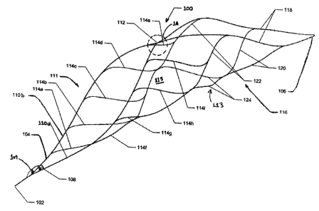
Figure 1 is a perspective view of showing the separation edge and frame of
the apparatus of the present invention;
Figure 1A is a detailed view of the deployment region shown in region 1A
of Figure 1;
Figure 2 is a bottom view of the apparatus of the present invention;
Figure 3 is an assembly view of the apparatus of the present invention; and
Figure 4 is a perspective view showing the apparatus of the present
invention deployed from a delivery device;
Figure 5 is a side, cutaway view showing a catheter deployed within a
conduit distally of matter to be removed there from;
Figure 6 is a side, cutaway view showing the apparatus of the present
invention is deployed within a conduit distally of matter to be removed there
from;
and
8
CA 02590977 2007-06-01
Figure 7 is a side, cutaway view showing the apparatus of the present
invention fully deployed within a conduit capturing the matter to be removed
there
from.
Detailed Description of the Preferred Embodiments
An apparatus for filtering and removing matter from the interior of a conduit
will be described with reference to Figures 1-7. As shown in Figure 1 the
apparatus 100 of the present invention generally comprises a separation edge
111 attached to a wire or tether 102, a frame 113 attached to the separation
edge
111, and, as shown in Figure 3, a membrane or net 130 disposed over the frame
113 enclosing its interior 115.
As shown in Figure 4, the membrane 130 includes openings along its
length that allows fluids found within the conduit to pass through the
interior 115
of the frame 113, but prevents matter 144 from escaping. For example, in one
embodiment of the invention membrane 130 is a blood permeable sac. If desired,
the membrane 130 may be attached to the separation edge 111 to provide
structural support thereto augmenting the support provided by the frame 113.
The membrane 130 may be constructed from a braided tube of material, wire, or
a thin metallic film. The metallic film can be set to a specific flat or three-
dimensional shape of cylindrical, non-cylindrical, or flat cross section. The
creation
of the film can be achieved with a wide variety of techniques such as Pulsed
Laser Ablation, Physical Vapor Deposition (PVD) including magnetron
sputtering,
Chemical Vapor Deposition (CVD), Molecular Beam Epitaxy (MBE), thermal
9
CA 02590977 2007-06-01
deposition (via electron beam or resistive heating), electroplating, dip
coating, spin
coating or other methods of depositing a metal. The thin films could also be
etched via chemical, laser or dry etch methods.
The frame 113, membrane 130 and separation edge 111 are preferably
constructed from a super-elastic material. One example of such super elastic
material is Nitinol (Ni-Ti). Ni-Ti is utilized in a wide variety of inedical
applications
due to its biomechanical compatibility, its biocompatibility, its fatigue
resistance,
its uniform plastic deformation, its magnetic resonance imaging compatibility,
its
ability to exert constant and gentle outward pressure, its dynamic
interference, its
1 o thermal deployment capability, its elastic deployment capability, its
hysteresis
characteristics, and is moderately radiopaque.
Nitinol exhibits shape memory and(or super-elastic characteristics. Shape
memory characteristics may be simplistically described as follows. A metallic
structure, for example, a Nitinol tube that is in an Austenitic phase may be
cooled
is to a temperature such that it is in the Martensitic phase. Once in the
Martensitic
phase, the Nitinol tube may be deformed into a particular configuration or
shape
by the application of stress. As long as the Nitinol tube is maintained in the
Martensitic phase, the Nitinol tube will remain in its deformed shape. If the
Nitinol
tube is heated to a temperature sufficient to cause the Nitinol tube to reach
the
2 o Austenitic phase, the Nitinol tube will return to its original or
programmed shape.
The original shape is programmed to be a particular shape by well-known
techniques.
Super-elastic characteristics may be simplistically described as follows. A
CA 02590977 2007-06-01
metallic structure for example, a Nitinol tube that is in an Austenitic phase
may be
deformed to a particular shape or configuration by the application of
mechanical
energy. The application of mechanical energy causes a stress induced
Martensitic phase transformation. In other words, the mechanical energy causes
the Nitinol tube to transform from the Austenitic phase to the Martensitic
phase.
By utilizing the appropriate measuring instruments, it can be determined that
the
stress from the mechanical energy causes the temperature to drop in the
Nitinol
tube. Once the mechanical energy or stress is released, the Nitinol tube
undergoes another mechanical phase transformation back to the Austenitic phase
1 o and thus its original or programmed shape. As described above, the
original
shape is programmed by well know techniques.
Medical devices constructed from Nitinol are typically utilized in both the
Martensitic phase andlor the Austenitic phase. The Martensitic phase is the
low
temperature phase. A material is in the Martensitic phase is typically very
soft
and malleable. These properties make it easier to shape or configure the
Nitinol
into complicated or complex structures. The Austenitic phase is the high
temperature phase. A material in the Austenitic phase is generally much
stronger
than the material in the Martensitic phase. Typically, many medical devices
are
cooled to the Martensitic phase for manipulation and loading into delivery
system
2 o and heated again for deployment. For example, apparatus 100 assumes a
first,
contracted shape for placement into a sheath 134. When the apparatus 100 is
deployed at body temperature, it returns to the Austenitic phase, or a second,
expanded shape as shown in Figure 4.
11
CA 02590977 2007-06-01
Although constructed from a super elastic material, the frame 113 and
membrane 130 expand axially at different rates during loading or compression
of
the apparatus 100 into the sheath 134. In order to maintain flexibility of the
apparatus 100 as it is loaded into the catheter 134, it is preferred that the
distal
ends of the frame and membrane 130 are not connected. Instead, the membrane
130 is attached directly to the wire or tether 102 and disposed over the frame
113.
As shown in Figure 1, the separation edge 111 comprises at least two
members 110a and 110b that are joined together. Alternatively, separation edge
1 o 111 may comprise a single hoop attached at its distal end to the wire or
tether
102. The proximal end of the separation edge 111 is connected to the distal
end
of wire or tether 102. Each member 110a, 110b may also be attached directly to
the wire at their proximal ends rather than to each other. Upon deployment
from
sheath 134, it is desired that the separation edge 111 maintain contact with
the
inside of the conduit forming a tight seal therewith. In one embodiment of the
invention, the members 110a and 110b are arcuate to accommodate generally
eccentric conduits. For example, the distance (D) between the peaks of the
members 110a and 100b ranges from 1.00mm to 7.00mm. An opening 136 is
defined between the members 110a and 110b serving as an inlet through which
matter passes into the interior 115 after it is dislodged from the walls of a
conduit
Alternatively, each of the members 110a and 110b may have a different shape
so as to conform the separation edge to the shape of the conduit 140.
As shown in Figures 1 and 4 Members 110a and 110b are joined together
12
CA 02590977 2007-06-01
at their distal ends and at their proximal ends such that there is an acute
separation angle 108 between the proximal ends of members 110a and 110b. In
addition, separation edge 111 is attached to the wire 102 at an angle 109
providing an offset cutting surface. As the separation edge 111 is pulled
toward
matter to be removed from the wall of a conduit or vessel, the proximal end of
separation edge 111, which is attached to the wire 102 and is more rigid, will
be
the first to contact the matter. The increased rigidity of the proximal end of
separation edge 111 along with the acute separation angle 108 will apply
shearing
force to remove the matter from the wall of the conduit. As the separation
edge is
1 o further articulated proximally toward the sheath 134, the slanted or
offset distal
portion of the separation edge 111 will urge the matter away from the wall of
the
conduit and into the interior 115 of the frame 113 through inlet 136.
The apparatus 100 is sized to allow for passage into small conduits such
as blood vessels found within the vasculature of the brain. In addition, the
apparatus 100 must also deploy to the proper configuration to ensure
filtration or
removal of matter 144 from within conduit 140. For example, apparatus 100 will
have the ability to be compressed down to sizes of a microcatheter inner
diameter
of between .018"-.021" and still deploy or expand to sizes between .197"-
.276."
Due to the relatively small dimensions of apparatus 100, the separation edge
111
2 o experiences high stress and strain due to the force required for removing
matter
that is entrapped within, or fixed to, the walls 142 of a conduit 140. Without
proper support, the separation edge 111 would experience strain to the point
where it assumes its first, contracted shape and fails to remove the matter
from
13
CA 02590977 2007-06-01
the conduit 140. Increasing the modulus of elasticity of the separation edge
111 is not an option since this would inhibit the deliverability and
deployment of
the apparatus 100 within small conduits. Thus, the separation edge 111
requires
additional structure that will resist strain, but not inhibit deployment and
deliverability.
Frame 113 is linked to the separation edge 111 and provides support
thereto preventing deformation as edge 111 engages matter 144 and removes it
from the wall 142 of the conduit 140. One example of a frame 113 that provides
the desired support is shown if Figures 2 and 3. The frame 113 comprises a
lo plurality of struts that are attached to the separation members 110a and
110b.
A first group of struts 114 are connected to the separation edge 111 and to
a second group of struts 116 that are joined at the distal end 106 of the
frame
113. As seen in Figure 3, struts 114a-114e are evenly distributed along the
length of member 110b. In particular, strut 114a is attached to the proximal
end
of member 110b, struts 114b and 114c are attached to the mid portion of member
110b, and struts 114d and 114e are attached to the distal portion of member
110b. Struts 114f and 114g are attached to the mid portion of member 110a and
struts 114h and 114i are attached to the distal portion of member 110a. In
order
to optimize deployment of the apparatus 100, there are no struts attached to
the
proximal portion of member 110a. Although this is a preferred configuration,
it
may be necessary to provide additional support to separation edge 111 in which
case additional struts 114 can be attached to member 110a or either of members
14
CA 02590977 2007-06-01
110a and 110b. First struts 114 are attached to strut the second set of struts
116, in particular, strut sets 122 and 126 described below.
The first group of struts 114 distributes forces from the members 110a and
110b to the distal end of frame 113 via the second group of struts located
distally
of the separation edge 111. The second group of struts 116 comprises an outer
strut set 118 that spans from the distal end 106 of the frame 113 to the
distal end
of separation edge 111 and a plurality of inner strut sets 120, 122, 124 and
126
interspersed between the outer strut set 118 as described herein. The distal
end
of each strut of set 120 is connected to the distal portion of outer strut set
118
lo while the distal end of each strut of set 122 is connected to the proximal
portion of
outer strut set 118. The proximal ends of the struts of set 126 are connected
together and the distal ends of each strut of set 126 are connected to the
proximal
end of strut sets 122 and 124. The distal ends of the struts of set 124 are
connected together and to the proximal ends of the struts of set 120 that are
also
joined together.
Frame 113 forms a scaffold or space truss that distributes forces across
the second struts 116. Although a particular form of truss is illustrated
frame 113
may take on a variety of spatial configurations known in the art. In addition,
the
strut configuration within the frame 113 can be varied. Yet another option is
to
vary the thickness or width of the struts. Alternatively, the materials used
to
construct the frame 113 or separation edge 111 can be varied throughout the
apparatus to achieve the necessary rigidity and flexibility of apparatus 100.
CA 02590977 2007-06-01
When the apparatus 100 is delivered to a targeted site in the conduit 140
via a sheath 134or other delivery device it is contracted within the sheath
134 to a
first diameter. Contracting the frame 113 and separation edge 111 to a small
diameter increases stiffness thereby limiting the minimum delivery profile
achievable. In addition, the apparatus 100 may deform when contracted
jeopardizing optimal deployment. If the apparatus 100 fails to properly deploy
the separation edge 111 will not assume the proper cutting angle and will fail
to
seal against the walls of the conduit potentially allowing matter to escape
downstream. Of even greater concern is if the separation edge 111 deforms into
1o a traumatic configuration causing damage to the walls of the conduit 140.
In
order to ensure proper deployment of the apparatus, at least one deployment
section 112 is disposed along the separation edge 111. The deployment section
112 allows the apparatus 100 to assume its first contracted shape, without
increasing stiffness, and allows for deployment in a predictable fashion. If
desired, a plurality of deployment sections 112 can be distributed along the
frame
113 and separation edge 111 depending upon the delivery profile.
In one embodiment of the invention, the deployment section 112 comprises
a pre-formed point deformation such as a kink as shown in greater detail in
Figure
1A. The prior art discusses kinks as being detrimental to loading and
deployment
of the apparatus 100 from a sheath 134. In contrast to the prior art, the
present
invention employs a kink specifically formed in an area that will assure that
the
apparatus 100 is folded within the sheath 134 and deployed in a predictable
manner. As shown in figure 1 A, the kink 112 is located between the point
where
16
CA 02590977 2007-06-01
outer strut set 118 and members 110a and 110b are joined together. The kink
112 has substantially the same, or greater, thickness (t) as members 110a and
110b and struts 118. Kink 112 is in the form of a pre-bend that will allow the
apparatus 100 to compress within the sheath 134 without increasing stiffness
and
to deploy to the desired configuration. Moreover, kink 112 will allow for
reloading
of the apparatus within a sheath or delivery device 134
Altematively, the deployment section 112 comprises a material interposed
between frame 113 and separation edge 111 that is more flexible than the
material that frame 113 and separation edge 111 is constructed form. In this
1o instance the more flexible material allows predictable bending at the point
where
folding of the apparatus 100 occurs when it is contracted into catheter 134.
In
yet another embodiment, the modulus of elasticity of the frame 113 and the
separation edge 111 may be varied such that bending occurs at deployment
section 112 disposed there between.
When employed as a filter, the apparatus 100 is positioned distally of the
matter 144 to be removed where it ensures that pieces of the matter 144 do not
escape downstream as another device acts upon and removes the matter 144
from conduit 140. When employed to actively remove the matter 144, the
apparatus 100 is first positioned distally of the matter 144 and is then
pulled
proximally whereby the apparatus engages the matter 144 and detaches it from
the wall 142 of the conduit.
As shown in Figure 5, a sheath or catheter 134 is guided through conduit
140 to a position distal of the matter 144 to be removed. The matter 144 may
be
17
CA 02590977 2007-06-01
attached to the wall 142 of the conduit 140 and usually occludes substantially
all
of conduit 140. Thus, the apparatus 100 is compressed to a small diameter to
allow for the use of a low profile sheath 134 to navigate around matter 144.
As
shown in Figures 5 and 6, when the sheath 134 is in position, the apparatus
100
is pushed from the sheath via actuation of the wire or tether 102.
Alternatively,
the wire 102 may be held in place to prevent movement and the sheath 134 may
be withdrawn exposing the apparatus 100. The distal end of the apparatus 106
emerges from the sheath 134. The distal end 106 of the apparatus 100 includes
atraumatic tip that will not puncture the walfs 142 of the conduit.
Thereafter, the
1 o super elastic frame 113 and separation edge 111 emerge from the sheath 134
resuming their remembered or deployed shape. A set of marker bands 146 on
the sheath 134 and apparatus 100 allows for alignment of the apparatus 100
into
a position to ensure optimal filtration or alignment of the separation edge
111 with
the matter 144.
As shown in Figure 7, once the apparatus 100 has been properly aligned, it
is pulled proximally, towards sheath 134. The separation edge 111 engages the
matter 144 and slides, or cuts, it away from the wall 142 of the conduit 140.
The
matter 144 passes through inlet 136 and into the interior of the frame 115
where it
is captured. Membrane 130 prevents pieces of matter 144 that may have
2 o become dislodged from escaping the interior 115. Fluid from the conduit
140 is
free to pass through the interstices of membrane 130 ensuring that proper flow
of
fluid through conduit 140 is maintained. Thereafter, the sheath 134 and
18
CA 02590977 2007-06-01
apparatus 100 are withdrawn proximally through the conduit 140 to a point
where
the matter 144 is removed.
Although the present invention has been described above with respect to
particular preferred embodiments, it will be apparent to those skilled in the
art that
numerous modifications and variations can be made to these designs without
departing from the spirit or essential attributes of the present invention.
Accordingly, reference should be made to the appended claims, rather than to
the
foregoing specification, as indicating the scope of the invention. The
descriptions
provided are for illustrative purposes and are not intended to limit the
invention
i o nor are they intended in any way to restrict the scope, field of use or
constitute
any manifest words of exclusion.
19