Note: Descriptions are shown in the official language in which they were submitted.
CA 02591545 2007-06-18
WO 2006/068808 PCT/US2005/043850
METHOD AND SYSTEM FOR TRANSCERVICAL TUBAL OCCLUSION
TECHNICAL FIELD
[0001] This invention relates to a medical device and procedure.
BACKGROUND
[0002] Female sterilization typically involves occluding the fallopian tubes
to
prevent sperm access to an egg within a female's fallopian tube. One
conventional
female sterilization procedure is laparoscopic tubal occlusion. In this
procedure, an
incision is made in the abdominal wall to provide access to the fallopian
tubes. The tubes
are surgically occluded with the aid of a laparoscope, for example, using
bipolar or
monopolar coagulation. Laparoscopic tubal occlusion is invasive and requires
multiple
incisions and passing of several instruments and a gaseous distension medium
into the
patient's abdomen. Thermal and mechanical injury to the surrounding tissues
and organs
has been reported.
[0003] Minimally invasive transcervical approaches to female sterilization
have
been used more recently. One such procedure involves placing small, flexible
devices
into the fallopian tubes; the devices are inserted transcervically into the
uterine cavity
providing access to the fallopian tubes. The devices are made from polyester
fibers and
metals and once in place, body tissue grows into the devices and blocks the
fallopian
tubes. The devices permanently remain in the patient's body, which has raised
concerns
about the long term effects of the implanted devices as well as restrictions
on potential
subsequent surgical interventions within the uterus, given the conductive
metallic
components in the devices.
[0004] A monopolar radio frequency technique has been investigated that
included passing a small diameter wire (an active electrode) transcervically
through the
uterine cavity and the tubal ostium to the fallopian tubes. A large, passive
electrode is
positioned externally. The current path between the two electrodes is not well
defined
and can lead to inadvertent burns. The technique was not successful and was
abandoned.
It could manage neither the varying thicknesses of endometrial tissue at the
tubal ostium,
nor the required tight tolerance on the depth of destruction within the
fallopian tubes.
I
CA 02591545 2007-06-18
WO 2006/068808 PCT/US2005/043850
SUIVIMARY
[0005] This invention relates to a medical device and procedure. In general,
in
one aspect, the invention features a method for fallopian tubal occlusion. A
tubal
occlusion device is inserted into a uterine cavity. The device includes an RF
applicator
head including an electrode carrier with one or more bipolar electrodes
thereon. During
insertion, the RF applicator head is in a closed position. The RF applicator
head is
positioned at a tubal ostium of a fallopian tube such that a distal tip of the
RF applicator
head advances into the tubal ostium. The RF applicator head is deployed into
an open
position such that the RF applicator head approximates the shape of the
uterine cavity in a
region of the tubal ostiuin. Current is passed through the one or more bipolar
electrodes
to the tubal ostium to destroy tissue to a known depth, which precipitates a
healing
response in surrounding tissue that over time scars and occludes the fallopian
tube.
[0006] Implementations of the invention can include one or more of the
following
features. Passing current through the one or more bipolar electrodes to the
tubal ostium to
destroy tissue can include vaporizing endometrium and destroying superficial
myometrium. Inserting a tubal occlusion device into a uterine cavity can
include
inserting the tubal occlusion device with the RF applicator head in a closed
position, and
before passing current through the one or more bipolar electrodes, deploying
the RF
applicator head into the' open position. Suction can be applied through the
electrode
carrier to draw the surrounding tissue into contact with the electrodes, and
to draw
moisture generated during ablation away from the electrodes to substantially
prevent the
formation of a low impedance liquid layer at the electrodes. Passing current
through the
one or more bipolar electrodes can include delivering radio frequency energy
to the one
or more bipolar electrodes.
[0007] The method can further include automatically terminating the flow of
current into the tissue once ablation has approximately reached a
predetermined depth of
ablation. Before positioning the RF applicator head at the tubal ostium, the
uterine cavity
can be insufflated. Insufflation is ceased before passing current through the
one or more
bipolar electrodes, allowing the uterine cavity to collapse onto the RF
applicator head.
Deploying the RF applicator head into an open position can include removing a
sheath to
expose the electrode carrier. The electrode carrier can include a fabric
having conductive
metallized regions and one or more non-conductive regions formed thereon to
create the
one or more bipolar electrodes. The method can further include advancing an
illuminator
and an optical instrument into the uterine cavity. Positioning the RF
applicator head at
2
CA 02591545 2007-06-18
WO 2006/068808 PCT/US2005/043850
the tubal ostium of a fallopian tube can include using the optical instrument
to visualize
the tubal ostium.
[0008] In general, in another aspect, the invention features a system for
fallopian
tubal occlusion. The system includes a tubal occlusion device, a source of
radio
frequency energy, a controller and a vacuum source. The tubal occlusion device
has a
distal end and a proximal end, the distal end including an electrode carrier
with one or
more bipolar electrodes thereon. In an open condition the distal end is shaped
to
approximate a uterine cavity in a region of a tubal ostium of a fallopian tube
to be
occluded. The source of radio frequency energy is electrically coupled to the
one or more
bipolar electrodes. The controller is configured to control the delivery of
radio frequency
energy to the one or more bipolar electrodes such that passing radio frequency
energy
through the one or more bipolar electrodes to the tubal ostium can be
controlled to
destroy tissue to a known depth, which precipitates a healing response in
surrounding
tissue that over time scars and occludes the fallopian tube. The vacuum source
is
operable to draw the tissue into contact with the one or more bipolar
electrodes and to
draw moisture generated during delivery of the radio frequency energy to the
bipolar
electrodes away from the bipolar electrodes. This can substantially eliminate
liquid
surrounding the bipolar electrodes.
[0009] Implementations of the invention can include one or more of the
following
features. Passing radio frequency energy through the one or more bipolar
electrodes to
the tubal ostium destroying tissue can include vaporizing endometrium and
destroying
superficial myometrium. The electrode carrier can include a structural support
member
within a fabric sheath having conductive metallized regions and having one or
more non-
conductive regions formed thereon to create the one or more bipolar
electrodes. The
structural support member can include flexible members movable between a
closed
condition and the open condition. The system can further include an
illumination source
electrically coupled to the distal end of the tubal occlusion device to
illuminate the uterus,
and an optical instrument electrically coupled to the distal end of the tubal
occlusion
device to provide images of the uterus.
[0010] In general, in another aspect, the invention features an apparatus for
occluding a fallopian tube. The apparatus includes an elongate member, an
electrode
carrier and a tube. The elongate member has a distal end, a proximal end and a
hollow
central interior. The electrode carrier is attached to the distal end of the
elongate member
and has one or more bipolar electrodes formed thereon. The electrode carrier
is operable
3
CA 02591545 2007-06-18
WO 2006/068808 PCT/US2005/043850
to couple to a radio frequency energy generator and is movable between a
closed position
in which the electrode carrier is collapsed for insertion into a uterine
cavity, and an open
position in which a distal end of the electrode carrier is shaped to fit
within a tubal ostium
of a fallopian tube. The hollow central interior of the elongate member is
operable to
couple to a vacuum source and to draw moisture away from the one or more
electrodes.
[0011] Implementations of the invention can include one or more of the
following
features. The apparatus can further include an illuminator attached to the
distal end of the
elongate member and operable to couple to an illumination source, and an
optical
instrument attached to the distal end of the elongate member and operable
couple to an
image display device. The electrode carrier can include a structural support
member
within a fabric sheath having conductive metallized regions and have one or
more non-
conductive regions formed thereon to create the one or more bipolar electrodes
The
structural support member can include flexible members movable between a
closed
condition and the open condition.
[0012] Implementations of the invention can realize one or more of the
following
advantages. The tubal occlusion procedure described is minimally invasive: the
tubal
occlusion device can be introduced into the patient's uterine cavity
transcervically and
requires no abdominal incision. The procedure does not leave any foreign
objects in the
patient's body, minimizing the risk of infection and eliminating the need to
restrict
subsequent surgical intervention options. The procedure can be performed
quickly, the
actual duration of ablation being approximately one minute per fallopian tube.
Because
the RF energy is limited to the region of ablation, there is less risk of
damage to other
organs during the procedure. The system and procedure automatically compensate
for
varying endometrial thicknesses, facilitating the proper, contoured depth of
tissue
destruction in the region of the tubal opening. Further, unlike the technique
described
above that implanted permanent devices in the fallopian tubes, there is no
need to
navigate a catheter through the fallopian tubes, which are prone to spasm,
inhibiting the
placement of permanent devices, making such a procedure difficult to achieve.
[0013] The details of one or more embodiments of the invention are set forth
in
the accompanying drawings and the description below. Other features, objects,
and
advantages of the invention will be apparent from the description and
drawings, and from
the claims.
4
CA 02591545 2007-06-18
WO 2006/068808 PCT/US2005/043850
DESCRIPTION OF DRAWINGS
[0014] FIG lA is a schematic representation of a uterus.
[0015] FIG 1B is a schematic representation of a RF applicator head positioned
in
a tubal ostium.
[0016] FIG 1C is a schematic representation of a region of ablated tissue in a
uterus and tubal ostium.
[0017] FICi. 2 shows a side view of a tubal occlusion device.
[0018] FIC~ 3A shows a top view of the tubal occlusion device of FIG 2 with a
RF
applicator head in a closed position.
[0019] FIG 3B shows a top view of the tubal occlusion device of FIG 2 with the
RF applicator head in an open position.
[0020] FIGS. 4A and 4B show one embodiment of a structural body of a RF
applicator head in closed and open positions respectively.
[0021] FICz 4C is a schematic representation of a RF applicator head in an
open
position.
[0022] FIC~ 4D is a schematic representation of center lines of electrodes of
the
RF applicator head of FIG 4C.
[0023] FIG 4E is a cross-sectional view of a main body of the tubal occlusion
device of FIGS. 2 and 3.
[0024] FIGS. 5A-D are schematic representations of cross-sectional views
showing electrodes in contact with tissue for ablation.
[0025] FIG 6 is a flowchart showing a process for tubal occlusion.
[0026] FIGS. 7A-D are schematic representations of steps of a process for
tubal
occlusion.
[0027] FIG 8 is a schematic representation of an alternative embodiment of a
structural body of a RF applicator head.
[0028] Like reference symbols in the various drawings indicate like elements.
DETAILED DESCRIPTION
[0029] A method and system for occlusion of a female's fallopian tubes is
described that provides a minimally invasive alternative for female
sterilization.
Referring to FIG. 1A, a schematic representation of a uterus 3 is shown,
including a
uterine cavity 5 surrounded by uterine tissue, namely endometrial tissue 7a
and
myometrial tissue 7b. The fallopian tubes 11 connect to the uterine cavity 5
at the tubal
CA 02591545 2007-06-18
WO 2006/068808 PCT/US2005/043850
ostia 9. Occluding the tubal ostia 9 prevents sperm from entering the
fallopian tubes 11
and fertilizing an egg, thereby sterilizing the female.
[0030] Referring to FIG. 1B, a RF (radio frequency) applicator head 2 can be
introduced transcervically into the uterine cavity and positioned at a tubal
ostium 9.
Transmitting RF energy through the RF applicator head 2 ablates the uterine
tissue 7a, 7b
and tissue within the tubal ostium 9, as shown schematically by the region 11
in FIG. 1 C.
Following the destruction of tissue at the tubal ostium 9, the healing
response occludes
the tubal ostium 9 and the adjacent portion of the fallopian tube 11 resulting
in
sterilization. Referring again to FIG. 1C, the targeted destruction from A-A
to B is
approximately 1.5 to 2.5 millimeters, from A-A to C is approximately 10 to 20
millimeters, and the depth D-D is typically approximately 2.0 to 3.5
millimeters.
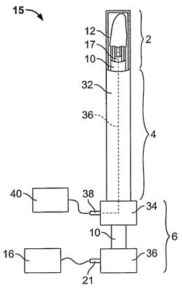
[0031] Referring to FIGS. 2, 3A and 3B, one embodiment of a tubal occlusion
device 15 is shown. The tubal occlusion device 15 includes generally three
major
components: the RF applicator head 2, a main body 4, and a handle 6. FIG. 2
shows a
side view of the tubal occlusion device 15 and FIGS. 3A and 3B show top views.
FIG.
3A shows the tubal occlusion device 15 with the RF applicator head 2 in a
closed position
within a sheath 32 and FIG. 3B shows the RF applicator head 2 in an open
position
outside of the sheath 32. The RF applicator head 2 includes an electrode
carrier 12
mounted to the distal end of the shaft 10 and electrodes 14 formed on the
surface of the
electrode carrier 12. An RF generator 16 can be electrically connected to the
electrodes
14 to provide mono-polar or bipolar RF energy to them.
[0032] The main body 4 includes a shaft 10. The shaft 10 is an elongate member
having a hollow interior. In one embodiment, the shaft 10 is approximately 30
centimeters long and has a cross-sectional diameter of approximately 4
millimeters.
Extending through the shaft 10 is a suction/insufflation tube 17 having a
plurality of holes
17a formed in its distal end (see FIGS. 4A and 4B).
[0033] Referring particularly to FIG. 3B, electrode leads 18a and 18b extend
through the shaft 10 from the distal end 20 to the proximal end 22 of the
shaft 10. At the
distal end 20 of the shaft 10, each of the leads 18a, 18b is coupled to a
respective one of
the electrodes 14. At the proximal end 22 of the shaft 10, the leads 18a, 18b
are
electrically connected to the RF generator 16 by an electrical connector 21.
During use,
the leads 18a, 18b carry RF energy from the RF generator 16 to the electrodes
14. Each
of the leads 18a, 18b is insulated, and the leads 18a and 18b can be connected
to opposite
terminals of the RF generator 16. When opposite polarity is applied to
alternating
6
CA 02591545 2007-06-18
WO 2006/068808 PCT/US2005/043850
electrodes or groups of electrodes, an electrode pair (i.e., one positively
charged and one
negatively charged electrode or group of electrodes) can be referred to as a
bipolar
electrode. Any references herein to a bipolar electrode refer to such an
electrode pair.
[0034] Referring to FIGS. 4A-C, the RF applicator head 2 can be shaped to
approximate the shape of the region to be ablated. The embodiment of the RF
applicator
head 2 shown in FIG. 4C has a V-shape which can fit within a corner of the
uterine cavity
and reach into the tubal ostium 9. FIGS. 4A and 4B show the RF applicator head
2
without the electrode carrier 12, thereby revealing the structural body 100 of
the RF
applicator head 2. A flexible member 19 is attached to the distal end of the
shaft 10 of the
main body and to the distal end of the tube 17. A flexure 22 is attached to
the tube 17 and
to an inner surface of the flexible member 19. In the closed position shown in
FIG. 4A,
the flexure 22 is compressed within the space formed between the inner surface
of the
flexible member 19 and the tube 17, and the shape of the structural body 100
of the RF
applicator head 2 is substantially cylindrical. In one embodiment, the flexure
22 and
flexible member 19 are made from stainless steel, are approximately 0.012
inches thick
and are substantially planar.
[0035] The RF applicator head 2 can be deployed into the open position shown
in
FIG. 4B by moving the tube 17 relative to the shaft 10. In one embodiment, the
shaft 10
is pulled toward the proximal end of the shaft, i.e., away from the RF
applicator head 2.
Movement of the shaft 10, which is connected to the flexible member 19, causes
the
flexible member 19 to also move in the same direction, causing the flexure 22
to move
laterally away from the tube 17. As shown in FIG. 4B, flexible member 19 is
deformed
outwardly, away from the tube 17, creating the V-shape at the distal end of
the RF
applicator head 2. The shape of the distal end differs depending on how much
the shaft
and tube 17 are moved relative to one another.
[0036] In an alternative embodiment, the tube 17 can be pushed toward the
proximal end of the flexible member 19, i.e., toward the RF applicator head 2,
thereby
moving the tube 17 relative to the shaft 10. The relative movement has the
same effect as
described above, that is, the flexible member 19 is deformed outwardly,
creating a V-
shape at the distal end.
[0037] FIG. 4C shows the distal end of the RF applicator head 2 with the
electrode carrier 12 over the structural body. The electrode carrier 12 can be
formed of a
fabric that is stretched over the structural body; the fabric is metallized in
the regions
forming the electrodes 14. The electrodes 14 are conductive and can alternate
between
7
CA 02591545 2007-06-18
WO 2006/068808 PCT/US2005/043850
positive and negative polarity (an electrode pair being a"bipolar electrode"
as described
above). In the embodiment depicted, there are four electrodes 14 (or 2 bipolar
electrodes), two on either face of the electrode carrier 12. A non-conductive
insulator 23
divides the electrode carrier 12 into the bipolar electrodes 14.
[0038] In one embodiment, the fabric is formed from a composite yarn with a
thermoplastic elastomer (TPE) core and multiple polyfilament nylon bundles
wound
around the TPE as a cover. The nylon bundles are plated with thin conductive
metal
layers. Preferably, the nylon is metallized, but not the TPE core. This
construction
facilitates stretching; the nylon windings open up their coils as the TPE core
is elongated,
without cracking the metallic layer. The TPE core facilitates recovery from
the stretched
position, pulling the nylon coils back into their initial configuration.
[0039] In an alternative embodiment, the electrode carrier 12 can be a sack
formed of a material that is non-conductive, that is permeable to moisture,
and that can be
compressed to a smaller volume and subsequently released to its natural size
upon
elimination of compression. Examples of materials for the electrode carrier 12
include
foam, cotton, fabric, or cotton-like material, or any other material having
the desired
characteristics. The electrodes 14 can be attached to the outer surface of the
electrode
carrier 12, e.g., by deposition or another attachment mechanism. The
electrodes 14 can
be made of lengths of silver, gold, platinum, or any other conductive
material. The
electrodes 14 can be formed on the electrode carrier 12 by electron beam
deposition, or
they can be formed into coiled wires and bonded to the electrode carrier 12
using a
flexible adhesive. Other means of attaching the electrodes, such as sewing
them onto the
surface of the electrode carrier 12, may alternatively be used.
[0040] Depth of destruction of the target tissue can be contoured to achieve
repeatable, predetermined depths. Variables such as the electrode
construction, power
applied to the electrodes (power density or power per unit surface area of the
electrode),
and the tissue impedance at which power is terminated can be used to affect
the depth of
tissue destruction, as discussed further below.
[0041] The spacing between the electrodes (i.e., the distance between the
centers
of adjacent electrodes) and the widths of the electrodes are selected so that
ablation will
reach predetermined depths within the tissue, particularly when maximum power
is
delivered through the electrodes. Maximum power is the level at which low
impedance,
low voltage ablation can be achieved. For example, referring to FIG. 4D, lines
19a and
19b represent center lines of the electrodes 14 of the RF applicator head 2 of
FIG. 4C, i.e.,
8
CA 02591545 2007-06-18
WO 2006/068808 PCT/US2005/043850
the spacing. The center lines diverge and are closest at the distal end I and
further apart at
the proximal end H. The closer the center lines the shallower the depth of
destruction.
That is, the depth of destruction at the distal end, which during operation is
positioned
within or closest to the tubal ostium 9, is least.
[0042] Referring to FIG. 5A, preferably each electrode is energized at a
polarity
opposite from that of its neighboring electrodes. By doing so, energy field
patterns,
designated 52, 53 and 54 in FIG. 5A, are generated between the electrode sites
and thus
help to direct the flow of current through the tissue T to form a region of
ablation A. As
can be seen in FIG. 5A, if electrode spacing is increased by energizing, for
example,
every third or fifth electrode rather than all electrodes, the energy patterns
will extend
more deeply into the tissue. See, for example, pattern 53 which results from
energization
of electrodes having a non-energized electrode between them, or pattern 54
which results
from energization of electrodes having two non-energized electrodes between
them.
[0043] The depth of ablation is also effected by the electrode density (i.e.,
the
percentage of the target tissue area which is in contact with active electrode
surfaces) and
may be regulated by pre-selecting the amount of this active electrode
coverage. For
example, the depth of ablation is much greater when the active electrode
surface covers
more than 10% of the target tissue than it is when the active electrode
surfaces covers
only 1% of the target tissue.
[0044] By way of illustration, by using 3-6 mm spacing and an electrode width
of
approximately 0.5-2.5 mm, delivery of approximately 20-40 watts over a 9-16
cm2 target
tissue area will cause ablation to a depth of approximately 5-7 millimeters
when the
active electrode surface covers more than 10% of the target tissue area. After
reaching
this ablation depth, the impedance of the tissue will become so great that
ablation will
self-terminate. By contrast, using the same power, spacing, electrode width,
and RF
frequency will produce an ablation depth of only 2-3 mm when the active
electrode
surfaces covers less than 1% of the target tissue area. This can be better
understood with
reference to FIG. 5B, in which high surface density electrodes are designated
51a and low
surface density electrodes are designated 5 lb. For purposes of this
comparison between
low and high surface density electrodes, each bracketed group of low density
electrodes is
considered to be a single electrode. Thus, the electrode widths W and spacings
S extend
as shown in FIG. 5B.
[0045] As is apparent from FIG. 513, the electrodes 51 a, which have more
active
area in contact with the underlying tissue T, produce a region of ablation Al
that extends
9
CA 02591545 2007-06-18
WO 2006/068808 PCT/US2005/043850
more deeply into the tissue T than the ablation region A2 produced by the low
density
electrodes 51b, even though the electrode spacings and widths are the same for
the high
and low density electrodes. Some examples of electrode widths, having spacings
with
more than 10% active electrode surface coverage, and their resultant ablation
depth, based
on an ablation area of 6 cm2 and a power of 20-40 watts, are given on the
following table:
ELECTRODE WIDTH SPACING APPROX. DEPTH
1 mm 1-2 mm 1-3 mm
1-2.5 mm 3-6 mm 5-7 mm
1-4.5 mm 8-10 mm 8-10 mm
[0046] Examples of electrode widths, having spacings with less than 1% active
electrode surface coverage, and their resultant ablation depth, based on an
ablation area of
6 cm2 and a power of 20-40 watts, are given on the following table:
ELECTRODE WIDTH SPACING APPROX. DEPTH
1 mm 1-2 mm 0.5-1 mm
1-2.5 mm 3-6 mm 2-3 mm
1-4.5 mm 8-10 mm 2-3 mm
[0047] Thus it can be seen that the depth of ablation is significantly less
when the
active electrode surface coverage is decreased.
[0048] Referring to FIG. 5C, if multiple, closely spaced, electrodes 51 are
provided on the electrode carrying member, a user may set the RF generator 16
to
energize electrodes which will produce a desired electrode spacing and active
electrode
area. For example, alternate electrodes may be energized as shown in FIG. 5C,
with the
first three energized electrodes having positive polarity, the second three
having negative
polarity, etc. All six electrodes together can be referred to as one bipolar
electrode. As
another example, shown in FIG. 5D, if greater ablation depth is desired the
first five
electrodes may be positively energized, and the seventh through eleventh
electrodes
negatively energized, with the sixth electrode remaining inactivated to
provide adequate
electrode spacing. Therefore, in one implementation, a user can control which
electrodes
are energized to produce a desired depth of destruction.
CA 02591545 2007-06-18
WO 2006/068808 PCT/US2005/043850
[0049] Referring again to FIGS. 3A and 3B, in one implementation, to achieve
the
desired depth of ablation, a controller included in the RF generator 16 can
monitor the
impedance of the tissue at the distal end of the RF applicator head 2 and
include an
automatic shut-off once a threshold impedance is detected. As the tissue is
desiccated by
the RF energy, fluid is lost and withdrawn from the region by a vacuum through
the tube
17, which can be connected to suction/insufflation unit 40 via
suction/insufflation port 38
(FIGS. 3A, 3B). The suction draws moisture released by tissue undergoing
ablation away
from the electrode carrier 12 and prevents formation of a low-impedance liquid
layer
around the electrodes 14 during ablation. As more tissue is desiccated, the
higher the
impedance experienced at the electrodes 14. By calibrating the RF generator
16, taking
into account system impedance (e.g., inductance in cabling etc.), a threshold
impedance
level can be set that corresponds to a desired depth of ablation.
[0050] Once the threshold impedance is detected, the controller shuts off the
RF
energy, preventing excess destruction of tissue. For example, when
transmitting RF
energy of 5.5 watts per square centimeter of tissue, an impedance of the
tissue of 50 ohms
can indicate a depth of destruction of approximately 3 to 4 millimeters at the
proximal
end H and approximately 2.5 millimeters at the distal end I. In an alternative
embodiment, the RF generator 16 can be configured such that above the
threshold
impedance level the RF generator's ability to deliver RF power is greatly
reduced, which
in effect automatically terminates energy delivery.
[0051] Referring again to FIGS. 3A and 3B, an introducer sheath 32 facilitates
insertion of the tubal occlusion device 15 into, and removal of the device
from, the
uterine cavity 5. The sheath 32 is a tubular member that is slidable over the
shaft 10. The
sheath 32 is slidable between a distal condition, shown in FIG. 3A, in which
the RF
applicator head 2 is compressed inside the sheath, and a proximal condition in
which the
sheath 32 is moved proximally to release the RF applicator head 2 from inside
the sheath
32 (FIG. 3). By compressing the electrode carrier 12 to a small volume, the RF
applicator
head 2 can be easily inserted transcervically into the uterine cavity 5.
[0052] During use, the sheath 32 is retracted from the electrode carrier 12,
for
example, by moving the distal handle member 34 towards the proximal handle
member
37 to slide the sheath 32 in the distal direction. Moving the distal handle
member 34
toward the proximal handle member 27 can also advance the shaft 10 in the
proximal
direction. The movement of the shaft 10 relative to the suction/insufflation
tube 17
causes the shaft 10 to pull proximally on the flexible member 19. Proximal
movement of
11
CA 02591545 2007-06-18
WO 2006/068808 PCT/US2005/043850
the flexible member 19 in turn pulls the flexure 22, causing it to move to the
opened
condition shown in FIG. 3B (see also FIG. 4B). In one embodiment, a locking
mechanism (not shown) is required to hold the shaft in the fully withdrawn
condition to
prevent inadvertent closure of the RF applicator head 2 during the ablation
procedure.
[0053] The amount by which the flexible member 19 is deformed outwardly from
the tube 17 can be controlled by manipulating the handle 6 to slide the shaft
10,
proximally or distally. The amount by which the shaft 10 is slid relative to
the tube 17
controls the shape of the flexible member 19.
[0054] As mentioned above, in an alternative embodiment, the handle 6 can be
configured so that the tube 17 can be moved distally relative to the shaft 10.
Distal
movement of the tube 17 in turn deforms the flexible member 19 outwardly. The
amount
by which the flexible member 19 is deformed outwardly from the tube 17 can be
controlled by manipulating the handle 6 to slide the tube 17 proximally or
distally, and
the amount by which the tube 17 moves relative to the shaft 10 controls the
shape of the
flexible member 19.
[0055] As shown in FIG. 3A, a flow pathway 36 is formed from the RF applicator
head 2 to the suction/insufflation port 38. The proximal end of the
suction/insufflation
tube 17 is fluidly coupled to the flow pathway so that gas fluid may be
introduced into, or
withdrawn from the suction/insufflation tube 17 via the suction/insufflation
port 38. For
example, suction may be applied to the fluid port 38 using a
suction/insufflation unit 40.
This causes water vapor within the uterine cavity 5 to pass through the
permeable
electrode carrier 12, into the suction/insufflation tube 17 via holes 17a,
through the tube
17, and through the suction/insufflation unit 40 via the port 38. If
insufflation of the
uterine cavity 5 is desired, insufflation gas, such as carbon dioxide, may be
introduced
into the suction/insufflation tube 17 via the port 38. The insufflation gas
travels through
the tube 17, through the holes 17a, and into the uterine cavity 5 through the
permeable
electrode carrying member 12.
[0056] One or more additional components can be provided for endoscopic
visualization purposes. For example, lumen 42, 44, and 46 may be formed in the
walls of
the introducer sheath 32 as shown in FIG. 4E. An optical instrument can be
used to
provide images from within the uterine cavity. For example, referring to FIGS.
3B and
4E, an imaging conduit, such as a fiberoptic bundle, extends through lumen 42
and is
coupled via a camera cable 43 to a camera 45. Images taken from the camera may
be
displayed on a monitor 47. An illumination fiber 50 can extend through lumen
44 and
12
CA 02591545 2007-06-18
WO 2006/068808 PCT/US2005/043850
couple to an illumination source 49. The optional third lumen 46 can be an
instrument
channel through which surgical instruments may be introduced into the uterine
cavity 5, if
necessary. In an alternative embodiment, one or more of the lumen 42, 44, 46
can be
formed in the walls of the shaft 10.
[0057] Because during use it is most desirable for the electrodes 14 on the
surface
of the electrode carrier 12 to be held in contact with the interior surface of
the uterine
cavity 5 and tubal ostia 9, the electrode carrier 12 may have additional
components inside
it that add structural integrity to the electrode carrying means when it is
deployed within
the body.
[0058] Referring to FIGS. lA-C, 5 and 6A-D, a process 58 for using the tubal
occlusion device 15 to sterilize a female shall be described. The tubal
occlusion device
15 is inserted through the vagina and cervix to the internal os 13 at the base
of the uterus
3 (step 59). A gas, e.g., carbon dioxide, is delivered into the uterine cavity
5 via the
suction/insufflation tube 17 from the suction/insufflation unit 40 to distend
the uterine
cavity 5 (step 60). The tubal occlusion device 15 is then advanced into the
uterine cavity
(step 61).
[0059] The user visualizes the target tubal ostium 9 on the monitor 47 using
images provided by the camera 45 (step 62). FIG. 7A is a schematic
representation of
what the user may see upon the tubal occlusion device 15 entering the uterine
cavity 5;
the tubal ostium 9 is a relatively small spot in the distance. As the tubal
occlusion device
advances toward the tubal ostium 9, the tubal ostium 9 is easier to visualize,
as shown
in FIG. 7B. The distal end of the RF applicator head 2, which is still within
the sheath 32,
is positioned at the tubal ostium 9, as depicted in FIG. 7C (step 63). The
sheath 32 is
withdrawn to expose the electrodes 14 (step 64) and the RF applicator head 2
is deployed
into the open position (step 65), as depicted in FIG. 7D.
[0060] Insufflation is ceased and the uterine cavity 5 is allowed to collapse
onto
the RF applicator head 2 (step 66). Vacuum can be applied to the RF applicator
head 2
via the suction/insufflation tube 17 to draw the surrounding tissue into
contact with the
electrodes 14 (step 67). The RF generator 16 is turned on to provide RF energy
to the
electrodes 14 (step 68). The RF energy is ceased once the desired amount of
tissue has
been ablated (step 69). In one implementation, 5.5 watts of RF power is
supplied for per
square centimeter of electrode surface area until the predetermined impedance
threshold
is reached, at which point power is terminated.
13
CA 02591545 2007-06-18
WO 2006/068808 PCT/US2005/043850
[0061] The uterine cavity 5 can be insufflated a second time, the RF
applicator
head 2 collapsed into a closed position and the tubal occlusion device 15
rotated
approximately 180 . The RF applicator head 2 can then be positioned at the
other tubal
ostium 9 and the above procedure repeated to ablate tissue at the other tubal
ostium 9.
The tubal occlusion device 15 is then closed and withdrawn from the patient's
body.
After ablation, healing and scarring responses of the tissue at the tubal
ostia 9
permanently occlude the fallopian tubes 11, without requiring any foreign
objects to
remain in the female's body and without any incisions into the female's
abdomen. The
procedure is fast, minimally invasive, and is highly effective at tubal
occlusion.
[0062] Referring to FIG. 8, an alternative embodiment of a structural body 70
of
the RF applicator head 2 is shown. The structural body 70 includes an external
hypotube
72 and an internal hypotube 74. If implementing the structural body 70 in the
embodiment of the tubal occlusion device 15 described above, the external
hypotube 72
can be the shaft 10 and the internal hypotube 74 can be the
suction/insufflation tube 17.
A cage 78 is formed over the internal hypotube 74 configured in a V-shape at
the distal
end 79 that can reach into a tubal ostium 9. The cage 78 can be a braided or
woven
structure made from a memory material, e.g., nitinol.
[0063] The cage 78 can be collapsed into a narrow cylindrical configuration by
moving the internal hypotube 74 relative to the external hypotube 72, e.g., by
pushing the
internal hypotube 74 distally away from the external hypotube 72. In a
collapsed state the
cage 78 can fit, for example, within the sheath 32 described above, when the
RF
applicator head 2 is placed in a closed position. Once the sheath 32 is
removed and the
internal hypotube 74 is moved back into the open position relative to the
external
hypotube 72, the nature of the material from which the cage 78 is made expands
the cage
78 into the desired shape that is depicted. An electrode carrier, such as the
electrode
carrier 12 made from a metallized fabric described above, can be fitted over
the structural
body 70, completing the RF applicator head.
[0064] A number of embodiments of the invention have been described.
Nevertheless, it will be understood that various modifications may be made
without
departing from the spirit and scope of the invention. Accordingly, other
embodiments are
within the scope of the following claims.
[0065] What is claimed is:
14