Note: Descriptions are shown in the official language in which they were submitted.
DEMANDES OU BREVETS VOLUMINEUX
LA PRESENTE PARTIE DE CETTE DEMANDE OU CE BREVETS
COMPREND PLUS D'UN TOME.
CECI EST LE TOME 1 DE 2
NOTE: Pour les tomes additionels, veillez contacter le Bureau Canadien des
Brevets.
JUMBO APPLICATIONS / PATENTS
THIS SECTION OF THE APPLICATION / PATENT CONTAINS MORE
THAN ONE VOLUME.
THIS IS VOLUME 1 OF 2
NOTE: For additional volumes please contact the Canadian Patent Office.
CA 02592043 2013-06-18
METHODS FOR REGULATION OF STEM CELLS
FIELD
[0002] The invention generally relates to methods for increasing the
successful activity
of stem cells, hematOpoietic progenitor/stem cells, mesenchymal
progenitor/stem cells,
mesodermal progenitor/stem cells, muscle progenitor/stem cells, or neural
progenitor/stem cells
in vivo in a mammalian subject. The invention further relates to methods of
treating an immune
related disease, a mesenchymal/mesoderm degenerative disease, or a
neurodegenerative disease
by administering one or more Wnt/P-catenin signal-, Notch signal-, or Hedgehog
signal-
promoting agents to a mammalian subject in need thereof.
BACKGROUND
[0003] Hematopoietic stems cells (HSCs) are rare cells of the hematopoietic
system
with the ability to self-renew and differentiate into all mature blood
lineages, thereby sustaining
hematopoietic homeostasis and immune function. HSC transplantation therapy has
been
effectively used to manage hematopoietic malignancies, bone
marrow/hematopoietic failure, and
immunodeficiency. Baron et al., Arch Med Res 34:528-44, 2003; Giralt, Curr
Hematol Rep
3:165-72, 2004; Vollweiler et al., Bone Marrow Transplant 32:1-7, 2003.
Despite successful
utility of HSCs, several clinical limitations remain. These include
availability of allogenic HSC
- 1 -
CA 02592043 2007-06-20
WO 2006/072016 PCT/US2005/047508
donors and inability to harvest adequate numbers of HSCs per donor. Moscardo
et al., Leuk
Lymphoma 45:11-8, 2004. Although autologous harvests of HSCs via G-CSF
mobilization to
the peripheral blood have alleviated some of the clinical burden for allogenic
HSC
transplantation, many patients remain refractory to mobilization and
subsequent HSC
reconstitution. Cohena and Nagler, Leuk Lymphoma 44:1287-99, 2003. Ex vivo
expansion of
HSCs has been suggested as a means to increase the number of available HSCs
for autologous or
allogenic transplantation. Unfortunately, current methods of ex vivo HSC
expansion have not
proven to benefit transplanted recipients, and experimental evidence suggests
that ex vivo culture
of HSCs negatively affects their hematopoietic reconstitution ability. Devine
et al., Bone
Marrow Transplant 31:241-52, 2003; Shih et al., J Hematother Stem Cell Res
9:621-8, 2000;
Srour et al., J Hematother 8:93-102, 1999.
[0004] Direct in vivo targeting of patient HSCs would provide a more
physiological
context to modulate HSC function as an alternative to HSC isolation and ex
vivo manipulation.
However, the current understanding of extrinsic regulators of HSCs has been
derived from
studies limited to ex vivo culture systems where HSCs are studied in
suboptimal and artificial
culture systems. As such, many factors implicated in regulating HSC self-
renewal in vitro are not
amenable to in vivo use.
[0005] Glycogen synthase kinase-3 (GSK-3) is a constitutively active
serine/threonine
kinase, originally identified as inactivating glycogen synthase. Frame and
Cohen, Biochem J
359:1-16, 2001; Cohen, Biochem Soc Trans 7:459-80, 1979; Embi et al., Eur J
Biochem
107:519-27, 1980. Inhibition of GSK-3 has been implicated in regulation of
several pathways,
including Wnt, Hedgehog, and Notch. Behrens et al., Science 280:596-599, 1998;
Yost et al.,
Genes Dev 10:1443-1454, 1996; Jia et al., Nature 416:548-552, 2002; Foltz et
al., Curr Biol
12:1006-1011, 2002; Espinosa et al., J Biol Chem 278:32227-35, 2003. Important
to HSCs,
these same pathways have recently been associated with HSC function by either
forced ectopic
overexpression of key upstream regulators of these pathways, or ligand
presentation in vitro.
Murdoch et al., PNAS 100:3422-3427, 2003; Reya et al., Nature 423:409-14,
2003; Bhardwaj et
al., Nat Immunol 2:172-80, 2001; Karanu et al., J Exp Med 192:1365-72, 2000;
Karanu et al.,
Blood 97:1960-7, 2001; Cline et al., Diabetes 51:2903-2910, 2002; Ring et al.,
Diabetes 52:588-
595, 2003.
[0006] Degenerative muscle diseases, such as muscular dystrophy (MD) include a
group of genetic diseases characterized by progressive weakness and
degeneration of the skeletal
muscles which control movement. There is no specific treatment for any of the
forms of MD.
Respiratory therapy, physical therapy to prevent painful muscle contractures,
orthopedic
- 2 -
CA 02592043 2007-06-20
WO 2006/072016 PCT/US2005/047508
appliances used for support, and corrective orthopedic surgery may improve the
quality of life in
some cases. Myopathy is a neuromuscular disorder in which the primary symptom
is muscle
weakness due to dysfunction of muscle fiber. Treatments for the myopathies
depend on the
disease or condition and specific causes. Supportive and symptomatic treatment
may be the only
treatment available or necessary for some disorders.
[0007] Regulators of hematopoietic stem cells (HSCs), stem cells, that elicit
their
effects in vivo have not been identified, limiting clinical manipulation of
HSCs to ex vivo
systems. Regulators of muscle progenitor cells or neural progenitor cells for
in vivo treatment of
degenerative muscle diseases or neurodegenerative diseases have not been
identified. A need
exists in the art for an improved therapy involving hematopoietic stem cell
for treatment of
immune related disease, and for an improved therapy involving stem cells,
muscle progenitor
cells or neural progenitor cells for treatment of degenerative muscle diseases
or
neurodegenerative diseases.
SUMMARY
[0008] The present invention relates to a method for increasing the successful
activity
of stem cells and progenitor cells, for example, hematopoietic progenitor/stem
cells (HSCs),
mesenchymal progenitor/stem cells, mesodermal progenitor/stem cells,
endothelial
progenitor/stem cells, or ectodermal or neural progenitor/stem cells in vivo
in a mammalian
subject comprising interacting one or more Wnt/13-catenin signal-, Notch
signal-, or Hedgehog
signal-promoting agents with the progenitor/stem cells in the mammalian
subject and increasing
the progenitor/stem cells in the mammalian subject. The progenitor/stem cells
can include, but
are not limited to, hematopoietic progenitor/stem cells (HSCs), stem cells,
mesenchymal
progenitor/stem cells, mesodermal progenitor/stem cells, endlthelial
progenitor/stem cells,
ectodermal progenitor/stem cells, muscle progenitor/stem cells, endodermal
progenitor/stem cells
or neural progenitor /stem cells The interacting of one or more Wnt/P-catenin
signal-, Notch
signal-, or Hedgehog signal-promoting agents with progenitor/stem cells, e.g.,
hematopoietic
progenitor/stem cells, occurs either by direct interaction of the signal
promoting agents with the
hematopoietic progenitor/stem cells or through an indirect interaction between
a second
signaling factor or cell type acting as an intermediate between the Wnt/P-
catenin signal-, Notch
signal-, or Hedgehog signal-promoting agents and the hematopoietic
progenitor/stem cells.
Whether the effect on the HSC, or progenitor/stem cell, is on progenitor/stem
cell proliferation,
survival, cell differentiation, or engrafttnent into the target tissue, the
net effect of activating the
Wnt/B-catenin signal is an increase in the measured stem cell/progenitor cell
activity. A
progenitor/stem cell, e.g., an hematopoietic progenitor/stem cell, can be
derived from a variety of
- 3 -
CA 02592043 2007-06-20
WO 2006/072016 PCT/US2005/047508
sources, including, but not limited to, adult bone marrow, umbilical cord
blood cells, or
embryonic stem cells, from a mammal, e.g, a human. A method of treating immune
related
disease is provided comprising administering one or more Wnt/f3-catenin signal-
, Notch signal-,
or Hedgehog signal-promoting agents to a mammalian subject and interacting the
agent with the
hematopoietic progenitor/stem cells of the mammalian subject. A method of
treating
degenerative disease is provided comprising administering one or more Wnt/13-
catenin
Notch signal-, or Hedgehog signal-promoting agents to a mammalian subject and
interacting the
agent with hematopoietic progenitor/stem cells, stem cells, mesenchymal
progenitor cells,
mesodermal progenitor cells, muscle progenitor cells, endothelial progenitor
cells, or neural
progenitor cells of the mammalian subject. The degenerative disease includes,
but is not limited
to, mesenchymal degenerative disease, mesodermal degenerative disease, muscle
degenerative
disease, endothelial degenerative disease, or neurodegenerative disease. A
method of treating
cytopenia in a mammalian subject is provided comprising administering one or
more Wnt/f3-
catenin signal-, Notch signal-, or Hedgehog signal-promoting agents to the
mammalian subject.
[0009] A method for increasing hematopoietic stem cells in vivo in a mammalian
subject is provided which comprises interacting one or more Wnt/P-catenin
signal-, Notch
signal-, or Hedgehog signal-promoting agents with the hematopoietic stem cells
in the
mammalian subject and increasing the hematopoietic stem cells in the mammalian
subject
compared to the hematopoietic stem cells in the mammalian'subject before
treatment. In one
aspect, the Wnt/I3-catenin signal- promoting agent is an agonist of one or
more of Wntl, Wnt2,
Wnt2b/13, Wnt3, Wnt3a, Wnt4, Wnt5a, Wnt5b, Wnt6, Wnt7a, Wnt7b, Wnt7c, Wnt8,
Wnt8a,
Wnt8b, Wnt8c, Wntl Oa, Wntl0b, Wntll, Wnt14, Wnt15, or Wnt16. In a further
aspect, the
Wnt/ p-catenin signal-promoting agent is an agonist of Wnt3a or Wnt8. In
another aspect, the
Notch signal-promoting agent is an agonist of Notch, Delta, Serrate, Jagged,
Deltex,
Mastermind, Enhancer of Split, Hesl, Split, Hairless, Suppressor of Hairless,
or RBP-Jk. In
another aspect, the Hedgehog signal-promoting agent is an agonist of Desert
hedgehog, Sonic
hedgehog, Indian hedgehog, Gli, Gli-1, Gli-3, Patched, or Patchedl.
[0010] The method for increasing hematopoietic stem cells in vivo in a
mammalian
subject further provides increasing hematopoietic stem cells in the subject as
a result of cell
proliferation, cell homing, decreased apoptosis, self renewal, or increased
cell survival. In a
further aspect, the hematopoietic stem cells comprise progenitor cells of
erythroid cells,
granulocyte cells, macrophage cells, granulocyte-macrophage cells, B cells, T
cells, and
multipotent mixed lineage colony types
- 4 -
CA 02592043 2007-06-20
WO 2006/072016 PCT/US2005/047508
[00111 The method for increasing hematopoietic stem cells in vivo in a
mammalian
subject further provides that the Wnt/13-catenin signal-, Notch signal-, or
Hedgehog signal-
promoting agent is a polypeptide, nucleic acid, small molecule, antisense
oligonucleotide,
ribozyme, RNAi construct, siRNA, shRNA, or antibody. In a one aspect, the Wnt
signal- or 13-
catenin signal-promoting agent is a polypeptide, for example, a wnt
polypeptide, a dishevelled
polypeptide, or a f3-catenin polypeptide. In a further aspect, the Notch
signal-promoting agent is
a notch polypeptide, delta polypeptide, serrate polypeptide, jagged
polypeptide, deltex
polypeptide, mastermind polypeptide, split polypeptide, hairless polypeptide,
RBP-Jk
polypeptide, or hesl polypeptide. In a further aspect, the Hedgehog signal-
promoting agent is a
desert hedgehog polypeptide, sonic hedgehog polypeptide, indian hedgehog
polypeptide, gli
polypeptide, gli-1 polypeptide, gli-3 polypeptide, patched polypeptide, or
patchedl polypeptide.
[0012] The method for increasing hematopoietic stem cells in vivo in a
mammalian
subject further provides that the Wnt/f3-catenin signal-, Notch signal-, or
Hedgehog signal-
promoting agent is a glycogen synthase kinase (GSK) inhibitor. In a further
aspect, the glycogen
synthase kinase (GSK) inhibitor is a GSK-3 inhibitor or a GSK-313 inhibitor.
[0013] A method is provided for treating an immune related disease in a
mammalian
subject in need thereof comprising administering one or more Wnt/13-catenin
signal-, Notch
signal-, or Hedgehog signal-promoting agents to the subject, and interacting
the one or more
Wnt/13-catenin signal-, Notch signal-, or Hedgehog signal-promoting agents
with hematopoietic
stem cells, and thereby increasing in vivo hematopoietic stem cells in the
subject to treat the
immune related disease compared to the hematopoietic stem cells in the
mammalian subject
before treatment. In a further aspect, the method comprises increasing
hematopoietic stem cells
in the subject as a result of cell proliferation, cell homing, decreased
apoptosis, self renewal, or
increased cell survival. In a further aspect, the immune related disease is
diabetes, graft vs. host
disease, immunodeficiency disease, hematopoietic malignancy, hematopoietic
failure, or
hematopoietic stem cell transplantation.
[0014] A method is provided for treating an immune related disease in a
mammalian
subject in need thereof comprising administering hematopoietic stem cells to
the subject,
administering to the subject one or more Wnt/13-catenin signal-, Notch signal-
, or Hedgehog
signal-promoting agents to contact the hematopoietic stem cells in the
subject, and interacting the
one or more Wnt/13-catenin signal-, Notch signal-, or Hedgehog signal-
promoting agents with
hematopoietic stem cells, and increasing in vivo hematopoietic stem cells in
the subject to treat
the immune related disease compared to the hematopoietic stem cells in the
mammalian subject
before treatment. In a further aspect, the method comprises increasing
hematopoietic stem cells
- 5 -
CA 02592043 2007-06-20
WO 2006/072016 PCT/US2005/047508
in the subject as a result of cell proliferation, cell homing, decreased
apoptosis, self renewal, or
increased cell survival. In one aspect, the hematopoietic stem cells are
neonatal cells, umbilical
cord blood cells, fetal liver cells, adult cells, bone marrow cells,
peripheral blood cells, or
embryonic stem cells. In a further aspect, the hematopoietic stem cells are
autologous or
allogeneic hematopoietic stem cells. In a further aspect, the immune related
disease is diabetes,
graft vs. host disease, immunodeficiency disease, hematopoietic malignancy,
hematopoietic
failure, or hematopoietic stem cell transplantation.
[0015] A method is provided for treating a degenerative disease in a mammalian
subject in need thereof comprising administering one or more Wnt/13-catenin
signal-, Notch
signal-, or Hedgehog signal-promoting agents to the subject, and increasing in
vivo one or more
mesenchymal progenitor/stem cells, mesodermal progenitor/stem cells or
endothelial
progenitor/stem cells in the subject to treat the degenerative disease
compared to the
progenitor/stem cells in the mammalian subject before treatment. In a further
aspect, the method
comprises increasing progenitor/stem cells in the subject as a result of cell
proliferation, cell
homing, decreased apoptosis, self renewal, or increased cell survival.
[0016] In a further aspect, the method comprises increasing in vivo one or
more
mesenchymal progenitor/stem cells or mesodermal progenitor/stem cells in the
subject to treat
degenerative muscle disease or to treat degenerative mesenchymal disease. In
one aspect, the
degenerative mesenchymal disease is treated by increasing or repairing bone,
chondrocytes/cartilage, skeletal muscle, endothelial cells, or adipose cells.
In a further aspect,
the method comprises increasing in vivo one or more endothelial
progenitor/stem cells in the
subject to treat degenerative endothelial disease. In one aspect, the
degenerative endothelial
disease is treated by increasing vascularization or increasing angiogenesis.
[0017] In one aspect, the degenerative muscle disease is muscular dystrophy,
duchenne
muscular dystrophy, facioscapulohumeral muscular dystrophy, myotonic muscular
dystrophy,
congenital myopathy, or mitochonclrial myopathy. In a further aspect, the
degenerative muscle
disease is familial cardiomyopathy, dilated cardiomyopathy, hypertrophic
cardiomyopathy,
restrictive cardiomyopathy, or coronary artery disease with resultant ischemic
cardiomyopathy.
In a further aspect, the method comprises increasing in vivo one or more
mesenchymal
progenitor/stem cells or mesodermal progenitor/stem cells in the subject to
treat degenerative
liver disease, nephritic disease, cirrhosis, alcoholic cirrhosis, fatty liver,
alcoholic hepatitis, viral
hepatitis, liver carcinoma, post necrotic cirrhosis, biliary cirrhosis,
hepatocellular injury or a
biliary tract disorder. In a further aspect, the method comprises increasing
in vivo one or more
mesenchymal progenitor/stem cells or mesodermal progenitor/stem cells in the
subject to treat
- 6 -
CA 02592043 2007-06-20
WO 2006/072016 PCT/US2005/047508
degenerative pancreatic disease, diabetes, diabetes related disorder,
hyperglycemia,
hyperinsulinaemia, hyperlipidaemia, insulin resistance, impaired glucose
metabolism, obesity,
diabetic retinopathy, macular degeneration, cataracts, diabetic nephropathy,
glomerulosclerosis,
diabetic neuropathy, erectile dysfunction, premenstrual syndrome, vascular
restenosis, ulcerative
colitis, coronary heart disease, hypertension, angina pectoris, myocardial
infarction, stroke, skin
and connective tissue disorders, foot ulcerations, metabolic acidosis,
arthritis, or osteoporosis.
[0018] A method is provided for treating a degenerative disease in a mammalian
subject in need thereof comprising administering one or more mesenchymal
progenitor/stem
cells, mesodermal progenitor/stem cells or endothelial progenitor/stem cells
to the subject,
administering to the subject one or more Wnt/13-catenin signaling, Notch
signaling or Hedgehog
signaling promoting agents to contact the progenitor/ stem cells in the
subject, and interacting the
one or more Wnt/f3-catenin signaling, Notch signaling or Hedgehog signaling
promoting agents
with the progenitor/ stem cells, and increasing in vivo progenitor/ stem cells
in the subject to treat
the degenerative disease compared to the progenitor/stem cells in the
mammalian subject before
treatment. In one aspect, the progenitor/stem cells are neonatal cells,
umbilical cord blood cells,
fetal liver cells, adult cells, bone marrow cells, peripheral blood cells, or
embryonic stem cells.
In a further aspect, the muscle progenitor cells or stem cells are autologous
or allogeneic muscle
progenitor cells/stem cells. In a further aspect, the method comprises
increasing progenitor/stem
cells in the subject as a result of cell proliferation, cell homing, decreased
apoptosis, self
renewal, or increased cell survival.
[0019] In a further aspect, the method comprises increasing in vivo one or
more
mesenchymal progenitor/stem cells or mesodermal progenitor/stem cells in the
subject to treat
degenerative muscle disease, muscular dystrophy, duchenne muscular dystrophy,
facioscapulohumeral muscular dystrophy, myotonic muscular dystrophy,
congenital myopathy,
or mitochondrial myopathy. In one aspect, the degenerative muscle disease is
familial
cardiomyopathy, dilated cardiomyopathy, hypertrophic cardiomyopathy,
restrictive
cardiomyopathy, or coronary artery disease with resultant ischemic
cardiomyopathy. In a further
aspect, the method comprises increasing in vivo one or more mesenchymal
progenitor/stem cells
or mesodermal progenitor/stem cells in the subject to treat degenerative liver
disease, nephritic
disease, cirrhosis, alcoholic cirrhosis, fatty liver, alcoholic hepatitis,
viral hepatitis, liver
carcinoma, post necrotic cirrhosis, biliary cirrhosis, hepatocellular injury
or a biliary tract
disorder.
[0020] In a further aspect, the method comprises increasing in vivo one or
more
mesodermal progenitor/stem cells in the subject to treat degenerative
pancreatic disease,
-.7-.
CA 02592043 2007-06-20
WO 2006/072016 PCT/US2005/047508
diabetes, diabetes related disorder, hyperglycemia, hyperinsulinaemia,
hyperlipidaemia, insulin
resistance, impaired glucose metabolism, obesity, diabetic retinopathy,
macular degeneration,
cataracts, diabetic nephropathy, glomerulosclerosis, diabetic neuropathy,
erectile dysfunction,
premenstrual syndrome, vascular restenosis, ulcerative colitis, coronary heart
disease,
hypertension, angina pectoris, myocardial infarction, stroke, skin and
connective tissue disorders,
foot ulcerations, metabolic acidosis, arthritis, or osteoporosis.
[0021] In a further aspect, the method comprises increasing in vivo one or
more
mesenchymal progenitor/stem cells in the subject to treat degenerative
mesenchymal disease. In
one aspect, the degenerative mesenchymal disease is treated by increasing or
repairing bone,
increasing or repairing chondrocytes/cartilage, increasing or repairing
skeletal muscle, increasing
or repairing endothelial cells, or increasing or repairing adipose cells. In a
further aspect, the
method comprises increasing in vivo one or more endothelial progenitor/stem
cells in the subject
to treat degenerative endothelial disease. In one aspect, the degenerative
endothelial disease is
treated by increasing vascularization or increasing angiogenesis.
[0022] A method is provided for treating a neurodegenerative disease in a
mammalian
subject in need thereof comprising administering one or more Wnt/f3-catenin
signal-, Notch
signal-, or Hedgehog signal- promoting agents to the subject, and increasing
in vivo one or more
neural progenitor/stem cells in the subject to treat the neurodegenerative
disease compared to the
neural progenitor/stem cells in the mammalian subject before treatment. In a
further aspect, the
method comprises increasing neural progenitor/stem cells in the subject as a
result of cell
proliferation, cell homing, decreased apoptosis, self renewal, or increased
cell survival. In one
aspect, the neurodegenerative disease is Alzheimer's disease, Parkinson's
disease, Huntington's
disease, multiple sclerosis (MS), or amyotrophic lateral sclerosis.
[00231 A method is provided for treating a neurodegenerative disease in a
mammalian
subject in need thereof comprising administering neural progenitor/stem cells
to the subject,
administering to the subject one or more Wnt/13-catenin signal-, Notch signal-
, or Hedgehog
signal- promoting agents to contact the neural progenitor/stem cells in the
subject, and
interacting the one or more Wnt/I3-catenin signal-, Notch signal-, or Hedgehog
signal- promoting
agents with the neural progenitor/stem cells, and increasing in vivo neural
progenitor/stem cells
in the subject to treat the neurodegenerative disease compared to the neural
progenitor/stem cells
in the mammalian subject before treatment. In one aspect, neural
progenitor/stem cells are
autologous or allogeneic progenitor/stem cells. In a further aspect, the
neural progenitor/stem
cells are neonatal cells, umbilical cord blood cells, adult cells, bone marrow
cells, peripheral
blood cells, or embryonic stem cells. In a further aspect, the method
comprises increasing neural
- 8 -
CA 02592043 2007-06-20
WO 2006/072016 PCT/US2005/047508
progenitor/stem cells in the subjectas a result of cell proliferation, cell
homing, decreased
apoptosis, self renewal, or increased cell survival. In one aspect, the
neurodegenerative disease
is Alzheimer's disease, Parkinson's disease, Huntington's disease, multiple
sclerosis (MS), or
amyotrophic lateral sclerosis.
[0024] A method is provided for treating leukemia disease in a mammalian
subject in
need thereof comprising administering one or more Wnt/13-catenin signal-,
Notch signal-, or
Hedgehog signal- promoting agents to the subject, and decreasing in vivo
proliferation of
leukemic progenitor/stem cells in the subject to treat the leukemia disease
compared to the
leukemic progenitor/stem cells in the mammalian subject before treatment.
[0025] A method is provided for treating leukemia disease in a mammalian
subject in
need thereof comprising administering hematopoietic progenitor/stem cells to
the subject,
administering to the subject one or more Wnt/13-catenin signal-, Notch signal-
, or Hedgehog
signal- promoting agents to contact the hematopoietic progenitor/stem cells in
the subject, and
interacting the one or more Wnt/13-catenin signal-, Notch signal-, or Hedgehog
signal- promoting
agents with the hematopoietic progenitor/stem cells, and decreasing in vivo
proliferation of
leukemic progenitor/stem cells in the subject to treat the leukemia disease
compared to the
leukemic progenitor/stem cells in the mammalian subject before treatment. In
one aspect, the
leukemia disease is chronic myelogenous leukemia.
[0026] For the methods provided for treating an immune related disease in a
mammalian subject, for treating a degenerative disease related to
hematopoietic progenitor/stem
cells, mesenchymal progenitor/stem cells, mesodermal progenitor/stem cells or
endothelial
progenitor/stem cells, for treating a neurodegenerative disease, or for
treating a leukemic disease,
one or more Wnt/13-catenin signaling, Notch signaling or Hedgehog signaling
promoting agents
are administered to the mammalian subject to contact the progenitor/ stem
cells in the subject. In
one aspect, the Wnt signal- or P-catenin signal-promoting agent is an agonist
of one or more of
Wntl, Wnt2, Wnt2b/13, Wnt3, Wnt3a, Wnt4, Wnt5a, Wnt5b, Wnt6, Wnt7a, Wnt7b,
Wnt7c,
Wnt8, Wnt8a, Wnt8b, Wnt8c, Wntl Oa, Wntl Ob, Wntl 1, Wnt14, Wnt15, or Wnt16.
In a further
aspect, the Notch signal-promoting agent is an agonist of Notch, Delta,
Serrate, Jagged, Deltex,
Mastermind, Enhancer of Split, Hesl, Split, Hairless, Suppressor of Hairless,
or RBP-Jk. In a
further aspect, the Hedgehog signal-promoting agent is an agonist of Desert
hedgehog, Sonic
hedgehog, Indian hedgehog, Gli, Gli-1, Gli-3, Patched, or Patchedl.
[0027] In one aspect, the Wnt/f3-catenin signal-, Notch signal-, or Hedgehog
signal-
promoting agent is a polypeptide, nucleic acid, small molecule, antisense
oligonucleotide,
ribozyme, RNAi construct, siRNA, shRNA, or antibody. In a further aspect, the
Wnt/f3-catenin
- 9 -
CA 02592043 2007-06-20
WO 2006/072016 PCT/US2005/047508
signal-, Notch signal-, or Hedgehog signal-promoting agent is a polypeptide.
The Wnt signal- or
13-catenin signal-promoting agent provided in a further aspect is a wnt
polypeptide, a dishevelled
polypeptide, or a 13-catenin polypeptide.
[0028] In a further aspect, the Wnt/13-catenin signal-, Notch signal-, or
Hedgehog
signal-promoting agent is a glycogen syrithase kinase (GSK) inhibitor. In a
further aspect, the
glycogen synthase kinase (GSK) inhibitor is a GSK-3 inhibitor or a GSK-313
inhibitor.
[0029] A method of treating cytopenia in a mammalian subject is provided
comprising
interacting one or more Wnt/13-catenin signal-, Notch signal-, or Hedgehog
signal-promoting
agents with the hematopoietic stem cells in the mammalian subject and
increasing the
hematopoietic stem cells in the mammalian subject to treat cytopenia compared
to the
hematopoietic stem cells in the mammalian subject before treatment. In one
aspect, the
cytopenia results from irradiation of the mammalian subject. In another
aspect, the cytopenia
results from environmental irradiation. In a detailed aspect, the cytopenia
results from
irradiation for cancer therapy. In a further detailed aspect, the cytopenia
results from cancer
chemotherapy.
[0030] A method is provided for identifying a test compound that increases
hematopoietic progenitor/stem cells in a mammalian subject comprising
interacting a test
compound to a hematopoietic progenitor/stem cell in a cell-based assay system,
assaying for an
effect of the test compound on Wnt/13-catenin signaling, Notch signaling or
Hedgehog signaling
in the cell-based assay system and on modulating hematopoietic progenitor/stem
cells, thereby
identifying compounds that increase hematopoietic progenitor/stem cells in the
mammalian
subject compared to the hematopoietic stem cells in the mammalian subject
before treatment.
[0031] A method is provided for screening drug candidates in a mammalian
subject
comprising administering a therapeutically effective amount of a compound to
the mammalian
subject wherein the compound acts as an activator of Wnt/13-catenin signaling,
Notch signaling or
Hedgehog signaling, and wherein the compound increases hematopoietic
progenitor/stem cells in
a cell-based assay system compared to the hematopoietic stem cells in the
mammalian subject
before treatment.
[0032] A method is provided for screening drug candidates in a mammalian
subject
comprising administering a therapeutically effective amount of a compound to
the mammalian
subject wherein the compound acts as an activator of Wnt/I3-catenin signaling,
Notch signaling or
Hedgehog signaling, and wherein the compound increases hematopoietic
progenitor/stem cells in
the mammalian subject compared to the hematopoietic stem cells in the
mammalian subject
before treatment.
-10-
CA 02592043 2014-01-23
In one aspect, the invention relates to a composition comprising a glycogen
synthase kinase-3 (GSK-3) inhibitor for use in increasing an adult
hematopoietic stem cell or
progenitor cell population in a mammalian subject, wherein the GSK-3 inhibitor
is a small
organic molecule that binds to and inhibits the expression or activity of GSK-
3.
In another aspect, the invention relates to a use of a glycogen synthase
kinase-3
(GSK-3) inhibitor in the preparation of a medicament for increasing an adult
hematopoietic
stem cell or progenitor cell population in a mammalian subject, wherein the
GSK-3 inhibitor
is a small organic molecule that binds to and inhibits the expression or
activity of GSK-3.
In another aspect, the invention relates to a use of a small molecule glycogen
synthase kinase-3 (GSK-3) inhibitor for increasing an endogenous adult
hematopoietic stem
cell or progenitor cell population in a mammalian subject in need of an
increase in its
endogenous adult hematopoietic stem cell or progenitor cell population.
In another aspect, the invention relates to a use of a Wnt/I3-catenin signal-
promoting agent in the preparation of a medicament for increasing engraftment
of a
population of hematopoietic stem or progenitor cells in a mammalian subject
wherein the
medicament comprises a population of hematopoietic stem or progenitor cells
treated in
vitro with the Wnt/f3 catenin signal-promoting agent and wherein the
population of
hematopoietic stem or progenitor cells is suitable for administration to a
mammalian subject
in need of an increase in hematopoietic stem or progenitor cells
In another aspect, the invention relates to a use of a Wnt/13-catenin signal-
promoting agent for increasing engraftment of a population of hematopoietic
stem or
progenitor cells in a mammalian subject in need of an increase in
hematopoietic stem or
progenitor cells, wherein the population of hematopoietic stem or progenitor
cells is for
administration to the mammalian subject and is treated in vitro with the
Wnt/I3-catenin
signal-promoting agent.
In another aspect, the invention relates to a Wnt/13-catenin signal-promoting
agent
for use in increasing engraftment of a population of hematopoietic stem or
progenitor cells
in a mammalian subject in need of an increase in hematopoietic stem or
progenitor cells,
wherein the population of hematopoietic stem or progenitor cells is for
administration to the
mammalian subject and is treated in vitro with the Wnt/13-catenin signal-
promoting agent.
- 10a -
CA 02592043 2007-06-20
WO 2006/072016 PCT/US2005/047508
BRIEF DESCRIPTION OF THE DRAWINGS
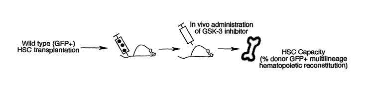
[0033] Figures la, lb, lc, id, le, if, lg, lh, ii, lj and lk show that in vivo
administration of GSK-3 inhibitor augments wild type HSC repopulating
capacity.
[0034] Figures 2a, 2b, 2c, 2d, and 2e show that in vivo administration of GSK-
3
inhibitor augments human neonatal and adult HSC capacity.
[0035] Figures 3a, 3b, 3c ,3d ,3e ,3f ,3g ,3h ,and 3i show in vivo
administration of
GSK-3 inhibitor expands a subset of Lin-c-Kit+Sca-1+ cells with progenitor
capacity but not
secondary reconstitution potential.
[0036] Figures 4a, 4b, 4c ,4d ,4e ,4f ,4g ,4h, 4i, 4j,and 4k show that in vivo
administration of GSK-3 inhibitor to TOP-gal mice enhances HSC activity and
regulates targets
of the Wnt, Notch and Hedgehog pathways.
[0037] Figures 5a, 5b, 5c, and 5d show that in vivo administration of GSK-3
inhibitor to
Ptc-1+/-lacz mice enhances HSC activity and decreases Hedgehog signaling
targets.
[0038] Figures 6a, 6b, 6c, and 6d show that in vitro effects of GSK-3
inhibitor on
purified HSCs.
[0039] Figure 7 shows a proposed model for the functional and molecular
effects of
GSK-3 inhibition on mammalian HSCs.
DETAILED DESCRIPTION
[0040] The invention generally relates to a method for increasing the
successful activity
of stem cells and progenitor cells, for example, hematopoietic progenitor/stem
cells (HSCs),
mesenchymal progenitor/stem cells, mesodermal progenitor/stem cells,
endothelial
progenitor/stem cells, or ectodermal or neural progenitor/stem cells in vivo
in a mammalian
subject comprising interacting one or more Wnt/13-catenin signal-, Notch
signal-, or Hedgehog
signal-promoting agents with the progenitor/stem cells in the mammalian
subject and increasing
the progenitor/stem cells in the mammalian subject. The progenitor/stem cells
can include, but
are not limited to, hematopoietic progenitor/stem cells (HSCs), stem cells,
mesenchymal
progenitor/stem cells, mesodermal progenitor/stem cells, endothelial
progenitor/stem cells,
ectodermal progenitor/stem cells, muscle progenitor/stem cells, endodermal
progenitor/stem cells
or neural progenitor /stem cells The interacting of one or more Wnt/I3-catenin
signal-, Notch
signal-, or Hedgehog signal-promoting agents with progenitor/stem cells, e.g.,
hematopoietic
progenitor/stem cells, occurs either by direct interaction of the signal
promoting agents with the
hematopoietic progenitor/stem cells or through an indirect interaction between
a second
signaling factor or cell type acting as an intermediate between the Wnt/13-
catenin signal-, Notch
signal-, or Hedgehog signal-promoting agents and the hematopoietic
progenitor/stem cells.
- 11 -
CA 02592043 2007-06-20
WO 2006/072016 PCT/US2005/047508
Whether the effect on the HSC, or progenitor/stem cell, is on progenitor/stem
cell proliferation,
survival, cell differentiation, or engraftment into the target tissue, the net
effect of activating the
Wnt/B-catenin signal is an increase in the measured stem cell/progenitor cell
activity. A
progenitor/stem cell, e.g., an hematopoietic progenitor/stem cell, can be
derived from a variety of
sources, including, but not limited to, adult bone marrow, umbilical cord
blood cells, or
embryonic stem cells, from a mammal, e.g., a human. A method of treating
immune related
disease is provided comprising administering one or more Wnt/fl-catenin signal-
, Notch signal-,
or Hedgehog signal-promoting agents to a mammalian subject and interacting the
agent with the
hematopoietic progenitor/stem cells of the mammalian subject. A method of
treating
degenerative disease is provided comprising administering one or more Wnt/B-
catenin signal-,
Notch signal-, or Hedgehog signal-promoting agents to a mammalian subject and
interacting the
agent with hematopoietic progenitor/stem cells, stem cells, mesenchymal
progenitor cells,
mesodermal progenitor cells, muscle progenitor cells, endothelial progenitor
cells, or neural
progenitor cells of the mammalian subject. The degenerative disease includes,
but is not limited
to, mesenchymal degenerative disease, mesodermal degenerative disease, muscle
degenerative
disease, endothelial degenerative disease, or neurodegenerative disease.
[0041] Regulators of hematopoietic progenitor/stem cells (HSCs) that elicit
their effects
in vivo have yet to be identified, limiting clinical manipulation of HSCs to
ex vivo systems. HSC
function can be augmented by administration of Wnt/f3-catenin signal-, Notch
signal-, or
Hedgehog signal-promoting agents, e.g., glycogen synthase kinase-3 (GSK-3)
inhibitors, to
recipient mice transplanted with either wild-type mouse reconstituting HSCs or
human HSCs.
Utilizing mouse reporter models and direct treatment of purified HSCs, GSK-3
inhibitors proved
to enhance HSC activity and modulate gene targets of the Wnt, Hedgehog, and
Notch pathways,
without affecting more mature hematopoietic cells. This study establishes GSK-
3 as a specific
modulator of HSC activity, capable of Wnt, Hedgehog, and Notch pathway
regulation.
Administration of GSK-3 inhibitors of any chemical nature can provide a
clinical approach to
enhance HSC capacity in vivo, thereby providing an alternative to ex vivo
manipulation that
necessitates the removal of HSCs from their physiological environment.
[0042] The present invention demonstrates that in vivo administration of one
or more
Wnt/B-catenin signal-, Notch signal-, or Hedgehog signal-promoting agents,
e.g., GSK-3
inhibitor, increases repopulating function of transplanted wild-type mouse
HSCs, and augments
human neonatal and adult HSC capacity in vivo. In the present invention, the
role of ATP-
competitive GSK-3 inhibitors has been investigated in the regulation of mouse
and human HSCs.
The findings demonstrate that GSK-3 inhibitors augment HSC function in vivo
and modulate
-12-
CA 02592043 2007-06-20
WO 2006/072016 PCT/US2005/047508
Wnt, Hedgehog, and Notch targets specifically in HSCs, thereby providing a
potent and unique
approach to directly enhance HSC function in vivo.
[0043] It is to be understood that this invention is not limited to particular
methods,
reagents, compounds, compositions or biological systems, which can, of course,
vary. It is also to
be understood that the terminology used herein is for the purpose of
describing particular
embodiments only, and is not intended to be limiting. As used in this
specification and the
appended claims, the singular forms "a", "an" and "the" include plural
referents unless the
content clearly dictates otherwise. Thus, for example, reference to "a cell"
includes a
combination of two or more cells, and the like.
[0044] The term "about" as used herein when referring to a measurable value
such as
an amount, a temporal duration, and the like, is meant to encompass variations
of 20% or
10%, more preferably 5%, even more preferably 1%, and still more preferably
0.1% from
the specified value, as such variations are appropriate to perform the
disclosed methods.
[0045] Unless defined otherwise, all technical and scientific terms used
herein have the
same meaning as commonly understood by one of ordinary skill in the art to
which the invention
pertains. Although any methods and materials similar or equivalent to those
described herein can
be used in the practice for testing of the present invention, the preferred
materials and methods
are described herein. In describing and claiming the present invention, the
following terminology
will be used.
[0046] "Increasing hematopoietic stem cells in vivo in a mammalian subject" or
"increasing hematopoietic stem cells in a mammalian subject" or "increasing
the successful
activity of progenitor/stem cells, or hematopoietic progenitor/stem cells in
vivo in a mammalian
subject" or "increasing successful activity of the HSC or progenitor/stem
cell" refers to
increasing an aspect of the life cycle of the progenitor/stem cell or the
hematopoietic
progenitor/stem cell (HSC), for example, as a result of a process, including
but not limited to,
cell proliferation, cell homing to the desired target tissue (e.g.,
transplanted HSCs are provided
intravenously and become established in the bone marrow), decreased apoptosis,
self renewal, or
increased cell survival. An increase in hematopoietic progenitor/stem cells in
vivo can be
measured by a cellular assay as disclosed herein (e.g., in vivo hematopoietic
stem cell
repopulation assay, or hematopoeitic colony-forming unit (CPU) assay) or other
cellular assay
known in the art. Increased hematopoietic progenitor/stem cells in vivo in a
mammalian subject
or increased successful activity can be measured by comparing the fold
increase in HSCs in a
mammalian subject treated by administering one or more Wnt/13-catenin signal-,
Notch signal-,
or Hedgehog signal-promoting agents to the mammalian subject compared to HSCs
in a
- 13 -
CA 02592043 2007-06-20
WO 2006/072016 PCT/US2005/047508
mammalian subject in the absence of such treatment, as measured by any of the
cellular activity
assays for HSCs or progenitor/stem cells discussed herein or known to one
skilled in the art..
The baseline number of HSCs in a mammalian subject is considered the the
successful activity of
HSCs in a mammalian subject in the absence of such treatment. The increase in
HSCs in the
treated mammalian subject by administering one or more Wnt/f3-catenin signal-,
Notch signal-,
or Hedgehog signal-promoting agents can be, for example, at least 1.5 fold, at
least 2-fold, at
least 4-fold, at least 8-fold, or at least 10-fold compared to HSCs in the
mammalian subject
before treatment.
[0047] "Increasing in vivo mesenchymal progenitor/stem cells or mesodermal
progenitor/stem cells in a mammalian subject" or "increasing mesenchymal
progenitor/stem cells
or mesodermal progenitor/stem cells in a mammalian subject" or "increasing in
vivo neural
progenitor/stem cells in a mammalian subject" or "increasing neural
progenitor/stem cells in a
mammalian subject" or or "increasing the successful activity of mesenchymal
progenitor/stem
cells, mesodermal progenitor/stem cells or neural progenitor cells in vivo in
a mammalian
subject" refers to increasing an aspect of the life cycle of the mesenchymal
progenitor/stem cells,
mesodermal progenitor/stem cells, neural progenitor cells, muscle progenitor
cells or stem cells,
for example, as a result of a cellular process, including but not limited to,
cell proliferation, cell
homing to the desired target tissue (e.g., transplanted stem cells, muscle
progenitor cells, or
neural progenitor cells are provided intravenously and become established in
the bone marrow,
muscle, or nerve tissue), decreased apoptosis, self renewal, or increased cell
survival. An
increase in mesenchymal progenitor/stem cells, mesodermal progenitor/stem
cells, neural
progenitor cells, muscle progenitor cells or stem cells in vivo can be
measured by a cellular assay
as disclosed herein (e.g., in vivo hematopoietic stem cell repopulation assay,
hematopoeitic
colony-forming unit (CFU) assay, in vivo stem cell or progenitor cell
repopulation assay, stem
cell or progenitor cell colony-forming unit (CFU) assay or other cellular
assay known in the art).
Increased mesenchymal progenitor/stem cells, mesodermal progenitor/stem cells,
neural
progenitor cells, muscle progenitor cells or stem cells in vivo in a mammalian
subject can be
measured by comparing the fold increase in mesenchymal progenitor/stem cells,
mesodermal
progenitor/stem cells neural progenitor cells, muscle progenitor cells or stem
cells in a
mammalian subject treated by administering one or more Wnt/13-catenin signal-,
Notch signal-,
or Hedgehog signal-promoting agents to the mammalian subject compared to the
number of
mesenchymal progenitor/stem cells, mesodermal progenitor/stem cells, neural
progenitor cells,
muscle progenitor cells or stem cells in a mammalian subject in the absence of
such treatment.
The increase in mesenchymal progenitor/stem cells, mesodermal progenitor/stem
cells neural
- 14 -
CA 02592043 2007-06-20
WO 2006/072016 PCT/US2005/047508
progenitor cells, muscle progenitor cells or stem cells in the mammalian
subject treated by
administering one or more Wnt/13-catenin signal-, Notch signal-, or Hedgehog
signal-promoting
agents can be, for example, at least 1.5 fold, at least 2-fold, at least 4-
fold, at least 8-fold, or at
least 10 fold compared to progenitor/stem cells in the mammalian subject
before treatment.
[0048] "Interacting one or more Wnt/p-catenin signal-, Notch signal-, or
Hedgehog
signal-promoting agents with the hematopoietic stem cells in the mammalian
subject" refers to
either (1) direct contact between the agent and the cell or (2) an indirect
interaction between the
agent and the cell through an intermediary molecule or intermediary cell type.
The interacting
step can refer to directly contacting the hematopoietic progenitor/stem cell
with the Wnt/P-
catenin signal-, Notch signal-, or Hedgehog signal-promoting agents to induce
Wnt/13-catenin
signaling, Notch signaling or Hedgehog signaling within the progenitor/stem
cell, for example,
through receptor/ ligand interaction, intracellular signaling, transcriptional
regulation of gene
expression, cell-cell interaction or intercellular signaling.
[0049] Alternatively, the "interacting" step refers to an indirect interaction
between a
hematopoietic progenitor/stem cell and the Wnt/13-catenin signal-, Notch
signal-, or Hedgehog
signal-promoting agents mediated through a third component, for example,
through an
intermediary signaling molecule, receptor, ligand, growth factor, or cell
type, that affects, or is
affected, by Wnt/P-catenin signaling, Notch signaling or Hedgehog signaling.
For example,
"interacting one or more Wnt/P-catenin signal-, Notch signal-, or Hedgehog
signal-promoting
agents with the hematopoietic progenitor/stem cells in the mammalian subject"
can occur by an
indirect interaction between hematopoietic progenitor/stem cells, stem cells,
muscle progenitor
cells, neural progenitor cells, with the Wnt/P-catenin signal-, Notch signal-,
or Hedgehog signal-
promoting agents. The indirect interaction can occur through intermediary
signaling molecules
for example, growth factors, ligands, receptors or through other cell types
that transmit the
intermediary signals. The intermediary signaling molecules, growth factors,
transcription
factors, ligands, or receptors that increase HSCs, mesenchymal progenitor/stem
cells or
mesodermal progenitor/stem cells neural progenitor/stem cells, muscle
progenitor/stem cells or
stem cells in vivo in a mammalian subject include, but are not limited to,
Notch, Notchl, Jagged-
1, Delta, Delta-1, Delta-4, Oct-3/4, Rex-1, Nanog, LIF-STAT2, STAT5, STAT5A,
sonic
hedgehog, bone morphogenetic proteins, cyclin-dependent kinase inhibitor,
p2lciP1iwan, HoxB4,
or cytokines, e.g., SCF, Fit-3L, G-CSF, IL-3, 1L-6 or IL-11. An indirect
interaction between
hematopoietic progenitor/stem cells and the Wnt/p-catenin signal-, Notch
signal-, or Hedgehog
signal-promoting agents can occur by contacting hematopoietic progenitor/stem
cells with a
number of different growth factors or in a number of different cell types. The
cell types include,
- 15 -
CA 02592043 2007-06-20
WO 2006/072016 PCT/US2005/047508
but are not limited to, stromal cells in bone marrow, any cells in muscle, and
all neural cells
including glial cells and astrocytes.
[0050] "Wnt signal- or P-catenin signal-promoting agent" refers to an agonist
of the
Wnt signaling pathway, including but not limited to an agonist of one or more
of Wntl, Wnt2,
Wnt2b/13, Wnt3, Wnt3a, Wnt4, Wnt5a, Wnt5b, Wnt6, Wnt7a, Wnt7b, Wnt7c, Wnt8,
Wnt8a,
Wnt8b, Wnt8c, Wntl Oa, Wntl Ob, Wntl 1, Wnt14, Wnt15, or Wnt16. "Wnt signal-
or 13-catenin
signal-promoting agent" refers to one or more of the following polypeptides or
a fragment
thereof: a Dkk polypeptide, a crescent polypeptide, a cerberus polypeptide, an
axin polypeptide,
a Frzb polypeptide, a glycogen synthase kinase polypeptide, a T-cell factor
polypeptide, or a
dominant negative dishevelled polypeptide.
[0051] "Wnt/f3-catenin signal-, Notch signal-, or Hedgehog signal-promoting
agent"
further refers to agonists or antagonists of positive or negative signaling
molecules, respectively,
of the Wnt/13-catenin signaling, Notch signaling or Hedgehog signaling
pathway. Signaling
molecules of the Wnt signaling pathway include, but are not limited to, 13-
catenin, tumor
suppressor gene product adenomatous polyposis coll (APC), axin, glycogen
synthase kinase
(GSK) -313, TCF/LEF transcription factors, crescent, groucho, CBP, frizzled
receptor, frizzled
related proteins, LRP, LRP5, LRP6, kremin, Dvl/Dsh (disheveled), dickkopf, GSK-
3 binding
protein (GBP), FRAT/GBP, and any of the Wnt signaling pathway factors listed
at
http://www.stanford.edu/¨musse/pathways/ce112.html.
[0052] "13-catenin signal-promoting agent" refers to agonists or antagonists
of positive
or negative signaling molecules, respectively, of 0-catenin signaling, e.g.,
any agent that
activates B-catenin signaling through inhibition of GSK-3 in the presence or
absence of Wnt
signaling. For example, activation of B-catenin signaling in the absence of
Wnt signaling can
occur by activation of integrin linked kinase, activation of p53 leading to
activation of Siahl, or
activation of FGF signaling. "I3-catenin signal-promoting agent" further
refers to any signaling
molecule that activates 13-catenin target genes and is achieved by inhibition
of GSK-3 that can
have therapeutic potential. "13-catenin signal-promoting agent" further refers
to any signaling
molecule that activates B-catenin target genes independent of GSK-3 that can
have therapeutic
potential. Activation of B-catenin target genes without inhibiting GSK-3 can
be achieved by
inhibition (for example, by drug therapy, RNAi therapy or gene therapy) of any
inhibitor of13-
catenin function, including, but not limited to, APC, Axin, Chibby, ICAT,
Groucho, CtBP.
[0053] "Wnt signal- or 13-catenin signal-promoting agent" refers to one or
more of the
following: a nucleic acid comprising a nucleotide sequence that encodes a Wnt
polypeptide, a
polypeptide comprising an amino acid sequence of a Wnt polypeptide, a nucleic
acid comprising
- 16 -
CA 02592043 2013-06-18
a nucleotide sequence that encodes an activated Wnt receptor, a polypeptide
comprising an
amino acid sequence of an activated Wnt receptor, a small organic molecule
that promotes
Wnt/P-catenin signaling, Notch signaling or Hedgehog signaling, a small
organic molecule that
inhibits the expression or activity of a Wnt, p-catenin, Notch or Hedgehog
antagonist, an
antisense oligonucleotide that inhibits expression of a Wnt, p-catenin, Notch
or Hedgehog
antagonist, a ribozyme that inhibits expression of a Wnt, f3-catenin, Notch or
Hedgehog
antagonist, an RNAi construct, siRNA, or shRNA that inhibits expression of a
Wnt, P-catenin,
Notch or Hedgehog antagonist, an antibody that binds to and inhibits the
activity of a Wnt, 13-
catenin, Notch or Hedgehog antagonist, a nucleic acid comprising a nucleotide
sequence that
encodes a p-catenin polypeptide, a polypeptide comprising an amino acid
sequence of a 13-
catenin polypeptide, a nucleic acid comprising a nucleotide sequence that
encodes a Lef-1
polypeptide, a polypeptide comprising an amino acid sequence of a Lef-1
polypeptide.
[0054] "Wnt/P-catenin signal-, Notch signal-, or Hedgehog signal-promoting
agent"
refers to one or more of the following: a nucleic acid comprising a nucleotide
sequence that
encodes a dominant negative GSK-3, GSK3a, or GSK3P polypeptide, a polypeptide
comprising
an amino acid sequence of a dominant negative GSK-3, GSK3a, or GSK313
polypeptide, a small
organic molecule that binds to and inhibits the expression or activity of GSK-
3, GSK3a, or
GSK313, an RNAi construct, siRNA, or shRNA that binds to and inhibits the
expression and/or
activity of GSK-3, GSK3a, or GSK313, an antisense oligonucleotide that binds
to and inhibits the
expression of GSK-3, GSK3a, or GSK313, an antibody that binds to and inhibits
the expression
and/or activity of GSK-3, GSK3a, or GSK3f3, a ribozyme that binds to and
inhibits the
expression of GSK-3, GSK3a, or GSK313, and any GSK-3-independent reagent that
activates B-
catenin target genes similar in effect to GSK-3 inhibition.
[0055] Exemplary Wnt/13-catenin signal-, Notch signal-, or Hedgehog signal-
promoting
agents include, but are not limited to, LiC1 or other GSK-3 inhibitors, as
exemplified in U.S.
patents 6,057,117 and 6,608,063; and U.S. applications 2004/0092535 and
2004/0209878; ATP-
competitive, selective GSK-3 inhibitors CH1R-911 and CHIR-837 (also referred
to as CT-99021
and CT-98023 respectively). Chiron Corporation (Emeryville, CA). These
inhibitors were
purified to >95% by high-performance liquid chromatography. CH1R-911 was
formulated in
10% captisol solution for administration in vivo by intraperitoneal injection,
with a half-maximal
effective concentration [EC50] of 766nM and >10,000 fold selectivity for GSK-
3. Ring et al.,
Diabetes 52: 588-595, 2003. CHIR-837 was formulated in DMSO for in vitro use,
with an EC50
of 375nM and >5,000 fold selectivity for GSK-3 Cline et al., Diabetes 51: 2903-
2910, 2002.
- 17 -
CA 02592043 2013-06-18
[0056] "Hedgehog compound" refers to a class of molecules of the hedgehog
family
that includes recombinant hedgehog protein, analogs, and derivatives of
hedgehog proteins, and
agonists and antagonists of hedgehog protein receptors and functional
equivalents of the
aforementioned. See, for example, PCT International Applications WO 95/18856
and WO
96/17924.
[0057] "Hedgehog agonist" refers to an agent which potentiates or
recapitulates the
bioactivity of hedgehog, such as to activate transcription of target genes.
Preferred hedgehog
agonists can be used to overcome a ptc gain-of-function and/or a smoothened
loss-of-function,
the latter also being referred to as smoothened agonists. "Hedgehog agonist"
refers not only to
any agent that may act by directly activating the normal function of the
hedgehog protein, but
also to any agent that activates the hedgehog signaling pathway, and thus
inhibits the function of
ptc.
[0058] Further hedgehog agonist proteins include, but are not limited to, TGF-
13
proteins, e.g., TGF- (31, bone morphogenic protein (BM?), e.g., BMP-4; tumor
necrosis factor,
(TNF) proteins, e.g., TNF-a; wnt family; and hedgehog proteins. Compounds may
also include
naturally occurring and synthetic agonists, antagonists, analogs and
derivatives of the above.
These molecules may interact with membrane proteins which initiate signal
transduction
pathways of Wnt, Hedgehog or Notch resulting in a biological response.
Therefore, in addition
to the above compounds, agonists and antagonists to these membrane binding
proteins including
those receptors, receptor agonists and receptor antagonists associated with
hedgehog binding
receptors and hedgehog signalling transduction pathways such as smoothened,
patched and gli
may have utility in regulating hematopoiesis and vascular growth.
[0059] Methods for treating disease in a mammalian subject or methods for
increasing
hematopoietic stem cells in vivo in a mammalian subject are provided by
administering a
therapeutic composition, for example, a polypeptide, nucleic acid, small
molecule, antisense
oligonucleotide, ribozyme, RNAi construct, siRNA, shRNA, or antibody, to the
mammalian
subject. For example, the therapeutic composition can be one or more small
molecule
modulators of Hedgehog signaling. The therapeutic composition can be a GSK-3
inhibitor.
Furthermore, therapeutic efficacy of the Hedgehog pathway antagonist,
cyclopamine, has been
studied in preclinical models of medulloblastoma, a common malignant brain
tumor in children.
Berman et al., Science 297: 1559-1561, 2002. Therapeutic efficacy of the
Hedgehog pathway
agonists have been studied for treatment of traumatic and chronic degenerative
conditions.
Hedgehog pathway agonists have been shown to target the protein Smoothened.
Both
antagonists and agonists of the Hedgehog pathway have been shown to target the
protein
- 18 -
CA 02592043 2013-06-18
Sznoothened. Therapeutic efficacy of a Hedgehog pathway agonist, SAG, a
chlorobenzothiophene-containing Hedgehog pathway agonist, binds to Smoothened
protein in a
manner that antagonizes cylcopamine action. King, Journal of Biology 1:8,
2002; Stecca et al.,
Journal of Biology 1:9, 2002; Frank-Kamenetsky, et al., Journal of Biology
1:10, 2002; Chen et
al., Proc. Natl. Acad. Sci USA 99: 14071-14076, 2002.
[0060] The present invention is directed to methods for increasing non-
terminally
differentiated cells in vivo, e.g., progenitor cells or stem cells, by
activating the Wnt/P-catenin
signaling, Notch signaling or Hedgehog signaling pathway in a progenitor/stem
cell such that the
differentiation of the progenitor/stem cell is inhibited without destroying
the ability of the cell to
proliferate. "precursor cells" or "progenitor/stem cell" shall mean any non-
terminally
differentiated cells. The present invention is also directed to methods for
increasing non-
terminally differentiated cells in vivo, e.g., progenitor cells or stem cells,
by activating the
Wnt/P-catenin signaling, Notch signaling or Hedgehog signaling pathway in the
cells such that
the differentiation of the progenitor/stem cell is inhibited without affecting
the mitotic activity of
the cells. Further, the progenitor/stem cells can be isolated from a cell
population, if desired,
before or after Wnt/P-catenin signaling, Notch signaling or Hedgehog signaling
pathway
activation.
[0061] Activation of Notch pathway is preferably achieved by contacting the
cell with a
Notch ligand, e.g., in soluble form or recombinantly expressed on a cell
surface or immobilized
on a solid surface, or by introducing into the cell a recombinant nucleic acid
expressing a
dominant active Notch mutant or an activating Notch ligand, or other molecule
that activates the
Notch pathway. Agonists of the Notch pathway are able to activate the Notch
pathway at the
level of protein--protein interaction or protein-DNA interaction. Agonists of
Notch include but
are not limited to proteins comprising the portions of toporythmic proteins
such as Delta or
Serrate or Jagged (Lindsell et al., Cell 80: 909-917, 1995) that mediate
binding to Notch, and
nucleic acids encoding the foregoing (which can be administered to express
their encoded
products in vivo).and proteins, nucleic acids, small molecules, or derivatives
thereof that regulate
activity or gene expression of these proteins. In a further embodiment, the
agonist is a protein or
derivative or fragment thereof comprising a functionally active fragment such
as a fragment of a
Notch ligand that mediates binding to a Notch protein. In another embodiment,
the agonist is a
human protein or portion thereof (e.g., human Delta). In another embodiment
the agonist is
Deltex or Suppressor of Hairless or a nucleic acid encoding the foregoing
(which can be
administered to express its encoded product in vivo).
-19-
CA 02592043 2007-06-20
WO 2006/072016 PCT/US2005/047508
[0062] The NOtc-li pliliWay is a signal transducing pathway comprising
elements which
interact, genetically and/or molecularly, with the Notch receptor protein. For
example, elements
which interact with the Notch protein on both a molecular and genetic basis
are, for example, and
not by way of limitation, Delta, Serrate and Deltex. Elements which interact
with the Notch
protein genetically are, for example, and not by way of limitation,
Mastermind, Hairless and
Suppressor of Hairless.
[0063] "Cytopenia" refers to a deficiency of a cellular element of the blood
including,
but not limited, to anemia (low erythrocyte count), neutropenia(low neutrophil
count),
leucopenia(low leucocyte count), thrombocytopenia (low platelet count)..
[0064] "Adult" refers to tissues and cells derived from or within an animal
subject at
any time after birth. "Embryonic" refers to tissues and cells derived from or
within an animal
subject at any time prior to birth.
[0065] "Blood development" refers to hematopoiesis and vascular growth.
"Vascular
growth" refers to at least one of vasculogenesis and angiogenesis and includes
formation of
capillaries, arteries, veins or lymphatic vessels.
[0066] "Hematopoiesis" refers to the process of production of progenitor/stem
cells
from which many cell types are derived. "Hematopoietic stem cell" refers to a
multipotential
precursor from which all classes of blood cell are derived; in addition,
mesenchymal
progenitor/stem cells, mesodermal progenitor/stem cells, endothelial
progenitor/stem cells, and
ectodermal or neural progenitor/stem cells can be derived from hematopoietic
stem cell.
"Definitive blood cells" refers to blood cells of the fetal or adult organism.
"Primitive blood
cells" refers to a transient population of blood cells forming during blood
development in the
embryo.
[0067] "Progenitor cells" or "stem cells" refers to undifferentiated cells
that are more
restricted in their potential to give rise to differentiated cell types
compared with a stem cell.
The progenitor cell or stem cell includes, but is not limited to,
hematopoietic progenitor/stem
cell, mesenchymal progenitor/stem cells, mesodermal progenitor/stem cells,
epithelial
progenitor/stem cell, kidney progenitor/stem cell, neural progenitor/stem
cell, skin
progenitor/stem cell, osteoblast progenitor/stem cell, chondrocyte
progenitor/stem cell, liver
progenitor/stem cell, or muscle progenitor/stem cell.
[0068] "Committed" refers to cells destined to differentiate along a specific
lineage
instead of retaining multipotency.
- 20 -
CA 02592043 2013-06-18
[0069] "Synergistic effect" refers to two or more compounds where little or no
biological effect is observed with the compounds alone but together the
compounds have a
potent biological effect.
[0070] Human hematopoietic progenitor/stem cells (HSCs) have been identified
in
bone marrow (BM), peripheral blood and umbilical cord blood (CB). The
functional capacity of
the cells derived from these tissues can differentiate to a hematopoietic cell
fate. In addition,
marrow-derived stromal cells were found to differentiate along the osteogenic
lineage. Further
studies indicated that multipotent mesenchymal progenitor/stem cells (MSCs)
reside within the
BM, and were capable of giving rise to adipose, bone, cartilage, skeletal
muscle and endothelial
cell lineages. These combined findings have led to the current notion that BM
is therefore a
source of both MSCs as well as HSCs. Similar to BM, human HSCs can also be
found in
umbilical CB and peripheral blood, however, studies aimed at isolating
mesenchymal
stem/progenitor cells from these alternative hematopoietic sources have
provided mixed results.
Cells from pre-term CB displayed mesenchymal properties while more recent
studies reported a
lack of MSCs from full term CB. Similarly, reports have demonstrated the
presence or absence
of mesenchymal precursors from peripheral blood. Jay et al. Cell Research 14:
268-282, 2004.
[0071] Human umbilical cord blood (CB) contains a combination of primitive
cells and
mature cells that have committed to the various hematopoietic lineages.
Studies have focused on
the characterization and clinical utility of progenitor/stem cells from CB
partly due to the ease of
obtaining this abundant cell source and the decreased immunogenicity of these
cells upon
allogenic transplantation. For hematopoietic cell fate, progenitors capable of
multi-lineage
hematopoiesis reside among cellular subsets of uncommitted CB cells that do
not express
specific hematopoietic lineage markers. These mature CB cells can be removed
based on the
surface expression of proteins associated with various hematopoietic lineages
to derive a
remaining subset of primitive cells referred to as the lineage depleted (Lin-)
fraction. Candidate
human HSCs have been shown to exclusively reside in the Lin" fraction and can
be further
enriched to Lin" subsets expressing CD34 but devoid of CD38 (Lin-CD34+CD38).
Subsequent
studies identified additional subpopulations of Lin" cells possessing
hematopoietic progenitor
function that was devoid of both CD34 and CD38 (Lin-CD34-CD38), indicating
that CD34 may
not be unique to the human HSC phenotype. This series of studies illustrates
the heterogeneity
of the Lin-CD34- population in human CB and suggests that additional
subpopulations may
remain to be identified within the Lin- population.
-21-
CA 02592043 2013-06-18
[0072] A population of cells in human CB devoid of the hematopoietic cell fate
marker,
CD45 has been identified. Functional analysis of the Lin-CD45-CD34- cells
revealed that similar
to CD45-CD34- cells from BM, these cells possess chondrocytic differentiation
potential and
hence share properties of mesenchymal progenitors. However, unlike BM-derived
mesenchymal
progenitor/stem cells, CB derived Lin-CD45-CD34" cells possess unique de novo
multi-lineage
hematopoietic progenitor capacity. The functional potential displayed by this
novel population
suggests that Lin-CD45-CD34- cells derived from human CB are potential
therapeutic targets for
cellular therapies for osteogenic as well as hematopoietic deficiencies and
represent a population
of human cells with unique developmental potential. Jay et al. Cell Research
14: 268-282,
2004.
[0073] Embryonic stem cells (ESCs), for example, mammalian or human embryonic
stem cells (hESCs), can be used to derive progenitor/stem cells including but
not limited to,
hematopoietic progenitor/stem cell, mesenchymal progenitor/stem cell,
mesodermal
progenitor/stem cell, endothelial progenitor/stem cell, or ectodermal or
neural progenitor/stem
cell. A subpopulation of primitive endothelial-like cells have been identified
that are derived
from human embryonic stem cells (bESCs) that express PECAM-1, Flk-1, and VE-
cadherin, but
not CD45 (CD45negPFV cells), and that are uniquely responsible for endothelial
and
hematopoietic development. Human hematopoiesis and endothelial maturation can
originate
from a subset of embryonic endothelial cells that possesses hemangioblastic
properties.
[0074] Therapeutic targets of corrective progenitor/stem cell gene therapy
include, but
are not limited to, allogeneic or autologous hematopoietic progenitor/stem
cell transplantation for
treatment of chronic myeloid leukemia, acute myeloid leukemia, non-Hodgkin's
lymphoma,
multiple myeloma, chronic lymphocytic leukemia, Hodgkin's disease,
myelodysplastic
syndrome.
[0075] A method of treating an immune related disease is provided wherein the
disease
is an immunodeficiency disease, for example, primary immunodeficiency, such as
ADA-
deficient SC1D, X-linked SOD, common variable immunodeficiency, chronic
granulomatous
disease (CGD), X-linked agammaglobulinemia, Wiskott-Aldrich syndrome;
hemoglobinopathy,
such as sickle cell anemia, p-thalassemia; other single-gene disorders, such
as Hurler's disease,
Gaucher's disease, hemophilia A, hemophilia B, a-1 antitrypsin deficiency; or
stem cell defects,
such as Fanconi anemia.
[0076] A method of treating a disease or conditions using methods of the
present
invention, include but are not limited to the following disease states. The
gene and genetic
perturbation are provided in parentheses. Schizophrenia (WNTI Elevated); Tetra-
amelia (WNT3
- 22 -
CA 02592043 2013-06-18
LOF); Intersex (WNT4 (OP); Kidney damage (WNT4 Elevated); Polycystic kidney
disease
(WNT4 Variable); Leukaemia (WNT5a LOF; reduced); Metastasis (WNT5a Elevated);
Osteoarthritis (sFRP3 SNP; reduced); FEVR (FZ4 LOF); Familial exudative
vitreoretinopathy
(LRP5, low bone mass LOF); High bone mass (LRP5 GOF); Lung cancer (DSHIDVL
Elevated);
Cancer (APC LOF); Cancer (AX1NLOF); Cancer (AX1N2, tooth agenesis LOF); Cancer
(0-
catenin GOF); Aggressive fibromatosis (0-catenin Elevated); Pulmonary fibrosis
(P-catenin
Elevated); Alzheimer's disease; Cardiovascular disease (Abbreviations: APC,
adenomatous
polyposis coli; DSHIDVL, Dishevelled; FZ, Frizzled; GOF, gain of function;
LOF, loss of
function; LRP, LDL-receptor-related protein; sFRP, secreted Frizzled-related
protein.) Moon et
al., Nature Reviews Genetics 5: 689-699, 2004.
[0077] Inhibitors of WNT/P-catenin signalling can be used for treatment of
cancers.
WNT/ P-catenin signalling seems to be involved in cancer progression, and not
just initiation.
Kim, et al.,. Mol. Cancer Ther. 1: 1355-1359, 2002; Gunther, etal., Genes Dev.
17: 488-501,
2003; Derksen, etal. Proc. Nall Acad. Sci. USA 101: 6122-6127, 2004.
Approaches to cancer
treatment include, but are not limited to, small-molecule inhibitors that
block interaction of 0-
catenin with TCF86 or CREB binding protein (CBP) (Emami, K. H. et al. Proc.
Nat! Acad. Sci.
USA, 2004), siRNAs (Giles, et al., Biochini. Biophys. Acta, 1653: 1-24, 2003)
and the
therapeutic use of antibodies against WNTs. He, et al.. Neoplasia 6, 7-14
(2004); You, et al.,
Oncogene 21 June 2004 [epub ahead of print],
[0078] Other potential therapeutic inhibitors of P-catenin signalling include
agents that
have no obvious link to the P-catenin pathway, such as extracellulax calcium,
non-steroidal anti-
inflammatory drugs, including exisulind, sulindac and aspirin, and the
tyrosine kinase inhibitor
STI571/Gleevac96. Conversely, activators of P-catenin signalling will probably
be useful in
treating osteoporosis and Alzheimer's disease, and might include activators of
LRP5 as well as
inhibitors of GSK3. Moon et al., Nature Reviews Genetics 5: 689-699, 2004.
[0079] A method of treating a degenerative muscle disease is provided wherein
the
disease is a muscular dystrophy or myopathy. Muscular dystrophy (MD) refers to
a group of
genetic diseases characterized by progressive weakness and degeneration of the
skeletal muscles
which control movement. There are many forms of muscular dystrophy, some
noticeable at birth
(congenital muscular dystrophy), others in adolescence (Becker MD). The 3 most
common
types are Duchenne, facioscapulohumeral, and myotonic. These three types
differ in terms of
pattern of inheritance, age of onset, rate of progression, and distribution of
weakness. Duchenne
muscular dystrophy primarily affects boys and is the result of mutations in
the gene that
- 23 -
CA 02592043 2007-06-20
WO 2006/072016 PCT/US2005/047508
regulates dystrophin - a protein involved in maintaining the integrity of
muscle fiber. Onset is
between 3-5 years and progresses rapidly. Facioscapulohumeral muscular
dystrophy appears in
adolescence and causes progressive weakness in facial muscles and certain
muscles in the arms
and legs. It progresses slowly and can vary in symptoms from mild to
disabling. Myotonic
muscular dystrophy varies in the age of onset and is characterized by myotonia
(prolonged
muscle spasm) in the fingers and facial muscles; a floppy-footed, high-
stepping gait; cataracts;
cardiac abnormalities; and endocrine disturbances.
[0080] "Myopathy" refers to neuromuscular disorders in which the primary
symptom is
muscle weakness due to dysfunction of muscle fiber. Other symptoms of myopathy
can include
include muscle cramps, stiffness, and spasm. Myopathies can be inherited (such
as the muscular
dystrophies) or acquired (such as common muscle cramps). Congenital myopathy
is
characterized by developmental delays in motor skills; skeletal and facial
abnormalities are
occasionally evident at birth. Muscular dystrophy is characterized by
progressive weakness in
voluntary muscles; sometimes evident at birth. Mitochondrial myopathy is
caused by genetic
abnormalities in mitochondria, cellular structures that control energy;
include Kearns-Sayre
syndrome, MELAS and MERRF. Glycogen storage diseases of muscle is caused by
mutations in
genes controlling enzymes that metabolize glycogen and glucose (blood sugar);
include Pompe's,
Andersen's and Cori's diseases. Myoglobinuria is caused by disorders in the
metabolism of a
fuel (myoglobin) necessary for muscle work; include McArdle, Tarui, and
DiMauro diseases.
Dermatomyositis is an inflammatory myopathy of skin and muscle. Myositis
ossificans is
characterized by bone growing in muscle tissue. Familial periodic paralysis is
characterized by
episodes of weakness in the arms and legs. Polymyositis, inclusion body
myositis, and related
myopathies are inflammatory myopathies of skeletal muscle. Neuromyotonia is
characterized by
alternating episodes of twitching and stiffness, and stiff-man syndrome is
characterized by
episodes of rigidity and reflex spasms. Common muscle cramps and stiffness,
and tetany is
characterized by prolonged spasms of the arms and legs
[00811 Cardionwopathy refers to a disease of the myocardium associated with
ventricular dysfunction as defined by the World Health Organization. Dilated
cardiomyopathy is
characterized by dilatation and impaired contractility of the left (or right)
ventricle. Presentation
is usually with heart failure. Arrhythmia, thromboembolism, and sudden death
are common.
Hypertrophic cardiomyopathy is characterized by left (or right) ventricular
hypertrophy, which is
usually asymmetric and involves the interventricular septum. Typically, left
ventricular volume
is reduced. Systolic gradients are sometimes present. Typical presentations
include dyspnea,
arrhythmia, and sudden death. Restrictive cardiomyopathy is characterized by
restrictive filling
- 24 -
CA 02592043 2007-06-20
WO 2006/072016 PCT/US2005/047508
of the left (or right) ventricle with normal or near normal ventricular
contractility and wall
thickness. Presentations are usually with heart failure. The cardiomyopathies
are not the only
causes of the heart failure syndrome. In western countries, coronary artery
disease with resultant
ischemic cardiomyopathy remains the primary cause of the heart failure
syndrome.
[0082] A method of treating a neurodegenerative disease is provided wherein
the
disease is a central nervous system disorder or peripheral nervous system
disorder. Central
nervous system disorders encompass numerous afflictions such as
neurodegenerative diseases
(e.g., Alzheimer's disease and Parkinson's disease), acute brain injury (e.g.,
stroke, head injury,
cerebral palsy) and a large number of CNS dysfunctions (e.g., depression,
epilepsy, and
schizophrenia). These diseases, which include Alzheimer's disease, multiple
sclerosis (MS),
Huntington's dsease, amyotrophic lateral sclerosis, and Parkinson's disease,
have been linked to
the degeneration of neural cells in particular locations of the CNS, leading
to the inability of
these cells or the brain region to carry out their intended function.
[0083] "Activators," "inhibitors," and "modulators" of Wnt/13-catenin
signaling, Notch
signaling or Hedgehog signaling are used to refer to activating, or modulating
molecules,
respectively, identified using in vitro and in vivo assays for Wnt/13-catenin
signal-, Notch signal-,
or Hedgehog signal-promoting agents, e.g., ligands, agonists, antagonists, and
their homologs
and mimetics. "Modulator" includes inhibitors and activators. Inhibitors are
agents that, e.g.,
bind to, partially or totally block stimulation, decrease, prevent, delay
activation, inactivate,
desensitize, or down regulate the activity of Wnt/13-catenin signaling, Notch
signaling or
Hedgehog signaling, e.g., antagonists. Activators are agents that, e.g., bind
to, stimulate,
increase, open, activate, facilitate, enhance activation, sensitize or up
regulate the activity of
Wnt/13-catenin signaling, Notch signaling or Hedgehog signaling, e.g.,
agonists. Modulators
include agents that, e.g., alter the interaction of Wnt, 13-catenin, Notch, or
Hedgehog with:
proteins that bind activators or inhibitors, receptors, including proteins,
peptides, lipids,
carbohydrates, polysaccharides, or combinations of the above, e.g.,
lipoproteins, glycoproteins,
and the like. Modulators include genetically modified versions of naturally-
occurring Wnt, 13-
catenin, Notch, or Hedgehog ligands, e.g., Wnt, 13-catenin, Notch, or Hedgehog
polypeptides
with altered activity, as well as naturally occurring and synthetic ligands,
antagonists, agonists,
small chemical molecules and the like. Such assays for inhibitors and
activators include, e.g.,
applying putative modulator compounds to a cell expressing Wnt, P-catenin,
Notch, or Hedgehog
and then determining the functional effects on Wnt/13-catenin signaling, Notch
signaling or
Hedgehog signaling, as described herein. Samples or assays comprising Wnt/13-
catenin signaling,
Notch signaling or Hedgehog signaling that are treated with a potential
activator, inhibitor, or
-25-
CA 02592043 2007-06-20
WO 2006/072016 PCT/US2005/047508
modulator are compared to control samples without the inhibitor, activator, or
modulator to
examine the extent of inhibition. Control samples (untreated with activators,
inhibitors, or
modulators) can be assigned a relative Wnt, P-catenin, Notch, or Hedgehog
activity value of
100%. Inhibition of Wnt/P-catenin signaling, Notch signaling or Hedgehog
signaling is achieved
when the Wnt, P-catenin, Notch, or Hedgehog activity value relative to the
control is about 80%,
optionally 50% or 25-0%. Activation of Wnt/P-catenin signaling, Notch
signaling or Hedgehog
signaling is achieved when the Wnt, P-catenin, Notch, or Hedgehog activity
value relative to the
control is 110%, optionally 150%, optionally 200-500%, or 1000-3000% higher.
Activation of
Wnt/P-catenin signaling, Notch signaling or Hedgehog signaling can be measured
in a cellular
assay for increasing hematopoietic progenitor/stem cells in vivo in a
mammalian subject or
increasing mesenchymal progenitor/stem cells, mesodermal progenitor/stem
cells, neural
progenitor/stem cells, muscle progenitor/stem cells or stem cells in vivo in a
mammalian subject,
as described herein.
[0084] "Agonist" is used in the broadest sense and includes any molecule that
mimics
or enhances a biological activity of Wnt, f3-catenin, Notch, or Hedgehog
polypeptides, or Wnt/f3-
catenin signaling, Notch signaling or Hedgehog signaling. Suitable agonist
molecules
specifically include agonist antibodies or antibody fragments, fragments or
amino acid sequence
variants of native Wnt, f3-catenin, Notch, or Hedgehog polypeptides, peptides,
antisense
oligonucleotides, small organic molecules, and the like. Methods for
identifying agonists of Wnt,
P-catenin, Notch, or Hedgehog polypeptides can comprise contacting a Wnt, P-
catenin, Notch, or
Hedgehog polypeptide with a candidate agonist molecule and measuring a
detectable change in
one or more biological activities normally associated with the Wnt/13-catenin
signaling, Notch
signaling or Hedgehog signaling, e.g., Wnt binding to the Frizzled receptor,
or intracellular
accumulation of P-catenin.
[0085] "Antagonist" is used in the broadest sense, and includes any molecule
that
partially or fully blocks, inhibits, or neutralizes an inhibitor of a
biological activity of a Wnt,
catenin, Notch, or Hedgehog polypeptide, or Wnt/13-catenin signaling, Notch
signaling or
Hedgehog signaling. Suitable antagonist molecules specifically include
antagonist antibodies or
antibody fragments, fragments or amino acid sequence variants of native
polypeptides, peptides,
antisense oligonucleotides, small organic molecules, and the like. Methods for
identifying
antagonists of an inhibitor of a biological activity of a Wnt, P-catenin,
Notch, or Hedgehog
polypeptide, or of Wnt- or P-catenin- signaling can comprise contacting Wnt, P-
catenin, Notch,
or Hedgehog polypeptides, or Wnt- or P-catenin-signaling polypeptides with a
candidate
- 26 -
CA 02592043 2007-06-20
WO 2006/072016 PCT/US2005/047508
antagonist molecule and measuring a detectable change in one or more
biological activities
normally associated with Wnt/P-catenin signaling, Notch signaling or Hedgehog
signaling.
[0086] "Signaling in cells" refers to the interaction of a ligand, such as an
endogenous
or exogenous ligand, e.g., Notch signal-promoting or Hedgehog signal-
promoting, or Wnt signal-
promoting or 13-catenin signal-promoting agents, with receptors, such as
Frizzled receptor,
resulting in cell signaling to produce a response, for example, increasing
expression of B-catenin
target genes resulting in increased hematopoietic progenitor/stem cells in
vivo in a mammalian
subject.
[0087] "Endogenous" refers a protein, nucleic acid, lipid or other component
produced
within the body or within cells or organs of the body of a mammalian subject
or originating
within cells or organs of the body of a mammalian subject.
[0088] "Exogenous" refers a protein, nucleic acid, lipid, or other component
originating
outside the body of a mammalian subject.
[0089] "Test compound" refers to a nucleic acid, DNA, RNA, protein,
polypeptide, or
small chemical entity that is determined to effect an increase or decrease in
a gene expression as
a result of signaling through the Wnt/13-catenin signaling, Notch signaling or
Hedgehog signaling
pathways. The test compound can be an antisense RNA, ribozyme, polypeptide, or
small
molecular chemical entity. "Test compound" can be any small chemical compound,
or a
biological entity, such as a protein, sugar, nucleic acid or lipid. Typically,
test compounds will be
small chemical molecules and polypeptides. A "test compound specific for
Wnt/13-catenin
signaling, Notch signaling or Hedgehog signaling" is determined to be a
modulator of Notch
signaling or Hedgehog signaling or Wnt signaling or 13-catenin signaling, for
example, resulting
in Wnt binding to the Frizzled receptor, or intracellular accumulation of B-
catenin.
[0090] "Cell-based assays" include Wnt/B-catenin signaling, Notch signaling or
Hedgehog signaling assays, for example, radioligand or fluorescent ligand
binding assays for
Wnt, 0-catenin, Notch, or Hedgehog to cells, plasma membranes, detergent-
solubilized plasma
membrane proteins, immobilized collagen (Alberdi, J Biol Chem. 274:31605-12,
1999; Meyer et
al., 2002); Wnt,13-catenin, Notch, or Hedgehog-affinity column chromatography
(Alberdi, J Biol
Chem. 274:31605-12, 1999; Aymerich etal., Invest Ophthalmol Vis Sci. 42:3287-
93, 2001);
Wnt,13-catenin, Notch, or Hedgehog blot using a radio- or fluorosceinated-
ligand (Aymerich et
al., Invest Ophthalmol Vis Sci. 42:3287-93, 2001; Meyer etal., 2002); Size-
exclusion
ultrafiltration (Alberdi et al., 1998, Biochem.; Meyer et al., 2002); or
ELISA. Cellular assay to
measure increasing hematopoietic progenitor/stem cells in vivo in a mammalian
subject or
increasing neural progenitor cells, muscle progenitor cells or stem cells in
vivo in a mammalian
- 27 -
CA 02592043 2013-06-18
subject, as described herein, include, but are not limited to, in vivo HSC
repopulation assay or
hematopoeitic colony-forming unit (CPU) assays. Multilineage donor HSC
reconstitution was
examined by flow cytometry for surface markers representing primitive (c-Kit+
Sca-l+),
myeloid (CD45+ CD11b+), erythroid (CD45- Ten 19+), B cell (CD45+ B220+), and T
cell
(CD45+ CD3+) lineages indicating multilineage donor HSC reconstitution.
Exemplary Wnt/j3-
catenin signaling, Notch signaling or Hedgehog signaling activity assays of
the present invention
are: Assays for Wnt-regulated target gene Axin2 quantified by real-time PCR;
Yan et al., Proc
Nati Acad Sci USA 98:14973-8, 2001; Jho et al., Mol Cell Biol 22:1172-83,2002.
Wnt-regulated
target gene CyclinD1 quantified by real-time PCR; Issack and Ziff, Cell Growth
Differ 9:837-
45, 1998. Notch regulated target gene, Hesl, quantified by real-time PCR;
Jarriault et al.,
Nature 377:355-8, 1995. Hedgehog regulated target genes, G1i3 and Patchedl
(Ptcl), quantified
by real-time PCR. Mango etal., 180 1:1996; Mango and Tabin, Proc Natl Acad Sci
USA
93:9346-9351, 1996. Further cell based assays
include but are not limited to, luciferase, green fluorescent protein (GFP),
or 13-galactosidase
reporter screens for 13-catenin responsive genes, for example using TOPFLASH
reporter. See,
for example, Veeman M. et al., Current Biology, 13: 680-685, 2003; Veeman M.
et al., Dev Cell.
5: 367-377, 2003.
[0091] In one aspect, a method of screening drug candidates for Wnt/13-catenin
signal-,
Notch signal-, or Hedgehog signal-promoting agents is provided and comprise,
for example, a B-
catenin-responsive gene reporter assay in which a reporter DNA construct
consists of TCF or
LEF binding sites upstream of a minimal promoter, and drives expression of
luciferase, GFP, or
13-galactosidase. Luciferase, GFP, or 13-ga1actosidase is used as a surrogate
to demonstrate
increased activity of13-catenin signaling, as measured by the reporter protein
fluorescence or
enzymatic means. Further methods of screening drug candidates for Wnt signal-
or 0-catenin
signal-promoting agents involve an assay to measure Wnt binding to the
Frizzled receptor, or
intracellular accumulation ofp-catenin. Wnt binding to the Frizzled receptor
or intracellular
accumulation of P-catenin can be assayed by either immobilizing the ligand or
the receptor. For
example, the assay can include immobilizing Frizzled receptor fused to a His
tag onto Ni-
activated NTA resin beads. Wnt can be added in an appropriate buffer and the
beads incubated
for a period of time at a given temperature. After washes to remove unbound
material, the bound
protein can be released with, for example, SDS, buffers with a high pH, and
the like and
analyzed. For example, the Wnt signal- or P-catenin signal-promoting agent
promotes Wnt
binding to the Frizzled receptor or intracellular accumulation of13-catenin.
-28-
CA 02592043 2013-06-18
[0092] "Interacting" refers to mixing a test compound, e.g., one or more
Wnt/13-catenin
signal-, Notch signal-, or Hedgehog signal-promoting agents, in a soluble form
into an assay
system, for example, a cell-based assay system, such that an effect upon
receptor-mediated
signaling or intracellular signaling can be measured. For example, the
"interacting" step can
occur directly by contacting the hematopoietic progenitor/stem cell, stem
cell, muscle progenitor
cell, or neural progenitor cell and the one or more Wnt/13-catenin signal-,
Notch signal-, or
Hedgehog signal-promoting agents to induce Wnt/13-catenin signaling, Notch
signaling or
Hedgehog signaling within the stem cell or progenitor cell. Alternatively, the
"interacting" step
can occur indirectly between a hematopoietic progenitor/stem cell, stem cell,
muscle progenitor
cell, or neural progenitor cell and the Wnt/13-catenin signal-, Notch signal-,
or Hedgehog signal-
promoting agents, for example, through an intermediary signaling molecule,
receptor, ligand,
growth factor, or cell type, that affects, or is affected by, Wnt/13-catenin
signaling, Notch
signaling or Hedgehog signaling.
[0093] "Signaling responsiveness" or "effective to activate signaling" or
"stimulating a
cell-based assay system" refers to the ability of Wnt/13-catenin signal-,
Notch signal-, or
Hedgehog signal-promoting agent to enhance an immune response, to increase
hematopoietic
progenitor/stem cells, stem cells, mesenchymal progenitor/stem cells,
mesodermal
progenitor/stem cells muscle progenitor cells, or neural progenitor cells in
vivo in a mammalian
subject, or treating an immune related disease or a degenerative muscle
disease or
neurodegenerative disease in a mammalian subject.
[0094] "Detecting an effect" refers to an effect measured in a cell-based
assay system.
For example, the effect detected can be Wnt/13-catenin signaling, Notch
signaling or Hedgehog
signaling in an assay system, for example, Wnt,13-catenin, Notch, or Hedgehog
cellular assay,
Frizzled receptor binding assay, Axin2 assay, or CyclinD1 assay, or13-catenin-
responsive gene
reporter assay. See, for example, Veeman M. et al., Current Biology, 13: 680-
685, 2003;
Veeman M. et al., Dev Cell. 5: 367-377, 2003.
[0095] "Assay being indicative of modulation" refers to results of a cell-
based assay
system indicating that cell activation by Wnt/13-catenin signal-, Notch signal-
, or Hedgehog
signal-promoting agents are indicated to treat an immune related disease in a
mammalian subject
by increasing hematopoietic progenitor/stem cells, stem cells, muscle
progenitor cells, or neural
progenitor cells in vivo in a mammalian subject or treating a degenerative
muscle disease or
neuro degenerative disease in a mammalian subject by increasing
progenitor/stem cells, muscle
progenitor cells, or neural progenitor cells.
- 29 -
CA 02592043 2007-06-20
WO 2006/072016 PCT/US2005/047508
[0096] "Biological activity" and "biologically active" with regard to Wnt/P-
catenin
signal-, Notch signal-, or Hedgehog signal-promoting agents of the present
invention refer to the
ability of the ligand molecule to specifically bind to and signal through a
native or recombinant
Wnt, P-catenin, Notch, or Hedgehog, or to block the ability of an inhibitor of
native or
recombinant Wnt, P-catenin, Notch, or Hedgehog polypeptides to participate in
signal
transduction. Thus, the (native and variant) Wnt/f3-catenin signal-, Notch
signal-, or Hedgehog
signal-promoting agents of the present invention include agonists of a native
or recombinant
Wnt, 13-catenin, Notch, or Hedgehog polypeptides and receptors or ligands
thereof. Preferred
biological activities of the Wnt/P-catenin signal-, Notch signal-, or Hedgehog
signal-promoting
agents of the present invention include the ability to induce or inhibit, for
example, enhancing an
immune response, or increasing hematopoietic progenitor/stem cells, stem
cells, muscle
progenitor cells, or neural progenitor cells in vivo in a mammalian subject or
treating a
degenerative muscle disease or neurodegenerative disease in a mammalian
subject. Accordingly,
the administration of the compounds or agents of the present invention can
prevent or delay, to
alleviate, or to arrest or inhibit development of the symptoms or conditions
associated with
immune related disease, a degenerative muscle disease or neurodegenerative
disease in a
mammalian subject.
[0097] "Signal transduction pathway" or "signal transduction event" refers to
at least
one biochemical reaction, but more commonly a series of biochemical reactions,
which result
from interaction of a cell with a stimulatory compound or agent. Thus, the
interaction of a
stimulatory compound with a cell generates a "signal" that is transmitted
through the signal
transduction pathway, ultimately resulting in a cellular response, e.g., an
immune response
described above.
[0098] "High affinity" for a ligand refers to an equilibrium association
constant (Ka) of
at least about 103M-1, at least about 104M-1, at least about 105M-1, at least
about 106M-1, at least
about 107 M-1, at least about 108M-1, at least about 109M-1, at least about
1010 M-1, at least about
1011M-1, or at least about 1012M-1 or greater, e.g., up to 1013M-1 or 1014M-1
or greater. However,
"high affinity" binding can vary for other ligands.
[0099] "Ka", as used herein, is intended to refer to the equilibrium
association constant
of a particular ligand-receptor interaction, e.g., antibody-antigen
interaction. This constant has
units of 1/M.
[0100] "Kd", as used herein, is intended to refer to the equilibrium
dissociation constant
of a particular ligand-receptor interaction. This constant has units of M.
-30-
CA 02592043 2007-06-20
WO 2006/072016 PCT/US2005/047508
[0101] "ka", as used herein, is intended to refer to the kinetic association
constant of a
particular ligand-receptor interaction. This constant has units of 1/Ms.
[0102] "lcd", as used herein, is intended to refer to the kinetic dissociation
constant of a
particular ligand-receptor interaction. This constant has units of 1/s.
[0103] "Particular ligand-receptor interactions" refers to the experimental
conditions
under which the equilibrium and kinetic constants are measured.
[0104] "Isotype" refers to the antibody class that is encoded by heavy chain
constant
region genes. Heavy chains are classified as gamma, mu, alpha, delta, or
epsilon, and define the
antibody's isotype as IgG, IgM, IgA, IgD and IgE, respectively. Additional
structural variations
characterize distinct subtypes of IgG (e.g., IgGi, IgG2, IgG3 and IgG4) and
IgA (e.g., IgAi and
IgA2)
[0105] The ability of a molecule to bind to Wnt, P-catenin, Notch, or Hedgehog
can be
determined, for example, by the ability of the putative ligand to modulate Wnt
binding to the
Frizzled receptor, by measuring intracellular accumulation of 13-catenin,
Axin2 assay, or
CyclinD1 assay. Specificity of binding can be determined by comparing binding
in the presence
or absence of the putative ligand.
[0106] "Control sequences" or "regulatory sequences" refers to DNA sequences
necessary for the expression of an operably linked coding sequence in a
particular host organism.
The control sequences that are suitable for prokaryotes, for example, include
a promoter,
optionally an operator sequence, a ribosome binding site, and possibly, other
as yet poorly
understood sequences. Eukaryotic cells are known to utilize promoters,
polyadenylation signals,
and enhancers.
[0107] "Vector" refers to a nucleic acid molecule capable of transporting
another
nucleic acid to which it has been linked. One type of vector is a "plasmid",
which refers to a
circular double stranded DNA loop into which additional DNA segments can be
ligated. Another
type of vector is a viral vector, wherein additional DNA segments can be
ligated into the viral
genome. Certain vectors are capable of autonomous replication in a host cell
into which they are
introduced (e.g., bacterial vectors having a bacterial origin of replication
and episomal
mammalian vectors). Other vectors (e.g., non-episomal mammalian vectors) can
be integrated
into the genome of a host cell upon introduction into the host cell, and
thereby are replicated
along with the host genome. Moreover, certain vectors are capable of directing
the expression of
genes to which they are operatively linked. Such vectors are referred to
herein as "recombinant
expression vectors" (or simply, "expression vectors"). In general, expression
vectors of utility in
recombinant DNA techniques are often in the form of plasmids. In the present
specification,
- 31 -
CA 02592043 2007-06-20
WO 2006/072016 PCT/US2005/047508
"plasmid" and "vector" can be used interchangeably as the plasmid is the most
commonly used
form of vector. However, the invention is intended to include such other forms
of expression
vectors, such as viral vectors (e.g., replication defective retroviruses,
adenoviruses and adeno-
associated viruses), which serve equivalent functions.
[0108] A "label" is a composition detectable by spectroscopic, photochemical,
biochemical, immunochemical, or chemical means. For example, useful labels
include 32P,
fluorescent dyes, electron-dense reagents, enzymes (e.g., as commonly used in
an ELISA),
biotin, digoxigenin, or haptens and proteins for which antisera or monoclonal
antibodies are
available (e.g., the polypeptides of the invention can be made detectable,
e.g., by incorporating a
radiolabel into the peptide, and used to detect antibodies specifically
reactive with the peptide).
[0109] "Sorting" in the context of cells as used herein to refers to both
physical sorting
of the cells, as can be accomplished using, e.g., a fluorescence activated
cell sorter, as well as to
analysis of cells based on expression of cell surface markers, e.g., FACS
analysis in the absence
of sorting.
[0110] "Cell," "cell line," and "cell culture" are used interchangeably and
all such
designations include progeny. Thus, the words "transforrnants" and
"transformed cells" include
the primary subject cell and cultures derived therefrom without regard for the
number of
transfers. It is also understood that all progeny cannot be precisely
identical in DNA content, due
to deliberate or inadvertent mutations. Mutant progeny that have the same
function or biological
activity as screened for in the originally transformed cell are included.
Where distinct
designations are intended, it will be clear from the context.
[0111] "Receptor" denotes a cell-associated protein, for example Frizzled
receptor, that
binds to a bioactive molecule termed a "ligand." This interaction mediates the
effect of the ligand
on the cell. Receptors can be membrane bound, cytosolic or nuclear; monomeric
(e.g., Frizzled
receptor, thyroid stimulating hormone receptor, beta-adrenergic receptor) or
multimeric (e.g.,
PDGF receptor, growth hormone receptor, IL-3 receptor, GM-CSF receptor, G-CSF
receptor,
erythropoietin receptor and IL-6 receptor). Membrane-bound receptors, for
example Frizzled
receptor, are characterized by a multi-domain structure comprising an
extracellular ligand-
binding domain and an intracellular effector domain that is typically involved
in signal
transduction. In certain membrane-bound receptors, the extracellular ligand-
binding domain and
the intracellular effector domain are located in separate polypeptides that
comprise the complete
functional receptor.
[0112] In general, the binding of ligand to receptor results in a
conformational change
in the receptor that causes an interaction between the effector domain and
other molecule(s) in
- 32 -
CA 02592043 2007-06-20
WO 2006/072016 PCT/US2005/047508
the cell, which in turn leads to an alteration in the metabolism of the cell.
Metabolic events that
are often linked to receptor-ligand interactions include gene transcription,
phosphorylation,
dephosphorylation, increases in cyclic AMP production, mobilization of
cellular calcium,
mobilization of membrane lipids, cell adhesion, hydrolysis of inositol lipids
and hydrolysis of
phospholipids.
[0113] "Treatment" or "treating" refers to any indicia of success in the
treatment or
amelioration of an injury, pathology or condition, including any objective or
subjective
parameter such as abatement; remission; diminishing of symptoms or making the
injury,
pathology, or condition more tolerable to the patient; slowing in the rate of
degeneration or
decline; making the final point of degeneration less debilitating; or
improving a subject's
physical or mental well-being. The treatment or amelioration of symptoms can
be based on
objective or subjective parameters; including the results of a physical
examination. Accordingly,
"treatment" or "treating" includes the administration of the compounds or
agents of the present
invention to inhibit or enhance an immune response, or treat immune related
disease, diabetes,
graft vs. host disease, immunodeficiency disease, hematopoietic malignancy,
hematopoietic
failure, hematopoietic progenitor/stem cell transplantation, or a muscle
degenerative disease. It
also includes the administration of the compounds of the present invention to
enhance an
immune response in a subject toward infection with a pathogen. Accordingly,
"treatment" or
"treating" includes the administration of the compounds or agents of the
present invention to
prevent or delay, to alleviate, or to arrest or inhibit development of the
symptoms or conditions
associated with immune related disease, diabetes, graft vs. host disease,
immunodeficiency
disease, hematopoietic malignancy, hematopoietic failure, hematopoietic
progenitor/stem cell
transplantation, a muscle degenerative disease, or other disorders.
"Therapeutic effect" refers to
the reduction, elimination, or prevention of the disease, symptoms of the
disease, or side effects
of the disease in the subject.
[0114] "Concomitant administration" of a known drug with a compound of the
present
invention means administration of the drug and the compound at such time that
both the known
drug and the compound will have a therapeutic effect or diagnostic effect.
Such concomitant
administration can involve concurrent (i.e. at the same time), prior, or
subsequent administration
of the drug with respect to the administration of a compound of the present
invention. A person
of ordinary skill in the art, would have n9 difficulty determining the
appropriate timing, sequence
and dosages of administration for particular drugs and compounds of the
present invention.
[0115] "Subject", "mammalian subject" or "patient" refers to any mammalian
patient or
subject to which the compositions of the invention can be administered.
"Mammal" or
- 33 -
CA 02592043 2007-06-20
WO 2006/072016 PCT/US2005/047508
"mammalian" refers to human patients and non-human primates, as well as
experimental animals
such as rabbits, rats, and mice, and other animals. In an exemplary
embodiment, of the present
invention, to identify subject patients for treatment according to the methods
of the invention,
accepted screening methods are employed to determine risk factors associated
with a targeted or
suspected disease or condition or to determine the status of an existing
disease or condition in a
subject. These screening methods include, for example, conventional work-ups
to determine risk
factors that can be associated with the targeted or suspected disease or
condition. These and
other routine methods allow the clinician to select patients in need of
therapy using the methods
and formulations of the invention.
[0116] By "solid phase" is meant a non-aqueous matrix to which a reagent of
interest
(e.g., Wnt, 0-catenin, or Frizzled receptor, or an antibody thereto) can
adhere. Examples of solid
phases encompassed herein include those formed partially or entirely of glass
(e.g., controlled
pore glass), polysaccharides (e.g., agarose), polyacrylamides, polystyrene,
polyvinyl alcohol and
silicones. In certain embodiments, depending on the context, the solid phase
can comprise the
well of an assay plate; in others it is a purification column (e.g. ,an
affinity chromatography
column). This term also includes a discontinuous solid phase of discrete
particles, such as those
described in U.S. Pat. No. 4,275,149.
[0117] "Specifically (or selectively) binds" to an antibody refers to a
binding reaction
that is determinative of the presence of the protein in a heterogeneous
population of proteins and
other biologics. Thus, under designated immunoassay conditions, the specified
antibodies bind
to a particular protein at least two times the background and do not
substantially bind in a
significant amount to other proteins present in the sample.
[0118] "Specifically bind(s)" or "bind(s) specifically" when referring to a
peptide refers
to a peptide molecule which has intermediate or high binding affmity,
exclusively or
predominately, to a target molecule. The phrase "specifically binds to" refers
to a binding
reaction which is determinative of the presence of a target protein in the
presence of a
heterogeneous population of proteins and other biologics. Thus, under
designated assay
conditions, the specified binding moieties bind preferentially to a particular
target protein and do
not bind in a significant amount to other components present in a test sample.
Specific binding
to a target protein under such conditions can require a binding moiety that is
selected for its
specificity for a particular target antigen. A variety of assay formats can be
used to select ligands
that are specifically reactive with a particular protein. For example, solid-
phase ELISA
immunoassays, immunoprecipitation, Biacore and Western blot are used to
identify peptides that
specifically affect Wnt signaling, f3-catenin signaling, or Frizzled receptor
proteins. Typically a
-34-
CA 02592043 2007-06-20
WO 2006/072016 PCT/US2005/047508
specific or selective reaction will be at least twice background signal or
noise and more typically
more than 10 times background. Specific binding between a monovalent peptide
and Wnt
signaling, f3-catenin signaling, or Frizzled receptor proteins means a binding
affinity of at least
103 M-1, and preferably 105, 106, 107, 108, 109 or 1010 1V1-1. The binding
affinity of Wnt to the
Frizzled receptor is between about 106 M-1 to about 1010 M-1.
[0119] The present invention is based on the discovery that in vivo
administration of
one or more Wnt/13-catenin signal-, Notch signal-, or Hedgehog signal-
promoting agents, e.g.,
GSK-3 inhibitor, increases repopulating function of transplanted wild-type
mouse HSCs, and
augments human neonatal and adult HSC capacity in vivo. In the present
invention, the role of
ATP-competitive GSK-3 inhibitors has been investigated in the regulation of
mouse and human
HSCs. The findings demonstrate that GSK-3 inhibitors augment HSC function in
vivo and
modulate Wnt, Hedgehog, and Notch targets specifically in HSCs, thereby
providing a potent
and unique approach to directly enhance HSC function in vivo.
[0120] This invention relies on routine techniques in the field of recombinant
genetics.
Basic texts disclosing the general methods of use in this invention include
Sambrook et al.,
Molecular Cloning, A Laboratory Manual, 2nd ed., 1989; Kriegler, Gene Transfer
and
Expression: A Laboratory Manual, 1990; and Ausubel et al., eds.õ Current
Protocols in
Molecular Biology, 1994.
[0121] Wnt, P-catenin, Notch, or Hedgehog nucleic acids, polymorphic variants,
orthologs, and alleles that are substantially identical to sequences provided
herein can be isolated
using Wnt, P-catenin, Notch, or Hedgehog nucleic acid probes and
oligonucleotides under
stringent hybridization conditions, by screening libraries. Alternatively,
expression libraries can
be used to clone Wnt, P-catenin, Notch, or Hedgehog receptor protein,
polymorphic variants,
orthologs, and alleles by detecting expressed homologs immunologically with
antisera or
purified antibodies made against human Wnt, P-catenin, Notch, or Hedgehog
portions thereof.
Identification of Compounds for Treatment and Prophylaxis of Disease
(A) Identification of Bioactive Agents
[0122] Identifying bioactive agents that modulate Wnt/P-catenin signaling,
Notch
signaling or Hedgehog signaling, the information is used in a wide variety of
ways. In one
method, one of several cellular assays, e.g., Wnt/P-catenin signaling, Notch
signaling or
Hedgehog signaling assay, can be used in conjunction with high throughput
screening
techniques, to allow monitoring for antagonists or agonists of Wnt/p-catenin
signaling, Notch
signaling or Hedgehog signaling after treatment with a candidate agent,
Zlokarnik, et al., Science
- 35 -
CA 02592043 2013-06-18
279:84-8, 1998; and Heid et al., Genome Res. 6:986, 1996.
In one method, the candidate agents are added to cells.
[0123] "Candidate bioactive agent" or "drug candidate" or grammatical
equivalents as
used herein describes any molecule, e.g., protein, oligopeptide, small organic
molecule,
polysaccharide, polynucleotide, to be tested for bioactive agents that are
capable of directly or
indirectly altering the activity of Wnt/P-catenin signaling, Notch signaling
or Hedgehog
signaling. In one methods, the bioactive agents modulate Wnt/13-catenin
signaling, Notch
signaling or Hedgehog signaling. In a further embodiment of the method, the
candidate agents
induce an antagonist or agonist effect in a Wnt/P-catenin signaling, Notch
signaling or Hedgehog
signaling assay, as further described below. Generally a plurality of assay
mixtures are run in
parallel with different agent concentrations to obtain a differential response
to the various
concentrations. Typically, one of these concentrations serves as a negative
control, i.e., at zero
concentration or below the level of detection.
[0124] Candidate agents encompass numerous chemical classes, though typically
they
are organic molecules, e.g., small organic compounds having a molecular weight
of more than
100 and less than about 2,500 daltons. Candidate agents comprise functional
groups necessary
for structural interaction with proteins, particularly hydrogen bonding, and
typically include at
least an amine, carbonyl, hydroxyl or carboxyl group, for example, at least
two of the functional
chemical groups. The candidate agents often comprise cyclical carbon or
heterocyclic structures
and/or aromatic or polyaromatic structures substituted with one or more of the
above functional
groups. Candidate agents are also found among biomolecules including peptides,
saccharides,
fatty acids, steroids, purines, pyrimidines, derivatives, structural analogs
or combinations thereof.
In a further embodiment, candidate agents are peptides.
[0125] Candidate agents are obtained from a wide variety of sources including
libraries
of synthetic or natural compounds. For example, numerous means are available
for random and
directed synthesis of a wide variety of organic compounds and biomolecules,
including
expression of randomized oligonucleotides. Alternatively, libraries of natural
compounds in the
form of bacterial, fungal, plant and animal extracts are available or readily
produced.
Additionally, natural or synthetically produced libraries and compounds are
readily modified
through conventional chemical, physical and biochemical means. Known
pharmacological
agents can be subjected to directed or random chemical modifications, such as
acylation,
alkylation, esterification, amidification to produce structural analogs.
[0126] In some embodiments, the candidate bioactive agents are proteins. By
"protein"
herein is meant at least two covalently attached amino acids, which includes
proteins,
- 36 -
CA 02592043 2007-06-20
WO 2006/072016 PCT/US2005/047508
polypeptides, oligopeptides and peptides. The protein can be made up of
naturally occurring
amino acids and peptide bonds, or synthetic peptidomimetic structures. Thus
"amino acid", or
"peptide residue", as used herein means both naturally occurring and synthetic
amino acids. For
example, homo-phenylalanine, citrulline and noreleucine are considered amino
acids for the
purposes of the methods herein. "Amino acid" also includes imino acid residues
such as proline
and hydroxyproline. The side chains can be in either the (R) or the (S)
configuration. In further
embodiments, the amino acids are in the (S) or (L)-configuration. If non-
naturally occurring side
chains are used, non-amino acid substituents can be used, for example to
prevent or retard in vivo
degradations.
[0127] In one method, the candidate bioactive agents are naturally occurring
proteins or
fragments of naturally occurring proteins. Thus, for example, cellular
extracts containing
proteins, or random or directed digests of proteinaceous cellular extracts,
can be used. In this
way libraries of procaryotic and eucaryotic proteins can be made for screening
using the methods
herein. The libraries can be bacterial, fungal, viral, and mammalian proteins,
and human
proteins.
[0128] In some methods, the candidate bioactive agents are peptides of from
about 5 to
about 30 amino acids, typically from about 5 to about 20 amino acids, and
typically from about
7 to about 15 being. The peptides can be digests of naturally occurring
proteins as is outlined
above, random peptides, or "biased" random peptides. By "randomized" or
grammatical
equivalents herein is meant that each nucleic acid and peptide consists of
essentially random
nucleotides and amino acids, respectively. Since generally these random
peptides (or nucleic
acids, discussed below) are chemically synthesized, they can incorporate any
nucleotide or
amino acid at any position. The synthetic process can be designed to generate
randomized
proteins or nucleic acids, to allow the formation of all or most of the
possible combinations over
the length of the sequence, thus forming a library of randomized candidate
bioactive
proteinaceous agents.
[0129] In some methods, the library can be fully randomized, with no sequence
preferences or constants at any position. In other methods, the library can be
biased. Some
positions within the sequence are either held constant, or are selected from a
limited number of
possibilities. For example, in some methods, the nucleotides or amino acid
residues are
randomized within a defined class, for example, of hydrophobic amino acids,
hydrophilic
residues, sterically biased (either small or large) residues, towards the
creation of nucleic acid
binding domains, the creation of cysteines, for cross-linking, prolines for SH-
3 domains, serines,
-37-
CA 02592043 2013-06-18
threonines, tyrosines or histklines for phosphorylation sites, or to purines.
In other methods, the
candidate bioactive agents are nucleic acids, as defined above.
[0130] As described above generally for proteins, nucleic acid candidate
bioactive
agents can be naturally occurring nucleic acids, random nucleic acids, or
"biased" random
nucleic acids. For example, digests of procaryotic or eucaryotic genomes can
be used as is
outlined above for proteins.
[0131] In some methods, the candidate bioactive agents are organic chemical
moieties.
(B) Drug Screening Methods
[0132] Several different drag screening methods can be accomplished to
identify drugs
or bioactive agents that act as Wnt/13-catenin signal-, Notch signal-, or
Hedgehog signal-
promoting agents. One such method is the screening of candidate agents that
can act as agonists
of Wnt/I3-catenin signaling, Notch signaling or Hedgehog signaling, thus
generating the
associated phenotype. Similarly, candidate agents that can act as an agonist
to Wnt/f3-catenin
signaling, Notch signaling or Hedgehog signaling, as shown herein, are
expected to result in the
immunostimulant phenotype, upon challenge with a pathogen. Thus, in some
methods,
candidate agents can be determined that mimic or alter Wnt/13-catenin
signaling, Notch signaling
or Hedgehog signaling.
[0133] In other methods, screening can be done to alter the biological
function of
Wnt/13-catenin signaling, Notch signaling or Hedgehog signaling. Again, having
identified the
importance of a Wnt binding to the Frizzled receptor, or intracellular
accumulation of 13-catenin,
screening for agents that bind and/or modulate the biological activity of the
Wnt/f3-catenin
signaling, Notch signaling or Hedgehog signaling can be performed as outlined
below.
[0134] Thus, screening of candidate agents that modulate Wnt/13-catenin
signaling,
Notch signaling or Hedgehog signaling either at the level of gene expression
or protein level can
be accomplished.
[0135] In some methods, a candidate agent can be administered in any one of
several
cellular assays, e.g., Wnt/13-catenin signaling, Notch signaling or Hedgehog
signaling assay. By
"administration" or "contacting" herein is meant that the candidate agent is
added to the cells in
such a manner as to allow the agent to act upon the cell, whether by uptake
and intracellular
action, or by action at the cell surface. In some embodiments, nucleic acid
encoding a
proteinaceous candidate agent (i.e., a peptide) can be put into a viral
construct such as a
retroviral construct and added to the cell, such that expression of the
peptide agent is
accomplished; see PCT US97/01019.
- 38 -
CA 02592043 2007-06-20
WO 2006/072016 PCT/US2005/047508
[0136] Once the candidate agent has been administered to the cells, the cells
can be
washed if desired and are allowed to incubate under physiological conditions
for some period of
time. The cells are then harvested and a new gene expression profile is
generated, as outlined
herein.
[0137] For example, Wnt/13-catenin signal-, Notch signal-, or Hedgehog signal-
promoting agents can be screened for agents that produce an immune-stimulating
phenotype. A
change in a binding assay or cellular assay indicates that the agent has an
effect on Wnt/f3-catenin
signaling, Notch signaling or Hedgehog signaling activity. In one method, an
immunosuppressive or immune-stimulating profile is induced or maintained,
before, during,
and/or after stimulation with ligand. By defining such a signature for
inhibiting or enhancing an
immune response, or treating autoimmune disease, neoplastic disease, systemic
lupus
erthymatosus, or allogeneic tissue rejection, screens for new drugs that mimic
the an
immunosuppressive or immune-stimulating phenotype can be devised. With this
approach, the
drug target need not be known and need not be represented in the original
expression screening
platform, nor does the level of transcript for the target protein need to
change. In some methods,
the agent acts as an agonist or antagonist in one of several cellular or
binding assays, e.g., Wnt/13-
catenin signaling, Notch signaling or Hedgehog signaling assay.
[0138] In some methods, screens can be done on individual genes and gene
products.
After having identified a cellular or binding assay as indicative of
inhibition or enhancement of
an immune response, or treatment of autoimmune disease, neoplastic disease,
systemic lupus
erthyrnatosus, or allogeneic tissue rejection, screening of modulators of
cellular or binding assay
can be completed.
[0139] Thus, in some methods, screening for modulators of cellular or binding
assay
can be completed. This will be done as outlined above, but in general a few
cellular or binding
assay are evaluated. In some methods, screens are designed to first find
candidate agents that
can affect a cellular activity or binding assay, and then these agents can be
used in other assays
that evaluate the ability of the candidate agent to modulate Wnt/I3-catenin
signaling, Notch
signaling or Hedgehog signaling.
[0140] In general, purified or isolated gene product can be used for binding
assays; that
is, the gene'products of Wnt/13-catenin signaling, Notch signaling or Hedgehog
signaling are
made. Using the nucleic acids of the methods and compositions herein which
encode Wnt, f3-
catenin, Notch, or Hedgehog polypeptides, or compounds of Wnt- or 13-catenin-
signaling, a
variety of expression vectors can be made. The expression vectors can be
either self-replicating
extrachromosomal vectors or vectors which integrate into a host genome.
Generally, these
- 39 -
CA 02592043 2007-06-20
WO 2006/072016 PCT/US2005/047508
expression vectors include transcriptional and translational regulatory
nucleic acid operably
linked to the nucleic acid encoding a Wnt signaling or 13-catenin protein.
"Control sequences"
refers to DNA sequences necessary for the expression of an operably linked
coding sequence in a
particular host organism. The control sequences that are suitable for
prokaryotes, for example,
include a promoter, optionally an operator sequence, and a ribosome binding
site. Eukaryotic
cells are known to utilize promoters, polyadenylation signals, and enhancers.
[0141] Nucleic acid is "operably linked" when it is placed into a functional
relationship
with another nucleic acid sequence. For example, DNA for a presequence or
secretory leader is
operably linked to DNA for a polypeptide if it is expressed as a preprotein
that participates in the
secretion of the polypeptide; a promoter or enhancer is operably linked to a
coding sequence if it
affects the transcription of the sequence; or a ribosome binding site is
operably linked to a coding
sequence if it is positioned so as to facilitate translation. Generally,
'operably linked" means
that the DNA sequences being linked are contiguous, and, in the case of a
secretory leader,
contiguous and in reading phase. However, enhancers do not have to be
contiguous. Linking is
accomplished by ligation at convenient restriction sites. If such sites do not
exist, the synthetic
oligonucleotide adaptors or linkers are used in accordance with conventional
practice. The
transcriptional and translational regulatory nucleic acid will generally be
appropriate to the host
cell used to express Wnt/13-catenin signal-, Notch signal-, or Hedgehog signal-
promoting protein;
for example, transcriptional and translational regulatory nucleic acid
sequences from Bacillus are
used to express the protein in Bacillus. Numerous types of appropriate
expression vectors, and
suitable regulatory sequences are known in the art for a variety of host
cells.
[0142] In general, the transcriptional and translational regulatory sequences
can
include, but are not limited to, promoter sequences, ribosomal binding sites,
transcriptional start
and stop sequences, translational start and stop sequences, and enhancer or
activator sequences.
In one method, the regulatory sequences include a promoter and transcriptional
start and stop
sequences.
[0143] Promoter sequences encode either constitutive or inducible promoters.
The
promoters can be either naturally occurring promoters or hybrid promoters.
Hybrid promoters,
which combine elements of more than one promoter, are also known in the art,
and are useful in
the methods herein.
[0144] In addition, the expression vector can comprise additional elements.
For
example, the expression vector can have two replication systems, thus allowing
it to be
maintained in two organisms, for example in mammalian or insect cells for
expression and in a
procaryotic host for cloning and amplification. Furthermore, for integrating
expression vectors,
-40 -
CA 02592043 2013-06-18
the expression vector contains at least one sequence homologous to the host
cell genome, and
typically two homologous sequences which flank the expression construct. The
integrating
vector can be directed to a specific locus in the host cell by selecting the
appropriate homologous
sequence for inclusion in the vector. Constructs for integrating vectors are
well known in the art.
Methods to effect homologous recombination are described in PCT US93/03868 and
PCT
US98/05223.
[0145] In some methods, the expression vector contains a selectable marker
gene to
allow the selection of transformed host cells. Selection genes are well known
in the art and will
vary with the host cell used.
[0146] One expression vector system is a retroviral vector system such as is
generally
described in PCT/US97/01019 and PCT/US97/01048.
[0147] The Wnt/I3-catenin signal-, Notch signal-, or Hedgehog signal-promoting
proteins of the present methods and compositions are produced by culturing a
host cell
transformed with an expression vector containing nucleic acid encoding Wnt/I3-
catenin
Notch signal-, or Hedgehog signal-promoting polypeptide, under the appropriate
conditions to
induce or cause expression of the protein. The conditions appropriate for
Wnt43-catenin signal-,
Notch signal-, or Hedgehog signal-promoting polypeptide expression will vary
with the choice
of the expression vector and the host cell, and will be easily ascertained by
one skilled in the art
through routine experimentation. For example, the use of constitutive
promoters in the
expression vector will require optimizing the growth and proliferation of the
host cell, while the
use of an inducible promoter requires the appropriate growth conditions for
induction. In some
methods, the timing of the harvest is important. For example, the baculoviral
systems used in
insect cell expression are lytic viruses, and thus harvest time selection can
be crucial for product
yield.
[0148] Appropriate host cells include yeast, bacteria, archebacteria, fungi,
and insect
and animal cells, including mammalian cells. Of particular interest are
Drosophila melanogaster
cells, Saccharomyces cerevisiae and other yeasts, E. coli, Bacillus subtilis,
SF9 cells, C129 cells,
293 cells, Neurospora, BBK., CHO, COS, and HeLa cells. In some methods,
hematopoietic
progenitor/stem cells or neural progenitor cells, muscle progenitor cells are
host cells as provided
herein, which for example, include non-recombinant cell lines, such as primary
cell lines. In
addition, purified primary hematopoietic stem cells or neural progenitor
cells, muscle progenitor
cells for INF assay derived from either transgenic or non-transgenic strains
can also be used.
The host cell can alternatively be an cell type known to have immunodeficiency
disorder.
- 41 -
CA 02592043 2007-06-20
WO 2006/072016 PCT/US2005/047508
[0149] In one method, the Wnt/f3-catenin signal-, Notch signal-, or Hedgehog
signal-
promoting proteins are expressed in mammalian cells. Mammalian expression
systems can
include retroviral systems. A mammalian promoter is any DNA sequence capable
of binding
mammalian RNA polymerase and initiating the downstream (3') transcription of a
coding
sequence for Wnt/P-catenin signal-, Notch signal-, or Hedgehog signal-
promoting protein into
mRNA. A promoter will have a transcription initiating region, which is usually
placed proximal
to the 5' end of the coding sequence, and a TATA box, using a located 25-30
base pairs upstream
of the transcription initiation site. The TATA box is thought to direct RNA
polymerase II to
begin RNA synthesis at the correct site. A mammalian promoter will also
contain an upstream
promoter element (enhancer element), typically located within 100 to 200 base
pairs upstream of
the TATA box. An upstream promoter element determines the rate at which
transcription is
initiated and can act in either orientation. Of particular use as mammalian
promoters are the
promoters from mammalian viral genes, since the viral genes are often highly
expressed and
have a broad host range. Examples include the SV40 early promoter, mouse
mammary tumor
virus LTR promoter, adenovirus major late promoter, herpes simplex virus
promoter, and the
CMV promoter.
[0150] Typically, transcription termination and polyadenylation sequences
recognized
by mammalian cells are regulatory regions located 3' to the translation stop
codon and thus,
together with the promoter elements, flank the coding sequence. The 3'
terminus of the mature
mRNA is formed by site-specific post-translational cleavage and
polyadenylation. Examples of
transcription terminator and polyadenlytion signals include those derived form
SV40.
[0151] The methods of introducing nucleic acid into mammalian hosts, as well
as other
hosts, is well known in the art, and will vary with the host cell used.
Techniques include
dextran-mediated transfection, calcium phosphate precipitation, polybrene
mediated transfection,
protoplast fusion, electroporation, viral infection, encapsulation of the
polynucleotide(s) in
liposomes, and direct microinjection of the DNA into nuclei.
[0152] In some methods, Wnt/P-catenin signal-, Notch signal-, or Hedgehog
signal-
promoting proteins are expressed in bacterial systems which are well known in
the art.
[0153] In other methods, Wnt/P-catenin signal-, Notch signal-, or Hedgehog
signal-
promoting proteins can be produced in insect cells. Expression vectors for the
transformation of
insect cells, and in particular, baculovirus-based expression vectors, are
well known in the art.
[0154] In some methods, Wnt/13-catenin signal-, Notch signal-, or Hedgehog
signal-
promoting proteins are produced in yeast cells. Yeast expression systems are
well known in the
art, and include expression vectors for Saccharomyces cerevisiae, Candida
albicans and C.
- 42 -
CA 02592043 2007-06-20
WO 2006/072016 PCT/US2005/047508
inaltosa, Hanseizula polymorpha, Kluyveromyces fragilis and K lactis, Pichia
guillerimondii and
P. pastoris, Schizosaccharomyces pombe, and Yarrowia lipolytica.
[0155] A Wnt/f3-catenin signal-, Notch signal-, or Hedgehog signal-promoting
protein
can also be made as a fusion protein, using techniques well known in the art.
For example, for
the creation of monoclonal antibodies, if the desired epitope is small, the
protein can be fused to
a carrier protein to form an immunogen. Alternatively, Wnt/f3-catenin signal-,
Notch signal-, or
Hedgehog signal-promoting protein can be made as a fusion protein to increase
expression. For
example, when a protein is a shorter peptide, the nucleic acid encoding the
peptide can be linked
to other nucleic acid for expression purposes. Similarly, Wnt/I3-catenin
signal-, Notch signal-, or
Hedgehog signal-promoting proteins of the methods and compositions herein can
be linked to
protein labels, such as green fluorescent protein (GFP), red fluorescent
protein (RFP), yellow
fluorescent protein (YFP), and blue fluorescent protein (BFP).
[0156] In one embodiment, the proteins are recombinant. A "recombinant
protein" is a
protein made using recombinant techniques, i.e., through the expression of a
recombinant nucleic
acid as depicted above. A recombinant protein is distinguished from naturally
occurring protein
by at least one or more characteristics. For example, the protein can be
isolated or purified away
from some or all of the proteins and compounds with which it is normally
associated in its wild
type host, and thus can be substantially pure. For example, an isolated
protein is unaccompanied
by at least some of the material with which it is normally associated in its
natural state, typically
constituting at least about 0.5%, typically at least about 5% by weight of the
total protein in a
given sample. A substantially pure protein comprises at least about 75% by
weight of the total
protein, at least about 80%, and typically at least about 90%. The definition
includes the
production of Wnt/f3-catenin signal-, Notch signal-, or Hedgehog signal-
promoting protein from
one organism in a different organism or host cell. Alternatively, the protein
can be made at a
significantly higher concentration than is normally seen, through the use of a
inducible promoter
or high expression promoter, such that the protein is made at increased
concentration levels.
Alternatively, the protein can be in a form not normally found in nature, as
in the addition of an
epitope tag or amino acid substitutions, insertions and deletions, as
discussed below.
[0157] In some methods, when the Wnt/13-catenin signal-, Notch signal-, or
Hedgehog
signal-promoting protein is to be used to generate antibodies, the protein
must share at least one
epitope or determinant with the full length transcription product of the
nucleic acids. By
"epitope" or "determinant" herein is meant a portion of a protein which will
bind an antibody.
Thus, in most instances, antibodies made to a smaller protein should be able
to bind to the full
- 43 -
CA 02592043 2013-06-18
length protein. In one embodiment, the epitope is unique; that is, antibodies
generated to a
unique epitope show little or no cross-reactivity.
[0158] In some methods, the antibodies provided herein can be capable of
reducing or
eliminating the biological function of a Wnt/13-catenin signal-, Notch signal-
, or Hedgehog
signal-promoting protein, as is described below. The addition of antibodies
(either polyclonal or
monoclonal) to the protein (or cells containing the protein) can reduce or
eliminate the protein's
activity. Generally, at least a 25% decrease in activity is observed, with
typically at least about
50% and typically about a 95-100% decrease being observed.
[0159] In addition, the proteins can be variant proteins, comprising one more
amino
acid substitutions, insertions and deletions.
[0160] In one method, a Wnt/I3-catenin signal-, Notch signal-, or Hedgehog
signal-
promoting protein is purified or isolated after expression. Proteins can be
isolated or purified in
a variety of ways. Standard purification methods include electrophoretic,
molecular,
immunological and chromatographic techniques, including ion exchange,
hydrophobic, affinity,
and reverse-phase HPLC chromatography, and chromatofocusing. For example, a
Wnt/I3-catenin
signal-, Notch signal-, or Hedgehog signal-promoting protein can be purified
using a standard
anti-Wnt, anti-13-catenin, anti-Notch, or anti-Hedgehog protein antibody
column. Ultrafiltration
and diafiltration techniques, in conjunction with protein concentration, are
also useful. For
general guidance in suitable purification techniques, see Scopes, Protein
Purification, Springer-
Verlag, NY, 1982. The degree of purification
necessary will vary depending on the use of the protein. In some instances no
purification will
be necessary.
[0161] Once the gene product of the Wnt/3-catenin signal-, Notch signal-, or
Hedgehog
signal-promoting gene is made, binding assays can be done. These methods
comprise combining
a Wnt/O-catenin signal-, Notch signal-, or Hedgehog signal-promoting protein
and a candidate
bioactive agent, and determining the binding of the candidate agent to the
Wnt/fl-catenin signal-,
Notch signal-, or Hedgehog signal-promoting protein. Methods utilize a human
Wnt/P-catenin
signal-, Notch signal-, or Hedgehog signal-promoting protein, although other
mammalian
proteins can also be used, including rodents (mice, rats, hamsters, guinea
pigs), farm animals
(cows, sheep, pigs, horses) and primates. These latter methods can be used for
the development
of animal models of human disease. In some methods, variant or derivative
Wnt/13-catenin
signal-, Notch signal-, or Hedgehog signal-promoting proteins can be used,
including deletion
Wnt/13-catenin signal-, Notch signal-, or Hedgehog signal-promoting proteins
as outlined above.
-44 -
CA 02592043 2007-06-20
WO 2006/072016 PCT/US2005/047508
[0162] The assays herein utilize Wnt/P-catenin signal-, Notch signal-, or
Hedgehog
signal-promoting proteins as defined herein. In some assays, portions of
proteins can be utilized.
In other assays, portions having different activities can be used. In
addition, the assays described
herein can utilize either isolated Wnt/P-catenin signal-, Notch signal-, or
Hedgehog signal-
promoting proteins or cells comprising the Wnt/P-catenin signal-, Notch signal-
, or Hedgehog
signal-promoting proteins. In some methods, the protein or the candidate agent
is non-diffusably
bound to an insoluble support having isolated sample receiving areas (e.g., a
microtiter plate or
an array). The insoluble supports can be made of any composition to which the
compositions
can be bound, is readily separated from soluble material, and is otherwise
compatible with the
overall method of screening. The surface of such supports can be solid or
porous and of any
convenient shape. Examples of suitable insoluble supports include microtiter
plates, arrays,
membranes and beads. These are typically made of glass, plastic (e.g.,
polystyrene),
polysaccharides, nylon or nitrocellulose, and tefionTM. Microtiter plates and
arrays are especially
convenient because a large number of assays can be carried out simultaneously,
using small
amounts of reagents and samples. In some cases magnetic beads and the like are
included. The
particular manner of binding of the composition is not crucial so long as it
is compatible with the
reagents and overall methods described herein, maintains the activity of the
composition and is
nondiffusable. Methods of binding include the use of antibodies (which do not
sterically block
either the ligand binding site or activation sequence when the protein is
bound to the support),
direct binding to ionic supports, chemical crosslinking, or by the synthesis
of the protein or agent
on the surface. Following binding of the protein or agent, excess unbound
material is removed
by washing. The sample receiving areas can then be blocked through incubation
with bovine
serum albumin (BSA), casein or other innocuous protein or other moiety. Also
included in the
methods and compositions herein are screening assays wherein solid supports
are not used.
[0163] In other methods, the Wnt/P-catenin signal-, Notch signal-, or Hedgehog
signal-
promoting protein is bound to the support, and a candidate bioactive agent is
added to the assay.
Alternatively, the candidate agent is bound to the support and the protein is
added. Novel
binding agents include specific antibodies, non-natural binding agents
identified in screens of
chemical libraries, and peptide analogs. Of particular interest are screening
assays for agents that
have a low toxicity for human cells. A wide variety of assays can be used for
this purpose,
including labeled in vitro protein-protein binding assays, electrophoretic
mobility shift assays,
immunoassays for protein binding, functional assays (such as phosphorylation
assays) and the
like.
- 45 -
CA 02592043 2007-06-20
WO 2006/072016 PCT/US2005/047508
r01641" The determination of the binding of the candidate bioactive agent to a
Wnt/f3-
catenin signal-, Notch signal-, or Hedgehog signal-promoting protein can be
done in a number of
ways. In some methods, the candidate bioactive agent is labeled, and binding
determined
directly. For example, this can be done by attaching all or a portion of a
Wnt/f3-catenin signal-,
Notch signal-, or Hedgehog signal-promoting protein to a solid support, adding
a labeled
candidate agent (for example a fluorescent label), washing off excess reagent,
and determining
whether the label is present on the solid support. Various blocking and
washing steps can be
utilized.
[0165] By "labeled" herein is meant that the compound is either directly or
indirectly
labeled with a label which provides a detectable signal, e.g., radioisotope,
fluorescers, enzyme,
antibodies, particles such as magnetic particles, chemiluminescers, or
specific binding molecules.
Specific binding molecules include pairs, such as biotin and streptavidin,
digoxin and
antidigoxin. For the specific binding members, the complementary member would
normally be
labeled with a molecule which provides for detection, in accordance with known
procedures, as
outlined above. The label can directly or indirectly provide a detectable
signal.
[0166] In some methods, only one of the components is labeled. For example,
the
proteins (or proteinaceous candidate agents) can be labeled at tyrosine
positions using 1251, or
with fluorophores. Alternatively, more than one component can be labeled with
different labels;
using 1251 for the proteins, for example, and a fluorophor for the candidate
agents.
[0167] In other methods, the binding of the candidate bioactive agent is
determined
through the use of competitive binding assays. In this method, the competitor
is a binding
moiety known to bind to the target molecule such as an antibody, peptide,
binding partner, or
ligand. Under certain circumstances, there can be competitive binding as
between the bioactive
agent and the binding moiety, with the binding moiety displacing the bioactive
agent. This assay
can be used to determine candidate agents which interfere with binding between
proteins and the
competitor.
[0168] In some methods, the candidate bioactive agent is labeled. Either the
candidate
bioactive agent, or the competitor, or both, is added first to the protein for
a time sufficient to
allow binding, if present. Incubations can be performed at any temperature
which facilitates
optimal activity, typically between about 4 C and 40 C. Incubation periods are
selected for
optimum activity, but can also be optimized to facilitate rapid high through
put screening.
Typically between 0.1 and 1 hour will be sufficient. Excess reagent is
generally removed or
washed away. The second component is then added, and the presence or absence
of the labeled
component is followed, to indicate binding.
- 46 -
CA 02592043 2007-06-20
WO 2006/072016 PCT/US2005/047508
[0169] In other methods, the competitor is added first, followed by the
candidate
bioactive agent. Displacement of the competitor is an indication that the
candidate bioactive
agent is binding to the Wnt/I3-catenin signal-, Notch signal-, or Hedgehog
signal-promoting
protein and thus is capable of binding to, and potentially modulating, the
activity of the protein.
In this method, either component can be labeled. For example, if the
competitor is labeled, the
presence of label in the wash solution indicates displacement by the agent.
Alternatively, if the
candidate bioactive agent is labeled, the presence of the label on the support
indicates
displacement.
[0170] In other methods, the candidate bioactive agent is added first, with
incubation
and washing, followed by the competitor. The absence of binding by the
competitor can indicate
that the bioactive agent is bound to the Wnt/P-catenin signal-, Notch signal-,
or Hedgehog signal-
promoting protein with a higher affinity. Thus, if the candidate bioactive
agent is labeled, the
presence of the label on the support, coupled with a lack of competitor
binding, can indicate that
the candidate agent is capable of binding to the protein.
[0171] Competitive binding methods can also be run as differential screens.
These
methods can comprise a Wnt/13-catenin signal-, Notch signal-, or Hedgehog
signal-promoting
protein and a competitor in a first sample. A second sample comprises a
candidate bioactive
agent, a Wnt/13-catenin signal-, Notch signal-, or Hedgehog signal-promoting
protein and a
competitor. The binding of the competitor is determined for both samples, and
a change, or
difference in binding between the two samples indicates the presence of an
agent capable of
binding to the Wnt/I3-catenin signal-, Notch signal-, or Hedgehog signal-
promoting protein and
potentially modulating its activity. If the binding of the competitor is
different in the second
sample relative to the first sample, the agent is capable of binding to the
protein.
[0172] Other methods utilize differential screening to identify drug
candidates that bind
to the native Wnt/13-catenin signal-, Notch signal-, or Hedgehog signal-
promoting protein, but
cannot bind to modified proteins. The structure of the protein can be modeled,
and used in
rational drug design to synthesize agents that interact with that site. Drug
candidates that affect
Wnt/13-catenin signaling, Notch signaling or Hedgehog signaling bioactivity
are also identified
by screening drugs for the ability to either enhance or reduce the activity of
the protein.
[01731 In some methods, screening for agents that modulate the activity of
proteins are
performed. In general, this will be done on the basis of the known biological
activity of the
Wnt/13-catenin signal-, Notch signal-, or Hedgehog signal-promoting protein.
In these methods, a
candidate bioactive agent is added to a sample of the protein, as above, and
an alteration in the
biological activity of the protein is determined. "Modulating the activity"
includes an increase in
-47 -
CA 02592043 2007-06-20
WO 2006/072016 PCT/US2005/047508
adrvit,a:"deadaS'e in adivity,-bfa change in the type or kind of activity
present. Thus, in these
methods, the candidate agent should both bind to a Wnt/p-catenin signal-,
Notch signal-, or
Hedgehog signal-promoting polypeptide (although this may not be necessary),
and alter its
biological or biochemical activity as defined herein. The methods include both
in vitro screening
methods, as are generally outlined above, and in vivo screening of cells for
alterations in the
presence, distribution, activity or amount of the protein.
[0174] Some methods comprise combining a Wnt/P-catenin signal-, Notch signal-,
or
Hedgehog signal-promoting polypeptide sample and a candidate bioactive agent,
then evaluating
the effect on Wnt/P-catenin signaling, Notch signaling or Hedgehog signaling
activity to inhibit
or enhance an immune response. By "Wnt/13-catenin signaling, Notch signaling
or Hedgehog
signaling activity" or grammatical equivalents herein is meant one of Wnt/P-
catenin signaling,
Notch signaling or Hedgehog signaling biological activities, including, but
not limited to, its
ability to affect immune activation or inhibition. One activity herein is the
capability to bind to a
target gene, or modulate Wnt/P-catenin signaling, Notch signaling or Hedgehog
signaling, for
example, wherein Wnt/P-catenin signaling, Notch signaling or Hedgehog
signaling is induced or
maintained.
[0175] In other methods, the activity of the Wnt/P-catenin signal-, Notch
signal-, or
Hedgehog signal-promoting protein is increased; in other methods, the activity
of the Wnt/P-
catenin signal-, Notch signal-, or Hedgehog signal-promoting protein is
decreased. Thus,
bioactive agents that are antagonists are useful in some methods, and
bioactive agents that are
agonists are useful in other methods.
[0176] Methods for screening for bioactive agents capable of modulating the
activity of
a Wnt/13-catenin signal-, Notch signal-, or Hedgehog signal-promoting protein
are provided.
These methods comprise adding a candidate bioactive agent, as defined above,
to a cell
comprising proteins. Cell types include almost any cell. The cells contain a
recombinant nucleic
acid that encodes a Wnt/P-catenin signal-, Notch signal-, or Hedgehog signal-
promoting protein.
In one method, a library of candidate agents are tested on a plurality of
cells. The effect of the
candidate agent on Wnt/P-catenin signaling, Notch signaling or Hedgehog
signaling activity is
then evaluated.
[0177] Positive controls and negative controls can be used in the assays. All
control and
test samples are performed in at least triplicate to obtain statistically
significant results.
Incubation of all samples is for a time sufficient for the binding of the
agent to the protein.
Following incubation, all samples are washed free of non-specifically bound
material and the
amount of bound, generally labeled agent determined. For example, where a
radiolabel is
-48 -
CA 02592043 2013-06-18
employed, the samples can be counted in a scintillation counter to determine
the amount of
bound compound.
[0178] A variety of other reagents can be included in the screening assays.
These
include reagents like salts, neutral proteins (e.g., albumin and detergents)
which can be used to
facilitate optimal protein-protein binding and/or reduce non-specific or
background interactions.
Reagents that otherwise improve the efficiency of the assay, (such as protease
inhibitors,
nuclease inhibitors, anti-microbial agents) can also be used. The mixture of
components can be
added in any order that provides for the requisite binding.
[0179] The components provided herein for the assays provided herein can also
be
combined to form kits. The kits can be based on the use of the protein and/or
the nucleic acid
encoding the Wnt/P-catenin signal-, Notch signal-, or Hedgehog signal-
promoting proteins.
Assays regarding the use of nucleic acids are further described below.
(C) Animal Models
[0180] In one method, nucleic acids which encode Wnt/P-catenin signal-, Notch
signal-
or Hedgehog signal-promoting proteins or their modified forms can also be used
to generate
either transgenic animals, including "knock-in" and "knock out" animals which,
in turn, are
useful in the development and screening of therapeutically useful reagents. A
non-human
transgenic animal (e.g., a mouse or rat) is an animal having cells that
contain a transgene, which
transgene is introduced into the animal or an ancestor of the animal at a
prenatal, e.g., an
embryonic stage. A transgene is a DNA which is integrated into the genome of a
cell from
which a transgenic animal develops, and can include both the addition of all
or part of a gene or
the deletion of all or part of a gene. In some methods, cDNA encoding a Wnt/P-
catenin signal-,
Notch signal-, or Hedgehog signal-promoting protein can be used to clone
genomic DNA
encoding a Wnt/p-catenin signal-, Notch signal-, or Hedgehog signal-promoting
protein in
accordance with established techniques and the genomic sequences used to
generate transgenic
animals that contain cells which either express (or overexpress) or suppress
the desired DNA.
Methods for generating transgenic animals, particularly animals such as mice
or rats, have
become conventional in the art and are described, for example, in U.S. Patent
Nos. 4,736,866 and
4,870,009. Typically, particular cells
would be targeted for a Wnt/P-catenin signal-, Notch signal-, or Hedgehog
signal-promoting
protein transgene incorporation with tissue-specific enhancers. Transgenic
animals that include a
copy of a transgene encoding a Wnt/P-catenin signal-, Notch signal-, or
Hedgehog signal-
promoting protein introduced into the germ line of the animal at an embryonic
stage can be used
- 49 -
CA 02592043 2013-06-18
to examine the-effect of increased expression of the desired nucleic acid.
Such animals can be
used as tester animals for reagents thought to confer protection from, for
example, pathological
conditions associated with its overexpression. In accordance with this facet,
an animal is treated
with the reagent and a reduced incidence of the pathological condition,
compared to untreated
animals bearing the transgene, would indicate a potential therapeutic
intervention for the
pathological condition. Similarly, non-human homologues of a Wnt/13-catenin
signal-, Notch
signal-, or Hedgehog signal-promoting protein can be used to construct a
transgenic animal
comprising a protein "knock out" animal which has a defective or altered gene
encoding a
Wnt/P-catenin signal-, Notch signal-, or Hedgehog signal-promoting protein as
a result of
homologous recombination between the endogenous gene encoding a Wnt/13-catenin
signal-,
Notch signal-, or Hedgehog signal-promoting protein and altered genomic DNA
encoding the
protein introduced into an embryonic cell of the animal. For example, cDNA
encoding a Wnt/f3-
catenin signal-, Notch signal-, or Hedgehog signal-promoting protein can be
used to clone
genomic DNA encoding the protein in accordance with established techniques. A
portion of the
genomic DNA encoding a Wnt/f3-catenin signal-, Notch signal-, or Hedgehog
signal-promoting
protein can be deleted or replaced with another gene, such as a gene encoding
a selectable
marker which can be used to monitor integration. Typically, several kilobases-
of unaltered
flanking DNA (both at the 5' and 3' ends) are included in the vector (see,
e.g., Thomas and
Capecchi, Cell 51:503, 1987, for a
description of
homologous recombination vectors). The vector is introduced into an embryonic
stem cell line
(e.g., by electroporation) and cells in which the introduced DNA has
homologously recombined
with the endogenous DNA are selected (see, e.g., Li et al., Cell 69:915,
1992.)
The selected cells are then injected into a blastocyst of an
animal (e.g., a mouse or rat) to form aggregation chimeras (see, e.g.,
Bradley, in
Teratocarcinomas and Embryonic Stem Cells: A Practical Approach, E. J.
Robertson, ed. (IRL,
Oxford, 1987), pp. 113-152). A chimeric embryo can then be implanted into a
suitable
pseudopregnant female foster animal and the embryo brought to tenn to create a
"knock out"
animal. Progeny harboring the homologously recombined DNA in their germ cells
can be
identified by standard techniques and used to breed animals in which all cells
of the animal
contain the homologously recombined DNA. Knockout animals can be characterized
for
instance, for their ability to defend against certain pathological conditions
and for their
development of pathological conditions due to absence of a Wnt/13-catenin
signal-, Notch signal-,
or Hedgehog signal-promoting polypeptide.
- 50 -
CA 02592043 2013-06-18
[0181] Animal models for Wnt/p-catenin signaling, Notch signaling or Hedgehog
signaling related disorders, or having a particular state of Wnt/P-catenin
signaling, Notch
signaling or Hedgehog signaling activity can include, for example, genetic
models. For example,
such animal models for immune related disease can include GFP/FVB mouse,
(Tsirigotis et al.,
Biotechniques 31:120-126, 128, 130, 2001); C57BL/6 mouse, Tg(Fos-lacZ)34Efu
(TOP-gal)
mouse, (DasGupta and Fuchs, Development 126:4557-4568, 1999); Ptc-l"1acz mouse
(Goodrich
et al., Science 277:1109-1113, 1997); and NOD/LtSz-scid/scid (NOD/SCID) mouse.
Other models can include studies involving
transplant rejection.
[0182] Animal models exhibiting Wnt/P-catenin signaling, Notch signaling or
Hedgehog signaling related disorder-like symptoms can be engineered by
utilizing, for example,
Wnt/P-catenin signal-, Notch signal-, or Hedgehog signal-promoting polypeptide
sequences in
conjunction with techniques for producing transgenic animals that are well
known to those of
skill in the art. For example, gene sequences can be introduced into, and
overexpressed in, the
genome of the animal of interest, or, if endogenous target gene sequences are
present, they can
either be overexpressed or, alternatively, can be disrupted in order to
underexpress or inactivate
target gene expression.
[0183] In order to overexpress a target gene sequence, the coding portion of
the target
gene sequence can be ligated to a regulatory sequence which is capable of
driving gene
expression in the animal and cell type of interest. Such regulatory regions
will be well known to
those of skill in the art, and can be utilized in the absence of undue
experimentation.
[0184] For underexpression of an endogenous target gene sequence, such a
sequence
can be isolated and engineered such that when reintroduced into the genome of
the animal of
interest, the endogenous target gene alleles will be inactivated. The
engineered target gene
sequence is introduced via gene targeting such that the endogenous target
sequence is disrupted
upon integration of the engineered target sequence into the animal's genome.
[0185] Animals of any species, including, but not limited to, mice, rats,
rabbits, guinea
pigs, pigs, micro-pigs, goats, and non-human primates, e.g., baboons, monkeys,
and chimpanzees
can be used to generate animal models of Wnt/p-catenin signaling, Notch
signaling or Hedgehog
signaling related disorders or being a perpetually desired state of the Wnt/P-
catenin signaling,
Notch signaling or Hedgehog signaling.
- 51 -
CA 02592043 2013-06-18
(DJ' .." NaclekAadVa-Sed Therapeutics
[0186] Nucleic acids encoding Wnt/13-catenin signal-, Notch signal-, or
Hedgehog
signal-promoting polypeptides, antagonists or agonists can also be used in
gene therapy.
Broadly speaking, a gene therapy vector is an exogenous polynucleotide which
produces a
medically useful phenotypic effect upon the mammalian cell(s) into which it is
transferred. A
vector can or can not have an origin of replication. For example, it is useful
to include an origin
of replication in a vector for propagation of the vector prior to
administration to a patient.
However, the origin of replication can often be removed before administration
if the vector is
designed to integrate into host chromosomal DNA or bind to host inRNA or DNA.
Vectors used
in gene therapy can be viral or nonviral. Viral vectors are usually introduced
into a patient as
components of a virus. Nonviral vectors, typically dsDNA, can be transferred
as naked DNA or
associated with a transfer-enhancing vehicle, such as a receptor-recognition
protein, lipoamine,
or cationic lipid.
[0187] Viral vectors, such as retroviruses, adenoviruses, adenoassociated
viruses and
herpes viruses, are often made up of two components, a modified viral genome
and a coat
structure surrounding it (see generally Smith et al., Ann. Rev. Microbiol.
49:807-838, 1995),
, although sometimes viral vectors are introduced
in naked form or coated with proteins other than viral proteins. Most current
vectors have coat
structures similar to a wildtype virus. This structure packages and protects
the viral nucleic acid
and provides the means to bind and enter target cells. However, the viral
nucleic acid in a vector
designed for gene therapy is changed in many ways. The goals of these changes
are to disable
growth of the virus in target cells while maintaining its ability to grow in
vector form in available
packaging or helper cells, to provide space within the viral genome for
insertion of exogenous
DNA sequences, and to incorporate new sequences that encode and enable
appropriate
expression of the gene of interest. Thus, vector nucleic acids generally
comprise two
components: essential cis-acting viral sequences for replication and packaging
in a helper line
and the transcription unit for the exogenous gene. Other viral functions are
expressed in trans in
a specific packaging or helper cell line.
[0188] Nonviral nucleic acid vectors used in gene therapy include plasmids,
RNAs,
antisense oligonucleotides (e.g., methylphosphonate or phosphorothiolate),
polyamide nucleic
acids, interfering RNA (RNAi), hairpin RNA, and yeast artificial chromosomes
(YACs). Such
vectors typically include an expression cassette for expressing a protein or
RNA. The promoter
in such an expression cassette can be constitutive, cell type-specific, stage-
specific, and/or
modulatable (e.g., by hormones such as glucocorticoids; MMTV promoter).
Transcription can
- 52 -
CA 02592043 2007-06-20
WO 2006/072016 PCT/US2005/047508
he inCrea-seRI Viliserfing '''''''''''''' sequence into the vector. Enhancers
are cis-acting
sequences of between 10 to 300bp that increase transcription by a promoter.
Enhancers can
effectively increase transcription when either 5' or 3' to the transcription
unit. They are also
effective if located within an intron or within the coding sequence itself.
Typically, viral
enhancers are used, including SV40 enhancers, cytomegalovirus enhancers,
polyoma enhancers,
and adenovirus enhancers. Enhancer sequences from mammalian systems are also
commonly
used, such as the mouse immunoglobulin heavy chain enhancer.
[0189] Gene therapy vectors can be delivered in vivo by administration to an
individual
patient, typically by systemic administration (e.g., intravenous,
intraperitoneal, intramuscular,
subdermal, or intracranial infusion) or topical application. Alternatively,
vectors can be
delivered to cells ex vivo, such as cells explanted from an individual patient
(e.g., lymphocytes,
bone marrow aspirates, tissue biopsy) or universal donor hematopoietic stem
cells, followed by
reimplantation of the cells into a patient, usually after selection for cells
which have incorporated
the vector.
Modulating Signaling in Wnt/P-catenin Signaling, Notch Signaling or Hedgehog
Signaling
Pathway
(A) Assays for Modulators of Wnt/fl-catenin signaling, Notch signaling or
Hedgehog
signaling
[0190] In numerous embodiments of this invention, the level of Wnt/p-catenin
signaling, Notch signaling or Hedgehog signaling will be modulated in a cell
by administering to
the cell, in vivo or in vitro, any of a large number of Wnt/P-catenin signal-,
Notch signal-, or
Hedgehog signal-promoting molecules, e.g., polypeptides, antibodies, amino
acids, nucleotides,
lipids, carbohydrates, or any organic or inorganic molecule.
[0191] To identify molecules capable of modulating Wnt/P-catenin signaling,
Notch
signaling or Hedgehog signaling, assays will be performed to detect the effect
of various
compounds on Wnt/P-catenin signaling, Notch signaling or Hedgehog signaling
activity in a cell.
Wnt/P-catenin signaling, Notch signaling or Hedgehog signaling can be assessed
using a variety
of in vitro and in vivo assays to determine functional, chemical, and physical
effects, e.g.,
measuring the binding of Wnt, P-catenin, Notch or Hedgehog to other molecules
(e.g.,
radioactive binding to Wnt, P-catenin, Notch or Hedgehog), measuring protein
and/or RNA
levels of Wnt/P-catenin signaling, Notch signaling or Hedgehog signaling that
provides an
immunosuppressive or immune-stimulating response, or measuring other aspects
of pathway
signaling, e.g., phosphorylation levels, transcription levels, receptor
activity, ligand binding and
- 53 -
CA 02592043 2007-06-20
WO 2006/072016 PCT/US2005/047508
the like. Such assays can be used to test for both activators and inhibitors
of Wnt/P-catenin
signaling, Notch signaling or Hedgehog signaling. Modulators thus identified
are useful for, e.g.,
many diagnostic and therapeutic applications.
[0192] The Wnt/P-catenin signaling, Notch signaling or Hedgehog signaling in
the
assay will typically be a recombinant or naturally occurring polypeptide or a
conservatively
modified variant thereof. Alternatively, the Wnt/P-catenin signaling, Notch
signaling or
Hedgehog signaling in the assay will be derived from a eukaryote and include
an amino acid
subsequence having amino acid sequence identity to the naturally occurring
Wnt/P-catenin
signaling, Notch signaling or Hedgehog signaling. Generally, the amino acid
sequence identity
will be at least 70%, optionally at least 75%, 85%, or 86%, 87%, 88%, 89%, 90
%, 91%, 92%,
93%, 94%, 95%, 96%, 97%, 98% or greater. Optionally, the polypeptide of the
assays will
comprise a domain of a Wnt/p-catenin signal-, Notch signal-, or Hedgehog
signal-promoting
polypeptide. In certain embodiments, a domain of Wnt, 0-catenin, Notch, or
Hedgehog protein is
bound to a solid substrate and used, e.g., to isolate any molecules that can
bind to and/or
modulate their activity. In certain embodiments, a domain of a Wnt/P-catenin
signal-, Notch
signal-, or Hedgehog signal-promoting polypeptide, e.g., an N-terminal domain,
a C-terminal
domain, is fused to a heterologous polypeptide, thereby forming a chimeric
polypeptide. Such
chimeric polypeptides are also useful, e.g., in assays to identify modulators
of an Wnt/p-catenin
signaling, Notch signaling or Hedgehog signaling.
[0193] "Identical" or percent "identity," in the context of two or more
nucleic acids or
polypeptide sequences, refers to two or more sequences or subsequences that
are the same or
have a specified percentage of amino acid residues or nucleotides that are the
same (i.e., about
60% identity, preferably 65%, 70%, 75%, 80%, 85%, 90%, 91%, 92%, 93%, 94%,
95%, 96%,
97%, 98%, 99%, or higher identity over a specified region (e.g., nucleotide
sequence encoding a
collectin described herein or amino acid sequence of a collectin described
herein), when
compared and aligned for maximum correspondence over a comparison window or
designated
region) as measured using a BLAST or BLAST 2.0 sequence comparison algorithms
with default
parameters described below, or by manual alignment and visual inspection (see,
e.g., NCBI web
site). Such sequences are then said to be "substantially identical." This term
also refers to, or
can be applied to, the compliment of a test sequence. The term also includes
sequences that have
deletions and/or additions, as well as those that have substitutions. As
described below, the
preferred algorithms can account for gaps and the like. Preferably, identity
exists over a region
that is at least about 25 amino acids or nucleotides in length, or more
preferably over a region
that is 50-100 amino acids or nucleotides in length.
- 54 -
CA 02592043 2013-06-18
[0194] For sequence comparison, typically one sequence acts as a reference
sequence,
to which test sequences are compared. When using a sequence comparison
algorithm, test and
reference sequences are entered into a computer, subsequence coordinates are
designated, if
necessary, and sequence algorithm program parameters are designated.
Preferably, default
program parameters can be used, or alternative parameters can be designated.
The sequence
comparison algorithm then calculates the percent sequence identities for the
test sequences
relative to the reference sequence, based on the program parameters.
[0195] A "comparison window," as used herein, includes reference to a segment
of any
one of the number of contiguous positions selected from the group consisting
of from 20 to 600,
usually about 50 to about 200, more usually about 100 to about 150 in which a
sequence can be
compared to a reference sequence of the same number of contiguous positions
after the two
sequences are optimally aligned. Methods of alignment of sequences for
comparison are well-
known in the art. Optimal alignment of sequences for comparison can be
conducted, e.g., by the
local homology algorithm of Smith & Waterman, Adv. App!. Math., 1981, 2:482 ,
by the
homology alignment algorithm of Needleman & Wunsch, J. MoL Biol., 1970,
48:443, by the
search for similarity method of Pearson & Lipman, Proc. Nat'l. Acad. Sci. USA,
1988, 85:2444,
by computerized implementations of these algorithms (GAP, BESTFIT, FASTA, and
TFASTA
in the Wisconsin Genetics Software Package, Genetics Computer Group, 575
Science Dr.,
Madison, WI), or by manual alignment and visual inspection (see, e.g., Current
Protocols in
Molecular Biology, Ausubel et al., eds. 1995 supplement)).
[0196] A preferred example of algorithm that is suitable for determining
percent
sequence identity and sequence similarity are the BLAST and BLAST 2.0
algorithms, which are
described in Altschul et al., Nuc. Acids Res., 1977, 25:3389-3402 and Altschul
et al., J MoL
Biol., 1990, 215:403-410, respectively. BLAST and BLAST 2.0 are used, with the
parameters
described herein, to determine percent sequence identity for the nucleic acids
and proteins of the
invention. Software for performing BLAST analyses is publicly available
through the National
Center for Biotechnology Information. This
algorithm involves
first identifying high scoring sequence pairs (HSPs) by identifying short
words of length W in
the query sequence, which either match or satisfy some positive-valued
threshold score T when
aligned with a word of the same length in a database sequence. T is referred
to as the
neighborhood word score threshold (Altschul et al., supra). These initial
neighborhood word
hits act as seeds for initiating searches to find longer HSPs containing them.
The word hits are
extended in both directions along each sequence for as far as the cumulative
alignment score can
be increased. Cumulative scores are calculated using, for nucleotide
sequences, the parameters
- 55 -
CA 02592043 2007-06-20
WO 2006/072016 PCT/US2005/047508
M (reWaref sccee for`a pair of matching residues; always > 0) and N (penalty
score for
mismatching residues; always <0). For amino acid sequences, a scoring matrix
is used to
calculate the cumulative score. Extension of the word hits in each direction
are halted when: the
cumulative alignment score falls off by the quantity X from its maximum
achieved value; the
cumulative score goes to zero or below, due to the accumulation of one or more
negative-scoring
residue alignments; or the end of either sequence is reached. The BLAST
algorithm parameters
W, T, and X determine the sensitivity and speed of the alignment. The BLASTN
program (for
nucleotide sequences) uses as defaults a wordlength (W) of 11, an expectation
(E) of 10, M=5,
N=-4 and a comparison of both strands. For amino acid sequences, the BLASTP
program uses
as defaults a wordlength of 3, and expectation (E) of 10, and the BLOSUM62
scoring matrix (see
Henikoff & Henikoff, Proc. Natl. Acad. Sci. USA 1989, 89:10915) alignments (B)
of 50,
expectation (E) of 10, M=5, N=-4, and a comparison of both strands.
[0197] "Polypeptide", "peptide", and "protein" are used interchangeably herein
to refer
to a polymer of amino acid residues. The terms apply to amino acid polymers in
which one or
more amino acid residue is an artificial chemical mimetic of a corresponding
naturally occurring
amino acid, as well as to naturally occurring amino acid polymers and non-
naturally occurring
amino acid polymer.
[0198] "Amino acid" refers to naturally occurring and synthetic amino acids,
as well as
amino acid analogs and amino acid mimetics that function in a manner similar
to the naturally
occurring amino acids. Naturally occurring amino acids are those encoded by
the genetic code,
as well as those amino acids that are later modified, e.g., hydroxyproline, 7-
carboxyglutamate,
and 0-phosphoserine. Amino acid analogs refers to compounds that have the same
basic
chemical structure as a naturally occurring amino acid, i.e., an a carbon that
is bound to a
hydrogen, a carboxyl group, an amino group, and an R group, e.g., homoserine,
norleucine,
methionine sulfoxide, methionine methyl sulfonium. Such analogs have modified
R groups
(e.g., norleucine) or modified peptide backbones, but retain the same basic
chemical structure as
a naturally occurring amino acid. Amino acid mimetics refers to chemical
compounds that have
a structure that is different from the general chemical structure of an amino
acid, but that
functions in a manner similar to a naturally occurring amino acid.
[0199] Amino acids can be referred to herein by either their commonly known
three
letter symbols or by the one-letter symbols recommended by the IUPAC-IUB
Biochemical
Nomenclature Commission. Nucleotides, likewise, can be referred to by their
commonly
accepted single-letter codes.
- 56 -
CA 02592043 2007-06-20
WO 2006/072016 PCT/US2005/047508
[0200] "Conservatively modified variants" applies to both amino acid and
nucleic acid
sequences. With respect to particular nucleic acid sequences, conservatively
modified variants
refers to those nucleic acids which encode identical or essentially identical
amino acid
sequences, or where the nucleic acid does not encode an amino acid sequence,
to essentially
identical sequences. Because of the degeneracy of the genetic code, a large
number of
functionally identical nucleic acids encode any given protein. For instance,
the codons GCA,
GCC, GCG and GCU all encode the amino acid alanine. Thus, at every position
where an
alanine is specified by a codon, the codon can be altered to any of the
corresponding codons
described without altering the encoded polypeptide. Such nucleic acid
variations are "silent
variations," which are one species of conservatively modified variations.
Every nucleic acid
sequence herein which encodes a polypeptide also describes every possible
silent variation of the
nucleic acid. One of skill will recognize that each codon in a nucleic acid
(except AUG, which is
ordinarily the only codon for methionine, and TGG, which is ordinarily the
only codon for
tryptophan) can be modified to yield a functionally identical molecule.
Accordingly, each silent
variation of a nucleic acid which encodes a polypeptide is implicit in each
described sequence
with respect to the expression product, but not with respect to actual probe
sequences.
[0201] As to amino acid sequences, one of skill will recognize that individual
substitutions, deletions or additions to a nucleic acid, peptide, polypeptide,
or protein sequence
which alters, adds or deletes a single amino acid or a small percentage of
amino acids in the
encoded sequence is a "conservatively modified variant" where the alteration
results in the
substitution of an amino acid with a chemically similar amino acid.
Conservative substitution
tables providing functionally similar amino acids are well known in the art.
Such conservatively
modified variants are in addition to and do not exclude polymorphic variants,
interspecies
homologs, and alleles of the invention.
[0202] The following eight groups each contain amino acids that are
conservative
substitutions for one another: 1) Alanine (A), Glycine (G); 2) Aspartic acid
(D), Glutamic acid
(E); 3) Asparagine (N), Glutamine (Q); 4) Arginine (R), Lysine (K); 5)
Isoleucine (I), Leucine
(L), Methionine (M), Valine (V); 6) Phenylalanine (F), Tyrosine (Y),
Tryptophan (W); 7) Serine
(S), Threonine (T); and 8) Cysteine (C), Methionine (M) (see, e.g., Creighton,
Proteins (1984)).
[0203] Macromolecular structures such as polypeptide structures can be
described in
terms of various levels of organization. For a general discussion of this
organization, see, e.g.,
Alberts et al., Molecular Biology of the Cell (3rd ed., 1994) and Cantor and
Schimmel,
Biophysical Chemistry Part I: The Conformation of Biological Macromolecules
(1980).
"Primary structure" refers to the amino acid sequence of a particular peptide.
"Secondary
- 57 -
CA 02592043 2007-06-20
WO 2006/072016 PCT/US2005/047508
structure" refers to locally ordered, three dimensional structures within a
polypeptide. These
structures are commonly known as domains, e.g., enzymatic domains,
extracellular domains,
transmembrane domains, pore domains, and cytoplasmic tail domains. Domains are
portions of
a polypeptide that form a compact unit of the polypeptide and are typically 15
to 350 amino
acids long. Exemplary domains include domains with enzymatic activity, e.g., a
kinase domain.
Typical domains are made up of sections of lesser organization such as
stretches of 13-sheet and
cc-helices. "Tertiary structure" refers to the complete three dimensional
structure of a
polypeptide monomer. "Quaternary structure" refers to the three dimensional
structure formed
by the noncovalent association of independent tertiary units. Anisotropic
terms are also known
as energy terms.
[0204] A particular nucleic acid sequence also implicitly encompasses "splice
variants." Similarly, a particular protein encoded by a nucleic acid
implicitly encompasses any
protein encoded by a splice variant of that nucleic acid. "Splice variants,"
as the name suggests,
are products of alternative splicing of a gene. After transcription, an
initial nucleic acid
transcript can be spliced such that different (alternate) nucleic acid splice
products encode
different polypeptides. Mechanisms for the production of splice variants vary,
but include
alternate splicing of exons. Alternate polypeptides derived from the same
nucleic acid by read-
through transcription are also encompassed by this definition. Any products of
a splicing
reaction, including recombinant forms of the splice products, are included in
this definition.
[0205] The phrase "stringent hybridization conditions" refers to conditions
under which
a probe will hybridize to its target subsequence, typically in a complex
mixture of nucleic acids,
but to no other sequences. Stringent conditions are sequence-dependent and
will be different in
different circumstances. Longer sequences hybridize specifically at higher
temperatures. An
extensive guide to the hybridization of nucleic acids is found in Tijssen,
Techniques in
Biochemistry and Molecular Biology--Hybridization with Nucleic Probes,
"Overview of
principles of hybridization and the strategy of nucleic acid assays" (1993).
Generally, stringent
conditions are selected to be about 5-10 C lower than the thermal melting
point (Tm) for the
specific sequence at a defined ionic strength pH. The Tm is the temperature
(under defined ionic
strength, pH, and nucleic concentration) at which 50% of the probes
complementary to the target
hybridize to the target sequence at equilibrium (as the target sequences are
present in excess, at
Tm, 50% of the probes are occupied at equilibrium). Stringent conditions can
also be achieved
with the addition of destabilizing agents such as formamide. For selective or
specific
hybridization, a positive signal is at least two times background, preferably
10 times background
hybridization. Exemplary stringent hybridization conditions can be as
following: 50%
- 58 -
CA 02592043 2007-06-20
WO 2006/072016 PCT/US2005/047508
formamide, 5x SSC, and 1% SDS, incubating at 42 C, or, 5x SSC, 1% SDS,
incubating at 65 C,
with wash in 0.2x SSC, and 0.1% SDS at 65 C.
[0206] Nucleic acids that do not hybridize to each other under stringent
conditions are
still substantially identical if the polypeptides which they encode are
substantially identical. This
occurs, for example, when a copy of a nucleic acid is created using the
maximum codon
degeneracy permitted by the genetic code. In such cases, the nucleic acids
typically hybridize
under moderately stringent hybridization conditions. Exemplary "moderately
stringent
hybridization conditions" include a hybridization in a buffer of 40%
formamide, 1 M NaC1, 1%
SDS at 37 C, and a wash in lx SSC at 45 C. A positive hybridization is at
least twice
background. Those of ordinary skill will readily recognize that alternative
hybridization and
wash conditions can be utilized to provide conditions of similar stringency.
Additional
guidelines for determining hybridization parameters are provided in numerous
reference, e.g.,
Ausubel et al, supra.
[0207] For PCR, a temperature of about 36 C is typical for low stringency
amplification, although annealing temperatures can vary between about 32 C and
48 C
depending on primer length. For high stringency PCR amplification, a
temperature of about
62 C is typical, although high stringency annealing temperatures can range
from about 50 C to
about 65 C, depending on the primer length and specificity. Typical cycle
conditions for both
high and low stringency amplifications include a denaturation phase of 90 C -
95 C for 30 sec -
2 min., an annealing phase lasting 30 sec. - 2 min., and an extension phase of
about 72 C for 1 -
2 min. Protocols and guidelines for low and high stringency amplification
reactions are
provided, e.g., in Innis et al. PCR Protocols, A Guide to Methods and
Applications, Academic
Press, Inc. N.Y. (1990).
[0208] Samples or assays that are treated with a potential Wnt/13-catenin
signaling,
Notch signaling or Hedgehog signaling inhibitor or activator are compared to
control samples
without the test compound, to examine the extent of modulation. Control
samples (untreated with
activators or inhibitors) are assigned a relative activity value of 100.
Inhibition of Wnt/P-catenin
signaling, Notch signaling or Hedgehog signaling is achieved when the Wnt/P-
catenin signaling,
Notch signaling or Hedgehog signaling activity value relative to the control
is about 90%,
optionally about 50%, optionally about 25-0%. Activation of a Wnt/P-catenin
signaling, Notch
signaling or Hedgehog signaling is achieved when the Wnt/P-catenin signaling,
Notch signaling
or Hedgehog signaling activity value relative to the control is about 110%,
optionally about
150%, 200-500%, or about 1000-2000%.
-59-
CA 02592043 2007-06-20
WO 2006/072016 PCT/US2005/047508
[0209] The effects of the test compounds upon the function of the polypeptides
can be
measured by examining any of the parameters described above. Any suitable
physiological
change that affects Wnt/[3-catenin signaling, Notch signaling or Hedgehog
signaling activity can
be used to assess the influence of a test compound on the polypeptides of this
invention. When
the functional consequences are determined using intact cells or animals, one
can also measure a
variety of effects such as changes in cell growth or changes in cell-cell
interactions.
[0210] Modulators of Wnt/I3-catenin signaling, Notch signaling or Hedgehog
signaling
that act by modulating gene expression can also be identified. For example, a
host cell containing
a Wnt signaling or I3-catenin protein of interest is contacted with a test
compound for a sufficient
time to effect any interactions, and then the level of gene expression is
measured. The amount of
time to effect such interactions can be empirically determined, such as by
running a time course
and measuring the level of transcription as a function of time. The amount of
transcription can be
measured using any method known to those of skill in the art to be suitable.
For example, mRNA
expression of the protein of interest can be detected using Northern blots or
by detecting their
polypeptide products using immunoassays.
(B) Assays for Wnt/13-catenin signaling, Notch signaling or Hedgehog signaling
Compounds
[0211] In certain embodiments, assays will be performed to identify molecules
that
physically interact with Wnt, 13-catenin, Notch or Hedgehog. Such molecules
can be any type of
molecule, including polypeptides, polynucleotides, amino acids, nucleotides,
carbohydrates,
lipids, or any other organic or inorganic molecule. Such molecules can
represent molecules that
normally interact with Wnt, f3-catenin, Notch or Hedgehog or can be synthetic
or other molecules
that are capable of interacting with Wnt, 13-catenin, Notch or Hedgehog and
that can potentially
be used as lead compounds to identify classes of molecules that can interact
with and/or
modulate Wnt/f3-catenin signaling, Notch signaling or Hedgehog signaling. Such
assays can
represent physical binding assays, such as affinity chromatography,
immunoprecipitation, two-
hybrid screens, or other binding assays, or can represent genetic assays.
[0212] In any of the binding or functional assays described herein, in vivo or
in vitro,
Wnt/13-catenin signaling, Notch signaling or Hedgehog signaling, or any
derivative, variation,
homolog, or fragment of Wnt signaling or 13-catenin, can be used. Preferably,
the Wnt/13-catenin
signaling, Notch signaling or Hedgehog signaling protein has at least about
85% identity to the
amino acid sequence of the naturally occurring Wnt/13-catenin signaling, Notch
signaling or
Hedgehog signaling protein. In numerous embodiments, a fragment of a Wnt,13-
catenin, Notch
- 60 -
CA 02592043 2007-06-20
WO 2006/072016 PCT/US2005/047508
or Hedgehog protein is used. Such fragments can be used alone, in combination
with other
Wnt/13-catenin signaling, Notch signaling or Hedgehog signaling protein
fragments, or in
combination with sequences from heterologous proteins, e.g., the fragments can
be fused to a
heterologous polypeptides, thereby forming a chimeric polypeptide.
[0213] Compounds that interact with Wnt/P-catenin signaling, Notch signaling
or
Hedgehog signaling can be isolated based on an ability to specifically bind to
a Wnt,13-catenin,
Notch or Hedgehog or fragment thereof. In numerous embodiments, the Wnt, P-
catenin, Notch
or Hedgehog or protein fragment will be attached to a solid support. In one
embodiment, affinity
columns are made using the Wnt, P-catenin, Notch or Hedgehog polypeptide, and
physically-
interacting molecules are identified. It will be apparent to one of skill that
chromatographic
techniques can be performed at any scale and using equipment from many
different
manufactures (e.g., Pharmacia Biotechnology). In addition, molecules that
interact with Wnt, 13-
catenin, Notch or Hedgehog in vivo can be identified by co-immunoprecipitation
or other
methods, i.e., immunoprecipitating Wnt, P-catenin, Notch or Hedgehog using
anti-Wnt or anti-13-
catenin antibodies from a cell or cell extract, and identifying compounds,
e.g., proteins, that are
precipitated along with the Wnt, P-catenin, Notch or Hedgehog. Such methods
are well known to
those of skill in the art and are taught, e.g., in Ausubel et al., 1994;
Sambrook et al., Molecular
Cloning: A Laboratory Manual, Cold Spring Harbor Press, NY., 1989; and Harlow
and Lane,
Antibodies: A Laboratory Manual, Cold Spring Harbor Press, NY., 1989.
(C) Increasing Wnt, fl-catenin, Notch or Hedgehog Protein Activity Levels in
Cells
[0214] In certain embodiments, this invention provides methods of treating
neoplastic
disease, allogeneic tissue rejection, or graft vs. host disease by increasing
Wnt, P-catenin, Notch
or Hedgehog, or Wnt/P-catenin signaling, Notch signaling or Hedgehog signaling
or protein
levels in a cell. Typically, such methods are used to increase a reduced level
of Wnt, P-catenin,
Notch or Hedgehog protein, e.g., a reduced level in a hematopoietic stem cell
or muscle
progenitor cell, and can be performed in any of a number of ways, e.g.,
increasing the copy
number of Wnt, P-catenin, Notch or Hedgehog genes or increasing the level of
Wnt, P-catenin,
Notch or Hedgehog mRNA, protein, or protein activity in a cell. Preferably,
the level of protein
activity is increased to a level typical of a normal, cell, but the level can
be increased to any level
that is sufficient to increase Wnt/P-catenin signaling, Notch signaling or
Hedgehog signaling in
an hematopoietic stem cell or muscle progenitor cell, including to levels
above or below those
typical of normal cells. Preferably, such methods involve the use of
activators of Wnt/13-catenin
signaling, Notch signaling or Hedgehog signaling, where an "activator of Wnt/P-
catenin
- 61 -
CA 02592043 2007-06-20
WO 2006/072016 PCT/US2005/047508
signaling, Notch signaling or Hedgehog signaling" is a molecule that acts to
increase Wnt, 13-
catenin, Notch or Hedgehog gene polynucleotide levels, polypeptide levels
and/or protein
activity. Such activators can include, but are not limited to, small molecule
activators of Wnt/f3-
catenin signaling, Notch signaling or Hedgehog signaling.
[0215] In preferred embodiments, Wnt, P-catenin, Notch or Hedgehog protein
levels or
Wnt/P-catenin signaling, Notch signaling or Hedgehog signaling will be
increased so as to
increase hematopoietic stem cells, stem cells, muscle progenitor cells, or
neural progenitor cells
in vivo in a mammalian subject or to treat a immune related disease,
degenerative muscle
disease, or neurodegenerative disease in a mammalian subject as a result of
decreased Wnt/f3-
catenin signaling, Notch signaling or Hedgehog signaling levels. The
proliferation of a cell refers
to the rate at which the cell or population of cells divides, or to the extent
to which the cell or
population of cells divides or increases in number. Proliferation can reflect
any of a number of
factors, including the rate of cell growth and division and the rate of cell
death. Without being
bound by the following offered theory, it is suggested that the amplification
and/or
overexpression of the Wnt/P-catenin signaling, Notch signaling or Hedgehog
signaling in
hematopoietic stem cells, stem cells, muscle progenitor cells, or neural
progenitor cells to treat
immune related disease or a degenerative muscle disease or neurodegenerative
disease in a
mammalian subject. Inhibition or activation of immune activity via Wnt, P-
catenin, Notch or
Hedgehog protein can act to treat immune related disease or a degenerative
muscle disease or
neurodegenerative disease in a mammalian subject. The ability of any of the
present compounds
to affect Wnt/P-catenin signaling, Notch signaling or Hedgehog signaling
activity can be
determined based on any of a number of factors, including, but not limited to,
a level of Wnt, p-
catenin, Notch or Hedgehog polynucleotide, e.g., mRNA or gDNA, the level of
Wnt, P-catenin,
Notch or Hedgehog polypeptide, the degree of binding of a compound to a Wnt, P-
catenin, Notch
or Hedgehog polynucleotide or polypeptide, Wnt, P-catenin, Notch or Hedgehog
protein
intracellular localization, or any functional properties of Wnt, P-catenin,
Notch or Hedgehog
protein, such as the ability of Wnt, P-catenin, Notch or Hedgehog protein
activity to enhance an
immune response, or treat immune related disease or a degenerative muscle
disease or
neurodegenerative disease in a mammalian subject.
(D) Regulators of Wnt, fl-catenin, Notch or Hedgehog Polynucleotides
[0216] In certain embodiments, Wnt/P-catenin signaling, Notch signaling or
Hedgehog
signaling activity is regulated by the use of antisense polynucleotide, i.e.,
a nucleic acid
complementary to, and which can preferably hybridize specifically to, a coding
mRNA nucleic
- 62 -
CA 02592043 2007-06-20
WO 2006/072016 PCT/US2005/047508
acid sequence, e.g., Wnt/f3-catenin signaling, Notch signaling or Hedgehog
signaling mRNA, or
a subsequence thereof. Binding of the antisense polynucleotide to the mRNA
reduces the
translation and/or stability of the Wnt/13-catenin signaling, Notch signaling
or Hedgehog
signaling mRNA.
[0217] In the context of this invention, antisense polynucleotides can
comprise
naturally-occurring nucleotides, or synthetic species formed from naturally-
occurring subunits or
their close homologs. Antisense polynucleotides can also have altered sugar
moieties or inter-
sugar linkages. Exemplary among these are the phosphorothioate and other
sulfur containing
species which are known for use in the art. All such analogs are comprehended
by this invention
so long as they function effectively to hybridize with Wnti13-catenin
signaling, Notch signaling or
Hedgehog signaling mRNA.
[0218] Such antisense polynucleotides can readily be synthesized using
recombinant
means, or can be synthesized in vitro. Equipment for such synthesis is sold by
several vendors,
including Applied Biosystems. The preparation of other oligonucleotides such
as
phosphorothioates and alkylated derivatives is also well known to those of
skill in the art.
[0219] In addition to antisense polynucleotides, ribozymes can be used to
target and
inhibit transcription of Wntif3-catenin signaling, Notch signaling or Hedgehog
signaling protein.
A ribozyme is an RNA molecule that catalytically cleaves other RNA molecules.
Different kinds
of ribozymes have been described, including group I ribozymes, hammerhead
ribozymes, hairpin
ribozymes, RNAse P, and axhead ribozymes (see, e.g., Castanotto et al., Adv.
in Pharmacology
25: 289-317, 1994 for a general review of the properties of different
ribozymes).
[0220] The general features of hairpin ribozymes are described, e.g., in
Hampel et al.,
NucL Acids Res., 18: 299-304, 1990; Hampel et al., European Patent Publication
No. 0 360 257,
1990; U.S. Patent No. 5,254,678. Methods of preparing are well known to those
of skill in the
art (see, e.g., Wong-Staal et al., WO 94/26877; Ojwang et al., Proc. Natl.
Acad. Sci. USA, 90:
6340-6344, 1993; Yamada et al., Human Gene Therapy 1: 39-45, 1994; Leavitt et
al., Proc. Natl.
Acad. Sci. USA, 92: 699-703, 1995; Leavitt et al., Human Gene Therapy 5: 1151-
120, 1994; and
Yamada et al., Virology 205: 121-126, 1994).
[0221] Wnt/13-catenin signaling, Notch signaling or Hedgehog signaling protein
activity
can also be increased by the addition of an activator or inhibitor of the Wnt,
f3-catenin, Notch or
Hedgehog protein. This can be accomplished in any of a number of ways,
including by providing
a dominant negative Wnt/13-catenin signaling, Notch signaling or Hedgehog
signaling
polypeptide, e.g., a form of Wntif3-catenin signaling, Notch signaling or
Hedgehog signaling
protein that itself has no activity and which, when present in the same cell
as a functional Wnt/13-
- 63 -
CA 02592043 2007-06-20
WO 2006/072016 PCT/US2005/047508
catenin signaling, Notch signaling or Hedgehog signaling protein, reduces or
eliminates the
Wnt/p-catenin signaling, Notch signaling or Hedgehog signaling protein
activity of the
functional Wnt/P-catenin signaling, Notch signaling or Hedgehog signaling
protein. Design of
dominant negative forms is well known to those of skill and is described,
e.g., in Herskowitz,
Nature 329:219-22, 1987. Also, inactive polypeptide variants (muteins) can be
used, e.g., by
screening for the ability to inhibit Wnt/P-catenin signaling, Notch signaling
or Hedgehog
signaling protein activity. Methods of making muteins are well known to those
of skill (see, e.g.,
U.S. Patent Nos. 5,486,463; 5,422,260; 5,116,943; 4,752,585; and 4,518,504).
In addition, any
small molecule, e.g., any peptide, amino acid, nucleotide, lipid,
carbohydrate, or any other
organic or inorganic molecule can be screened for the ability to bind to or
inhibit Wnt/f3-catenin
signaling, Notch signaling or Hedgehog signaling protein activity, as
described below.
(E) Modulators and Binding Compounds
[0222] The compounds tested as modulators of a Wnt/P-catenin signaling, Notch
signaling or Hedgehog signaling protein can be any small chemical compound, or
a biological
entity, such as a protein, sugar, nucleic acid or lipid. Typically, test
compounds will be small
chemical molecules and peptides. Essentially any chemical compound can be used
as a potential
modulator or binding compound in the assays of the invention, although most
often compounds
can be dissolved in aqueous or organic (especially DMSO-based) solutions. The
assays are
designed to screen large chemical libraries by automating the assay steps and
providing
- compounds from any convenient source to assays, which are typically run
in parallel (e.g., in
microtiter formats on microtiter plates in robotic assays). It will be
appreciated that there are
many suppliers of chemical compounds, including Sigma (St. Louis, MO), Aldrich
(St. Louis,
MO), Sigma-Aldrich (St. Loms, MO), Fluka Chemika-Biochemica Analytika (Buchs,
Switzerland) and the like.
[0223] In one preferred embodiment, high throughput screening methods involve
providing a combinatorial chemical or peptide library containing a large
number of potential
therapeutic compounds (potential modulator or binding compounds). Such
"combinatorial
chemical libraries" are then screened in one or more assays, as described
herein, to identify those
library members (particular chemical species or subclasses) that display a
desired characteristic
activity. The compounds thus identified can serve as conventional "lead
compounds" or can
themselves be used as potential or actual therapeutics.
[0224] A combinatorial chemical library is a collection of diverse chemical
compounds
generated by either chemical synthesis or biological synthesis, by combining a
number of
- 64 -
CA 02592043 2007-06-20
WO 2006/072016 PCT/US2005/047508
chemical "building blocks" such as reagents. For example, a linear
combinatorial chemical
library such as a polypeptide library is formed by combining a set of chemical
building blocks
(amino acids) in every possible way for a given compound length (i.e., the
number of amino
acids in a polypeptide compound). Millions of chemical compounds can be
synthesized through
such combinatorial mixing of chemical building blocks.
[0225] Preparation and screening of combinatorial chemical libraries is well
known to
those of skill in the art. Such combinatorial chemical libraries include, but
are not limited to,
peptide libraries (see, e.g., U.S. Patent No. 5,010,175; Furka, Int. J Pept.
Prot. Res. 37:487-493,
1991; and Houghton et al., Nature 354: 84-88, 1991). Other chemistries for
generating chemical
diversity libraries can also be used. Such chemistries include, but are not
limited to: peptoids
(e.g., PCT Publication No. WO 91/19735), encoded peptides (e.g., PCT
Publication No. WO
93/20242), random bio-oligomers (e.g., PCT Publication No. WO 92/00091),
benzodiazepines
(e.g., U.S. Patent No. 5,288,514), diversomers such as hydantoins,
benzodiazepines and
dipeptides (Hobbs et al., Proc. Nat. Acad. Sci. USA 90:6909-6913, 1993),
vinylogous
polypeptides (Hagihara et al., J Amer. Chem. Soc. 114:6568, 1992), nonpeptidal
peptidomimetics with glucose scaffolding (Hirschmann et al., J. Amer. Chem.
Soc. 114:9217-
9218, 1992), analogous organic syntheses of small compound libraries (Chen et
al., J. Amer.
Chem. Soc. 116:2661, 1994), oligocarbamates (Cho et al., Science 261:1303,
1993), and/or
peptidyl phosphonates (Campbell et al., J. Org. Chem. 59:658, 1994), nucleic
acid libraries (see
Ausubel, Berger and Sambrook, all supra), peptide nucleic acid libraries (see,
e.g., U.S. Patent
No. 5,539,083), antibody libraries (see, e.g., Vaughn et al., Nature
Biotechnology 14:309-314,
1996; and PCT/U596/10287), carbohydrate libraries (see, e.g., Liang et al.,
Science, 274:1520-
1522, 1996; and U.S. Patent No. 5,593,853), small organic molecule libraries
(see, e.g.,
benzodiazepines, Baum, C&EN, page 33, Jan 18, 1993; isoprenoids, U.S. Patent
5,569,588;
thiazolidinones and metathiazanones, U.S. Patent No. 5,549,974; pyrrolidines,
U.S. Patent Nos.
5,525,735 and 5,519,134; morpholino compounds, U.S. Patent No. 5,506,337;
benzodiazepines,
U.S. Patent No. 5,288,514, and the like).
[0226] Devices for the preparation of combinatorial libraries are commercially
available (see, e.g., 357 MPS, 390 MPS, Advanced Chem Tech, Louisville KY,
Symphony,
Rainin, Woburn, MA, 433A Applied Biosystems, Foster City, CA, 9050 Plus,
Millipore,
Bedford, MA). In addition, numerous combinatorial libraries are themselves
commercially
available (see, e.g., ComGenex, Princeton, N.J.; Tripos, Inc., St. Louis, MO;
3D
Pharmaceuticals, Exton, PA; Martek Biosciences, Columbia, MD, etc.).
- 65 -
CA 02592043 2007-06-20
WO 2006/072016 PCT/US2005/047508
(F) Solid state and soluble high throughput assays
[0227] In one embodiment, the invention provides soluble assays using
molecules such
as an N-terminal or C-terminal domain either alone or covalently linked to a
heterologous protein
to create a chimeric molecule. In another embodiment, the invention provides
solid phase based
iin vitro assays in a high throughput format, where a domain, chimeric
molecule, Wnt/13-catenin
signaling, Notch signaling or Hedgehog signaling protein, or cell or tissue
expressing a Wnt/f3-
catenin signaling, Notch signaling or Hedgehog signaling protein is attached
to a solid phase
substrate.
[0228] In the high throughput assays of the invention, it is possible to
screen up to
several thousand different modulators in a single day. In particular, each
well of a microtiter
plate can be used to run a separate assay against a selected potential
modulator, or, if
concentration or incubation time effects are to be observed, every 5-10 wells
can test a single
modulator. Thus, a single standard microtiter plate can assay about 100 (e.g.,
96) modulators. If
1536 well plates are used, then a single plate can easily assay from about 100
to about 1500
different compounds. It is possible to assay several different plates per day;
assay screens for up
to about 6,000-20,000 different compounds is possible using the integrated
systems of the
invention. More recently, microfluidic approaches to reagent manipulation have
been developed.
[0229] The molecule of interest can be bound to the solid state component,
directly or
indirectly, via covalent or non covalent linkage, e.g., via a tag. The tag can
be any of a variety of
components. In general, a molecule which binds the tag (a tag binder) is fixed
to a solid support,
and the tagged molecule of interest is attached to the solid support by
interaction of the tag and
the tag binder.
[0230] A number of tags and tag binders can be used, based upon known
molecular
interactions well described in the literature. For example, where a tag has a
natural binder, for
example, biotin, protein A, or protein G, it can be used in conjunction with
appropriate tag
binders (avidin, streptavidin, neutravidin, the Fc region of an
immunoglobulin, etc.) Antibodies
to molecules with natural binders such as biotin are also widely available and
appropriate tag
binders; see, SIGMA Immunochemicals 1998 catalogue SIGMA, St. Louis MO).
[0231] Similarly, any haptenic or antigenic compound can be used in
combination with
an appropriate antibody to form a tag/tag binder pair. Thousands of specific
antibodies are
commercially available and many additional antibodies are described in the
literature. For
example, in one common configuration, the tag is a first antibody and the tag
binder is a second
antibody which recognizes the first antibody.
- 66 -
CA 02592043 2007-06-20
WO 2006/072016 PCT/US2005/047508
[0232] Synthetic polymers, such as polyurethanes, polyesters, polycarbonates,
polyureas, polyamides, polyethyleneimines, polyarylene sulfides,
polysiloxanes, polyimides, and
polyacetates can also form an appropriate tag or tag binder. Many other
tag/tag binder pairs are
also useful in assay systems described herein, as would be apparent to one of
skill upon review
of this disclosure.
[0233] Common linkers such as peptides, polyethers, and the like can also
serve as tags,
and include polypeptide sequences, such as poly-gly sequences of between about
5 and 200
amino acids. Such flexible linkers are known to persons of skill in the art.
For example,
poly(ethelyne glycol) linkers are available from Shearwater Polymers, Inc.
Huntsville, Alabama.
These linkers optionally have amide linkages, sulfhydryl linkages, or
heterofunctional linkages.
[0234] Tag binders are fixed to solid substrates using any of a variety of
methods
currently available. Solid substrates are commonly derivatized or
functionalized by exposing all
or a portion of the substrate to a chemical reagent which fixes a chemical
group to the surface
which is reactive with a portion of the tag binder. For example, groups which
are suitable for
attachment to a longer chain portion would include amines, hydroxyl, thiol,
and carboxyl groups.
Aminoalkylsilanes and hydroxyalkylsilanes can be used to functionalize a
variety of surfaces,
such as glass surfaces. The construction of such solid phase biopolymer arrays
is well described
in the literature. See, e.g., Merrifield, J. Am. Chem. Soc. 85:2149-2154, 1993
(describing solid
phase synthesis of, e.g., peptides); Geysen et al., J. Immun. Meth. 102:259-
274, 1987 (describing
synthesis of solid phase components on pins); Frank & Doring, Tetrahedron
44:6031-6040, 1988
(describing synthesis of various peptide sequences on cellulose disks); Fodor
et al., Science 251:
767-777, 1991; Sheldon et al., Clinical Chemistry 39:718-719, 1993; and Kozal
et al., Nature
Medicine 2:753-759, 1996 (all describing arrays of biopolyrners fixed to solid
substrates).
Nonchemical approaches for fixing tag binders to substrates include other
common methods,
such as heat, cross-linking by UV radiation, and the like.
(G) Rational Drug Design Assays
[0235] Yet another assay for compounds that modulate Wnt/O-catenin signaling,
Notch
signaling or Hedgehog signaling protein activity involves computer assisted
drug design, in
which a computer system is used to generate a three-dimensional structure of a
Wnt/13-catenin
signaling, Notch signaling or Hedgehog signaling protein based on the
structural information
encoded by its amino acid sequence. The input amino acid sequence interacts
directly and
actively with a pre-established algorithm in a computer program to yield
secondary, tertiary, and
quaternary structural models of the protein. The models of the protein
structure are then
- 67 -
CA 02592043 2007-06-20
WO 2006/072016 PCT/US2005/047508
examined to identify regions of the structure that have the ability to bind.
These regions are then
used to identify compounds that bind to the protein.
[0236] The three-dimensional structural model of the protein is generated by
entering
protein amino acid sequences of at least 10 amino acid residues or
corresponding nucleic acid
sequences encoding a Wnt/13-catenin signaling, Notch signaling or Hedgehog
signaling
polypeptide into the computer system. The nucleotide sequence encoding the
polypeptide, or the
amino acid sequence thereof, and conservatively modified versions thereof, of
the naturally
occurring gene sequence. The amino acid sequence represents the primary
sequence or
subsequence of the protein, which encodes the structural information of the
protein. At least 10
residues of the amino acid sequence (or a nucleotide sequence encoding 10
amino acids) are
entered into the computer system from computer keyboards, computer readable
substrates that
include, but are not limited to, electronic storage media (e.g., magnetic
diskettes, tapes,
cartridges, and chips), optical media (e.g., CD ROM), information distributed
by intemet sites,
and by RAM. The three-dimensional structural model of the protein is then
generated by the
interaction of the amino acid sequence and the computer system, using software
known to those
of skill in the art.
[0237] The amino acid sequence represents a primary structure that encodes the
information necessary to form the secondary, tertiary and quaternary structure
of the protein of
interest. The software looks at certain parameters encoded by the primary
sequence to generate
the structural model. These parameters are referred to as "energy terms," and
primarily include
electrostatic potentials, hydrophobic potentials, solvent accessible surfaces,
and hydrogen
bonding. Secondary energy terms include van der Waals potentials. Biological
molecules form
the structures that minimize the energy terms in a cumulative fashion. The
computer program is
therefore using these terms encoded by the primary structure or amino acid
sequence to create
the secondary structural model.
[0238] The tertiary structure of the protein encoded by the secondary
structure is then
formed on the basis of the energy terms of the secondary structure. The user
at this point can
enter additional variables such as whether the protein is membrane bound or
soluble, its location
in the body, and its cellular location, e.g., cytoplasmic, surface, or
nuclear. These variables along
with the energy terms of the secondary structure are used to form the model of
the tertiary
structure. In modeling the tertiary structure, the computer program matches
hydrophobic faces of
secondary structure with like, and hydrophilic faces of secondary structure
with like.
[0239] Once the structure has been generated, potential modulator binding
regions are
identified by the computer system. Three-dimensional structures for potential
modulators are
- 68 -
CA 02592043 2007-06-20
WO 2006/072016 PCT/US2005/047508
generated by entering amino acid or nucleotide sequences or chemical formulas
of compounds,
as described above. The three-dimensional structure of the potential modulator
is then compared
to that of the Wnt/13-catenin signaling, Notch signaling or Hedgehog signaling
protein to identify
compounds that bind to the protein. Binding affinity between the protein and
compound is
determined using energy terms to determine which compounds have an enhanced
probability of
binding to the protein.
[0240] Computer systems are also used to screen for mutations, polymorphic
variants,
alleles and interspecies homologs of Wnt/f3-catenin signaling, Notch signaling
or Hedgehog
signaling genes. Such mutations can be associated with disease states or
genetic traits.
GeneChipTM and related technology can also be used to screen for mutations,
polymorphic
variants, alleles and interspecies homologs. Once the variants are identified,
diagnostic assays
can be used to identify patients having such mutated genes. Identification of
the mutated Wnt/f3-
catenin signaling, Notch signaling or Hedgehog signaling genes involves
receiving input of a
first nucleic acid or amino acid sequence of the naturally occurring Wnt/f3-
catenin signaling,
Notch signaling or Hedgehog signaling induced gene, respectively, and
conservatively modified
versions thereof. The sequence is entered into the computer system as
described above. The first
nucleic acid or amino acid sequence is then compared to a second nucleic acid
or amino acid
sequence that has substantial identity to the first sequence. The second
sequence is entered into
the computer system in the manner described above. Once the first and second
sequences are
compared, nucleotide or amino acid differences between the sequences are
identified. Such
sequences can represent allelic differences in various Wnt/f3-catenin
signaling, Notch signaling
or Hedgehog signaling genes, and mutations associated with disease states and
genetic traits.
Diagnostic Methods
[0241] In addition to assays, the creation of animal models, and nucleic acid
based
therepeutics, identification of important genes allows the use of these genes
in diagnosis (e.g.,
diagnosis of cell states and abnormal cell conditions). Disorders based on
mutant or variant
Wnt/13-catenin signaling, Notch signaling or Hedgehog signaling genes can be
determined.
Methods for identifying cells containing variant Wnt/f3-catenin signaling,
Notch signaling or
Hedgehog signaling genes comprising determining all or part of the sequence of
at least one
endogeneous genes in a cell are provided. As will be appreciated by those in
the art, this can be
done using any number of sequencing techniques. Methods of identifying the
genotype of an
individual comprising determining all or part of the sequence of at least one
Wnt/f3-catenin
signaling, Notch signaling or Hedgehog signaling gene of the individual are
also provided. This
- 69 -
CA 02592043 2007-06-20
WO 2006/072016 PCT/US2005/047508
is generally done in at least one tissue of the individual, and can include
the evaluation of a
number of tissues or different samples of the same tissue. The method can
include comparing
the sequence of the sequenced mutant Wnt gene or P-catenin gene to a known Wnt
gene or 0-
catenin gene, i.e., a wild-type gene.
[0242] The sequence of all or part of the Wnt/P-catenin signaling, Notch
signaling or
Hedgehog signaling gene can then be compared to the sequence of a known Wnt/P-
catenin
signaling, Notch signaling or Hedgehog signaling gene to determine if any
differences exist.
This can be done using any number of known sequence identity programs, such as
Bestfit, and
others outlined herein. In some methods, the presence of a difference in the
sequence between
the Wnt/f3-catenin signaling, Notch signaling or Hedgehog signaling gene of
the patient and the
known Wnt/P-catenin signaling, Notch signaling or Hedgehog signaling gene is
indicative of a
disease state or a propensity for a disease state, as outlined herein.
[0243] Similarly, diagnosis of hematopoietic stem cell or muscle progenitor
cell states
can be done using the methods and compositions herein. By evaluating the gene
expression
profile of hematopoietic stem cells or neural progenitor cells, muscle
progenitor cells from a
patient, the hematopoietic stem cell or muscle progenitor cell state can be
determined. This is
particularly useful to verify the action of a drug, for example an
immunosuppressive drug. Other
methods comprise administering the drug to a patient and removing a cell
sample, particularly of
hematopoietic stem cells or neural progenitor cells, muscle progenitor cells,
from the patient.
The gene expression profile of the cell is then evaluated, as outlined herein,
for example by
comparing it to the expression profile from an equivalent sample from a
healthy individual. In
this manner, both the efficacy (i.e., whether the correct expression profile
is being generated
from the drug) and the dose (is the dosage correct to result in the correct
expression profile) can
be verified.
[0244] The present discovery relating to the role of Wnt/P-catenin signaling,
Notch
signaling or Hedgehog signaling in enhancing an immune response, or e.g.,
treating immune
related disease, degenerative muscle disease or neurodegenerative disease in a
mammalian
subject. In one method, the Wnt/P-catenin signaling, Notch signaling or
Hedgehog signaling
proteins, and particularly Wnt/P-catenin signaling, Notch signaling or
Hedgehog signaling
protein fragments, are useful in the study or treatment of conditions which
are mediated by
various disease states, i.e., to diagnose, treat or prevent immune-mediated
disorders. Thus,
"immune-mediated disorders" or "disease states" can include conditions
involving, for example,
inhibition or enhancement of an immune response, autoimmune disease,
neoplastic disease,
systemic lupus erthymatosus, or allogeneic tissue rejection.
- 70 -
CA 02592043 2013-06-18
[0245] Methods of modulating immune-regulatory states in cells or organisms
are
provided. Some methods comprise administering to a cell an anti-Wnt signaling
promoting
protein antibody or anti- 13-catenin signal-promoting protein antibody or
other agent identified
herein or by the methods provided herein, that reduces or eliminates the
biological activity of the
endogeneous Wnt/f3-catenin signaling, Notch signaling or Hedgehog signaling
protein.
Alternatively, the methods comprise administering to a cell or organism a
recombinant nucleic
acid encoding a Wnt/I3-catenin signal-, Notch signal-, or Hedgehog signal-
promoting protein or
modulator including anti-sense nucleic acids. As will be appreciated by those
in the art, this can
be accomplished in any number of ways. In some methods, the activity Wnt/I3-
catenin signaling,
Notch signaling or Hedgehog signaling is increased by increasing the amount or
activity of
Wnt/P-catenin signal-, Notch signal-, or Hedgehog signal-promoting protein in
the cell, for
example by overexpressing the endogeneous Wnt/I3-catenin signal-, Notch signal-
, or Hedgehog
signal-promoting protein or by administering a Wnt/13-catenin signal-, Notch
signal-, or
Hedgehog signal-promoting gene, using known gene therapy techniques, for
example. In one
method, the gene therapy techniques include the incorporation of the exogenous
gene using
enhanced homologous recombination (EHR), for example as described in
PCT/1JS93/03868.
[0246] Methods for diagnosing a hematopoietic stem cell or muscle progenitor
cell
activity related condition in an individual are provided. The methods comprise
measuring the
activity of Wnt/I3-catenin signal-, Notch signal-, or Hedgehog signal-
promoting protein in a
tissue from the individual or patient, which can include a measurement of the
amount or specific
activity of the protein. This activity is compared to the activity of Wnt/13-
catenin signal-, Notch
signal-, or Hedgehog signal-promoting protein from either an unaffected second
individual or
from an unaffected tissue from the first individual. When these activities are
different, the first
individual can be at risk for a hematopoietic stem cell or muscle progenitor
cell activity mediated
disorder.
[0247] Furthermore, nucleotide sequences encoding a Wnt/13-catenin signal-,
Notch
signal-, or Hedgehog signal-promoting protein can also be used to construct
hybridization probes
for mapping the gene which encodes that Wnt/13-catenin signal-, Notch signal-,
or Hedgehog
signal-promoting protein and for the genetic analysis of individuals with
genetic disorders. The
nucleotide sequences provided herein can be mapped to a chromosome and
specific regions of a
chromosome using known techniques, such as in situ hybridization, linkage
analysis against
known chromosomal markers, and hybridization screening with libraries.
-71-
CA 02592043 2007-06-20
WO 2006/072016 PCT/US2005/047508
Antibodies
[0248] "Antibody" is used in the broadest sense and specifically covers
polyclonal
antibodies, monoclonal antibodies, antibody compositions with polyepitopic
specificity,
bispecific antibodies, diabodies, and single-chain molecules, as well as
antibody fragments (e.g.,
Fab, F(ab )2, and Fv), so long as they exhibit the desired biological
activity. Antibodies can be
labeled/conjugated to toxic or non-toxic moieties. Toxic moieties include, for
example, bacterial
toxins, viral toxins, radioisotopes, and the like. Antibodies can be labeled
for use in biological
assays (e.g., radioisotope labels, fluorescent labels) to aid in detection of
the antibody.
Antibodies can also be labeled/conjugated for diagnostic or therapeutic
purposes, e.g., with
radioactive isotopes that deliver radiation directly to a desired site for
applications such as
radioimmunotherapy (Garmestani. et al., Nuel Med Biol, 28:409, 2001), imaging
techniques and
radioimmunoguided surgery or labels that allow for iin vivo imaging or
detection of specific
antibody/antigen complexes. Antibodies can also be conjugated with toxins to
provide an
immunotoxin (see, Kreitman, RJ Adv Drug Del Rev, 31:53, 1998).
[0249] "Monoclonal antibody" refers to an antibody obtained from a population
of
substantially homogeneous antibodies, i.e., the individual antibodies
comprising the population
are identical except for possible naturally occurring mutations that can be
present in minor
amounts. Monoclonal antibodies are highly specific, being directed against a
single antigenic
site. Furthermore, in contrast to conventional (polyclonal) antibody
preparations which typically
include different antibodies directed against different determinants
(epitopes), each monoclonal
antibody is directed against a single determinant on the antigen. In addition
to their specificity,
the monoclonal antibodies are advantageous in that they are synthesized by the
hybridoma
culture, uncontaminated by other immuno globulins. The modifier "monoclonal"
indicates the
character of the antibody as being obtained from a substantially homogeneous
population of
antibodies, and is not to be construed as requiring production of the antibody
by any particular
method. For example, the monoclonal antibodies to be used in accordance with
the present
invention can be made by the hybridoma method first described by Kohler et
al., Nature, 256:
495, 1975, or can be made by recombinant DNA methods (see, e.g., U.S. Pat. No.
4,816,567,
Cabilly et al.). The "monoclonal antibodies" can also be isolated from phage
antibody libraries
using the techniques described in Clackson et al., 624-628, 1991, and Marks et
aL, J. MoL Biol.,
222:581-597, 1991, for example.
[0250] The monoclonal antibodies herein specifically include "chimeric"
antibodies
(immunoglobulins) in which a portion of the heavy and/or light chain is
identical with or
homologous to corresponding sequences in antibodies derived from a particular
species or
- 72 -
CA 02592043 2007-06-20
WO 2006/072016 PCT/US2005/047508
belonging to a particular antibody class or subclass, while the remainder of
the chain(s) is
identical with or homologous to corresponding sequences in antibodies derived
from another
species or belonging to another antibody class or subclass, as well as
fragments of such
antibodies, so long as they exhibit the desired biological activity. (Cabilly
et al., supra; Morrison
et al., Proc. Natl. Acad. Sci. USA, 81:6851-6855, 1984).
[0251] "Humanized" forms of non-human (e.g., murine) antibodies are chimeric
immunoglobulins, immunoglobulin chains or fragments thereof (such as Fv, Fab,
Fab', F(ab')2
or other antigen-binding subsequences of antibodies) which contain minimal
sequence derived
from non-human immunoglobulin. For the most part, humanized antibodies are
human
immunoglobulins (recipient antibody) in which residues from a complementary-
determining
region (CDR) of the recipient are replaced by residues from a CDR of a non-
human species
(donor antibody) such as mouse, rat or rabbit having the desired specificity,
affinity, and
capacity. In some instances, Fv framework region (FR) residues of the human
immunoglobulin
are replaced by corresponding non-human residues. Furthermore, humanized
antibodies can
comprise residues which are found neither in the recipient antibody nor in the
imported CDR or
framework sequences. These modifications are made to further refine and
optimize antibody
performance. In general, the humanized antibody will comprise substantially
all of at least one,
and typically two, variable domains, in which all or substantially all of the
CDR regions
correspond to those of a non-human immunoglobulin and all or substantially all
of the FR
regions are those of a human immunoglobulin sequence. The humanized antibody
optimally also
will comprise at least a portion of an immunoglobulin constant region (Fc),
typically that of a
human immunoglobulin. For further details, see Jones et al., Nature 321:522-
525, 1986;
Reichmann et al., Nature 332:323-329, 1988; and Presta, Curr. Op. Struct.
Biol. 2:593-596,
1992. The humanized antibody includes a PrimatizedTM antibody wherein the
antigen-binding
region of the antibody is derived from an antibody produced by immunizing
macaque monkeys
with the antigen of interest.
[0252] Amino acids from the variable regions of the mature heavy and light
chains of
immunoglobulins are designated Hx and Lx respectively, where x is a number
designating the
position of an amino acids according to the scheme of Kabat et al., 1987 and
1991, Sequences of
Proteins of Immunological Interest (National Institutes of Health, Bethesda,
MD). Kabat et al.
list many amino acid sequences for antibodies for each subclass, and list the
most commonly
occurring amino acid for each residue position in that subclass. Kabat et al.
use a method for
assigning a residue number to each amino acid in a listed sequence, and this
method for
assigning residue numbers has become standard in the field. Kabat et al. 's
scheme is extendible
- 73 -
CA 02592043 2007-06-20
WO 2006/072016 PCT/US2005/047508
to other antibodies not included in the compendium by aligning the antibody in
question with
one of the consensus sequences in Kabat etal. The use of the Kabat etal.
numbering system
readily identifies amino acids at equivalent positions in different
antibodies. For example, an
amino acid at the L50 position of a human antibody occupies the equivalence
position to an
amino acid position L50 of a mouse antibody
[0253] "Non-immunogenic in a human" means that upon contacting the polypeptide
of
interest in a physiologically acceptable carrier and in a therapeutically
effective amount with the
appropriate tissue of a human, no state of sensitivity or resistance to the
polypeptide of interest is
demonstrable upon the second administration of the polypeptide of interest
after an appropriate
latent period (e.g., 8 to 14 days).
[0254] "Neutralizing antibody" refers to an antibody which is able to block or
significantly reduce an effector function of wild type or mutant Wnt/f3-
catenin signal-, Notch
signal-, or Hedgehog signal-promoting. For example, a neutralizing antibody
can inhibit or
reduce Wnt, f3-catenin, Notch, or Hedgehog activation by an agonist antibody,
as determined, for
example, in a Wnt/13-catenin signaling, Notch signaling or Hedgehog signaling
assay, or other
assays taught herein or known in the art.
[0255] In some methods, the Wnt/13-catenin signal-, Notch signal-, or Hedgehog
signal-
promoting proteins can be used to generate polyclonal and monoclonal
antibodies to Wnt/f3-
catenin signal-, Notch signal-, or Hedgehog signal-promoting proteins, which
are useful as
described herein. A number of immunogens are used to produce antibodies that
specifically bind
Wnt/13-catenin signal-, Notch signal-, or Hedgehog signal-promoting
polypeptides. Full-length
Wnt/13-catenin signal-, Notch signal-, or Hedgehog signal-promoting
polypeptides are suitable
immunogens. Typically, the immunogen of interest is a peptide of at least
about 3 amino acids,
more typically the peptide is at least 5 amino acids in length, the fragment
is at least 10 amino
acids in length and typically the fragment is at least 15 amino acids in
length. The peptides can
be coupled to a carrier protein (e.g., as a fusion protein), or are
recombinantly expressed in an
immunization vector. Antigenic determinants on peptides to which antibodies
bind are typically
3 to 10 amino acids in length. Naturally occurring polypeptides are also used
either in pure or
impure form. Recombinant polypeptides are expressed in eukaryotic or
prokaryotic cells and
purified using standard techniques. The polypeptide, or a synthetic version
thereof, is then
injected into an animal capable of producing antibodies. Either monoclonal or
polyclonal
antibodies can be generated for subsequent use in immunoassays to measure the
presence and
quantity of the polypeptide.
- 74 -
CA 02592043 2013-06-18
[0256] These antibodies find use in a number of applications. For example, the
Wnt/P-
catenin signal-, Notch signal-, or Hedgehog signal-promoting antibodies can be
coupled to
standard affinity chromatography columns and used to purify Wnt/P-catenin
signal-, Notch
signal-, or Hedgehog signal-promoting proteins as further described below. The
antibodies can
also be used as blocking polypeptides, as outlined above, since they will
specifically bind to the
Wnt/P-catenin signal-, Notch signal-, or Hedgehog signal-promoting protein.
[0257] The anti-Wnt protein or anti-p-catenin protein antibodies can comprise
polyclonal antibodies. Methods for producing polyclonal antibodies are known
to those of skill
in the art. In brief, an immunogen, for example, a purified polypeptide, a
polypeptide coupled to
an appropriate carrier (e.g., GST and keyhole limpet hemocyanin), or a
polypeptide incorporated
into an immunization vector such as a recombinant vaccinia virus (see, U.S.
Patent No.
4,722,848) is mixed with an adjuvant and animals are immunized with the
mixture. The
animal's immune response to the immunogen preparation is monitored by taking
test bleeds and
determining the titer of reactivity to the polypeptide of interest. When
appropriately high titers
of antibody to the immunogen are obtained, blood is collected from the animal
and antisera are
prepared. Further fractionation of the antisera to enrich for antibodies
reactive to the polypeptide
is performed where desired. See, e.g., Coligan, Current Protocols in
Immunology,
Wiley/Greene, NY, 1991; and Harlow and Lane, supra.
[0258] Antibodies, including binding fragments and single chain recombinant
versions
thereof, against predetermined fragments of Wnt/P-catenin signal-, Notch
signal-, or Hedgehog
signal-promoting proteins are raised by immunizing animals, e.g., with
conjugates of the
fragments with carrier proteins as described above.
[0259] The phrase "immune cell response" refers to the response of immune
system
cells to external or internal stimuli (e.g., antigen, cytokines, chemokines,
and other cells)
producing biochemical changes in the immune cells that result in immune cell
migration, killing
of target cells, phagocytosis, production of antibodies, other soluble
effectors of the immune
response, and the like.
[0260] "Immune response" refers to the concerted action of lymphocytes,
antigen
presenting cells, phagocytic cells, granulocytes, and soluble macromolecules
produced by the
above cells or the liver (including antibodies, cytokines, and complement)
that results in
selective damage to, destruction of, or elimination from the human body of
invading pathogens,
cells or tissues infected with pathogens, cancerous cells, allogeneic tissue
rejection, or, in cases
of autoimmtuaity or pathological inflammation, normal human cells or tissues.
- 75 -
CA 02592043 2013-06-18
[0261] Other suitable techniques involve selection of libraries of recombinant
antibodies in phage or similar vectors, for example, single chain Fv (scFv)
libraries. See, Huse et
al., Science 246:1275-1281, 1989; and Ward, et al., Nature 341:544-546, 1989.
[0262] The protocol described by Huse is rendered more efficient in
combination with
phage-display technology. See, e.g., Dower et aL, WO 91/17271; McCafferty et
al., WO
92/01047; and U.S. Patent Nos. 5,871,907; 5,858,657; 5,837,242; 5,733,743; and
5,565,332.
In these methods, libraries of phage are
produced in which members (display packages) display different antibodies on
their outer
surfaces. Antibodies are usually displayed as Fv or Fab fragments. Phage
displaying antibodies
with a desired specificity can be selected by affinity enrichment to the
antigen or fragment
thereof. Phage display combined with immunized transgenic non-human animals
expressing
human immunoglobulin genes can be used to obtain antigen specific antibodies
even when the
immune response to the antigen is weak.
[0263] Also, recombinant immunoglobulins can be produced. See, U.S. Patent
No. 4,816,567; and Queen et al., Proc. Natl Acad. Sci. USA 86:10029-10033,
1989.
[0264] Briefly, nucleic acids encoding light and heavy chain variable regions,
optionally linked to constant regions, are inserted into expression vectors.
The light and heavy
chains can be cloned in the same or different expression vectors. The DNA
segments encoding
antibody chains are operably linked to control sequences in the expression
vector(s) that ensure
the expression of antibody chains. Such control sequences include a signal
sequence, a
promoter, an enhancer, and a transcription termination sequence. Expression
vectors are
typically replicable in the host organisms either as episomes or as an
integral part of the host
chromosome.
[0265] E. coli is one procaryotic host useful for expressing antibodies. Other
microbial
hosts suitable for use include bacilli, such as Bacillus subtilus, and other
enterobacteriaceae, such
as Salmonella, Serratia, and various Pseudomonas species. In these prokaryotic
hosts, one can
also make expression vectors, which typically contain expression control
sequences compatible
with the host cell (e.g., an origin of replication) and regulatory sequences
such as a lactose
promoter system, a tryptophan (trp) promoter system, a beta-lactarnase
promoter system, or a
promoter system from phage lambda.
[0266] Other microbes, such as yeast, can also be used for expression.
Saccharomyces
is one host, with suitable vectors having expression control sequences, such
as promoters,
- 76 -
CA 02592043 2013-06-18
=
including 3-phosphoglycerate kinase or other glycolytic enzymes, and an origin
of replication,
termination sequences and the like as desired.
[0267] Mammalian tissue cell culture can also be used to express and produce
the
antibodies (See Winnacker, From Genes to Clones, VCH Publishers, N.Y., 1987,
incorporated
herein by reference in its entirety). Eukaryotic cells are useful because a
number of suitable host
cell lines capable of secreting intact antibodies have been developed.
Suitable host cells for
expressing nucleic acids encoding the immunoglobulins include: monkey kidney
CV1 line
transformed by SV40 (COS-7, ATCC CRL 1651); human embryonic kidney line (293)
(Graham
et al., J. Gen. ViroL 36:59, 1977); baby hamster kidney cells (MK, ATCC CCL
10); Chinese
hamster ovary-cells-DBFR (CHO, Urlaub and Chasin, Proc. NatL Acad. Sci. U.S.A.
77:4216,
1980); mouse sertoli cells (TM4, Mather, Biol. Reprod. 23:243-251, 1980);
monkey kidney cells
(CV1 ATCC CCL 70); african green monkey kidney cells (VERO-76, ATCC CRL 1587);
human cervical carcinoma cells (HELA, ATCC CCL 2); canine kidney cells (MDCK,
ATCC
CCL 34); buffalo rat liver cells (BRL 3A, ATCC CRL 1442); human lung cells
(W138, ATCC
CCL 75); human liver cells (Hep G2, HB 8065); mouse mammary tumor (MMT 060562,
ATCC
CCL51); and, TM cells (Mather et al., Annals N.Y. Acad. Sci. 383:44-46, 1982);
baculovirus
cells.
[0268] The vectors containing the polynucleotide sequences of interest (e.g.,
the heavy
and light chain encoding sequences and expression control sequences) can be
transferred into the
host cell. Calcium chloride transfection is commonly utilized for prokaryotic
cells, whereas
calcium phosphate treatment or electroporation can be used for other cellular
hosts. See
generally Sambrook et al., Molecular Cloning: A Laboratoiy Manual, Cold Spring
Harbor Press,
2d ed., 1989.
[0269] Once expressed, the whole antibodies, their dimers, individual light
and heavy
chains, or other immunoglobulin forms can be purified according to standard
procedures of the
art, including ammonium sulfate precipitation, affinity columns, column
chromatography, gel
electrophoresis and the like. See generally Scopes, Protein Purification,
Springer-Verlag, N.Y.,
1982.
Substantially pure immunoglobulins are of
at least about 90 to 95% homogeneity, and are typically 98 to 99% homogeneity
or more.
[0270] Frequently, the polypeptides and antibodies will be labeled by joining,
either
covalently or non-covalently, a substance which provides for a detectable
signal. A wide variety
of labels and conjugation techniques are known and are reported extensively in
both the
scientific and patent literature. Thus, an antibody used for detecting an
analyte can be directly
- 77 -
CA 02592043 2007-06-20
WO 2006/072016 PCT/US2005/047508
labeled with a detectable moiety, or can be indirectly labeled by, for
example, binding to the
antibody a secondary antibody that is, itself directly or indirectly labeled.
[0271] Antibodies are also used for affinity chromatography in isolating Wnt/D-
catenin
signal-, Notch signal-, or Hedgehog signal-promoting proteins. Columns are
prepared, e.g., with
the antibodies linked to a solid support, e.g., particles, such as agarose,
Sephadex, or the like,
where a cell lysate is passed through the column, washed, and treated with
increasing
concentrations of a mild denaturant, whereby purified Wnt/13-catenin signal-,
Notch signal-, or
Hedgehog signal-promoting polypeptides are released.
Effective Dosages
[0272] Effective doses of a composition of Wnt/f3-catenin signal-, Notch
signal-, or
Hedgehog signal-promoting agent for the treatment of disease, e.g., immune
related disease,
degenerative muscle disease or neurodegenerative disease, described herein
vary depending upon
many different factors, including means of administration, target site,
physiological state of the
patient, whether the patient is human or an animal, other medications
administered, and whether
treatment is prophylactic or therapeutic. Usually, the patient is a human but
nonhuman mammals
including transgenic mammals can also be treated. Treatment dosages need to be
titrated to
optimize safety and efficacy.
[0273] For administration with a Wnt/13-catenin signal-, Notch signal-, or
Hedgehog
signal-promoting agent, the dosage ranges from about 0.0001 to 100 mg/kg, and
more usually
0.01 to 5 mg/kg, of the host body weight. For example dosages can be 1 mg/kg
body weight, 10
mg/kg body weight or 30 mg/kg body weight, or within the range of 1-30 mg/kg
body weight.
An exemplary treatment dosage with a GSK-3 inhibitor is 30mg/kg body weight.
An exemplary
treatment regime entails administration once per every two weeks or once a
month or once every
3 to 6 months. In some methods, two or more Wnt/13-catenin signal-, Notch
signal-, or Hedgehog
signal-promoting agents are administered simultaneously, in which case the
dosage of each
Wnt/13-catenin signal-, Notch signal-, or Hedgehog signal-promoting agent
administered falls
within the ranges indicated. Multiple administrations of Wnt/O-catenin signal-
, Notch signal-, or
Hedgehog signal-promoting agent can occur. Intervals between single dosages
can be weekly,
monthly or yearly. Intervals can also be irregular as indicated by measuring
blood levels of the
Wnt/13-catenin signal-, Notch signal-, or Hedgehog signal-promoting agent in
the patient. In
some methods, dosage is adjusted to achieve a plasma enriched Wnt/13-catenin
signal-, Notch
signal-, or Hedgehog signal-promoting agent of 1-1000 ug/m1 and in some
methods 25-300
ug/ml. Alternatively, a Wnt/13-catenin signal-, Notch signal-, or Hedgehog
signal-promoting
agent can be administered as a sustained release formulation, in which case
less frequent
-78-
CA 02592043 2007-06-20
WO 2006/072016 PCT/US2005/047508
administration is required. Dosage and frequency vary depending on the half-
life of the Wnt/13-
catenin signal-, Notch signal-, or Hedgehog signal-promoting agent in the
patient. The dosage
and frequency of administration can vary depending on whether the treatment is
prophylactic or
therapeutic. In prophylactic applications, a relatively low dosage is
administered at relatively
infrequent intervals over a long period of time. Some patients continue to
receive treatment for
the rest of their lives. In therapeutic applications, a relatively high dosage
at relatively short
intervals is sometimes required until progression of the disease is reduced or
terminated, and
preferably until the patient shows partial or complete amelioration of
symptoms of disease.
Thereafter, the patent can be administered a prophylactic regime.
Routes of Administration
[0274] Compositions of a Wnt/I3-catenin signal-, Notch signal-, or Hedgehog
signal-
promoting agent for the treatment of disease, e.g., immune related disease,
degenerative muscle
disease or neurodegenerative disease, can be administered by intravesicular,
intrathecal,
parenteral, topical, intravenous, oral, inhalants, subcutaneous,
intraarterial, intracranial,
intraperitoneal, intranasal or intramuscular means. As a prophylactic/
adjuvant or for treatment
of disease, Wnt/13-catenin signal-, Notch signal-, or Hedgehog signal-
promoting agents target an
immune related disease, diabetes, graft vs. host disease, immunodeficiency
disease,
hematopoietic malignancy, hematopoietic failure, hematopoietic stem cell
transplantation, or a
muscle degenerative disease and/or therapeutic treatment. The most typical
route of
administration of an immunogenic agent is subcutaneous or intravenous although
other routes
can be equally effective. The next most common route is intramuscular
injection. This type of
injection is most typically performed in the arm or leg muscles. In some
methods, agents are
injected directly into a particular tissue where deposits have accumulated,
for example injection
into the bone marrow. Intramuscular injection on intravenous infusion are
preferred for
administration of a Wnt/f3-catenin signal-, Notch signal-, or Hedgehog signal-
promoting agent.
[0275] Agents of the invention can optionally be administered in combination
with
other agents that are at least partly effective in treating various diseases
including immune
related disease, degenerative muscle disease or neurodegenerative disease.
Formulation
[0276] Compositions of a Wnt/f3-catenin signal-, Notch signal-, or Hedgehog
signal-
promoting agent for the treatment of disease, e.g., immune related disease,
degenerative muscle
disease or neurodegenerative disease.
- 79 -
CA 02592043 2007-06-20
WO 2006/072016 PCT/US2005/047508
[0277] Compositions of a Wnt/13-catenin signal-, Notch signal-, or Hedgehog
signal-
promoting agent for the treatment of disease, e.g., immune related disease,
degenerative muscle
disease or neurodegenerative disease, are often administered as pharmaceutical
compositions
comprising an active therapeutic agent, i.e., and a variety of other
pharmaceutically acceptable
components. See, e.g., Alfonso R Gennaro (ed), Remington: The Science and
Practice of
Pharmacy, (Formerly Remington's Pharmaceutical Sciences) 20th ed., Lippincott,
Williams &
Wilkins, 2003, incorporated herein by reference in its entirety. The preferred
form depends on
the intended mode of administration and therapeutic application. The
compositions can also
include, depending on the formulation desired, pharmaceutically-acceptable,
non-toxic carriers
or diluents, which are defined as vehicles commonly used to formulate
pharmaceutical
compositions for animal or human administration. The diluent is selected so as
not to affect the
biological activity of the combination. Examples of such diluents are
distilled water,
physiological phosphate-buffered saline, Ringer's solutions, dextrose
solution, and Hank's
solution. In addition, the pharmaceutical composition or formulation can also
include other
carriers, adjuvants, or nontoxic, nontherapeutic, nonimmunogenic stabilizers
and the like.
[0278] Pharmaceutical compositions can also include large, slowly metabolized
macromolecules such as proteins, polysaccharides such as chitosan, polylactic
acids,
polyglycolic acids and copolymers (such as latex functionalized SepharoseTM,
agarose, cellulose,
and the like), polymeric amino acids, amino acid copolymers, and lipid
aggregates (such as oil
droplets or liposomes). Additionally, these carriers can function as
immunostimulating agents
(i.e., adjuvants).
[0279] For parenteral administration, compositions of the invention can be
administered
as injectable dosages of a solution or suspension of the substance in a
physiologically acceptable
diluent with a pharmaceutical carrier that can be a sterile liquid such as
water oils, saline,
glycerol, or ethanol. Additionally, auxiliary substances, such as wetting or
emulsifying agents,
surfactants, pH buffering substances and the like can be present in
compositions. Other
components of pharmaceutical compositions are those of petroleum, animal,
vegetable, or
synthetic origin, for example, peanut oil, soybean oil, and mineral oil. In
general, glycols such as
propylene glycol or polyethylene glycol are preferred liquid carriers,
particularly for injectable
solutions. Wnt/P-catenin signal-, Notch signal-, or Hedgehog signal-promoting
agent can be
administered in the form of a depot injection or implant preparation which can
be formulated in
such a manner as to permit a sustained release of the active ingredient. An
exemplary
composition comprises an Wnt/ii-catenin signal-, Notch signal-, or Hedgehog
signal-promoting
- 80 -
CA 02592043 2013-06-18
agent at 5 mg/mL, formulated in aqueous buffer consisting of 50 mM L-
histidine, 150 mM NaC1,
adjusted to pH 6.0 with HC1.
[0280] Typically, compositions are prepared as injectables, either as liquid
solutions or
suspensions; solid forms suitable for solution in, or suspension in, liquid
vehicles prior to
injection can also be prepared. The preparation also can be emulsified or
encapsulated in
liposomes or micro particles such as polylactide, polyglycolide, or copolymer
for enhanced
adjuvant effect, as discussed above. Langer, Science, 249: 1527, 1990; Hanes,
Advanced Drug
Delivery Reviews, 28: 97-119, 1997. The
agents of this invention can be administered in the form of a depot injection
or implant
preparation which can be formulated in such a manner as to permit a sustained
or pulsatile
release of the active ingredient.
[0281] Additional formulations suitable for other modes of administration
include oral,
intranasal, and pulmonary formulations, suppositories, and transdermal
applications.
[0282] For suppositories, binders and carriers include, for example,
polyalkylene
glycols or triglycerides; such suppositories can be formed from mixtures
containing the active
ingredient in the range of 0.5% to 10%, preferably 1%-2%. Oral formulations
include excipients,
such as pharmaceutical grades of mannitol, lactose, starch, magnesium
stearate, sodium
saccharine, cellulose, and magnesium carbonate. These compositions take the
form of solutions,
suspensions, tablets, pills, capsules, sustained release formulations or
powders and contain 10%-
95% of active ingredient, preferably 25%-70%.
[0283] Topical application can result in transdermal or intradermal delivery.
Topical
administration can be facilitated by co-administration of the agent with
cholera toxin or
detoxified derivatives or subunits thereof or other similar bacterial toxins.
Glenn, et al., Nature,
391: 851, 1998. Co-administration can be achieved by using the components as a
mixture or as
linked molecules obtained by chemical crosslinking or expression as a fusion
protein.
[0284] Alternatively, transdermal delivery can be achieved using a skin patch
or using
transferosomes. Paul, et al., Eur. Immunol., 25: 3521-24, 1995; Cevc, et al.,
Biochem.
Biophys. Acta., 1368: 201-15, 1998.
[0285] The pharmaceutical compositions generally comprise a composition of the
enriched Wnt/O-catenin signal-, Notch signal-, or Hedgehog signal-promoting
agent in a form
suitable for administration to a patient. The pharmaceutical compositions are
generally
formulated as sterile, substantially isotonic and in full compliance with all
Good Manufacturing
Practice (GMP) regulations of the 'U.S. Food and Drug Administration.
-81-
CA 02592043 2013-06-18
Toxicity
[0286] Preferably, a therapeutically effective dose of a composition of the
Wnt/r3-
catenin signal-, Notch signal-, or Hedgehog signal-promoting agent described
herein will provide
therapeutic benefit without causing substantial toxicity.
[0287] Toxicity of the Wnt/13-catenin signal-, Notch signal-, or Hedgehog
signal-
promoting agent described herein can be determined by standard pharmaceutical
procedures in
cell cultures or experimental animals, e.g., by determining the LD50 (the dose
lethal to 50% of
the population) or the LD100 (the dose lethal to 100% of the population). The
dose ratio between
toxic and therapeutic effect is the therapeutic index. The data obtained from
these cell culture
assays and animal studies can be used in formulating a dosage range that is
not toxic for use in
human. The dosage of the Wnt/13-catenin signal-, Notch signal-, or Hedgehog
signal-promoting
agent described herein lies preferably within a range of circulating
concentrations that include
the effective dose with little or no toxicity. The dosage can vary within this
range depending
upon the dosage form employed and the route of administration utilized. The
exact formulation,
route of administration and dosage can be chosen by the individual physician
in view of the
patient's condition. (See, e.g., Fingl, et al., The Pharmacological Basis Of
Therapeutics, Ch. 1,
1975).
Kits
[0288] Also within the scope of the invention are kits comprising the
compositions
(e.g., a Wnt/O-catenin signal-, Notch signal-, or Hedgehog signal-promoting
agent) of the
invention and instructions for use. The kit can further contain a least one
additional reagent, or
one or more additional human antibodies of the invention (e.g., a human
antibody having a
complementary activity which binds to an epitope in the antigen distinct from
the first human
antibody). Kits typically include a label indicating the intended use of the
contents of the kit. The
term label includes any writing, or recorded material supplied on or with the
kit, or which
otherwise accompanies the kit.
[0289] Other embodiments and uses will be apparent to one skilled in the art
in light of
the present disclosures.
=
- 82 -
CA 02592043 2007-06-20
WO 2006/072016 PCT/US2005/047508
EXEMPLARY EMBODIMENTS
EXAMPLE 1
Administration of GSK-3 Inhibitor to HSC Transplanted Recipients Augments
Donor
HSC Function In Vivo
[0290] HSCs possess multi-lineage hematopoietic in vivo repopulation capacity
upon
intravenous transplant into recipient mice. Morrison and Weissman, Immunity
1:661-73, 1994.
As illustrated in Fig. la, HSCs were isolated from wild-type GFP/FVB mice, and
transplanted
into recipient mice treated with or without GSK-3 inhibitor. In vivo HSC
activity was evaluated
by donor (GFP+) multilineage repopulation of recipients. Only the Lin¨c-
Kit+Sca-1+
subpopulation of bone marrow cells was capable of engrafting sublethally
irradiated NOD-SCID
mice (Fig. li, 1j, and lk), indicating that transplantation into NOD-SCID
recipients exclusively
detects primitive hematopoietic cells. As in vivo administration of GSK-3
inhibitor (CHM-911)
has been demonstrated to enhance glucose disposal in rodent models of type 2
diabetes, an
optimized dose of 30mg/kg of GSK-3 inhibitor was identified to allow
peripheral tissue
distribution of the inhibitor, while maintaining a normoglycemic state in
transplanted recipients
(Fig. lb). Ring et al., Diabetes 52:588-595, 2003; Cline et al., Diabetes
51:2903-2910, 2002.
[0291] Functional capacity of donor HSC repopulation was increased by more
than
22.5% in mice treated with GSK-3 inhibitor (Fig. lc). The total number of
donor GFP+ cells in
recipient mice was also greater following administration of GSK-3 inhibitor in
vivo (Fig. 1c,
inset), indicative of robust enhancement of HSC proliferative reconstitution.
Donor-derived
GFP+ cells resulting from transplanted HSCs (Fig. id, gated R1) were examined
by flow
cytometry for surface markers representing primitive (c-Kit+ Sca-l+), myeloid
(CD45+
CD11b+), erythroid (CD45- Ten 19+), B cell (CD45+ B220+), and T cell (CD45+
CD3+)
lineages indicating multilineage donor HSC reconstitution. Recipients treated
with or without
GSK-3 inhibitor contained similar multilineage donor-derived cells indicative
of HSC function
(Fig. id, representative). The developmental program of HSCs was not affected
by GSK-3
inhibitor, as observed by the similar frequency of reconstituting cell types
in GSK-3 inhibitor
treated mice compared to control treated recipients (Fig. le). These results
demonstrate that in
vivo administration of GSK-3 inhibitor to HSC transplanted recipients is
capable of robust
augmentation of transplanted HSC function.
[0292] To further examine the potential utility of GSK-3 inhibitor
administration on
reconstitution properties of transplanted HSCs, the recovery of peripheral
blood counts was
analyzed. In transplanted mice receiving GSK-3 inhibitor treatment, the
numbers of donor GFP+
- 83 -
CA 02592043 2007-06-20
WO 2006/072016 PCT/US2005/047508
blood neutrophils (CD l'3+) (Fig. if) and megakaryocytes (CD41+) (Fig. 1g)
were notably
increased at 2 and 4 weeks after transplant, suggesting that administration of
GSK-3 inhibitor
enhances cytopenic recovery. To evaluate effects of GSK-3 inhibitor on
sustained long-term
activity of HSCs, the kinetics of donor HSC reconstitution at 6, 8, and 11
weeks after transplant
was evaluated. GSK-3 inhibitor treatment was able to sustain enhanced
multilineage
hematopoietic repopulation for as long as 11 weeks after transplantation with
as few as 25,000
and up to 250,000 Lin- cells (Fig. 1h). The composition of mature cells in the
hematopoietic
graft remained unchanged. These observations indicate that administration of a
GSK-3 inhibitor
is capable of improving short-term neutrophil and megakaryocyte recovery, and
enhancing
sustained long-term hematopoietic repopulating capacity in vivo, independent
of the number of
HSCs transplanted.
[0293] Figure 1 shows that in vivo administration of GSK-3 inhibitor augments
wild
type HSC repopulating capacity. (a) Schematic illustration of experimental
design to test effects
of in vivo administration of GSK-3 inhibitor CHIR-911 on the multilineage
repopulating capacity
of GFP/FVB HSCs in recipient mice. (b) Blood glucose levels in peripheral
blood of recipient
mice following 5-week in vivo administration of 30mg/kg GSK-3 inhibitor or
vehicle control.
Error bars represent SEM (n=6). (c) Percentage of GFP+ donor-derived cells in
the bone marrow
of recipient mice treated in vivo with GSK-3 inhibitor or vehicle control.
Each symbol ( )
represents the percentage of donor-derived cells in a single transplanted
mouse. Horizontal lines
represent average level of HSC repopulation for each treatment (n=7) *p<0.05.
(d)
Representative analysis of multilineage differentiation of GFP+ HSCs.
Repopulating donor-
derived GFP+ cells were gated (R1) and analyzed for presence of surface
markers representing
primitive (c-kit+sca-l+), myeloid (CD45+CD11b+), erythroid (CD45-Ter119+), B
cell
(CD45+B220+), and T cell (CD45+CD3+) lineages. (e) Average frequency of donor-
derived
GFP+ primitive, myeloid, erythroid, B cells and T cells in recipient mice
after in vivo
administration of GSK-3 inhibitor or vehicle control. Error bars represent SEM
(n=6). Figure
l(f) Total donor-derived GFP+ neutrophils in peripheral blood of recipient
mice at 2 and 4 weeks
post-transplant. Error bars represent SEM (n=4) *P<0.05. (g) Total donor-
derived GFP+
megakaryocytes in peripheral blood of recipient mice at 2 and 4 wks post-
transplant. Error bars
represent SEM (n=4) *P<0.05. (h) Total number of GFP+ donor-derived cells in
the bone
marrow of recipient mice transplanted with 25,000 or 250,000 Lin¨ BM cells,
treated with GSK-
3 inhibitor or vehicle control for 6-11 weeks post-transplant. Each circle
represents a single
transplanted mouse. Figure li. Progenitor (CFU) capacity and NOD/SCID
repopulation activity
of primitive mouse bone marrow subsets. Lineage-depleted (Lin-) bone marrow
cells from
- 84-
CA 02592043 2007-06-20
WO 2006/072016 PCT/US2005/047508
GFP/FVB mice were FACS sorted based on expression of c-Kit and Sea-1,
isolating the most
primitive Lin-c-Kit+Sca-1+ population and the non-c-Kit+Sca-1+ "rest" of the
Lin- cells. (j) Total
progenitor (CFU) capacity of 2,500 or 100,000 of the Lin- "rest" cells, or
2,500 Lin-c-Kit+Sca-1+
cells. Error bars represent SEM (n6). (k) Frequency of GFP+ donor-derived
cells in the bone
marrow of sublethally irradiated NOD/SCID recipient mice transplanted with
2,500-3,000 or
>100,000 Lin- "rest" cells, or 2,500-3,000 Lin-c-Kit+Sca-1+ cells. Error bars
represent SEM
(n=13).
EXAMPLE 2
Administration of GSK-3 Inhibitor Increases Human Neonatal and Adult HSC
Capacity In
Vivo
[0294] Effects of GSK-3 inhibitor on human HSCs were examined utilizing an
established pre-clinical in vivo model of human HSC function. Intravenous
transplantation of a
unique subset of primitive human hematopoietic cells has been shown to
repopulate and sustain
human hematopoietic reconstitution in the bone marrow of immune deficient
NOD/SCID
recipient mice. This reconstituting population of human hematopoietic cells is
operationally
defined as Scid-Repopulating Cells (SRCs), and accordingly represents
candidate human HSCs.
Larochelle etal., Nat Med 2:1329-37, 1996; Bhatia et al., Proc Natl Acad Sci
USA 94:5320-
5325, 1997; Bhatia et al., Nat Med 4:1038-45, 1998.
[0295] Primitive lineage-depleted (Lin-) neonatal umbilical cord blood (CB)
cells, or
adult mobilized peripheral blood (M-PB) mononuclear cells, were transplanted
into sublethally
irradiated recipients administered 30mg/kg GSK-3 inhibitor or vehicle (Fig.
2a). Human
multilineage repopulating capacity of transplanted HSCs was examined by flow
cytometry for
surface markers representing human lymphoid (CD19, CD20), myeloid (CD15,
CD33), and
primitive (CD34, CD38), hematopoietic subsets. All reconstituted recipients
were found to
contain multilineage human donor-derived cells indicative of human HSC
fimction (Fig. 2b).
[0296] Administration of GSK-3 inhibitor increased the frequency (Fig. 2c) and
total
number (Fig. 2d) of primitive human hematopoietic cells (CD45+CD34+) from CB-
HSCs
compared to control treated recipients, indicating that in vivo human HSC
capacity was
augmented by GSK-3 inhibitor. To further characterize the effect of GSK-3 on
primitive human
HSC capacity, progenitor potential was determined using functional clonogenic
assays of human
hematopoietic colony-forming units (CFU) derived from HSCs (HSC-CFU). Murdoch
et al.,
PNAS 100:3422-3427, 2003. Using fluorescence-activated cell sorting (FACS),
human CD45+
hematopoietic cells were isolated from human reconstituted recipient NOD/SCID
mice treated
- 85 -
CA 02592043 2007-06-20
WO 2006/072016 PCT/US2005/047508
with or without GSK-3 inhibitor to quantitate HSC-CFU potential. Both human CB-
HSC and
M-PB-HSC donor-derived cells isolated from GSK-3 inhibitor treated recipients
gave rise to
greater numbers of HSC-CFUs (Fig. 2e). Developmental potential of HSC-CFU
progenitors
(based on subtype analysis of erythroid, granulocyte, macrophage, granulocyte-
macrophage
colonies) was not altered by GSK-3 inhibitor treatment. Combined with effects
of GSK-3
inhibitor on reconstituting mouse HSCs (Fig. 1), these data indicate that GSK-
3 is a potent
regulator of transplanted mammalian HSCs.
[0297] Figure 2 shows that in vivo administration of GSK-3 inhibitor augments
human
neonatal and adult HSC capacity. (a) Schematic illustration of experimental
design to test effects
of in vivo administration of GSK-3 inhibitor on the frequency of human
hematopoietic
progenitor cells and progenitor function after repopulation of human neonatal
umbilical cord
blood (CB), or adult mobilized peripheral blood (MPB) HSCs in recipient mice.
(b)
Representative analysis of multilineage human CB-derived hematopoietic
repopulation in
recipient mice. Repopulating human CD45+ cells were gated (R1) and analyzed
for the presence
of B cells (CD19,CD20), myeloid (CD15,CD33), and primitive (CD34,CD38)
lineages. (c)
,
Representative analysis of the frequency of CD34+ cells within the human CB
CD45+ donor-
derived graft. (d) Average number of human CB-derived progenitors (CD34+) in
recipient mice
following in vivo administration of GSK-3 inhibitor or vehicle control.
*p<0.05. Error bars
represent SEM (n=4). (e) Total human progenitors (CFU) isolated from CB-
derived or MPB-
derived (inset) reconstituting HSCs after in vivo treatment with GSK-3
inhibitor or vehicle
control. Each symbol ( ) represents CFU capacity of human cells isolated from
a single
transplanted mouse. Horizontal lines represent average level of engraftment
for each recipient.
*p<0.05, n=7.
EXAMPLE 3
GSK-3 inhibitor expands progenitors but retains HSCs
[0298] Given the effects of GSK-3 inhibitor treatment on mouse and human HSCs
assayed in the NOD-SCID reconstitution model (Fig. 1, 2), the basis for
enhanced repopulation
was characterized. The effects of GSK-3 inhibition was evaluated on the
primitive
hematopoietic compartment in primary transplanted mice by analyzing the
frequency of
phenotypically primitive hematopoietic cells (Lin-c-Kit+Sca-l+), and
functional hematopoietic
progenitors (CFU) (Fig. 3a). The frequency of primitive Lin-c-Kit+Sca-1+ cells
in mice treated
with GSK-3 inhibitor or vehicle control was assessed by flow cytometry at 6
(Fig. 3b), 8, and 11
weeks after transplantation. Recipient mice treated with GSK-3 inhibitor
showed greater
numbers of primitive donor GFP+ cells up to 11 weeks after transplant (Fig.
3c). Donor GFP+
- 86 -
CA 02592043 2007-06-20
WO 2006/072016 PCT/US2005/047508
cells isolated from mice treated with GSK-3 inhibitor gave rise to
significantly greater numbers
(P<0.05) of HSC-CFUs (Fig. 3d), indicating that treatment with GSK-3 inhibitor
increases
progenitor capacity.
[0299] The results indicate that the regulatory effect of GSK-3 inhibitor
treatment
preferentially impacts the primitive hematopoietic compartment. As recovery of
circulating
blood cells is typically delayed following transplantation of highly purified
HSCs in the absence
of cotransplanted accessory cells, the biological benefit of GSK-3 inhibitor
administration during
this process was evaluated. Treatment with GSK-3 inhibitor increased survival
of recipient mice
transplanted with 1,000 Lin-c-Kit+Sca-1+ cells (Fig. 3e), and reduced the
number of days
necessary to achieve normal levels of circulating neutrophils and
megakaryocytes (Fig. 3e,
insets). In the absence of transplanted mouse HSCs, irradiated mice receiving
GSK-3 inhibitor
showed enhanced survival as compared to control-treated mice (Fig. 3i),
supporting the notion
that administration of the inhibitor may have a protective effect against
irradiation-induced
mortality by also acting on endogenous stem or progenitor cells that resist
irradiation insult.
Together, these results establish that GSK-3 inhibitor treatment enhances
survival and reduces
the number of days of cytopenia, strongly suggesting that administration of
GSK-3 inhibitor may
be clinically useful in accelerating short-term recovery during HSC
transplantation.
[0300] To examine the effects of GSK-3 inhibitor treatment on HSC repopulating
capacity in the absence of continued treatment, donor GFP+ cells isolated from
GSK-3 inhibitor
or control treated animals was transplanted into secondary, untreated
recipients. The
repopulation capacity in secondary recipients was unaffected by treatment of
primary recipients
with GSK-3 inhibitor (Fig. 3f, 3g). HSCs engrafting secondary recipients were
capable of
multilineage reconstitution ability and no difference was observed in numbers
of primitive c-
Kit+Sca-1+ cells generated (Fig. 3h). As secondary repopulating capacity was
comparable, it is
suggested that augmented reconstitution is dependent upon continued GSK-3
inhibitor
administration and does not induce expansion of HSCs, but rather enhances
progenitor
production while retaining the HSC pool.
[0301] Figure 3 shows in vivo administration of GSK-3 inhibitor expands a
subset of
Lin-c-Kit+Sca-1+ cells with progenitor capacity but not secondary
reconstitution potential. (a)
Experimental design to test effects of in vivo administration of GSK-3
inhibitor in primary
transplanted mice on the frequency of primitive Lin-c-Kit+Sca-1+ cells,
progenitor cell function
(CFU) and secondary repopulation capacity. (b) Representative analysis of c-
Kit, Sea-1
expression in GFP+ donor-derived cells at 6 weeks post-transplant. (c) Total
number of primitive
donor Lin-c-Kit+Sca-1+ cells in recipient mice at 6-11 weeks following
treatment with GSK-3
- 87 -
CA 02592043 2007-06-20
WO 2006/072016 PCT/US2005/047508
inhibitor or vehicle control. Each circle represents a single transplanted
mouse. (d) Total
hematopoietic progenitors (CFU) isolated from donor GFP+ reconstituting cells
after treatment
with GSK-3 inhibitor or vehicle control. Error bars indicate SEM (n=7)
*P<0.05. (e) Survival
curve indicating percentage of total surviving transplanted mice having
received 1,000 purified
Lin-c-Kit+Sca-1+ cells and treated with GSK-3 inhibitor (¨) or vehicle control
(¨). Total numbers
of peripheral blood neutrophils and megakaryocytes were assessed in
transplanted mice (inset).
n=13. Normal peripheral cell counts were established in untreated NOD/SCID
mice (n=3) and
cytopenic levels calculated as 20% of normal. (f) Percentage of GFP+ donor-
derived cells in
secondary recipient mice, treated in primary transplants with GSK-3 inhibitor
or vehicle control.
Each circle represents a single transplanted mouse. Horizontal lines represent
average level of
donor repopulation for each treatment (n=7). (g) Total GFP+ donor-derived
cells in secondary
recipient mice. Error bars represent SEM (n=7). (h) Average donor GFP+
multilineage
composition in secondary recipient mice. Error bars represent SEM (n=7).
Figure 3i.
Administration of GSK-3 inhibitor and survival following sublethal irradiation
in the absence of
transplanted mouse HSCs. NOD/SCID mice were sublethally irradiated and
administered 30
mg/kg GSK-3 inhibitor or vehicle control (captisol) by intraperitoneal
injection twice per week
for 6 weeks. Mice were monitored daily and survival calculated as a frequency
of total irradiated
mice (n=15 GSK-3 inhibitor treated, n=13 control treated).
EXAMPLE 4
GSK-3 Inhibitor Increases HSC Frequency and Function, and Specifically
Modulates Wnt,
Hedgehog, and Notch Pathways of HSCs in vivo
[0302] GSK-3 has been shown to be involved in the Wnt signaling pathway, and
has
been shown to affect human and mouse HSCs. Behrens et al., Science 280:596-
599, 1998; Yost
et al., Genes Dev 10:1443-1454, 1996; Murdoch et al., PNAS 100:3422-3427,
2003; Reya et al.,
Nature 423:409-14, 2003. Using transgenic mice with a LEF-1/TCF promoter ("TCF
optimal
promoter") driving expression of P-galactosidase (TOP-gal), the effects of in
vivo administration
of GSK-3 inhibitor on HSC frequency and function were examined, and impact on
the Wnt
pathway was examined. DasGupta and Fuchs, Development 126:4557-4568, 1999.
Transgenic
TOP-gal mice treated 3 times every 12 hours with 30mg/kg GSK-3 inhibitor
demonstrated an
increase in the frequency of HSCs (Lin-c-Kit+Sca-l+), compared to control
treatment (Fig. 4b).
Uchida and Weissman, J Exp Med 175:175-84, 1992. Furthermore, primitive (Lin-)
bone marrow
cells isolated from GSK-3 inhibitor treated TOP-gal mice revealed a greater
than 50% increase in
- 88 -
CA 02592043 2007-06-20
WO 2006/072016 PCT/US2005/047508
clonogenic progenitor capacity (CPU), as compared to control treated animals
(Fig. 4c). These
observations indicate that the effect of GSK-3 inhibitor administration in
vivo is not limited to
transplanted HSCs, but is also capable of directly affecting HSC function in
non-ablated hosts
where HSCs remain in their native bone marrow environment.
[0303] To elucidate the cellular basis for the effect of GSK-3 inhibitor on
enhanced
repopulating function the frequency, progenitor capacity, and cycling status
of cells within the
primitive Lin-c-Kit+Sca-1+ compartment was compared (Fig. 4a). Mice treated 3
times over 36 h
with 30 mg/kg GSK-3 inhibitor (CHlR-911) showed an increase in the frequency
(Fig. 4b) and
total number (Fig. 4c) of Lin-c-Kit+Sca-1 cells, and a greater than 50%
increase in CFU
capacity (Fig. 4c), as compared to control-treated mice. Apoptosis and cell-
cycle analysis of Lin-
c-Kit+Sca-1+ cells indicated that GSK-3 inhibitor treatment did not affect the
frequency of
primitive cells undergoing apoptosis (Fig. 4i), but significantly increased
the number of primitive
cells undergoing mitotic division, as determined by incorporation of BrdU
(BrdU+Lin-c-Kit+Sca-
1+ cells) (Fig. 4j). In addition, the frequency of cycling (S or G2/M),
compared to non-cycling
(GO or Gl) primitive cells was increased by in vivo treatment of GSK-3
inhibitor (Fig. 4k).
Consistent with the observed expansion of Lin-c-Kit+Sca-1+ cells (Fig. 4c),
these data suggest
that administration of GSK-3 inhibitor directly promotes proliferation and
overall expansion of
primitive cells to enhance hematopoietic repopulating activity in vivo.
[0304] To determine whether Wnt signaling in HSCs was affected by GSK-3
inhibitor
administration, the level of f3-galactosidase expression was used to assess
Wnt activation in
treated HSCs isolated from TOP-gal mice using flow cytometry and fluorescein
di-P-D-
Galactopyranoside (FDG) intracellular staining. Negative and positive staining
controls are
shown (Fig. 4d). Upon in vivo GSK-3 inhibitor treatment, Wnt signaling in HSCs
was increased
by up to 28% (Figs. 4e, 4f). In contrast, Wnt signaling was not significantly
affected in more
mature (Lin+ Sca-1-) hematopoietic subsets (Fig. 4f) the specific effect of
GSK-3 inhibitor
administration on Wnt signaling within HSCs. To further characterize the
effects of GSK-3
inhibitor on the Wnt pathway, Wnt-regulated target genes Axin2 and CyclinD1
were examined
by quantitative real-time PCR in highly purified HSC isolates (Fig. 4g). Yan
et al., Proc Natl
Acad Sci USA 98:14973-8, 2001; Jho et al., Mol Cell Biol 22:1172-83, 2002;
Issack and Ziff,
Cell Growth Differ 9:837-45, 1998. Axin2 was downregulated 1.5-fold, whereas
CyclinD1 was
upregulated by 2.5-fold after in vivo GSK-3 inhibitor treatment, compared to
control treated
purified HSCs (Fig. 4h), further supporting in vivo action of GSK-3 inhibitor
treatment on targets
of the Wnt pathway in HSCs. This profile of gene regulation suggests that GSK-
3 inhibition
- 89 -
CA 02592043 2007-06-20
WO 2006/072016 PCT/US2005/047508
activates the Notch pathway in the primitive hematopoietic compartment, in
contrast to the
apparent repression of the Hedgehog pathway.
[0305] In addition to modulating the Wnt pathway, GSK-3 has been associated
with
Notch and Hedgehog signaling both also implicated in self-renewal of HSCs.
Foltz et al., Curr
Biol 12:1006-1011, 2002; Espinosa et al., J Biol Chem 278:32227-35, 2003;
Bhardwaj et al., Nat
Immunol 2:172-80, 2001; Dyer et al., Development 128:1717-30, 2001; Jia et
al., Nature
416:548-552, 2002; Karanu et al., J Exp Med 192:1365-72, 2000; Karanu et al.,
Blood 97:1960-
7, 2001; Varnum-Finney etal., Nat Med 6:1278-81, 2000. To more broadly
investigate the
potential effects of GSK-3 inhibition on these pathways, expression of Notch
regulated gene
target, Hesl, and gene targets of the Hedgehog pathway, Gli3 and Patchedl
(Ptcl), were
quantitatively examined in purified HSCs derived from GSK-3 inhibitor treated
TOP-gal mice.
Jarriault et al., Nature 377:355-8, 1995; Mango et al., 180 1:1996; Mango and
Tabin, Proc Natl
Acad Sci USA 93:9346-9351, 1996. Expression of Hesl was increased, whereas
G1i3 was
increased 2.5-fold, and Ptcl expression downregulated 1.5-fold after GSK-3
inhibitor treatment
(Fig. 4h). This profile of gene regulation suggests that GSK-3 inhibition
activates the Notch
pathway, in contrast to apparent repression of the Hedgehog pathway in HSCs.
[0306] Figure 4 shows that in vivo administration of GSK-3 inhibitor to TOP-
gal mice
enhances HSC activity and regulates targets of the Wnt, Notch and Hedgehog
pathways. (a)
Schematic illustration of experimental design utilizing TOP-gal transgenic
mice to test effects of
in vivo administration of GSK-3 inhibitor on HSC frequency, activation of Wnt
signaling, and
progenitor function measured by an in vitro hematopoietic colony-forming unit
(CFU) assay. (b)
Frequency of HSCs following in vivo administration of GSK-3 inhibitor or
vehicle control.
represents SEM (n=3). (c) Hematopoeitic progenitor capacity of primitive (Lin-
)
hematopoietic cells following in vivo administration of GSK-3 inhibitor or
vehicle control. Error
bars represent SEM (n=3). (d) Establishing a flow cytometric assay for f3-
galactosidase using
intracellular FDG staining. Histograms showing mean fluorescence intensity
(MFI) of FDG for
unstained bone marrow cells and stained ROSA26 bone marrow cells. (e)
Representative
histograms showing Wnt activation in HSCs following in vivo administration of
GSK-3 inhibitor
or vehicle control, measured by MFI of FDG. (f) Activation of Wnt signaling in
HSCs and
mature hematopoietic lineages (Link Sca-1), following in vivo administration
of GSK-3 inhibitor
or vehicle control. *p<0.05, error bars represent SEM (n=4). (g) Real-time PCR
amplification
plots for Axin2, CyclinD1, Hesl, Gli3, Ptcl, and GAPDH from de novo isolated,
wild-type
mouse HSCs. Representative plots for isolation of Lin-c-Kit+Sca-1+ cells
following short-term
GSK-3 inhibitor treatment. (h) Relative expression of Axin2, CyclinD1, Hes1,
Gli3, and Ptcl in
- 90 -
CA 02592043 2007-06-20
WO 2006/072016 PCT/US2005/047508
TOP-gal HSCs following in vivo administration of GSK-3 inhibitor, relative to
vehicle control.
Replicates were averaged after normalization to levels of the housekeeping
gene GAPDH. Error
bars represent SEM (n=3). (i) Short-term in vivo administration of GSK-3
inhibitor expands and
increases cycling of primitive Lin-c-Kit+Sca-1+ cells. Representative plot and
average frequency
of live (Annexin V-) Lin-c-Kit+Sca-1+ cells following administration of GSK-3
inhibitor or
vehicle control. Error bars represent SEM (n=4). (j) Average frequency of Lin-
c-Kit+Sca-1+ cells
incorporating BrdU following administration of GSK-3 inhibitor or vehicle
control. Error bars
represent SEM (n=4) *P<0.05. (k) Representative plot and average frequency of
Lin-c-Kit+Sca-
1+ cells in GO/G1, S, and G2/M phases of the cell cycle following
administration of GSK-3
inhibitor or vehicle control. Error bars represent SEM (n=4) *P<0.05.
[0307] Due to contrasting effects on Notch and Wnt pathways versus the
Hedgehog
pathway, Hedgehog regulation in vivo using HSCs of transgenic Ptclz mice
having one copy
of Patchedl (Ptcl) replaced with a lacZ coding sequence was further
characterized (Fig. 5a).
Goodrich et al., Science 277:1109-1113, 1997. Similar to TOP-gal mice,
administration of GSK-
3 inhibitor in vivo resulted in a significant increase in hematopoietic
progenitors compared to
control treated Ptcl+/-lacz mice (Fig. 5b). The composition of clonogenic
progenitors detected
(represented by erythroid, bipotent granulocyte-macrophage, and multipotent
mixed lineage
colony types) was not affected by GSK-3 inhibitor treatment, compared to
control treatment
(Fig. 5b), indicating that the progenitor potential was not affected by the
GSK-3 inhibitor.
Administration of GSK-3 inhibitor in vivo resulted in a 16% increase in the
frequency of HSCs
compared to control treated mice (Fig. 5c). However, the Hedgehog pathway
itself was
significantly decreased in expanded HSCs derived from mice treated with GSK-3
inhibitor, but
was not affected in more abundant and mature (Lin+Sca-1-) hematopoietic cells
(Fig. 5d). The
specificity of GSK-3 inhibitor regulation of Ptcl+/-lacz HSCs is similar to
that observed in TOP-
gal mice used as reporters of Wnt signaling (Fig. 4f). Taken together, these
results suggest that
in vivo administration of GSK-3 inhibitor modulates downstream Wnt, Hedgehog,
and Notch
signals in HSCs, and increases frequency and function of HSCs in their native
environment.
[0308] Figure 5 shows that in vivo administration of GSK-3 inhibitor to Ptc-
1+/-lacz
mice enhances HSC activity and decreases Hedgehog signaling targets. (a)
Schematic illustration
of experimental design to test effects of in vivo administration of GSK-3
inhibitor to Ptc-l+macz
mice on progenitor function (CFU), frequency of HSCs, and activation of
Hedgehog signaling.
(b) Hematopoietic progenitor capacity of primitive (Lin-) hematopoietic cells
following in vivo
administration of GSK-3 inhibitor or vehicle control. *p<0.05, error bars
represent SEM (n=3).
Subtypes of hematopoietic colonies (BFU-E, GM-CFC, GEMM-CFC) are presented as
a
-91-
CA 02592043 2007-06-20
WO 2006/072016 PCT/US2005/047508
percentage of total CFU. Error bars represent SEM (n=3). (c) Frequency of HSCs
following in
vivo administration of GSK-3 inhibitor or vehicle control. represents SEM
(n=3). (d)
Hedgehog activation in HSCs and mature hematopoietic lineages (Lin+ Sca-1-),
following in
vivo administration of GSK-3 inhibitor or vehicle control. "p<0.01, error bars
represent SEM
(n=3).
EXAMPLE 5
HSC Isolation and In Vitro Culture Demonstrates GSK-3 Inhibitor Acts Directly
on HSCs
and Modulates Targets of Wnt, Hedgehog, and Notch Pathways
[0309] Recent studies have characterized the HSC microenvironment, indicating
that
genetic perturbation of the HSC niche via transgenic systems can have profound
effects on HSC
regulation. Calvi et al., Nature 425:841-6, 2003. Since GSK-3 inhibitor
administration affects
HSCs in vivo, it is possible that GSK-3 inhibitors act on the HSC
microenvironment to elicit
their effect on HSC function indirectly via cells in the HSC niche. To address
this possibility,
HSCs were isolated from GFP/FVB and C57BL/6 mice and treated with GSK-3
inhibitor in
vitro, thereby removing any potential indirect effects of the bone marrow
microenvironment.
GSK-3 inhibitor significantly increased the total cell proliferation of
purified GFP/FVB HSCs by
1.5-fold, and C57BL/6 HSCs by 3-fold (Fig. 6a), as compared to control treated
HSCs. The
frequency of expanded cells maintaining HSC phenotype (c-Kit+Sca-1+) was
similarly affected
(Fig. 6a). In addition, direct treatment of GFP/FVB and C57BL/6 HSCs with GSK-
3 inhibitor
demonstrated significant increases in total CFU capacity (Fig. 6b), similar to
effects of in vivo
GSK-3 administration (Figs 1-4).
[0310] To examine whether Wnt, Notch, and Hedgehog pathways were modulated by
GSK-3 inhibitor treatment in the absence of the bone marrow microenvironment,
purified Lin-c-
Kit+Sca-1+ cells with GSK-3 inhibitor (CHER-911 or CHlR-837) were treated for
24 h or 9 d, and
quantitatively exam- ined Wnt, Hedgehog, and Notch gene targets in Lin-c-
Kit+Sca-1+ cells
repurified from treated and untreated cultures (Fig. 6c, 6d). After 24 h of
stimulation with GSK-3
inhibitor, Wnt, Notch and Hedgehog target genes were modulated, indicating
upregulation of all
three signaling pathways in Lin-c-Kit+Sca-1+ cells (Fig. 6c). After 9-day
culture with GSK-3
inhibitor (Fig. 6d), target genes in re-isolated Lin-c-Kit+Sca-1+ cells showed
similar upregulation
of Wnt and Notch signaling, but downregulation of Hedgehog targets (Fig. 6d).
Pathway
regulation following 9 d GSK-3 inhibitor treatment in vitro is consistent with
the in vivo effect of
GSK-3 inhibitor administration on Lin-c-Kit+Sca-1+ cells (Fig. 4h). Together,
these results
indicate consistent activation of Wnt and Notch signaling through the duration
of GSK-3
- 92 -
CA 02592043 2007-06-20
WO 2006/072016 PCT/US2005/047508
inhibitor treatment, both in vitro and in vivo. Additionally, these results
suggest a switch from
activation of Hedgehog signaling at early time points of GSK-3 inhibitor
exposure (24 h in vitro)
to a repression of Hedgehog signaling later on (9 d in vitro). These results
show that direct in
vitro treatment of Lin-c-Kit+Sca-1+ cells with GSK-3 inhibitor augments
proliferation and CFU
output while modulating Wnt, Hedgehog, and Notch pathways, similar to in vivo
administration,
suggesting that GSK-3 inhibitors are capable of directly modulating HSC
function in the absence
of non-HSC elements comprising the bone marrow microenvironment.
[0311] Figure 6 shows that in vitro effects of GSK-3 inhibitor on purified
HSCs. (a)
Fold cell expansion of HSCs isolated from GFP/FVB and C57BL/6 mice after 9
days in vitro
culture with GSK-3 inhibitor or vehicle control. The post-culture frequency of
c-Kit+ Sca-1+
cells was multiplied by total cell expansion (black bars), and used to
calculate fold cell expansion
of the c-Kit+ Sca-1+ population (white bars). *p<0.05 vs. control culture,
error bars represent
SEM (n=3) (b) Total number of hematopoietic progenitors in the expanded
population after in
vitro culture of GFP/FVB and C57BL/6 HSCs with GSK-3 inhibitor. *p<0.05, error
bars
represent SEM (n=4) (c) Relative expression of Axin2, CyclinD1, Hesl, Gli3,
and Ptc1 in
GFP/FVB HSCs following 24-hour in vitro stimulation with GSK-3 inhibitor.
After GSK-3
inhibitor treatment, expression of Axin2 was downregulated 1.3-fold and
CyclinD1 was
upregulated 1.2-fold; Hesl was increased 1.4-fold; Gli3 was downregulated 1.3-
fold and Ptcl
upregulated 2.3-fold. Replicates were averaged after normalization to levels
of the housekeeping
gene GAPDH. Error bars represent SEM (n=2). (d) In vitro effects of GSK-3
inhibitor on
purified Lin-c-Kit+Sca-1+ cells. Relative expression of Axin2, Cyclin Dl, Hesl
, Gli3, and Ptcl
in GFP/FVB c-Kit+Sca-1+ cells following 9 d culture with GSK-3 inhibitor. Plot
shows re-
isolation of c-Kit+Sca-1+ cells following the culture period, prior to
analysis of gene expression.
Error bars represent SEM (n=2) .
EXAMPLE 6
In Vivo Administration of GSK-3 Inhibitor Increases Repopulating Function of
Transplanted Wild-Type Mouse HSCs, and Augments Human Neonatal and Adult HSC
Capacity In Vivo
[0312] The present invention demonstrates that in vivo administration of GSK-3
inhibitor increases hematopoietic repopulation in recipients transplanted with
mouse or human
HSCs. Administration of GSK-3 inhibitor shortened the neutrophil and
megakaryocyte recovery
period, improved survival of transplanted mice, and sustained enhanced long-
term HSC
repopulation. This effect was independent of numbers of transplanted cells and
levels of
- 93 -
CA 02592043 2007-06-20
WO 2006/072016 PCT/US2005/047508
reconstitution generated, thereby showing the broad benefit of GSK-3
inhibition on increasing
repopulating capacity. GSK-3 inhibitor treatment augmented HSC output of
primitive Lin-c-
Kit+Sca-1+ cells and progenitors without altering secondary repopulation,
suggesting that
although repopulating cells are not expanded, the HSC pool is maintained while
increasing
overall hematopoietic reconstitution. Notably, in vivo effects of GSK-3
inhibitor treatment were
specific, increasing cycling of primitive Lin-c-Kit Sca-1+ cells and
exclusively modulating gene
targets of the Wnt, Hedgehog, and Notch pathways within these cells. Based on
these
observations, a working model of the cellular and molecular basis of GSK-3
inhibition on
augmentation of transplanted HSC repopulation in vivo is proposed (Fig. 6e,
6f).
[03131 In the absence of GSK-3 inhibitor administration (Fig. 6e), mouse and
human
repopulating HSCs establish and maintain reconstituted hematopoiesis by
contributing progeny
to the mouse c-Kit+Sca-1+ or human CD34+ primitive hematopoietic compattinent.
This
compartment is composed of the HSC pool containing cells with secondary
repopulating ability,
and a larger pool of progenitors with similar mouse c-Kit+Sca-1+ or human
CD34+ phenotypes
that are sustained by transplanted parent HSCs. Upon in vivo administration of
GSK-3 inhibitor
(Fig. 6!"), target genes of the Wnt, Notch, and Hedgehog pathways are
specifically modulated in
cells within the primitive hematopoietic compartment, proliferation of
transplanted HSCs is
induced and these HSCs contribute normally to the HSC pool while promoting
production of
progenitors. Ultimately, administration of GSK-3 inhibitor enhances
hematopoietic repopulation
by increasing output of hematopoietic progenitors, thereby improving
neutrophil and
megakaryocyte recovery, and sustaining enhanced long-term HSC function through
retention of
the HSCs.
[0314] The findings underscore the involvement of multiple signaling pathways
in the
regulation of HSC fate decisions, and suggest a more universal role for GSK-3
in stem cell
regulation, a paradigm supported recently by observations that GSK-3
inhibitors sustain mouse
and human embryonic stem cells in vitro. Reya, T. Recent Frog Horm Res 58: 283-
295, 2003;
Sato, N., et al. Nat Med 10: 55-63, 2004. In mammalian HSCs, activation of the
Wnt, Notch,
and Hedgehog pathways have all independently been proposed to impact
repopulation capacity.
Murdoch, B. et al. PNAS 100: 3422-7, 2003; Reya, T. et al. Nature 423: 409-14,
2003; Karam,
F.N. et al. J Exp Med 192: 1365-72, 2000; Varnum-Finney, B. et al. Nat Med 6:
1278-81, 2000;
Bhardwaj, G. et al. Nat Immunol 2: 172-80, 2001. It is observed that GSK-3
inhibitor activates
Notch and Wnt signaling in primitive Lin-c-Kit+Sca-1+ cells, although,
notably, it downregulates
one of the established Wnt target genes, Axin2. Although ligand-mediated
activation of the
Notch and Wnt signaling pathways has been proposed to promote HSC self-renewal
and
- 94 -
CA 02592043 2007-06-20
WO 2006/072016 PCT/US2005/047508
expansion, genetic disruption of these pathways in recent conditional knockout
studies has
shown that they are not required for HSC function. Reya, T. et al. Nature 423:
409-14, 2003;
Varnum-Finney, B. etal. Nat Med 6: 1278-81, 2000; Mancini, S.J. et al. Blood
105: 2340-2,
2005; Cobas, M. et al. J Exp Med 199: 221-9, 2004. Accordingly, separate
categories for HSC
modulators may be necessary to delineate signaling pathways that are essential
versus those that
augment HSC function. Bhatia, M. Blood 105: 2340, 2005. It is suggested that
regulation of the
Notch, Wnt, and Hedgehog signaling pathways augments HSC capacity by enhancing
progenitor
output of repopulating cells, as opposed to promoting self-renewal and
expansion of HSCs.
Because previous reports implicating Wnt, Notch, and Hedgehog pathways in HSC
regulation
have not investigated specific effects on HSC self-renewal in definitive
secondary repopulation
experiments, it is difficult to establish a collective model for how these
pathways regulate HSCs.
[0315] In vivo administration of GSK-3 inhibitors in a clinical setting to
enhance long-
term HSC function and shorten post-transplant cytopenia offers a further use
for these well-
developed drugs currently being optimized as treatments for diabetes and
Alzheimer's disease.
Cohen, P. et al. Nat Rev Drug Dis 3: 479-87, 2004. In cases where the number
of autologous
HSCs collected are suboptimal, or in transplant recipients demonstrating poor
engraftment, direct
in vivo targeting of patient HSCs provides a more physiological context to
modulate HSC
repopulating capacity, as an alternative to HSC collection and ex vivo
expansion. The effects of
GSK-3 inhibitor administration on endogenous HSCs suggests an overall impact
on both
endogenous and exogenously supplied HSCs. Based on the results of this study,
a promising
specific application is to administer GSK-3 inhibitor to adult patients
transplanted with limited
numbers of cord blood-derived HSCs, with the goal providing the necessary
augmentation of
reconstitution to increase utility of cord blood transplantation without the
need for ex vivo
expansion or pooling of MHC-disparate CB samples. Barker, J .N. et al. Blood
105: 1343-7,
2005; Gluckman, E. Exp Hematol 28: 1197-205, 2000.
EXAMPLE 7
Use Of Wnt/P-Catenin Signal-, Notch Signal- or Hedgehog Signal Promoting
Agents to
Inhibit Proliferation and/or Induce Differentiation of Leukemic Stem Cells and
to Treat
Leukemic Disease
[0316] Compositions of Wnt/13-catenin signal-, Notch signal-, or Hedgehog
signal-
promoting agents, for example, GSK-3 inhibitors, can be used to inhibit stem
cell proliferation,
for example, leukemic stem cells, and to treat leukemic disease. Compositions
of Wnt/P-catenin
signal-, Notch signal-, or Hedgehog signal-promoting agents in combination
with compositions
- 95 -
CA 02592043 2013-06-18
including peptides and polypeptides which are inhibitors of stem cell
proliferation, for example,
polypeptides of a-hemoglobin in combination with opiate compounds. The
compositions can be
used to treat autoimmune disease, cancer, psoriasis, AIDS, anemia or pain. See
U.S. patent
6,784,155.
[0317] In human patients with early acute lymphoblastic leukemia (ALL) or
primitive
acute myeloid leukemia, the disease is known to originate and be sustained by
a transformed
hematopoietic stem cell (HSC). This transformed HSC or leukemic HSC possessing
cell cycle
regulation and differentiation potential is distinct from its normal
counterpart in the patients or
from HSCs transplanted into the patient from an allogenic source. One or more
Wnt/I3-catenin
signal-, Notch signal-, or Hedgehog signal-promoting agents, for example, GSK-
3 inhibitors,
will inhibit the proliferation of transformed HSC or leukemic HSC by inducing
senescence or
apoptotic programs, and/or induce differentiation of leukemic stem cells into
benign mature
blood cells that will terminally differentiate and be lost. This provides a
purging strategy to
eradicate leukemic stem cells, and/or induce normal HSC to reconstitute a non-
cancerous blood
system via targeted GSK-3 inhibition.
EXAMPLE 8
Methodology
[0318] Mice. GFP/FVB, C57BL/6, Tg(Fos-lacZ)34Efu (TOP-gal), Ptc-l+macz
(Stanford
University School of Medicine) and NOD/LtSz-scid/scid (NOD/SCID) mice were
used at 8-12
weeks of age. Tsirigotis et al., Biotechniques 31:120-126, 128, 130, 2001;
DasGupta and Fuchs,
Development 126:4557-4568, 1999; Goodrich et al., Science 277:1109-1113, 1997.
Mice were
bred and maintained in the animal barrier facility at Robarts Research
Institute (London, Ontario,
Canada), with the exception of Ptc-l+macz, bred and maintained in conventional
housing in the
University of Western Ontario animal care facility (London, Ontario, Canada).
[0319] GSK-3 Inhibitors. ATP-competitive, selective GSK-3 inhibitors CHIR-911
and
CHIR-837 (also referred to as CT-99021 and CT-98023 respectively). Chiron
Corporation
(Emeryville, CA). These inhibitors were purified to >95% by high-performance
liquid
chromatography. CHIR-911 was formulated in 10% captisol solution for
administration in vivo
by intraperitoneal injection, with a half-maximal effective concentration
[EC50] of 766nM and
>10,000 fold selectivity for GSK-3. Ring et al., Diabetes 52:588-595, 2003.
CH1R-837 was
formulated in DMSO for in vitro use, with an EC50 of 375nM and >5,000 fold
selectivity for
GSK-3 Cline et al., Diabetes 51:2903-2910, 2002.
- 96 -
CA 02592043 2007-06-20
WO 2006/072016 PCT/US2005/047508
[0320] Murine HSC isolation. A lineage-depleted (Lin-) population was purified
from
bone marrow cells using the StemSepTM murine hematopoietic progenitor
enrichment kit
(StemCell Technologies, Vancouver, BC, Canada). Cells expressing high levels
of c-Kit and
Sca-1 were further purified by cell sorting using a FACSVantage SE (Becton
Dickinson (BD),
Franklin Lakes, NJ).
[0321] Human hematopoietic cell purification. Human umbilical cord blood (CB)
mononuclear cells (MNCs) were isolated and enriched for a lineage-depleted
(Lin-) population
as described. Bhatia et al., Proc Natl Acad Sci USA 94:5320-5325, 1997. For
mobilized
peripheral blood (M-PB), the mobilization regimen consisted of 20 ,g/kg/d SCF
(Amgen,
Mississauga, ON, Canada) plus 10 ,g/kg/d Neupogen (G-CSF) administered
subcutaneously,
from day 1 to completion of apheresis (day 11). Whole blood was harvested at
11 days, and
MNCs collected by centrifugation on Ficoll-Paque (Pharmacia, Piscataway, NJ).
The patient had
achieved a total cell dose exceeding 2 x 10⁸ CD34+ cells per L.
[0322] In vivo HSC repopulation assay. Purified cell populations were
transplanted by
tail vein injection into sublethally irradiated (350 rads, 137Cesium) NOD/SC1D
mice according
to standard protocols 26. Mice received intraperitoneal injections of 30mg/kg
body weight
CHIR-911 (Chiron Corporation) or vehicle control (10% captisol) twice per week
for the
duration of the transplant period. Mice that received GFP/FVB BM-derived cells
were sacrificed
weeks after transplantation, and those transplanted with human cells were
sacrificed after 5-6
weeks. Engraftment was assessed by the presence of GFP+ donor mouse cells, or
CD45+ donor
human cells, in combination with analysis of mature, multilineage
hematopoietic cell surface
makers, on a FACSCalibur (BD).
[0323] Hematopoeitic colony-forming unit (CFU) assays. Mouse clonogenic
hematopoietic progenitor assays were performed by plating equal numbers of GSK-
3 inhibitor
treated, or control treated, cells into MethoCultTm GF M3434 (StemCell
Technologies), and
scoring colonies after 10-12 days incubation at 37 C and 5% CO2. Human CFU
assays were
performed by plating equal numbers of GSK-3 inhibitor treated or control
treated cells into
MethoCultTm H4434 (StemCell Technologies) containing 5Ong/mL human SCF
(Amgen),
lOng/mL GM-CSF, lOng/mL interleukin-3, and 3U/mL erythropoietin (all R&D
Systems,
Minneapolis, MN). Differential colony counts were assessed following
incubation for 10-14
days at 37 C and 5% CO2 as described previously. Bhatia et al., Nat Med 4:1038-
45, 1998.
[0324] Fluorescein di-fl-D-Galactopyranoside (FDG) staining. Primitive (Lin-)
hematopoietic cells isolated from TOP-gal or Ptc-1+/-lacz mice in PBS/5% FBS
were incubated 2
min at 37 C with an equal volume of 2mM FDG reagent (Marker Gene Technologies
Inc.,
- 97 -
CA 02592043 2007-06-20
WO 2006/072016 PCT/US2005/047508
Eugene, Oregon) in dH20. FDG loading was terminated by addition of cold
PBS/FBS. Cells
were then stained with fluorescent-conjugated antibodies against c-Kit and Sca-
1, the viability
dye 7-AAD (BD), and analyzed on a FACSCalibur (BD).
[0325] Real-time PCR analysis. Total RNA was extracted from a minimum of
30,000
de novo isolated HSCs, or cells re-isolated after culture based on expression
of high levels of c-
Kit and Sca-1, using the RNeasy kit (Qiagen, Mississauga, ON, Canada). First
strand cDNA
synthesis was performed (First-Strand cDNA Synthesis Kit, Amersham
Biosciences, Piscataway,
NJ), and resulting cDNA analyzed for differential gene expression by real-time
PCR using
SYBRGREEN double-stranded DNA binding dye and the Mx4000TM Multiplex
Quantitative
PCR System (Stratagene, La Jolla, CA). Comparative quantitation of transcripts
was assessed
relative to the housekeeping gene glyceraldehyde-3-phosphate dehydrogenase
(GAPDH). All
primer sequences are listed in Table 1. Amplified products were agarose gel
purified and
sequenced to verify specificity of gene amplification.
Table 1
Forward Primer Reverse Primer
SEQ ID NO:
(5' to 3') (5' to 3')
GAPDH TTCACCACCATGGAGAAGGC GGCATGGACTGTGGTCATGA 1,2
Ax1n2 AACCTATGCCCG1 I CCTCT CTGGTCACCCAACAAGGAGT 3,4
CyclinD1 AGTGCGTGCAGAAGGAGATT CACAACTTCTCGGCAGTCAA 5,6
Hes1 CTACCCCAGCCAGTGTCAAC ATGCCGGGAGCTATCTTTCT 7,8
Gli3 TGCCCATCAGCTACTCAGTG TTGTTGCAGAGTGAGGTTGC 9,10
Ptcl CTCAGGCAATACGAAGCACA GACAAGGAGCCAGAGTCCAG 11, 12
[0326] Serum-free HSC culture. Purified cells were cultured in BIT 9500
(StemCell
Technologies), diluted in Iscove's modified Dulbecco's medium (IMDM), and
supplemented
with P-mercaptoethanol (55 mM), L-glutamine (2mM, Gibco BRL, Gaithersburg,
MD),
10Ong/mL human IL-6 (R&D Systems), 5Ong/mL mouse SCF (StemCell Technologies),
10Ong/mL mouse IL-11 (StemCell Technologies), and 10Ong/mL human Flt-3 ligand
(R&D
Systems). Cells were cultured for 9 days with li.tM CHIR-837 (Chiron
Corporation), or vehicle
(DMSO) at 37 C and 5% CO2, and replenished with fresh media every 2 days.
[0327] Statistical analysis. Data was analyzed by paired, two-tailed Student t
tests, and
results considered significant when P<0.05. Error bars represent SEM.
[0328] When ranges are used herein for physical properties, such as molecular
weight,
or chemical properties, such as chemical formulae, all combinations and
subcombinations of
ranges and specific embodiments therein are intended to be included.
-98-
CA 02592043 2013-06-18
[0330] The scope of the claims should not be limited by the preferred
embodiments set
forth in the examples, but should be given the broadest interpretation
consistent with the
description as a whole.
- 99 -
DEMANDES OU BREVETS VOLUMINEUX
LA PRESENTE PARTIE DE CETTE DEMANDE OU CE BREVETS
COMPREND PLUS D'UN TOME.
CECI EST LE TOME 1 DE 2
NOTE: Pour les tomes additionels, veillez contacter le Bureau Canadien des
Brevets.
JUMBO APPLICATIONS / PATENTS
THIS SECTION OF THE APPLICATION / PATENT CONTAINS MORE
THAN ONE VOLUME.
THIS IS VOLUME 1 OF 2
NOTE: For additional volumes please contact the Canadian Patent Office.