Note: Descriptions are shown in the official language in which they were submitted.
CA 02592179 2010-07-22
COMPOSITIONS AND METHODS FOR DETECTING
GROUP B STREPTOCOCCI
Field of the Invention
The present invention relates to the field of biotechnology. More
specifically, the
invention relates to diagnostic assays for detecting the nucleic acids of
Group B streptococci
(GBS).
Background of the Invention
Streptococcus agalactiae, more commonly known as Group B Streptococcus, is one
of
the leading causes of septicemia and meningitis in newborns. The incidence of
Group B
disease is estimated to be 2-5 infants per 1000 live births. The early onset
form of this disease
occurs during the first week of life and has a mortality rate greater than
50%. When the onset
occurs after day seven, the mortality rate drops to 14-23%. Newborns are at
increased risk for
Group B disease if they are bom to women who are colonized with Group B
streptococci in the
vaginal or anorectal areas and who experience prolonged or difficult labor and
delivery. As a
strategy for prevention of Group B disease, the CDC has recommended that all
pregnant
women be screened for anogenital Group B colonization at 35 to 37 weeks
gestation, so that
intrapartum antimicrobial prophylaxis can be offered to all women identified
as carriers.
Screening is accomplished by obtaining vaginal and/or anorectal swabs and
culturing them in
recommended media such as Lim broth, incubated for 18 to 24 hours. Group B
Streptococcus
can also cause serious illness in adults but it far less common than in
newborns.
Presumptive identification of Streptococcus agalactiae was traditionally made
by
physiological and biochemical methods. These include gram strain, catalase
reaction,
hemolytic activity on sheep blood agar plates, hippurate or L-pyrrolidonyl-(3-
naphthylamide
(PYR) hydrolysis, CAMP and bile esculin tests. Because some Group B
Streptococcus
colonies are non-hemolytic on sheep blood agar, confirmative identification of
Group B
Streptococcus required a combination of biochemical tests and/or serological
tests.
1
CA 02592179 2010-07-22
More recently, DNA probe tests have aided in the identification of Group B
Streptococcus from culture. The DNA probe tests use nucleic acid hybridization
for the
qualitative detection of Group B Streptococcal DNA and RNA. Such tests offer a
non-
subjective, accurate and rapid identification method for definitively
identifying Streptococcus
agalactiae.
The present invention improves upon the DNA probe tests by: increasing the
sensitivity, precision and specific detection of Group B streptococci;
providing for the ability of
qualitative and quantitative measurements; and, increasing the speed of
detection of low target
copy levels due to the combination of amplification and detection in real-
time.
Summary of the Invention
Various embodiments of this invention provide a hybridization assay probe for
amplifying and detecting a Streptococcus agalactiae nucleic acid, the probe
having a
maximum length of 30 bases that comprises a target-complementary sequence and
optionally one or more base sequences that are not complementary to the
Streptococcus
agalactiae nucleic acid that is to be detected, said target-complementary
sequence of said
probe comprising 16 contiguous bases contained within SEQ ID NO:3, or the full
complement thereof; said amplifying and detecting allowing for presence of RNA
and
DNA equivalents and nucleotide analogs, provided that the RNA or DNA
equivalents
have a same degree of complementarity to the Streptococcus agalactiae nucleic
acid as
said target-complementary sequence, and the nucleotide analogs do not inhibit
hybridization capability of the probe with the Streptococcus agalactiae
nucleic acid.
Various embodiments of this invention provide a kit for amplifying and
detecting a
Streptococcus agalactiae nucleic acid comprising: a first primer for
participating in a
nucleic acid amplification reaction with a Streptococcus agalactiae nucleic
acid,
comprising a 3' terminal target-complementary sequence and optionally a first
primer
upstream sequence that is not complementary to the Streptococcus agalactiae
nucleic acid
that is to be amplified, said 3' terminal target-complementary sequence of
said first primer
comprising 25 contiguous bases contained within SEQ ID NO:2, or the full
complement
thereof, allowing for presence of RNA and DNA equivalents and nucleotide
analogs,
provided the reaction that the RNA or DNA equivalents have a same degree of
complementarity to the Streptococcus agalactiae nucleic acid as the target-
complementary
sequence of the first primer, and the nucleotide analogs do not inhibit
hybridization
2
CA 02592179 2010-07-22
capability of the first primer with the Streptococcus agalactiae nucleic acid;
a second
primer for participating in a nucleic acid amplification reaction with a
Streptococcus
agalactiae nucleic acid, comprising a 3' terminal target-complementary
sequence and
optionally a second primer upstream sequence that is not complementary to the
Streptococcus agalactiae nucleic acid that is to be amplified, said 3'
terminal target-
complementary sequence of said second primer comprising 20 contiguous bases
contained
within SEQ ID NO: I, or the full complement thereof, the reaction allowing for
presence of
RNA and DNA equivalents and nucleotide analogs, provided that the RNA or DNA
equivalents have a same degree of complementarity to the Streptococcus
agalactiae
1o nucleic acid as the target-complementary sequence of the first primer, and
the nucleotide
analogs do not inhibit hybridization capability of the second primer with the
Streptococcus
agalactiae nucleic acid; and a probe that comprises a target-complementary
sequence and
optionally one or more base sequences that are not complementary to the
Streptococcus
agalactiae nucleic acid that is to be detected, said target-complementary
sequence of said
probe comprising 16 contiguous bases contained within SEQ ID NO:3, or the full
complement thereof, said detecting allowing for presence of RNA and DNA
equivalents
and nucleotide analogs, provided that the RNA or DNA equivalents have a same
degree of
complementarity to the Streptococcus agalactiae nucleic acid as the target-
complementary
sequence of the probe, and the nucleotide analogs do not inhibit hybridization
capability of
the probe with the Streptococcus agalactiae nucleic acid.
Various embodiments of this invention provide use of a probe or kit of this
invention for amplifying and detecting said Streptococcus agalactiae nucleic
acid.
2a
CA 02592179 2010-07-22
A first aspect of the invention relates to a hybridization assay probe for
detecting a
Streptococcus agalactiae nucleic acid. This hybridization assay probe includes
a probe
sequence that has a target-complementary sequence of bases, and optionally one
or more base
15 sequences that are not complementary to the nucleic acid that is to be
detected. The target-
complementary sequence of bases consists of 16-20 contiguous bases contained
within the
sequence of SEQ ID NO:3, or the complement thereof, allowing for the presence
of RNA and
DNA equivalents and nucleotide analogs. In general, the hybridization assay
probe can have a
length of up to 30 bases. In a preferred embodiment, the probe sequence
includes the optional
20 base sequences that are not complementary to the nucleic acid that is to be
detected. More
preferably, the hybridization assay probe includes a detectable label. For
example, the probe
may include a fluorophore moiety and a quencher moiety. In such an instance,
the
hybridization assay probe can be a molecular beacon. An exemplary molecular
beacon can
include a target-complementary sequence of bases selected from the group
consisting of SEQ
25 ID NO:14, SEQ ID NO:15, SEQ ID NO:16 and SEQ ID NO:17. In accordance with
another
preferred embodiment, the probe sequence does not include the optional base
sequences that
are not complementary to the nucleic acid that is to be detected. More
preferably, the
hybridization assay probe includes a detectable label. This detectable label
can be either a
chemiluminescent label or a fluorescent label.
30 Another aspect of the invention relates to a kit for amplifying a
Streptococcus
agalactiae nucleic acid sequence that may be present in a biological sample.
The kit contains a
first primer that has a 3' terminal target-complementary sequence, and
optionally a first primer
2b
CA 02592179 2007-06-26
WO 2006/086438 PCT/US2006/004372
upstream sequence that is not complementary to the target nucleic acid
sequence that is to be
amplified. The 3' terminal target-complementary sequence of this first primer
includes 25
contiguous bases contained within SEQ ID NO:2, allowing for the presence of
RNA and DNA
equivalents and nucleotide analogs. Also included in the kit is a second
primer that has a 3'
terminal target-complementary sequence, and optionally a second primer
upstream sequence
that is not complementary to the target nucleic acid sequence that is to be
amplified. The 3'
terminal target-complementary sequence of the second primer includes 20
contiguous bases
contained within SEQ ID NO:1, allowing for the presence of RNA and DNA
equivalents and
nucleotide analogs. In a preferred embodiment of the kit, the first primer and
the second primer
are each up to 58 bases in length. In another preferred embodiment, the 3'
terminal target-
complementary sequence of the first primer and the 3' terminal target-
complementary sequence
of the second primer are each up to 31 bases in length. When this is the case,
it is more
preferable for the 3' terminal target-complementary sequence of the second
primer to be up to
21 bases in length. Still more preferably, the first primer includes a first
primer upstream
sequence, such as a promoter sequence for T7 RNA polymerase. In accordance
with another
preferred embodiment of the kit, when the 3' terminal target-complementary
sequence of the
first primer is up to 31 bases in length, and when the 3' terminal target-
complementary
sequence of the second primer is up to 21 bases in length, the 3' terminal
target-complementary
sequence of the first primer is preferably selected from the group consisting
of SEQ ID NO:8,
SEQ ID NO:9 and SEQ ID NO: 10, and the 3' terminal target-complementary
sequence of the
second primer is preferably selected from the group consisting of SEQ ID NO:4,
SEQ ID NO:5
and SEQ ID NO:6.
Definitions
The following terms have the following meanings for the purpose of this
disclosure,
unless expressly stated to the contrary herein.
As used herein, a "biological sample" is any tissue or polynucleotide-
containing
material obtained from a human, animal, or environmental sample. Biological
samples in
accordance with the invention include peripheral blood, plasma, serum or other
body fluid,
bone marrow or other organ, biopsy tissues, or other materials of biological
origin. A
biological sample may be treated to disrupt tissue or cell structure, thereby
releasing
intracellular components into a solution which may contain enzymes, buffers,
salts, detergents,
3
CA 02592179 2007-06-26
WO 2006/086438 PCT/US2006/004372
and the like.
As used herein, "polynucleotide" means either RNA or DNA, along with any
synthetic
nucleotide analogs or other molecules that may be present in the sequence and
that do not
prevent hybridization of the polynucleotide with a second molecule having a
complementary
sequence.
As used herein, a "detectable label" is a chemical species that can be
detected or can
lead to a detectable response. Detectable labels in accordance with the
invention can be linked
to polynucleotide probes either directly or indirectly, and include
radioisotopes, enzymes,
haptens, chromophores such as dyes or particles that impart a detectable color
(e.g., latex beads
or metal particles), luminescent compounds (e.g., bioluminescent,
phosphorescent or
chemiluminescent moieties), and fluorescent compounds.
A "homogeneous detectable label" refers to a label that can be detected in a
homogeneous fashion by determining whether the label is on a probe hybridized
to a target
sequence. That is, homogeneous detectable labels can be detected without
physically removing
hybridized from unhybridized forms of the label or labeled probe. Homogeneous
detectable
labels are preferred when using labeled probes for detecting GBS nucleic
acids. Examples of
homogeneous labels have been described in detail by Arnold et al., U.S. Patent
No. 5,283,174;
Woodhead et al., U.S. Patent No. 5,656,207; and, Nelson et al., U.S. Patent
No. 5,658,737.
Preferred labels for use in homogenous assays include chemiluminescent
compounds (see, e.g.,
Woodhead et al., U.S. Patent No. 5,656,207; Nelson et al., U.S. Patent No.
5,658,737; and,
Arnold et al., U.S. Patent No. 5,639,604). Preferred chemiluminescent labels
are acridinium
ester (AE) compounds, such as standard AE or derivatives thereof (e.g.,
naphthyl-AE, ortho-
AE, 1- or 3-methyl-AE, 2,7-dimethyl-AE, 4,5-dimethyl-AE, ortho-dibromo-AE,
ortho-
dimethyl-AE, meta-dimethyl-AE, ortho-methoxy-AE, ortho-methoxy(cinnamyl)-AE,
ortho-
methyl-AE, ortho-fluoro-AE, 1- or 3-methyl-ortho-fluoro-AE, 1- or 3-methyl-
meta-difluoro-
AE, and 2-methyl-AE).
A "homogeneous assay" refers to a detection procedure that does not require
physical
separation of hybridized probe from unhybridized probe prior to determining
the extent of
specific probe hybridization. Exemplary homogeneous assays, such as those
described herein,
can employ molecular beacons or other self-reporting probes that emit
fluorescent signals when
hybridized to an appropriate target, chemiluminescent acridinium ester labels
that can be
selectively destroyed by chemical means unless present in a hybrid duplex, and
other
4
CA 02592179 2007-06-26
WO 2006/086438 PCT/US2006/004372
homogeneously detectable labels that will be familiar to those having an
ordinary level of skill
in the art.
As used herein, "amplification" refers to an in vitro procedure for obtaining
multiple
copies of a target nucleic acid sequence, its complement or fragments thereof.
By "target nucleic acid" or "target" is meant a nucleic acid containing a
target nucleic
acid sequence. In general, a target nucleic acid sequence that is to be
amplified will be
positioned between two oppositely disposed primers, and will include the
portion of the target
nucleic acid that is fully complementary to each of the primers.
By "target nucleic acid sequence" or "target sequence" or "target region" is
meant a
specific deoxyribonucleotide or ribonucleotide sequence comprising all or part
of the
nucleotide sequence of a single-stranded nucleic acid molecule, and the
deoxyribonucleotide or
ribonucleotide sequence complementary thereto.
By "transcription associated amplification" is meant any type of nucleic acid
amplification that uses an RNA polymerase to produce multiple RNA transcripts
from a nucleic
acid template. One example of a transcription associated amplification method,
called
"Transcription Mediated Amplification" (TMA), generally employs an RNA
polymerase, a
DNA polymerase, deoxyribonucleoside triphosphates, ribonucleoside
triphosphates, and a
promoter-template complementary oligonucleotide, and optionally may include
one or more
analogous oligonucleotides. Variations of TMA are well known in the art as
disclosed in detail
in Burg et al., U.S. Patent No. 5,437,990; Kacian et al., U.S. Patent Nos.
5,399,491 and
5,554,516; Kacian et al., PCT Int'l Publ. No. WO 93/22461; Gingeras et al.,
PCT Int'l Publ.
No. WO 88/01302; Gingeras et al., PCT Int'l Publ. No. WO 88/10315; Malek et
al., U.S.
Patent No. 5,130,238; Urdea et al., U.S. Patent Nos. 4,868,105 and 5,124,246;
McDonough et
al., PCT Int'l Publ. No. WO 94/03472; and, Ryder et al., PCT Int'l Publ. No.
WO 95/03430.
The methods of Kacian et al. are preferred for conducting nucleic acid
amplification
procedures of the type disclosed herein.
As used herein, an "oligonucleotide" or "oligomer" is a polymeric chain of at
least two,
generally between about five and about 100, chemical subunits, each subunit
comprising a
nucleotide base moiety, a sugar moiety, and a linking moiety that joins the
subunits in a linear
spacial configuration. Common nucleotide base moieties are guanine (G),
adenine (A),
cytosine (C), thymine (T) and uracil (U), although other rare or modified
nucleotide bases able
to hydrogen bond are well known to those skilled in the art. Oligonucleotides
may optionally
5
CA 02592179 2007-06-26
WO 2006/086438 PCT/US2006/004372
include analogs of any of the sugar moieties, the base moieties, and the
backbone constituents.
Preferred oligonucleotides of the present invention fall in a size range of
about 10 to about 100
residues. Oligonucleotides may be purified from naturally occurring sources,
but preferably are
synthesized using any of a variety of well known enzymatic or chemical
methods.
As used herein, a "probe" is an oligonucleotide that hybridizes specifically
to a target
sequence in a nucleic acid, preferably in an amplified nucleic acid, under
conditions that
promote hybridization, to form a detectable hybrid. A probe optionally may
contain a
detectable moiety which either may be attached to the end(s) of the probe or
may be internal.
The nucleotides of the probe that combine with the target polynucleotide need
not be strictly
contiguous, as may be the case with a detectable moiety internal to the
sequence of the probe.
Detection may either be direct (i.e., resulting from a probe hybridizing
directly to the target
sequence or amplified nucleic acid) or indirect (i.e., resulting from a probe
hybridizing to an
intermediate molecular structure that links the probe to the target sequence
or amplified nucleic
acid). The "target" of a probe generally refers to a sequence contained within
an amplified
nucleic acid sequence which hybridizes specifically to at least a portion of a
probe
oligonucleotide using standard hydrogen bonding (i.e., base pairing). A probe
may comprise
target-specific sequences and optionally other sequences that are non-
complementary to the
target sequence that is to be detected. These non-complementary sequences may
comprise a
promoter sequence, a restriction endonuclease recognition site, or sequences
that contribute to
three-dimensional conformation of the probe (see, e.g., Lizardi et al., U.S.
Patent Nos.
5,118,801 and 5,312,728). Sequences that are "sufficiently complementary"
allow stable
hybridization of a probe oligonucleotide to a target sequence that is not
completely
complementary to the probe's target-specific sequence.
As used herein, an "amplification primer' 'is an oligonucleotide that
hybridizes to a
target nucleic acid, or its complement, and participates in a nucleic acid
amplification reaction.
For example, amplification primers, or more simply "primers," may be
optionally modified
oligonucleotides that are capable of hybridizing to a template nucleic acid
and that have a 3'
end that can be extended by a DNA polymerase activity. In general, a primer
will have a
downstream sequence that is complementary to GBS nucleic acids, and optionally
an upstream
sequence that is not complementary to GBS nucleic acids. The optional upstream
sequence
may, for example, serve as an RNA polymerase promoter or contain restriction
endonuclease
cleavage sites.
6
CA 02592179 2007-06-26
WO 2006/086438 PCT/US2006/004372
By "substantially homologous," "substantially corresponding" or "substantially
corresponds" is meant that the subject oligonucleotide has a base sequence
containing an at
least 10 contiguous base region that is at least 70% homologous, preferably at
least 80%
homologous, more preferably at least 90% homologous, and most preferably 100%
homologous, to an at least 10 contiguous base region present in a reference
base sequence
(excluding RNA and DNA equivalents). Those skilled in the art will readily
appreciate
modifications that could be made to the hybridization assay conditions at
various percentages
of homology to permit hybridization of the oligonucleotide to the target
sequence while
preventing unacceptable levels of non-specific hybridization. The degree of
similarity is
determined by comparing the order of nucleobases making up the two sequences
and does not
take into consideration other structural differences which may exist between
the two sequences,
provided the structural differences do not prevent hydrogen bonding with
complementary
bases. The degree of homology between two sequences can also be expressed in
terms of the
number of base mismatches present in each set of at least 10 contiguous bases
being compared,
which may range from 0-2 base differences.
By "substantially complementary" is meant that the subject oligonucleotide has
a base
sequence containing an at least 10 contiguous base region that is at least 70%
complementary,
preferably at least 80% complementary, more preferably at least 90%
complementary, and most
preferably 100% complementary, to an at least 10 contiguous base region
present in a target
nucleic acid sequence (excluding RNA and DNA equivalents). Those skilled in
the art will
readily appreciate modifications that could be made to the hybridization assay
conditions at
various percentages of complementarity to permit hybridization of the
oligonucleotide to the
target sequence while preventing unacceptable levels of non-specific
hybridization. The degree
of complementarity is determined by comparing the order of nucleobases making
up the two
sequences and does not take into consideration other structural differences
which may exist
between the two sequences, provided the structural differences do not prevent
hydrogen
bonding with complementary bases. The degree of complementarity between two
sequences
can also be expressed in terms of the number of base mismatches present in
each set of at least
10 contiguous bases being compared, which may range from 0-2 base mismatches.
By "sufficiently complementary" is meant a contiguous nucleic acid base
sequence that
is capable of hybridizing to another base sequence by hydrogen bonding between
a series of
complementary bases. Complementary base sequences may be complementary at each
position
7
CA 02592179 2007-06-26
WO 2006/086438 PCT/US2006/004372
in the base sequence of an oligonucleotide using standard base pairing (e.g.,
G:C, A:T or A:U
pairing) or may contain one or more residues that are not complementary using
standard
hydrogen bonding (including abasic nucleotides), but in which the entire
complementary base
sequence is capable of specifically hybridizing with another base sequence
under appropriate
hybridization conditions. Contiguous bases are preferably at least about 80%,
more preferably
at least about 90%, and most preferably about 100%, complementary to a
sequence to which an
oligonucleotide is intended to specifically hybridize. Appropriate
hybridization conditions are
well known to those skilled in the art, can be predicted readily based on base
sequence
composition, or can be determined empirically by using routine testing (see,
e.g., Sambrook et
al., Molecular Cloning: A Laboratory Manual, 2 d ed. (Cold Spring Harbor
Laboratory Press,
Cold Spring Harbor, NY, 1989) at 1.90-1.91, 7.37-7.57, 9.47-9.51 and 11.47-
11.57,
particularly at 9.50-9.51, 11.12-11.13, 11.45-11.47 and 11.55-11.57).
By "capture oligonucleotide" is meant at least one nucleic acid
oligonucleotide that
provides means for specifically joining a target sequence and an immobilized
oligonucleotide
due to base pair hybridization. A capture oligonucleotide preferably includes
two binding
regions: a target sequence-binding region and an immobilized probe-binding
region. Usually
the two binding regions are contiguous on the same oligonucleotide, although
the capture
oligonucleotide may include a target sequence-binding region and an
immobilized probe-
binding region that are present on two different oligonucleotides joined
together by one or more
linkers. For example, an immobilized probe-binding region may be present on a
first
oligonucleotide, the target sequence-binding region may be present on a second
oligonucleotide, and the two different oligonucleotides are joined by hydrogen
bonding with a
linker that is a third oligonucleotide containing sequences that hybridize
specifically to the
sequences of the first and second oligonucleotides.
By "immobilized probe" or "immobilized nucleic acid" is meant a nucleic acid
that
joins, directly or indirectly, a capture oligonucleotide to an immobilized
support. An
immobilized probe is an oligonucleotide joined to a solid support that
facilitates separation of
bound target sequence from unbound material in a sample.
By "separating" or "purifying" is meant that one or more components of the
biological
sample are removed from one or more other components of the sample. Sample
components
include nucleic acids in a generally aqueous solution phase which may also
include materials
such as proteins, carbohydrates, lipids, and labeled probes. Preferably, the
separating or
8
CA 02592179 2007-06-26
WO 2006/086438 PCT/US2006/004372
purifying step removes at least about 70%, more preferably at least about 90%,
and even more
preferably at least about 95%, of the other components present in the sample.
By "RNA and DNA equivalents" or "RNA and DNA equivalent bases" is meant
molecules, such as RNA and DNA, having the same complementary base pair
hybridization
properties. RNA and DNA equivalents have different sugar moieties (i.e.,
ribose versus
deoxyribose) and may differ by the presence of uracil in RNA and thymine in
DNA. The
differences between RNA and DNA equivalents do not contribute to differences
in homology
because the equivalents have the same degree of complementarity to a
particular sequence.
By "consisting essentially of' is meant that additional component(s),
composition(s) or
method step(s) that do not materially change the basic and novel
characteristics of the present
invention may be included in the compositions or kits or methods of the
present invention.
Such characteristics include the ability to selectively detect GBS nucleic
acids in biological
samples such as whole blood or plasma. Any component(s), composition(s) or
method step(s)
that have a material effect on the basic and novel characteristics of the
present invention would
fall outside of this term.
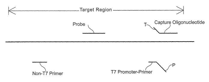
Brief Description of the Drawings
Figure 1 is a schematic diagram illustrating the various polynucleotides that
can be used
for detecting a target region within the GBS nucleic acid (represented by a
thick horizontal
line). Positions of the following nucleic acids are shown relative to the
target region: "Non-T7
Primer" and "T7 Promoter-Primer" represent two amplification primers used for
conducting
TMA, where "P" indicates the promoter sequence of the T7 promoter-primer; and
"Probe"
refers to the probe used for detecting amplified nucleic acid.
Detailed Description of the Invention
Disclosed herein are compositions, methods and kits for selectively detecting
GBS
nucleic acids in biological samples such as blood, plasma, serum or other body
fluid, or tissue.
The primers, probes and methods of the invention can be used in diagnostic
applications.
Introduction and Overview
The present invention includes compositions (primers and probes), methods and
kits
that are particularly useful for detecting GBS nucleic acids in a biological
sample. To design
oligonucleotide sequences appropriate for such uses, known GBS nucleic acid
sequences were
9
CA 02592179 2010-07-22
first compared to identify candidate regions of the bacterial genome that
could serve as targets
in a diagnostic assay. As a result of these comparisons, three different
regions of the GBS
genome (SEQ ID NOs:1-3) were selected as targets for detection using the
primers and probes
shown schematically in Figure 1. Portions of sequences containing relatively
few variants
between the compared sequences were chosen as starting points for designing
synthetic
oligonucleotides suitable for use in amplification and detection of amplified
sequences.
Based on these analyses, the amplification primer and probe sequences
presented below
were designed. Those having an ordinary level of skill in the art will
appreciate that any primer
sequences specific for GBS or other bacterial target, with or without a T7
promoter sequence,
may be used as primers in the various primer-based in vitro amplification
methods described
below. It is also contemplated that oligonucleotides having the sequences
disclosed herein
could serve alternative functions in assays for detecting GBS nucleic acids.
For example, the
hybridization probes disclosed herein could be used as amplification primers,
and the
amplification primers disclosed herein could be used as hybridization probes
in alternative
detection assays. It is further contemplated that capture oligonucleotides may
be used to
hybridize to and capture a target nucleic acid prior to amplification.
The amplification primers disclosed herein are particularly contemplated as
components
of multiplex amplification reactions wherein several amplicon species can be
produced from an
assortment of target-specific primers. For example, it is contemplated that
certain preferred
GBS-specific primers disclosed herein can be used in multiplex amplification
reactions that are
capable of amplifying polynucleotides of unrelated bacteria without
substantially
compromising the sensitivities of those assays.
Useful Amplification Methods
Amplification methods useful in connection with the present invention include
Transcription Mediated Amplification (TMA), Nucleic Acid Sequence-Based
Amplification
(NASBA), the Polymerase Chain Reaction (PCR), Strand Displacement
Amplification (SDA),
and amplification methods using self-replicating polynucleotide molecules and
replication
enzymes such as MDV-1 RNA and Q-beta enzyme. Methods for carrying out these
various
amplification techniques can be found respectively in U.S. Patent No.
5,399,491; published
European Patent Appl. No. EP 0 525 882; U.S. Patent No. 4,965,188; U.S. Patent
No.
5,455,166; U.S. Patent No. 5,472,840; and, Lizardi et al., BioTechnology
6:1197 (1988).
CA 02592179 2010-07-22
In a highly preferred embodiment of the invention, GBS nucleic acid sequences
are
amplified using a TMA protocol. According to this protocol, the reverse
transcriptase which
provides the DNA polymerase activity also possesses an endogenous RNase H
activity. One of
the primers used in this procedure contains a promoter sequence positioned
upstream of a
sequence that is complementary to one strand of a target nucleic acid that is
to be amplified. In
the first step of the amplification, a promoter-primer hybridizes to the GBS
target at a defined
site. Reverse transcriptase creates a complementary DNA copy of the target RNA
by extension
from the 3' end of the promoter-primer. Following interaction of an opposite
strand primer
with the newly synthesized DNA strand, a second strand of DNA is synthesized
from the end of
the primer by reverse transcriptase, thereby creating a double-stranded DNA
molecule. RNA
polymerase recognizes the promoter sequence in this double-stranded DNA
template and
initiates transcription. Each of the newly synthesized RNA amplicons re-enters
the TMA
process and serves as a template for a new round of replication, thereby
leading to an
exponential expansion of the RNA amplicon. Since each of the DNA templates can
make 100-
1000 copies of RNA amplicon, this expansion can result in the production of 10
billion
amplicons in less than one hour. The entire process is autocatalytic and is
performed at a
constant temperature.
Structural Features of Primers
As indicated above, a "primer" refers to an optionally modified
oligonucleotide that is
capable of participating in a nucleic acid amplification reaction. Preferred
primers are capable
of hybridizing to a template nucleic acid and have a 3' end that can be
extended by a DNA
polymerase activity. The 5' region of the primer may be non-complementary to
the target
nucleic acid. If the 5' non-complementary region includes a promoter sequence,
it is referred to
as a "promoter-primer." Those skilled in the art will appreciate that any
oligonucleotide that
can function as a primer (i.e., an oligonucleotide that hybridizes
specifically to a target
sequence and has a 3' end capable of extension by a DNA polymerase activity)
can be modified
to include a 5' promoter sequence, and thus could function as a promoter-
primer. Similarly,
any promoter-primer can be modified by removal of, or synthesis without, a
promoter sequence
and still function as a primer.
Nucleotide base moieties of primers may be modified (e.g., by the addition of
propyne
groups), so long as the modified base moiety retains the ability to form a non-
covalent
11
CA 02592179 2010-07-22
association with G, A, C, T or U, and so long as an oligonucleotide comprising
at least one
modified nucleotide base moiety or analog is not sterically prevented from
hybridizing with a
single-stranded nucleic acid. As indicated below in connection with the
chemical composition
of useful probes, the nitrogenous bases of primers in accordance with the
invention may be
conventional bases (A, G, C, T, U), known analogs thereof (e.g., inosine or
"I" having
hypoxanthine as its base moiety; see The Biochemistry of the Nucleic Acids 5-
36, Adams et al.,
ed., 1 Ph ed., 1992), known derivatives of purine or pyrimidine bases (e.g.,
N4-methyl
deoxygaunosine, deaza- or aza-purines and deaza- or aza-pyrimidines,
pyrimidine bases having
substituent groups at the 5 or 6 position, purine bases having an altered or a
replacement
substituent at the 2, 6 or 8 positions, 2-amino-6-methylaminopurine, 06-
methylguanine, 4-thio-
pyrimidines, 4-amino-pyrimidines, 4-dimethylhydrazine-pyrimidines, and 04-
alkyl-pyrimidines
(see Cook, PCT Int'l Pub. No. WO 93/13121)), and "abasic" residues where the
backbone
includes no nitrogenous base for one or more residues of the polymer (see
Arnold et al., U.S.
Patent No. 5,585,481). Common sugar moieties that comprise the primer backbone
include
ribose and deoxyribose, although 2'-O-methyl ribose (2'-OMe), halogenated
sugars, and other
modified sugar moieties may also be used. Usually, the linking group of the
primer backbone
is a phosphorus-containing moiety, most commonly a phosphodiester linkage,
although other
linkages, such as, for example, phosphorothioates, methylphosphonates, and non-
phosphorus-
containing linkages such as the linkages found in "locked nucleic acids" (LNA)
and the
peptide-like linkages found in "peptide nucleic acids" (PNA) also are intended
for use in the
assay disclosed herein.
Useful Probe Labeling Systems and Detectable Moieties
Essentially any labeling and detection system that can be used for monitoring
specific
nucleic acid hybridization can be used in conjunction with the present
invention. Included
among the collection of useful labels are radiolabels, enzymes, haptens,
linked
oligonucleotides, chemiluminescent molecules, fluorescent moieties (either
alone or in
combination with "quencher" moieties), and redox-active moieties that are
amenable to
electronic detection methods. Preferred chemiluminescent molecules include
acridinium esters
of the type disclosed in Arnold et al., U.S. Patent No. 5,283,174 for use in
connection with
homogenous protection assays, and of the type disclosed in Woodhead et al.,
U.S. Patent No.
5,656,207 for use in connection with assays that quantify multiple targets in
a single reaction.
12
CA 02592179 2010-07-22
Preferred electronic labeling and detection approaches are disclosed in U.S.
Patent Nos.
5,591,578 and 5,770,369, and PCT Int'l Publ. No. WO 98/57158.
Redox active moieties useful as labels in the present
invention include transition metals such as Cd, Mg, Cu, Co, Pd, Zn, Fe, and
Ru.
Particularly preferred detectable labels for probes in accordance with the
present
invention are detectable in homogeneous assay systems (i.e., where, in a
mixture, bound
labeled probe exhibits a detectable change, such as stability or differential
degradation,
compared to unbound labeled probe). While other homogeneously detectable
labels, such as
fluorescent labels and electronically detectable labels, are intended for use
in the practice of the
present invention, a preferred label for use in homogenous assays is a
chemiluminescent
compound (e.g., as described in Woodhead et al., U.S. Patent No. 5,656,207;
Nelson et al., U.S.
Patent No. 5,658,737; or Arnold et al., U.S. Patent No. 5,639,604).
Particularly preferred
chemiluminescent labels include acridinium ester (AE) compounds, such as
standard AE or
derivatives thereof, such as naphthyl-AE, ortho-AE, 1- or 3-methyl-AE, 2,7-
dimethyl-AE, 4,5-
dimethyl-AE, ortho-dibromo-AE, ortho-dimethyl-AE, meta-dimethyl-AE, ortho-
methoxy-AE,
ortho-methoxy(cinnamyl)-AE, ortho-methyl-AE, ortho-fluoro-AE, 1- or 3-methyl-
ortho-fluoro-
AE, 1- or 3-methyl-meta-difluoro-AE, and 2-methyl-AE. The acridinium ester can
be joined to
the probe by means of a non-nucleotide linker. For example, detection probes
can be labeled
with chemiluminescent acridinium ester compounds that are attached via a
linker substantially
as described in U.S. Patent No. 5,585,481 and U.S. Patent No. 5,639,604,
particularly at
column 10, line 6 to column 11, line 3, and Example 8.
In some applications, probes exhibiting at least some degree of self-
complementarity
are desirable to facilitate detection of probe:target duplexes in a test
sample without first
requiring the removal of unhybridized probe prior to detection. By way of
example, structures
referred to as "molecular torches" are designed to include distinct regions of
self-
complementarity (coined "the target binding domain" and "the target closing
domain") which
are connected by a joining region and which hybridize to one another under
predetermined
hybridization assay conditions. When exposed to denaturing conditions, the two
complementary regions of the molecular torch, which may be fully or partially
complementary,
melt, leaving the target binding domain available for hybridization to a
target sequence when
the predetermined hybridization assay conditions are restored. Molecular
torches are designed
13
CA 02592179 2011-09-29
so that the target binding domain favors hybridization to the target sequence
over the target
closing domain. The target binding domain and the target closing domain of a
molecular torch
include interacting labels (e.g., a fluorescent/quencher pair) positioned so
that a different signal
is produced when the molecular torch is self-hybridized as opposed to when the
molecular
torch is hybridized to a target nucleic acid, thereby permitting detection of
probe:target
duplexes in a test sample in the presence of unhybridized probe having a
viable label associated
therewith. Molecular torches are fully described in U.S. Patent No. 6,361,945.
Another example of a self-complementary hybridization assay probe that may be
used
in conjunction with the invention is a structure commonly referred to as a
"molecular beacon."
Molecular beacons comprise nucleic acid molecules having a target
complementary sequence,
an affinity pair (or nucleic acid arms) that holds the probe in a closed
conformation in the
absence of a target nucleic acid sequence, and a label pair that interacts
when the probe is in a
closed conformation. Hybridization of the molecular beacon target
complementary sequence to
the target nucleic acid separates the members of the affinity pair, thereby
shifting the probe to
an open conformation. The shift to the open conformation is detectable due to
reduced
interaction of the label pair, which may be, for example, a fluorophore and a
quencher (e.g.,
DABCYL and EDANS). Molecular beacons are fully described in U.S. Patent No.
5,925,517.
Molecular beacons useful for
detecting GBS-specific nucleic acid sequences may be created by appending to
either end of
one of the probe sequences disclosed herein, a first nucleic acid arm
comprising a fluorophore
and a second nucleic acid arm comprising a quencher moiety. In this
configuration, the GBS-
specific probe sequence disclosed herein serves as the target-complementary
"loop" portion of
the resulting molecular beacon.
Molecular beacons are preferably labeled with an interactive pair of
detectable labels.
Preferred detectable labels interact with each other by FRET or non-FRET
energy transfer
mechanisms. Fluorescence resonance energy transfer (FRET) involves the
radiationless
transmission of energy quanta from the site of absorption to the site of its
utilization in the
molecule or system of molecules by resonance interaction between chromophores,
over
distances considerably greater than interatomic distances, without conversion
to thermal
energy, and without the donor and acceptor coming into kinetic collision. The
"donor" is the
moiety that initially absorbs the energy, and the "acceptor" is the moiety to
which the energy is
14
CA 02592179 2007-06-26
WO 2006/086438 PCT/US2006/004372
subsequently transferred. In addition to FRET, there are at least three other
"non-FRET"
energy transfer processes by which excitation energy can be transferred from a
donor to an
acceptor molecule.
When two labels are held sufficiently close such that energy emitted by one
label can be
received or absorbed by the second label, whether by a FRET or non-FRET
mechanism, the
two labels are said to be in an "energy transfer relationship." This is the
case, for example,
when a molecular beacon is maintained in the closed state by formation of a
stem duplex and
fluorescent emission from a fluorophore attached to one arm of the molecular
beacon is
quenched by a quencher moiety on the other arm.
Highly preferred label moieties for the invented molecular beacons include a
fluorophore and a second moiety having fluorescence quenching properties
(i.e., a "quencher").
In this embodiment, the characteristic signal is likely fluorescence of a
particular wavelength,
but alternatively could be a visible light signal. When fluorescence is
involved, changes in
emission are preferably due to FRET, or to radiative energy transfer or non-
FRET modes.
When a molecular beacon having a pair of interactive labels in the closed
state is stimulated by
an appropriate frequency of light, a fluorescent signal is generated at a
first level, which may be
very low. When this same molecular beacon is in the open state and is
stimulated by an
appropriate frequency of light, the fluorophore and the quencher moieties are
sufficiently
separated from each other such that energy transfer between them is
substantially precluded.
Under that condition, the quencher moiety is unable to quench the fluorescence
from the
fluorophore moiety. If the fluorophore is stimulated by light energy of an
appropriate
wavelength, a fluorescent signal of a second level, higher than the first
level, will be generated.
The difference between the two levels of fluorescence is detectable and
measurable. Using
fluorophore and quencher moieties in this manner, the molecular beacon is only
"on" in the
"open" conformation and indicates that the probe is bound to the target by
emanating an easily
detectable signal. The conformational state of the probe alters the signal
generated from the
probe by regulating the interaction between the label moieties.
Examples of donor/acceptor label pairs that may be used in connection with the
invention, making no attempt to distinguish FRET from non-FRET pairs, include
fluorescein/tetramethylrhodamine, IAEDANS/fluorescein, EDANS/DABCYL,
coumarin/DABCYL, fluorescein/fluorescein, BODIPY FL/BODIPY FL,
fluoresceinlDABCYL,
lucifer yellow/DABCYL, BODIPY/DABCYL, eosine/DABCYL, erythrosine/DABCYL,
CA 02592179 2007-06-26
WO 2006/086438 PCT/US2006/004372
tetramethylrhodamine/DABCYL, Texas Red/DABCYL, CY5BH1, CY5/BH2, CY3BH1,
CY3/BH2, and fluorescein/QSY7 dye. Those having an ordinary level of skill in
the art will
understand that when donor and acceptor dyes are different, energy transfer
can be detected by
the appearance of sensitized fluorescence of the acceptor or by quenching of
donor
fluorescence. When the donor and acceptor species are the same, energy can be
detected by the
resulting fluorescence depolarization. Non-fluorescent acceptors such as
DABCYL and the
QSY 7 dyes advantageously eliminate the potential problem of background
fluorescence
resulting from direct (i.e., non-sensitized) acceptor excitation. Preferred
fluorophore moieties
that can be used as one member of a donor-acceptor pair include fluorescein,
ROX, and the CY
dyes (such as CY5). Highly preferred quencher moieties that can be used as
another member of
a donor-acceptor pair include DABCYL and the BLACK HOLE QUENCHER moieties
which
are available from Biosearch Technologies, Inc. (Novato, CA).
Synthetic techniques and methods of bonding labels to nucleic acids and
detecting
labels are well known in the art (see, e.g., Sambrook et al., Molecular
Cloning: A Laboratory
Manual, 2nd ed. (Cold Spring Harbor Laboratory Press, Cold Spring Harbor, NY,
1989),
Chapter 10; Nelson et al., U.S. Patent No. 5,658,737; Woodhead et al., U.S.
Patent No.
5,656,207; Hogan et al., U.S. Patent No. 5,547,842; Arnold et al., U.S. Patent
No. 5,283,174;
Kourilsky et al., U.S. Patent No. 4,581,333; and, Becker et al., European
Patent Appl. No. EP 0
747 706).
Chemical Composition of Probes
Probes in accordance with the invention comprise polynucleotides or
polynucleotide
analogs, and optionally carry a detectable label covalently bound thereto.
Nucleosides or
nucleoside analogs of the probe comprise nitrogenous heterocyclic bases or
base analogs,
where the nucleosides are linked together, for example, by phosphodiester
bonds to form a
polynucleotide. Accordingly, a probe may comprise conventional ribonucleic
acid (RNA)
and/or deoxyribonucleic acid (DNA), but also may comprise chemical analogs of
these
molecules. The probe backbone may be made up from a variety of linkages known
in the art,
including one or more sugar-phosphodiester linkages, locked nucleic acid (LNA)
bonds,
peptide-nucleic acid bonds (sometimes referred to as "peptide nucleic acids"
as described in
Hyldig-Nielsen et al., PCT Int'l Publ. No. WO 95/32305), phosphorothioate
linkages,
methylphosphonate linkages, or combinations thereof. Sugar moieties of the
probe may be
either ribose or deoxyribose, or similar compounds having known substitutions,
such as, for
16
CA 02592179 2010-07-22
example, 2'-O-methyl ribose and 2' halide substitutions (e.g., 2'-F). The
nitrogenous bases
may be conventional bases (A, G, C, T, U), known analogs thereof (e.g.,
inosine or "I"; see The
Biochemistry of the Nucleic Acids 5-36, Adams et al., ed., 11th ed., 1992),
known derivatives of
purine or pyrimidine bases (e.g., N4-methyl deoxyguanosine, deaza- or aza-
purines and deaza-
or aza-pyrimidines, pyrimidine bases having substituent groups at the 5 or 6
position, purine
bases having an altered or a replacement substituent at the 2, 6 or 8
positions, 2-amino-6-
methylaminopurine, O6-methylguanine, 4-thio-pyrimidines, 4-amino-pyrimidines,
4-
dimethylhydrazine-pyrimidines, and 04-alkyl-pyrimidines (see Cook, PCT Int'l
Publ. No. WO
93/13121)), and "abasic" residues where the backbone includes no nitrogenous
base for one or
more residues of the polymer (see Arnold et al., U.S. Patent No. 5,585,481). A
probe may
comprise only conventional sugars, bases and linkages found in RNA and DNA, or
may
include both conventional components and substitutions (e.g., conventional
bases linked via a
methoxy backbone, or a nucleic acid including conventional bases and one or
more base
analogs).
While oligonucleotide probes of different lengths and base composition may be
used for
detecting GBS nucleic acids, preferred probes in this invention have lengths
of up to 30
nucleotides, and more preferably within the length range of 26 to 30
nucleotides. However, the
specific probe sequences described below may also be provided in a nucleic
acid cloning vector
or transcript or other longer nucleic acid and still be used for detecting GBS
nucleic acids.
Selection of GBS-Specific Amplification Primers and Detection Probes
Useful guidelines for designing amplification primers and probes with desired
characteristics are described herein. The optimal sites for amplifying and
probing GBS nucleic
acids are three conserved regions of the GBS genome, each greater than about
20 bases in
length, within about 250 bases of contiguous sequence. The degree of
amplification observed
with a set of primers, including one or more promoter-primers, depends on
several factors
including the ability of the oligonucleotides to hybridize to their
complementary sequences and
their ability to be extended enzymatically. Because the extent and specificity
of hybridization
reactions are affected by a number of factors, manipulation of those factors
will determine the
exact sensitivity and specificity of a particular oligonucleotide, whether
perfectly
complementary to its target or not. The effects of varying assay conditions
are known to those
skilled in the art, and are described in Hogan et al,, U.S. Patent No.
5,840,488.
17
CA 02592179 2007-06-26
WO 2006/086438 PCT/US2006/004372
The length of the target nucleic acid sequence and, accordingly, the length of
the primer
sequence or probe sequence can be important. In some cases, there may be
several sequences
from a particular target region, varying in location and length, which will
yield primers or
probes having the desired hybridization characteristics. While it is possible
for nucleic acids
that are not perfectly complementary to hybridize, the longest stretch of
perfectly homologous
base sequence will normally determine hybrid stability.
Amplification primers and probes should be positioned to minimize the
stability of an
oligonucleotide:non-target nucleic acid hybrid. It is preferred that the
amplification primers
and probes are able to distinguish between target and non-target sequences. In
designing
primers and probes, the differences in melting temperature, represented by T.
values, should be
as large as possible (e.g., at least 2 , and preferably 5 Q.
The degree of non-specific extension (primer-dimer or non-target copying) can
also
affect amplification efficiency. For this reason, primers are selected to have
low self- or cross-
complementarity, particularly at the 3' ends of the sequence. Long homopolymer
tracts and
high GC content are avoided to reduce spurious primer extension. Commercially
available
computer software can aid in this aspect of the design. Available computer
programs include
MacDNASISTM 2.0 (Hitachi Software Engineering American Ltd.) and OLIGO ver.
6.6
(Molecular Biology Insights; Cascade, CO).
Those having an ordinary level of skill in the art will appreciate that
hybridization
involves the association of two single strands of complementary nucleic acid
to form a
hydrogen bonded double strand. It is implicit that if one of the two strands
is wholly or
partially involved in a hybrid, then that strand will be less able to
participate in formation of a
new hybrid. By designing primers and probes so that substantial portions of
the sequences of
interest are single stranded, the rate and extent of hybridization may be
greatly increased. If the
target is an integrated genomic sequence, then it will naturally occur in a
double stranded form
(as is the case with the product of the polymerase chain reaction). These
double-stranded
targets are naturally inhibitory to hybridization with a probe and require
denaturation prior to
the hybridization step.
The rate at which a polynucleotide hybridizes to its target is a measure of
the thermal
stability of the target secondary structure in the target-binding region. The
standard
measurement of hybridization rate is the Cot112, which is measured as moles of
nucleotide per
liter multiplied by seconds. Thus, it is the concentration of probe multiplied
by the time at
18
CA 02592179 2007-06-26
WO 2006/086438 PCT/US2006/004372
which 50% of maximal hybridization occurs at that concentration. This value is
determined by
hybridizing various amounts of polynucleotide to a constant amount of target
for a fixed time.
The Cot112 is found graphically by standard procedures familiar to those
having an ordinary level
of skill in the art.
Preferred Amplification Primers
Primers useful for conducting amplification reactions can have different
lengths to
accommodate the presence of extraneous sequences that do not participate in
target binding and
that may not substantially affect amplification or detection procedures. For
example, promoter-
primers useful for performing amplification reactions in accordance with the
invention have at
least a minimal sequence that hybridizes to the GBS target nucleic acid and a
promoter
sequence positioned upstream of that minimal sequence. However, insertion of
sequences
between the target binding sequence and the promoter sequence could change the
length of the
primer without compromising its utility in the amplification reaction.
Additionally, the lengths
of the amplification primers and probes are matters of choice so long as the
sequences of these
oligonucleotides conform to the minimal essential requirements for hybridizing
the desired
complementary sequence.
Tables 1 and 2 present specific examples of oligonucleotide sequences that
were used as
primers for amplifying GBS nucleic acids. Table 1 presents the sequences of
GBS target-
complementary primers to one strand of the GBS nucleic acid. All of the
illustrative primers
presented in Table 1 have target-complementary sequences contained within the
sequence of
SEQ ID NO:1.
Table 1
Oligonucleotide Sequences of Amplification Primers
Sequence SEQ ID NO:
AAGTGTCTGGTCAAACAGTGA SEQ ID NO:4
TGGTCAAACAGTGAGGTGTGA SEQ ID NO:5
GTGAGGTGTGATATGAGTCA SEQ ID NO:6
Table 2 presents the sequences of both the GBS target-complementary primers
and the
corresponding promoter-primers to the opposing strand of the GBS nucleic acid.
As indicated
above, all promoter-primers included sequences complementary to a GBS target
sequence at
their 3' ends and the T7 promoter sequence AATTTAATACGACTCACTATAGGGAGA
19
CA 02592179 2007-06-26
WO 2006/086438 PCT/US2006/004372
(SEQ ID NO:7) at their 5' ends. Primers identified by SEQ ID NOs:11-13 in
Table 2 are
promoter-primers corresponding to the GBS target-complementary primers
identified as SEQ
ID NOs:8-10, respectively. All of the illustrative primers presented in Table
2 have target-
complementary sequences contained within the sequence of SEQ ID NO:2.
Table 2
Oligonucleotide Sequences of Amplification Primers
Sequence SEQ ID NO:
TCCTTAACGAGAGTTCTCTCGCTCA SEQ ID NO:8
GGGCCATTTTGCCGAGTTCCTTAACGAGA SEQ ID NO:9
AAGTTACGGGGCCATTTTGCCGAGTTCCTTA SEQ ID NO:I0
AATTTAATACGACTCACTATAGGGAGATCCTTA SEQ ID NO: 11
ACGAGAGTTCTCTCGCTCA
AATTTAATACGACTCACTATAGGGAGAGGGCCA SEQ ID NO:12
TTTTGCCGAGTTCCTTAACGAGA
AATTTAATACGACTCACTATAGGGAGAAAGTTA SEQ ID NO:13
CGGGGCCATTTTGCCGAGTTCCTTA
Preferred sets of primers for amplifying GBS nucleic acid sequences include a
first
primer that hybridizes a GBS target sequence, such as one of the primers
listed in Table 2, and
a second primer that is complementary to the sequence of an extension product
of the first
primer, such as one of the primers listed in Table 1. In a highly preferred
embodiment, the first
primer is a promoter-primer that includes a T7 promoter sequence at its 5'
end.
Preferred Detection Probes
Another aspect of the invention relates to oligonucleotides that can be used
as
hybridization probes for detecting GBS nucleic acids. Methods for amplifying a
target nucleic
acid sequence present in a GBS nucleic acid can include an optional further
step for detecting
amplicons. This detection procedure includes a step for contacting a test
sample with a
hybridization assay probe that preferentially hybridizes to the target nucleic
acid sequence, or
the complement thereof, under stringent hybridization conditions, thereby
forming a
probe:target duplex that is stable for detection. Next there is a step for
determining whether the
hybrid is present in the test sample as an indication of the presence or
absence of GBS nucleic
acids in the test sample. This may involve detecting the probe:target duplex,
and preferably
involves homogeneous assay systems.
CA 02592179 2007-06-26
WO 2006/086438 PCT/US2006/004372
Hybridization assay probes useful for detecting GBS nucleic acid sequences
include a
sequence of bases substantially complementary to a GBS target nucleic acid
sequence. Thus,
probes of the invention hybridize to one strand of a GBS target nucleic acid
sequence, or the
complement thereof. These probes may optionally have additional bases outside
of the targeted
nucleic acid region, which may or may not be complementary to the GBS nucleic
acid.
Preferred probes are sufficiently homologous to the target nucleic acid to
hybridize
under stringent hybridization conditions corresponding to about 60 C and a
salt concentration
in the range of 0.6-0.9 M for probes labeled with chemiluminescent molecules
and
corresponding to about 42 C and a salt concentration in the range of 20-100
mM for molecular
beacon probes. Preferred salts include lithium, magnesium and potassium
chlorides, but other
salts such as sodium chloride and sodium citrate also can be used in the
hybridization solution.
Example high stringency hybridization conditions are alternatively provided by
0.48 M sodium
phosphate buffer, 0.1% sodium dodecyl sulfate and 1 mM each of EDTA and EGTA,
or by 0.6
M LiCl, I% lithium lauryl sulfate, 60 mM lithium succinate and 10 mM each of
EDTA and
EGTA.
Probes in accordance with the invention have sequences complementary to, or
corresponding to, a domain of the GBS genome. Molecular beacon probes that are
preferred
for detecting GBS nucleic acid sequences have a probe sequence, which includes
the target-
complementary sequence of bases together with any base sequences that are not
complementary
to the nucleic acid that is to be detected, in the length range of from 26-30
nucleotides.
Specific molecular beacon probes that are preferred for detecting GBS nucleic
acid sequences
have target-complementary sequences in the length range of from 16-20
nucleotides. Of
course, these target-complementary sequences may be linear sequences, or may
be contained in
the structure of a molecular beacon or other construct having one or more
optional nucleic acid
sequences that are non-complementary to the GBS target sequence that is to be
detected. As
indicated above, probes may be made of DNA, RNA, a combination DNA and RNA, a
nucleic
acid analog, or contain one or more modified nucleosides (e.g., a
ribonucleoside having a 2'-O-
methyl substitution to the ribofuranosyl moiety).
Simply stated, preferred probes for detecting target nucleic acids of interest
in
connection with the present invention include sequences that are contained
within one or more
of several defined probe domains, or the complements thereof, allowing for the
presence of
RNA and DNA equivalents and nucleotide analogs. For example, preferred
hybridization assay
21
CA 02592179 2007-06-26
WO 2006/086438 PCT/US2006/004372
probes for detecting GBS nucleic acids can include target-complementary
sequences of bases
contained within the sequence of SEQ ID NO:3. Optional sequences which are not
complementary to the nucleic acid sequence that is to be detected may be
linked to the target-
complementary sequence of the probe.
Certain preferred probes in accordance with the present invention include a
detectable
label. This label includes a fluorophore and a second moiety having
fluorescence quenching
properties.
Table 3 presents the GBS target-complementary oligonucleotide sequences
contained in
the loop portions of the molecular beacon probes and the corresponding
complete sequences of
the molecular beacon probes used for detecting GBS amplicons. Each of the
molecular
beacons included a 5' CCGAG arm sequence and a 3' CUCGG arm sequence appended
to the
GBS target-complementary sequence contained in the loop portion of the
molecular beacon.
Loop portions identified by SEQ ID NOs:14-17 in Table 3 correspond to the
molecular beacons
identified as SEQ ID NOs: 18-21, respectively. All of the GBS-specific
molecular beacons used
in the procedure had target-complementary sequences that included 16-20
contiguous
nucleotides contained within the sequence of SEQ ID NO:3, allowing for the
presence of RNA
and DNA equivalents. The target-complementary sequences presented in Table 3
were
independently incorporated into the loop regions of molecular beacons. Each of
the molecular
beacons used in the procedure included a fluorescein fluorophore at its 5'-end
and a DABCYL
quencher moiety at its 3'-end.
Table 3
Oligonucleotide Sequences of GBS-Specific Molecular Beacons
Sequence SEQ ID NO:
UACAUAUACUCUACCC SEQ ID NO:14
AUACAUAUACUCUACCC SEQ ID NO:15
GAUACAUAUACUCUACCC SEQ ID NO:16
GCGAUACAUAUACUCUACCC SEQ ID NO:17
CCGAG-UACAUAUACUCUACCC-CUCGG SEQ ID NO: 18
CCGAG-AUACAUAUACUCUACCC-CUCGG SEQ ID NO:19
CCGAG-GAUACAUAUACUCUACCC-CUCGG SEQ ID NO:20
CCGAG-GCGAUACAUAUACUCUACCC-CUCGG SEQ ID NO:21
22
CA 02592179 2007-06-26
WO 2006/086438 PCT/US2006/004372
Since alternative probes for detecting GBS nucleic acid sequences can
hybridize to the
opposite-sense GBS strand, the present invention also includes
oligonucleotides that are
complementary to the sequences presented in Table 3.
As indicated above, any number of different backbone structures can be used as
a
scaffold for the oligonucleotide sequences of the invented hybridization
probes. In certain
highly preferred embodiments, the probe sequence used for detecting GBS
amplicons includes
a methoxy backbone or at least one methoxy linkage in the nucleic acid
backbone.
Preferred Methods for Amplifying and Detecting GBS Polynucleotide Sequences
Preferred methods of the present invention are described and illustrated by
the
Examples presented below. Figure 1 schematically illustrates one system that
may be used for
detecting a target region of the GBS nucleic acid (shown by a thick solid
horizontal line). This
system includes at least three oligonucleotides (shown by the shorter solid
lines): one T7
promoter-primer which includes a sequence that hybridizes specifically to a
GBS sequence in
the target region and a T7 promoter sequence ("P") which, when double-
stranded, serves as a
functional promoter for T7 RNA polymerase; one non-T7 primer which includes a
sequence
that hybridizes specifically to a first strand cDNA made from the target
region sequence using
the T7 promoter-primer; and, one labeled probe which includes a sequence that
hybridizes
specifically to a portion of the target region that is amplified using the two
primers.
As indicated above, amplifying the target region using the two primers can be
accomplished by any of a variety of known nucleic acid amplification reactions
that will be
familiar to those having an ordinary level of skill in the art. In a preferred
embodiment, a
transcription associated amplification reaction, such as TMA, is employed. In
such an
embodiment, many strands of nucleic acid are produced from a single copy of
target nucleic
acid, thus permitting detection of the target by detecting probes that are
bound to the amplified
sequences. Preferably, transcription associated amplification uses two types
of primers (one
being referred to as a promoter-primer because it contains a promoter
sequence, labeled "P" in
Figure 1, for an RNA polymerase), two enzymes (a reverse transcriptase and an
RNA
polymerase), and substrates (deoxyribonucleoside triphosphates, ribonucleoside
triphosphates)
with appropriate salts and buffers in solution to produce multiple RNA
transcripts from a
nucleic acid template.
Referring to Figure 1, during transcription mediated amplification, the target
nucleic
acid is hybridized to a first primer shown as a T7 promoter-primer. Using
reverse transcriptase,
23
CA 02592179 2007-06-26
WO 2006/086438 PCT/US2006/004372
a complementary DNA strand is synthesized from the T7 promoter-primer using
the target
RNA as a template. A second primer, shown as a non-T7 primer, hybridizes to
the newly
synthesized DNA strand and is extended by the action of a reverse
transcriptase to form a DNA
duplex, thereby forming a double-stranded T7 promoter region. T7 RNA
polymerase then
generates multiple RNA transcripts by using this functional T7 promoter. The
autocatalytic
mechanism of TMA employs repetitive hybridization and polymerization steps
following a
cDNA synthesis step using the RNA transcripts as templates to produce
additional transcripts,
thereby amplifying target region-specific nucleic acid sequences.
The detecting step uses at least one detection probe that binds specifically
to the
amplified RNA transcripts or amplicons described above. Preferably, the
detection probe is
labeled with a label that can be detected using a homogeneous detection
system. For example,
the labeled probe can be labeled with an acridinium ester compound from which
a
chemiluminescent signal may be produced and detected, as described above.
Alternatively, the
labeled probe may comprise a fluorophore, or fluorophore and quencher
moieties. A molecular
beacon is one embodiment of such a labeled probe that may be used in a
homogeneous
detection system.
Kits for Detecting GBS Nucleic Acids
The present invention also embraces kits for performing polynucleotide
amplification
reactions using bacterial nucleic acid templates. Certain preferred kits will
contain a
hybridization assay probe that includes a target-complementary sequence of
bases, and
optionally including primers or other ancillary oligonucleotides for
amplifying the target that is
to be detected. Other preferred kits will contain a pair of oligonucleotide
primers that may be
used for amplifying target nucleic acids in an in vitro amplification
reaction. Exemplary kits
include first and second amplification oligonucleotides that are complementary
to opposite
strands of a GBS nucleic acid sequence that is to be amplified. The kits may
further contain
one or more oligonucleotide detection probes. Still other kits in accordance
with the invention
may additionally include capture oligonucleotides for purifying GBS template
nucleic acids
away from other species prior to amplification.
The general principles of the present invention may be more fully appreciated
by
reference to the following non-limiting Examples.
Example 1 describes procedures that identified some of the hybridization
probes, which
subsequently were used in assays for detecting GBS nucleic acids. One
synthetic RNA
24
CA 02592179 2007-06-26
WO 2006/086438 PCT/US2006/004372
oligonucleotide served as a target for binding the probes.
Example 1
Oligonucleotides for Detecting GBS Nucleic Acids
Synthetic molecular beacons were prepared according to standard laboratory
procedures
using 2'-OMe nucleotide analogs. The sequences of the synthetic molecular
beacons are
shown in Table 3.
Hybridization reactions included 10 pmol/reaction of the molecular beacon and
30
pmol/reaction of the synthetic GBS RNA target oligonucleotide as given in
Table 4.
Table 4
Synthetic Target Sequence
Target Sequence SEQ ID NO:
GCGGUACGGGUAGAGUAUAUGUAUCGCUAGAAGCU SEQ ID NO:22
Hybridization reactions of the molecular beacons in the absence or presence of
the
synthetic GBS RNA target oligonucleotide were carried out at 60 C for 10
minutes, followed
by incubation at 42 C for 60 minutes in 100 1 reaction volumes of a TRIS-
buffered solution
that included 20 MM MgC12.
Fluorescence was measured every 30 seconds at 42 C for 99 cycles using a Rotor-
Gene
2000 instrument (Corbett Research, Sydney, Australia). Results from the
fluorescent reactions
were measured in relative fluorescence units (RFU). After completion of the
hybridization
reactions, the reaction temperature was increased in one degree Celsius
increments from 42-
99 C holding for 15 seconds at each step, and the resulting RFUs were measured
to determine
the melting temperatures (Tm) of the molecular beacons using the data analysis
software
provided by the Rotor-Gene 2000 instrument. Representative results for the
hybridization
reactions and melting temperature measurements are summarized in Table 5.
Numerical values shown in Table 5 indicate the average signal/noise (S/N)
ratio values
calculated from the measured endpoint RFUs in the presence of target divided
by the measured
endpoint RFUs in the absence of target. The calculated melting temperature of
the molecular
beacons in the absence of target is useful to determine the stability of the
stem structure of the
molecular beacon, whereas the melting temperature of the molecular beacon
hybridized to the
target sequence provides information about the stability of the hybrid.
CA 02592179 2007-06-26
WO 2006/086438 PCT/US2006/004372
Table 5
Melting Temperatures and Hybrid Stability of Molecular Beacons
Molecular T,õ w/o Target T. w/ Target S/N Ratio
Beacon ( C) ( C)
SEQ ID NO:18 73.7 62.9 28.4
SEQ ID NO: 19 74.0 70.2 11.4
SEQ ID NO:20 85.4 67.8 15.7
SEQ ID NO:21 83.0 75.8 23.7
The results presented in Table 5 showed that each molecular beacon gave strong
S/N
ratio values following binding to the synthetic GBS RNA target
oligonucleotide. In addition,
the melting temperatures of the molecular beacons in the absence of target
demonstrated that
the molecular beacons have stable stem structures, which prevent unspecific
"opening" of the
molecular beacons at lower temperatures. The high melting temperatures of the
molecular
beacons in the presence of the synthetic GBS RNA target oligonucleotide showed
that a stable
hybrid was formed under the experimental conditions.
Amplicon production was monitored as a function of time in real-time
amplification
procedures. Amplicon-specific molecular beacons that were included in the
amplification
reactions provided a means for continuous monitoring of amplicon synthesis.
Fluorescent
emissions that increased with time indicated the production of amplicons that
hybridized to the
molecular beacon and caused a detectable transition to the open conformation
of the molecular
beacon.
Molecular beacons comprise nucleic acid molecules having a target-
complementary
sequence, an affinity pair (or nucleic acid arms) that interact to form a stem
structure by
complementary base pairing in the absence of a target (i.e., the closed
conformation), and a
paired set of labels that interact when the probe is in the closed
conformation. Those having an
ordinary level of skill in the art will understand that the target-
complementary sequence
contained within the structure of a molecular beacon is generally in the form
of a
single-stranded loop region of the probe. Hybridization of the target nucleic
acid and the
target-complementary sequence of the probe causes the members of the affinity
pair to separate,
thereby shifting the probe to the open conformation. This shift is detectable
by virtue of
reduced interaction between the members of the label pair, which may be, for
example, a
26
CA 02592179 2011-09-29
fluorophore and a quencher. Molecular beacons are fully described in U.S.
Patent No.
5,925,517.
Commercially available software was used to analyze time-dependent results
obtained
using molecular beacons that were specific for amplicons derived from the GBS
nucleic acid.
Results from these analyses indicated a substantially linear relationship
between the number of
target copies included in an amplification reaction and the time at which the
fluorescent signal
exceeded a background threshold (i.e., time-of-emergence). As confirmed by the
results
presented below, these procedures were useful for quantifying analyte target
amounts over a
very broad range. More particularly, when known amounts of analyte
polynucleotides are used
as calibration standards, it is possible to determine the amount of analyte
present in a test
sample by comparing the measured time-of-emergence with the standard curve.
The fact that the amplification reaction used in the below-described
procedures operated
at constant temperature and without interruption for a separate detection
step, so that
amplification and detection took place simultaneously, imposed strict
requirements on the
molecular beacons. More specifically, success in the procedure required that
the molecular
beacon bind amplicon without inhibiting subsequent use of the amplicon as a
template in the
exponential amplification mechanism. Indeed, the finding that an amplification
reaction could
proceed efficiently in the presence of a molecular beacon indicated that
interaction of the probe
with its target did not irreversibly inhibit or poison the amplification
reaction.
Example 2 describes procedures wherein molecular beacon probes, each labeled
with an
interactive fluorophore/quencher pair, were used for monitoring time-dependent
amplicon
production in TMA reactions. Although the molecular beacons described in
Example 2
hybridized to only one strand of the amplified nucleic acid product,
complementary probe
sequences also would be expected to hybridize to the opposite nucleic acid
strand, and so fall
within the scope of the invention.
Example 2
Real-Time Monitoring of Amplicon Production
Molecular beacons having binding specificity for the GBS amplicon were
synthesized
by standard solid-phase phosphite triester chemistry using 3' quencher-linked
controlled pore
glass (CPG) and 5' fluorophore-labeled phosphoramidite on a Perkin-Elmer
(Foster City, CA)
EXPEDITE model 8909 automated synthesizer. Fluorescein was used as the
fluorophore, and
DABCYL was used as the quencher for construction of the molecular beacons. All
of the
27
CA 02592179 2007-06-26
WO 2006/086438 PCT/US2006/004372
molecular beacons were constructed using 2'-OMe nucleotide analogs. The CPG
and
phosphoramidite reagents were purchased from Glen Research Corporation
(Sterling, VA).
Following synthesis, the probes were deprotected and cleaved from the solid
support matrix by
treatment with concentrated ammonium hydroxide (30%) for two hours at 60 C.
Next, the
probes were purified using polyacrylamide gel electrophoresis followed by HPLC
using
standard procedures that will be familiar to those having an ordinary level of
skill in the art.
The nucleic acid target used in the real-time amplification and detection
procedures was
purified rRNA of known concentration. Different target concentrations were
tested in
triplicate. Molecular beacons were used at a level of 0.2 pmol/ l (3
pmol/reaction). Reactions
for amplifying GBS nucleic acids were conducted using from as low as 50
template
copies/reaction up to as high as 5x10⁸ template copies/reaction.
Reactions containing 3 pmol/reaction of T7 primer, 3 pmol/reaction of non-T7
primer,
target polynucleotide, and 3 pmol/reaction molecular beacon in 15 l of a
buffered solution
(final concentration: 50 mM Tris HC1(pH 8.2 to 8.5), 35 mM ICI, 4 mM GTP, 4 mM
ATP, 4
mM UTP, 4 mM CTP, 1 mM dATP, 1 mM dTTP, 1 mM dCTP, 1 mM dGTP, 20 MM MgCl2,
mM N-Acetyl-L-Cysteine, and 5% (w/v) glycerol) were incubated in a dry heat
block for 10
minutes at 60 C to facilitate primer annealing. Following the 60 C incubation
step, reactions
were transferred to a 42 C heat block and then incubated for 2 minutes. Five
microliter
aliquots of an enzyme reagent that included both MMLV reverse transcriptase
and T7 RNA
20 polymerase enzymes were added to each of the reactions using a repeat
pipettor. Tubes were
vortexed briefly and then transferred to a Rotor-Gene 2000 (Corbett Research;
Sydney,
Australia) rotor that had been pre-warmed to 42 C. Amplification reactions
were carried out at
42 C, fluorescence readings were taken every 30 seconds for 60 cycles, and the
results
analyzed in real-time using standard software that was bundled with the RG2000
instrument.
Representative results from this procedure using different molecular beacons
and different
primer combinations are summarized in Tables 6 and 7, respectively.
28
CA 02592179 2007-06-26
WO 2006/086438 PCT/US2006/004372
Table 6
Measured Time-of-Emergence During Real-Time Detection
Different Molecular Beacons
Time-of-Emergence with
Molecular Different Primer Combinations
(minutes)
Beacons
r-i N M
vD
z z z
a a a
SEQ ID NO: 18 26.5 17.4 ND
SEQ ID NO: 19 4.6 6.0 3.0
ND = Not Detected
The results presented in Table 6 confirmed that the amplification reactions
containing
3 primer combinations, different GBS-specific molecular beacons and a fixed
target
concentration (5 x 10⁸ copies/rxn) desirably produced a fluorescent signal
that increased with
time until reaching a plateau except with primer combination SEQ ID NOs:4 & 13
and
molecular beacon SEQ ID NO: 18. Each of the molecular beacons used in the
procedure
included a fluorescein fluorophore at its 5'-end and a DABCYL quencher moiety
at its 3'-end.
All results were based on reactions that were included in triplicate.
29
CA 02592179 2007-06-26
WO 2006/086438 PCT/US2006/004372
Table 7
Measured Time-of-Emergence During Real-Time Detection
Different Primer Combinations
Time-of-Emergence Measured Using Molecular Beacon
SEQ ID NO: 19 and Different Primer Combinations
(minutes)
GBS Target
(copies/rxn) N M N M N M
88 0i a3 ~3 X23 -18 d
Zt d- Ln kn kn 10 10 110
O Cl) Cl) Cl) O O O O
z z z z z z z z z
a o' a a a CW11 a a a
5x10⁸ NT NT 6.0 NT NT NT NT NT NT
5x 10' NT NT 8.5 NT NT NT NT NT NT
5x10⁶ 30.3 21.0 12.2 NT NT NT NT NT NT
5x10⁵ ND 26.6 15.7 NT NT NT NT NT NT
5x10⁴ ND 32.7 19.3 NT NT NT NT NT NT
5x10³ ND 39.8 23.8 48.9 32.1 23.6 35.7 39.7 22.9
5x10² ND 50.8 34.7 29.5 47.7* 33.2 44.6 50.3 27.2
5x101 ND 55.6 44.1 47.0 53.6f 49.3 ND 58.1* 35.0
NT = Not Tested
ND = Not Detected
*Only 2/3 replicates detected
The results shown in Table 7 confirmed that the amplification reactions
containing
different primer combinations and a fixed molecular beacon (SEQ ID NO: 19)
desirably
produced a fluorescent signal that increased with time until reaching a
plateau. All results were
based on reactions that were included in triplicate. The results also showed
that this particular
molecular beacon was able to detect amplification product down to 50
copies/reaction. Only
primer combinations SEQ ID NOs:5 & 12 and SEQ ID NOs:6 & 12 detected 2/3
replicates at
the 50 copies/reaction level, while none of the 3 replicates were detected
with primer
combination SEQ ID NOs:6 & 11 at this level.
Each of the primer combinations tested gave at least some level of time-
dependent
analyte detection. The different primer combinations tested in the procedure
behaved
CA 02592179 2007-06-26
WO 2006/086438 PCT/US2006/004372
somewhat differently in the real-time assay format. For example, reactions
that included
primer combinations SEQ ID NOs:4 & 13 and SEQ ID NOs:6 & 13 gave exceedingly
rapid
detection. The results presented in Table 7 further illustrate how the above-
described primers
and molecular beacons could be used in a highly sensitive assay for detecting
GBS nucleic
acids at very low levels of input template.
31
CA 02592179 2010-07-22
SEQUENCE LISTING
<110> GEN-PROBE INCORPORATED
POLLNER, Reinhold B.
KAMANTIGUE,.Edgar
<120> compositions and methods for Detecting Group B Streptococci
<130> GP173-PCT
<140> To Be Assigned
<141> 2006-02-07
<150> 60/650,501
<151> 2005-02-07
<160> 22
<170> Patentln version 3.3
<210> 1
<211> 37
<212> DNA
<213> Streptococcus agalactiae
<400> 1
aagtgtctgg tcaaacagtg aggtgtgata tgagtca 37
<210> 2
<211> 50
<212> DNA
<213> Streptococcus agalactiae
<400> 2
tgagcgagag aactctcgtt aaggaactcg gcaaaatggc cccgtaactt 50
<210> 3
<211> 20
<212> DNA
<213> Streptococcus agalactiae
<400> 3
gcgatacata tactctaccc 20
<210> 4
<211> 21
<212> DNA
<213> Streptococcus agalactiae
<400> 4
aagtgtctgg tcaaacagtg a 21
<210> 5
<211> 21
<212> DNA
<213> Streptococcus agalactiae
<400> 5
tggtcaaaca gtgaggtgtg a 21
<210> 6
<211> 20
<212> DNA
32
CA 02592179 2010-07-22
<213> streptococcus agalactiae
<400> 6
gtgaggtgtg atatgagtca 20
<210> 7
<211> 27
<212> DNA
<213> streptococcus agalactiae
<400> 7
aatttaatac gactcactat agggaga 27
<210> 8
<211> 25
<212> DNA
<213> Streptococcus agalactiae
<400> 8
tccttaacga gagttctctc gctca 25
<210> 9
<211> 29
<212> DNA
<213> streptococcus agalactiae
<400> 9
gggccatttt gccgagttcc ttaacgaga 29
<210> 10
<211> 31
<212> DNA
<213> Streptococcus agalactiae
<400> 10
aagttacggg gccattttgc cgagttcctt a 31
<210> 11
<211> 52
<212> DNA
<213> Streptococcus agalactiae
<400> 11
aatttaatac gactcactat agggagatcc ttaacgagag ttctctcgct ca 52
<210> 12
<211> 56
<212> DNA
<213> Streptococcus agalactiae
<400> 12
aatttaatac gactcactat agggagaggg ccattttgcc gagttcctta acgaga 56
<210> 13
<211> 58
<212> DNA
<213> Streptococcus agalactiae
<400> 13
aatttaatac gactcactat agggagaaag ttacggggcc attttgccga gttcctta 58
33
CA 02592179 2010-07-22
<210> 14
<211> 16
<212> RNA
<213> streptococcus agalactiae
<400> 14
uacauauacu cuaccc 16
<210> 15
<211> 17
<212> RNA
<213> Streptococcus agalactiae
<400> 15
auacauauac ucuaccc 17
<210> 16
<211> 18
<212> RNA
<213> Streptococcus agalactiae
<400> 16
gauacauaua cucuaccc 18
<210> 17
<211> 20
<212> RNA
<213> Streptococcus agalactiae
<400> 17
gcgauacaua uacucuaccc 20
<210> 18
<211> 26
<212> RNA
<213> streptococcus agalactiae
<400> 18
ccgaguacau auacucuacc ccucgg 26
<210> 19
<211> 27
<212> RNA
<213> streptococcus agalactiae
<400> 19
ccgagauaca uauacucuac cccucgg 27
<210> 20
<211> 28
<212> RNA
<213> Streptococcus agalactiae
<400> 20
ccgaggauac auauacucua ccccucgg 28
<210> 21
<211> 30
<212> RNA
<213> streptococcus agalactiae
34
CA 02592179 2010-07-22
<400> 21
ccgaggcgau acauauacuc uaccccucgg 30
<210> 22
<211> 35
<212> RNA
<213> Streptococcus agalactiae
<400> 22
gcgguacggg uagaguauau guaucgcuag aagcu 35