Note: Descriptions are shown in the official language in which they were submitted.
CA 02592606 2007-06-28
1
FACET JOINT REPLACEMENT
FIELD OF THE INVENTION
The present invention relates to spinal instrumentation, and in particular to
various
devices that are adapted to mimic the natural function of the structural
posterior elements.
BACKGROUND OF THE INVENTION
The vertebrae in a patient's spinal column are linked to one another by the
disc and
the facet joints, which control movement of the vertebrae relative to one
another. Each
vertebra has a pair of articulating surfaces located on the left side, and a
pair of articulating
surfaces located on the right side, and each pair includes a superior
articular surface, which
faces upward, and an inferior articular surface, which faces downward.
Together the superior
and inferior articular surfaces of adjacent vertebra form a facet joint. Facet
joints are
synovial joints, which means that each joint is surrounded by a capsule of
connective tissue
and produces a fluid to nourish and lubricate the joint. The joint surfaces
are coated with
cartilage allowing the joints to move or articulate relative to one another.
Diseased, degenerated, impaired, or otherwise painful facet joints and/or
discs can
require surgery to restore function to the three joint complex. Subsequent
surgery may also
be required after a laminectomy, as a laminectomy predisposes the patient to
instability and
may lead to post-laminectomy kyphosis (abnormal forward curvature of the
spine), pain, and
neurological dysfunction. Damaged, diseased levels in the spine were
traditionally fused to
one another. While such a technique may relieve pain, it effectively prevents
motion between
at least two vertebrae. As a result, additional stress
CA 02592606 2007-06-28
WO 2006/073593 PCT/US2005/042123
-2-
may be applied to the adjoining levels, thereby potentially leading to further
damage.
More recently, techniques have been developed to restore normal function to
the
facet joints. One such technique involves covering the facet joint with a cap
to preserve
the bony and articular structure. Capping techniques, however, are limited in
use as they
will not remove the source of the pain in osteoarthritic joints. Caps are also
disadvantageous as they must be available in a variety of sizes and shapes to
accommodate the wide variability in the anatomical morphology of the facets.
Caps also
have a tendency to loosen over time, potentially resulting in additional
damage to the
joint and/or the bone support structure containing the cap.
Other techniques for restoring the normal function to the posterior element
involve arch replacement, in which superior and inferior prosthetic arches are
implanted
to extend across the vertebra. The arches may have rigid surfaces that can
articulate
relative to one another to replace the articulating function of the facet
joints. However,
aligning two articulating rigid surfaces for facet replacements can be very
difficult given
the variations in patient anatomy and various motion required (i.e., flexion,
extension,
lateral bending, and translations).
Accordingly, there remains a need for improved systems and methods that are
adapted to mimic the natural function of the facet joints.
SUMMARY OF THE INVENTION
The present invention provides various methods and devices for repairing
and/or
replacing a damaged facet joint, and optionally for replacing other posterior
elements,
including, for example, the lamina, the posterior ligaments, and/or other
features of a
patient's spinal column. In one exemplary embodiment, an implant for replacing
and/or
stabilizing one or more facet joints in a patient's spinal column is provided
and it
generally includes a first member that is adapted to couple to a first
vertebra, and a
second member that is adapted to couple to a second vertebra and that is
configured to
move relative to the first member to control movement of the adjacent
vertebrae. In
certain exemplary embodiments, the implant can be configured to limit axial
rotation
and shearing, while allowing or controlling flexion, extension, and lateral
bending.
CA 02592606 2007-06-28
WO 2006/073593 PCT/US2005/042123
-3-
While the implant can have a variety of configurations, in one exemplary
embodiment the implant can include a first member that is adapted to couple to
a first
vertebra and that has a bearing element rotatably disposed therein with an
opening
formed therethrough, and a second member that is adapted to couple to a second
vertebra adjacent to the first vertebra. The second member can include an
extension rod
that is adapted to extend through the opening formed in the bearing element to
control
movement between the first and second vertebrae.
While the first and second members can have a variety of configurations, in
one
exemplary embodiment the first member can be substantially U-shaped with
opposed
arms extending from a central portion, and the second member can be
substantially Y-
shaped with opposed arms extending from a terminal end of the extension rod.
In use,
each arm on the first and second members can be adapted to be received within
a
receiving head of a bone engaging element, such as a bone screw, to attach
each arm to a
vertebra. The implant can also include at least one compressive element
positioned
between the central portion of the first member and the central portion of the
second
member, and at least one compressive element positioned between the central
portion of
the second member and a terminal end of the extension rod. The compressive
element(s) can be adapted to facilitate controlled movement of the adjacent
vertebrae.
The bearing element can also have a variety of configurations, but in one
exemplary embodiment the bearing element can be a ball bearing having an
opening
formed therethrough. The opening formed through the bearing element can
include a
coating formed thereon that is adapted to reduce friction between the bearing
element
and the extension rod. The bearing element can also be disposed at various
locations on
the first member, but in one exemplary embodiment the bearing element can be
freely
rotatably disposed within the central portion of the first member. In
particular, the
central portion can include a substantially spherical opening formed therein
for rotatably
seating the bearing element.
In another embodiment of the invention, the extension rod can include at least
one stop member formed thereon and adapted to limit slidable movement of the
extension rod relative to the bearing element. For example, the extension rod
can
include first and second stop members formed on first and second terminal ends
thereof.
The stop member(s) can have a variety of configurations, and it can be formed
from a
CA 02592606 2007-06-28
WO 2006/073593 PCT/US2005/042123
-4-
variety of materials including, for example, a compressive material. In one
embodiment,
the stop member(s) can be in the form of a ring-shaped member that is disposed
around
the extension rod. An exemplary ring-shaped member has a diameter that is
greater than
a diameter of the opening in the bearing element.
In yet another embodiment, the first member can be substantially L-shaped with
a first portion that is adapted to mate to a bone engaging element, and a
second portion
having the bearing element rotatably disposed therein. The first portion of
the first
member can include an opening formed therein for receiving a portion of a
locking
mechanism adapted to couple the first portion of the first member to a bone
engaging
element. The first portion of the first member can also include an
articulating surface
formed thereon and that is adapted to be received within a complementary
surface
formed on a bone engaging element. In one exemplary embodiment, the
articulating
surface can be substantially spherical.
In another exemplary embodiment, the second member can be a substantially
elongate member having a first portion that is adapted to mate to a bone
engaging
element and a second portion that is adapted to be disposed through the
bearing element.
The first and second portions of the second member can be axially offset from
one
another. The second member can also include a stop formed thereon between the
first
and second portions. The stop can be adapted to limit movement of the second
portion
relative to the bearing.
One exemplary method for stabilizing the posterior element in adjacent
vertebrae
is also provided. The method can include coupling a first member to a first
vertebra and
a second member to a second vertebra such that an extension rod on the first
member
extends through a bearing element rotatably disposed within the second member
to
control movement of the first and second vertebrae relative to one another.
The method
can also include positioning the extension rod at a predetermined angle
relative to a
central axis of the first and second vertebrae.
In one exemplary embodiment, the first member can be coupled to the first
vertebra by implanting first and second bone engaging members in the first
vertebra and
mating a portion of the first member to the first and second bone engaging
members, and
the second member can be coupled to the second vertebra by implanting first
and second
bone engaging members in the second vertebra and mating a portion of the
second
CA 02592606 2007-06-28
WO 2006/073593 PCT/US2005/042123
-5-
member to the first and second bone engaging members. The first and second
bone
engaging members can be implanted an opposed lateral sides of each vertebra.
In another exemplary embodiment, the first member can be coupled to the first
vertebra by implanting a bone engaging member in the first vertebra and mating
a
portion of the first member to the bone engaging member, and the second member
can
be coupled to the second vertebra by implanting a bone engaging member in the
second
vertebra and mating a portion of the second member to the bone engaging
member.
In other aspects, an implant for stabilizing the spine is provided and it can
include a first member that is adapted to rigidly couple to a first vertebra,
and a second
member that is adapted to movably couple to a second vertebra. The second
member
can be slidably coupled to and movable relative to the first member to control
movement
of first and second vertebrae coupled thereto.
While the first and second members can have a variety of configurations, in
one
exemplary embodiment the first member can include a first lateral portion
having a
lumen extending therethrough, a second lateral portion having a lumen
extending
therethrough, and a connecting member extending between and coupled to the
first and
second lateral portions. The second member can include a first pin member
slidably
disposed through the first lateral portion, and a second pin member slidably
disposed
through the second lateral portion. In one exemplary embodiment, the first and
second
pin members each include a head formed on a terminal end thereof and adapted
to be
received within a portion of the lumen in the first and second lateral
portions. In certain
exemplary embodiments, the lumens in the first and second lateral portions can
each
include a stop formed therein and adapted to limit slidably movement of the
head of the
pin member.
In other embodiments, the implant can include at least one compressive member
disposed between the first and second members and adapted to compress to limit
extension of first and second vertebrae coupled to the first and second
members. In one
exemplary embodiment, the implant can include a compressive member disposed on
each pin member and adapted to compress as the pin member slidably moves
relative to
the first and second lateral portions of the first member.
CA 02592606 2007-06-28
WO 2006/073593 PCT/US2005/042123
-6-
A variety of techniques are also provided for movably coupled the second
member to a vertebra. In one embodiment, the pin members can couple to a
polyaxial
bone screw that allows movement of the second member relative to the vertebra.
In
another embodiment, a terminal end of each of the first and second pin members
can
include a spherical member formed thereon and adapted to be rotatably received
within a
fastening element, such as a monoaxial bone screw, for movably coupling the
pin
members to a second vertebra.
The connecting member that couples the first and second members can also have
a variety of configurations, but in one exemplary embodiment the connecting
member
can be in the form of an elongate bar having opposed terminal ends that are
adapted to
mate to the first and second lateral portions. The first and second lateral
portions can
include offset connectors formed thereon, and the offset connectors and the
opposed
terminal ends of the connecting member can include bores formed therein for
receiving a
bone screw to mate the first member to a first vertebra. In certain exemplary
embodiments, one or more washers can be provided to allow a bone screw
inserted
therethrough and through the offset connector and connecting member to be
positioned
at an angle relative to an axis of the bore formed in at least one of the
terminal ends of
the connecting member. The washer can have a variety of configurations
including, for
example, an angled configuration or a polyaxial configuration.
In another exemplary embodiment, an implant for stabilizing the spine is
provided and it includes a first member having a first portion adapted to
rigidly couple to
a first vertebra, and a second portion slidably movable with respect to the
first portion
and adapted to couple to a second vertebra, and a second member having a first
portion
adapted to rigidly couple to a first vertebra, and a second portion slidably
movable with
respect to the first portion and adapted to couple to a second vertebra. A
connecting
member can extend between and couple to the first and second members. The
first and
second members can also each include a compressible member disposed around a
portion of the second portion and adapted to compress upon slidable movement
of the
second portion relative to the first portion.
The second portion of each of the first and second members can have a variety
of
configurations, but in one exemplary embodiment the second portions can be in
the form
of pin members that are slidably disposed through the first portions. The pin
members
CA 02592606 2007-06-28
7
can mate to a second vertebra using a variety of techniques, but in one
exemplary
embodiment each pin member can be received within the head of a bone screw.
The head of
the bone screw can be polyaxial, or it can be rigid and the pin member can
include a
spherical member formed on the head thereof for providing a polyaxial
connection.
In another aspect, a method for stabilizing the posterior element in adjacent
vertebrae
is provided. The method can include rigidly coupling a first member to a first
vertebra, and
movably coupling a second member to a second vertebra. The second member can
be slidably
movable relative to the first member to control movement of the first and
second vertebrae
relative to one another. In one embodiment, the first and second members can
substantially
prevent axial rotation of the first and second vertebrae relative to one
another. In another
embodiment, the first and second members can limit extension of the first and
second
vertebrae relative to one another. In yet another embodiment, the first and
second members
can control lateral bending and flexion of the first and second vertebrae
relative to one
another.
In yet another exemplary embodiment, a method for stabilizing adjacent
vertebrae is
provided and includes accessing a spinal column having a dynamic implant with
first and
second members coupled to first and second adjacent vertebrae, the first and
second members
being movable relative to one another to control movement of the adjacent
vertebrae coupled
thereto, and coupling a locking mechanism to the first and second members to
substantially
prevent movement of the first and second members relative to one another,
thereby
converting the dynamic implant into a rigid implant.
Another aspect of the present invention is a use of the implant described
above for
stabilizing the spine.
BRIEF DESCRIPTION OF THE DRAWINGS
The invention will be more fully understood from the following detailed
description
taken in conjunction with the accompanying drawings, in which:
FIG. 1 A is perspective view of two exemplary posterior stabilizing implants
coupled
to adjacent vertebrae;
FIG. 1B is a side view of one of the posterior stabilizing implants shown in
FIG. lA
coupled to adjacent vertebrae;
CA 02592606 2007-06-28
WO 2006/073593 PCT/US2005/042123
-8-
FIG. 2A is a side view of a first member of one of the exemplary implants
shown
in FIG. lA;
FIG. 2B is a perspective view of one exemplary embodiment of a bone screw and
a locking mechanism for use with the first member shown in FIG. 2A;
FIG. 3 is a side view of a second member of one of the exemplary implants
shown in FIG. lA;
FIG. 4A is a side view of one of the posterior stabilizing implants shown in
FIG.
lA showing the adjacent vertebrae in a neutral position;
FIG. 4B is a side view of one of the posterior stabilizing implants shown in
FIG.
]A showing extension of the adjacent vertebrae;
FIG. 4C is a side view of one of the posterior stabilizing implants shown in
FIG.
lA showing flexion of the adjacent vertebrae;
FIG. 5A is a perspective view of another exemplary embodiment of a posterior
stabilizing implant coupled to adjacent vertebrae;
FIG. 5B is a side view of the posterior stabilizing implant shown in FIG. 5A;
FIG. 6 is a side view of a first member of the exemplary implant shown in FIG.
5A;
FIG. 7 is a side view of a second member of the exemplary implant shown in
FIG. 5A;
FIG. 8A is a side view of the posterior stabilizing implant shown in FIG. 5A
showing the adjacent vertebrae in a neutral position;
CA 02592606 2007-06-28
WO 2006/073593 PCT/US2005/042123
-9-
FIG. 8B is a side view of the posterior stabilizing implant shown in FIG. 5A
showing extension of the adjacent vertebrae;
FIG. 8C is a side view of the posterior stabilizing implant shown in FIG. 5A
showing flexion of the adjacent vertebrae;
FIG. 9A is a posterior view of another embodiment of a posterior stabilizing
implant having opposed lateral members that are connected to one another;
FIG. 9B is a top view of the posterior stabilizing implant shown in FIG. 9A;
FIG. 10A is a perspective view of one exemplary embodiment of one of the
lateral connectors of the posterior stabilizing implant shown in FIGS. 9A and
9B;
FIG. l OB is a cross-sectional view of the lateral connector shown in FIG. 10A
taken across line B-B;
FIG. 11 is a perspective view of one exemplary embodiment of a connecting
member of the posterior stabilizing implant shown in FIGS. 9A and 9B;
FIG. 12 is a perspective view of one exemplary embodiment of a bone screw for
mating the posterior stabilizing implant shown in FIGS. 9A and 9B to a
vertebra;
FIG. 13 is a perspective view of one exemplary embodiment of a washer of the
posterior stabilizing implant shown in FIGS. 9A and 9B;
FIG. 14 is a perspective view of another exemplary embodiment of a washer for
use with a posterior stabilizing implant;
FIG. 15 is a perspective view of one exemplary embodiment of one of the
sliding
pins shown in FIGS. 9A and 9B;
CA 02592606 2007-06-28
WO 2006/073593 PCT/US2005/042123
-10-
FIG. 16 is a perspective view of a prior art polyaxial bone screw for coupling
the
sliding pin shown in FIG. 15 to bone;
FIG. 17A is a perspective posterior view of another exemplary embodiment of a
posterior stabilizing implant coupled to adjacent vertebrae;
FIG. 17B is a top view of the posterior stabilizing implant shown in FIG. 17A;
FIG. 17C is a cross-sectional view of the posterior stabilizing implant shown
in
FIG. 17B taken across line A-A;
FIG. 17D is an exploded view of a portion of the posterior stabilizing implant
shown in FIGS. 17A-17C;
FIG. 18A is a partially cross-sectional side view of another embodiment of a
bone screw for mating a sliding pin of a posterior stabilizing implant to a
vertebra,
showing a sleeve disposed within a head of the bone screw;
FIG. 18B is a partially cross-sectional side view of another embodiment of a
bone screw for mating a sliding pin of a posterior stabilizing implant to a
vertebra,
showing a partial opening formed in a head of the bone screw for receiving a
housing;
FIG. 18C is a partially cross-sectional side view of another embodiment of a
bone screw for mating a sliding pin of a posterior stabilizing implant to a
vertebra,
showing an outer locking nut for engage a housing disposed within a head of
the bone
screw;
FIG. 18D is a partially cross-sectional side view of another embodiment of a
bone screw for mating a sliding pin of a posterior stabilizing implant to a
vertebra,
showing an inner locking nut for engage a housing disposed within a head of
the bone
screw;
CA 02592606 2007-06-28
WO 2006/073593 PCT/US2005/042123
-11-
FIG. 18E is a partially cross-sectional side view of another embodiment of a
bone screw for mating a sliding pin of a posterior stabilizing implant to a
vertebra,
showing a clamp mechanism for engage a housing disposed within a head of the
bone
screw;
FIG. 18F is a partially cross-sectional side view of another embodiment of a
bone screw for mating a sliding pin of a posterior stabilizing implant to a
vertebra,
showing a universal joint for allowing movement of a portion of a posterior
stabilizing
implant coupled thereto;
FIG. 19A is a posterior view of the posterior stabilizing implant shown in
FIGS.
9A and 9B, showing the implant in use mated to adjacent vertebrae;
FIG. 19B is a side view of the implant and vertebrae shown in FIG. 19A;
FIG. 20A is a side view of posterior stabilizing implant shown in FIG. 1 B
showing a locking mechanism adapted to prevent movement of the posterior
stabilizing
implant;
FIG. 20B is a side view of the locking mechanism shown in FIG. 20A; and
FIG. 20C is a top view of the locking mechanism shown in FIG. 20B.
DETAILED DESCRIPTION OF THE INVENTION
The present invention provides various methods and devices for replacing
damaged, injured, diseased, or otherwise unhealthy posterior elements, such as
the facet
joints, the lamina, the posterior ligaments, and/or other features of a
patient's spinal
column. In one exemplary embodiment, a posterior stabilizing implant is
provided and
it includes at least two members that are adapted to move relative to one
another to
mimic the natural function of the spine by allowing or controlling flexion,
extension, and
lateral bending of the spine, preferably while substantially restricting
posterior-anterior
shear and rotation of the spine. A person skilled in the art will appreciate
that, while the
CA 02592606 2007-06-28
WO 2006/073593 PCT/US2005/042123
-12-
methods and devices are especially configured for use in restoring and/or
replacing the
facet joints and optionally other posterior elements of a patient's spine, the
methods and
devices can be used for a variety of other purposes in a variety of other
surgical
procedures.
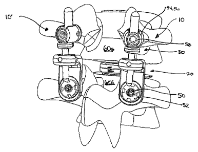
FIGS. IA-4C illustrate one exemplary embodiment of a posterior stabilizing
implant. While two implants 10, 10' are shown coupled to opposed lateral sides
of two
adjacent vertebrae 60s, 60i, only one implant 10 will be discussed herein. A
person
skilled in the art will understand that the implants 10, 10' can have
substantially the same
configuration. Moreover, while only two implants 10, 10' are shown, additional
implants can be coupled to additional vertebrae located along the patient's
spinal
column. FIGS. lA-1B also illustrate an artificial disc I implanted between the
adjacent
vertebrae 60s, 60i. A person skilled in the art will appreciate that the
posterior
stabilizing implants disclosed herein can be used with a natural disc or with
an artificial
disc. In an exemplary embodiment, where an artificial disc is used, the disc
is preferably
one which allows movement of the adjacent vertebrae 60s, 60i relative to one
another.
By way of non-limiting example, one exemplary artificial disc for use with the
present
invention is the CharitdTM Artificial Disc available from DePuy Spine, Inc.
As shown in FIGS. lA-IB, the implant 10 can include a first member 20 that is
coupled to a first vertebra, e.g., the inferior vertebra 60i, and a second
member 30 that is
coupled to a second vertebra, e.g., the superior vertebra 60s. While not
shown, the first
and second members 20, 30 can be reversed such that the first member 20 is
coupled to
the superior vertebra 60s and the second member 30 is coupled to the inferior
vertebra
60i. The first and second members 20, 30 can also be movably coupled to one
another.
In particular, the first member 20 can include a bearing element 22 movably
disposed
therein, and the second member 30 can include an extension rod 32 that is
adapted to
slidably extend through the bearing element 22. In use, the bearing element 22
and the
extension rod 32 cooperate to control movement of the superior and inferior
vertebrae
60s, 60i relative to one another, and in particular they allow flexion,
extension, and
lateral bending of the vertebrae 60s, 60i, while substantially restricting
posterior-anterior
shear and rotation of the vertebrae 60s, 60i.
CA 02592606 2007-06-28
WO 2006/073593 PCT/US2005/042123
- 13-
The first member 20 of the implant 10, which is shown in more detail in FIG.
2A, can have a variety of configurations. In the illustrated exemplary
embodiment,
however, the first member 20 is substantially L-shaped and it includes a first
portion 20a
that is adapted to mate to a vertebra, e.g., the inferior vertebra 60i, and a
second portion
20b having the bearing element 22 disposed therein. The exemplary first and
second
portions 20a, 20b each have a substantially planar configuration, and each
portion 20a,
20b can be positioned at an angle relative to one another. For example, the
first and
second portions 20a, 20b can be substantially perpendicular to one another.
The
configuration of each portion 20a, 20b relative to one another can, however,
vary
depending on the intended use.
As noted above, the first portion 20a is adapted to mate to a vertebra. While
various techniques can be used to allow the first portion 20a to mate to a
vertebra, in the
illustrated exemplary embodiment the first portion 20a includes an opening 24
extending
therethrough for receiving a portion of a fastening element and/or a bone
engaging
element. The opening 24 can vary in shape and size depending on the type of
bone
engaging element and fastening element being used. In an exemplary embodiment,
as
shown in FIG. 2B, the bone engaging element is a bone screw 50 and the
fastening
element is a locking nut 52 that is adapted to engage the bone screw 50 to
lock the first
portion 20a of the first element 20 relative to the vertebra 60i. In
particular, the bone
screw 50 has a threaded shank 50a that is adapted to extend into the vertebra
60i, a
receiving head 50b formed on the threaded shank 50a, and a threaded central
shaft 50c
that extends from the receiving head 50b through the opening 24 in the first
portion 20a
and that mates to the locking nut 52. In one exemplary embodiment the
receiving head
50b can have a shape that is configured to seat a posterior surface or
articulating surface
26 of the first portion 20a of the first member 20 such that a position of the
first member
20 relative to the bone screw 50 can be adjusted. For example, the receiving
head 50b
can include a substantially spherical recess 51 formed therein, and the
articulating
surface 26 of the first portion 20a of the first member 20 can be
substantially spherical,
as shown in FIG. 2A. As a result, the first member 20 can be angularly
adjustable
relative to the bone screw 50, and in particular relative to the vertebra 60i.
Such a
configuration allows the bearing element 22 of the second portion 20b of the
first
member 20 to be positioned as desired, as will be discussed in more detail
below.
CA 02592606 2007-06-28
WO 2006/073593 PCT/US2005/042123
-14-
The second portion 20b of the first member 20 can also have a variety of
configurations, but as noted above the exemplary second portion 20b includes a
bearing
element 22 disposed therein for receiving the extension rod 32 on the second
member
30. Various bearing elements 22 known in the art can be used, but in the
illustrated
embodiment the bearing element 22 is a standard ball bearing that includes an
opening
22i formed therethrough. The bearing element 22 can be disposed within the
second
portion 20b of the first member 20 using a variety of techniques, but in an
exemplary
embodiment the bearing element 22 is preferably freely rotatable relative to
the second
portion 20b of the first member 20. This will allow the bearing element 22 to
pivot/rotate as the first and second members 20, 22 move relative to one
another as a
result of movement of the vertebrae 60s, 60i relative to one another. As shown
in FIG.
2A, the bearing element 22 is disposed within a spherical recess 28 that is
formed within
and extends through an insert 27, and the insert 27 in turn is disposed within
an opening
25 formed in the second portion 20b. A person skilled in the art will
understand that the
bearing element 22 can be directly disposed within a recess formed within the
second
portion 20b, and the use of an insert 27 is not necessary.
In order to facilitate free rotation/movement of the bearing element 22 within
the
recess 28, the bearing element 22 and/or the recess 28 can include a coating
to reduce
friction and reduce wear. The opening 22i in the bearing element 22 can also
include a
coating formed therein to reduce friction and wear on the bearing element 22
caused by
movement of the extension rod 32 therethrough. Suitable exemplary materials
for
coating the bearing element 22, the recess 28, and/or the extension rod 32
include, by
way of non-limiting example, titanium nitrite coating, titanium carbon-nitrite
coating,
diamond-like carbon coating, and other similar materials. The bearing element
22, the
recess 28, and/or the extension rod 32, which will be discussed in more detail
below, can
also be formed from certain materials that are adapted to withstand wear, such
as, for
example, stainless steel, titanium, cobalt chrome, plastics such as
polyethylene and
polyurethane, and various ceramics.
The second member 30 of the implant 10 can also have a variety of
configurations, but in one exemplary embodiment, as shown in more detail in
FIG. 3, the
second member 30 can have a substantially elongate shape with first and second
portions
30a, 30b. The first portion 30a can be adapted to couple to a bone engaging
element for
CA 02592606 2007-06-28
WO 2006/073593 PCT/US2005/042123
- 15 -
mating the first portion 30a to a vertebra, e.g., the superior vertebra 60s,
and the second
portion 30b can form the extension rod 32 that is adapted to extend through
the opening
22i formed in the bearing element 22. The first and second portions 30a, 30b
can be
coaxial with one another, but in an exemplary embodiment the first and second
portions
30a, 30b are axially offset from one another. In particular, the axis A, of
the first
portion 30a can be spaced a distance D apart from the axis A2 of the second
portion 30b.
While the distance can vary, in one exemplary embodiment the distance D can be
in the
range of about 2 mm to 10 mm. Such a configuration will facilitate positioning
of the
second portion 30b, e.g., the extension rod 32, relative to the bearing
element 22, and it
can also allow the extension rod 32 to move relative to the bearing element 22
without
abutting against or otherwise coming into contact with the first portion 20a
of the first
member 20.
As noted above, the first portion 30a of the second member 30 can be adapted
to
couple to a bone engaging element to mate the first portion 30a to the
superior vertebra
60s. Accordingly, the first portion 30a can have a variety of configurations
depending
on the type of bone engaging element used. In the exemplary embodiment shown
in
FIGS. 1A and 1B, the bone engaging element is a bone screw 54 having a shank
(not
shown) that threads into the vertebra 60s, and a U-shaped receiving head 56.
Accordingly, the first portion 30a can be in the form of a rod that is adapted
to seat
within the receiving head 56. A locking element, such as a set screw, can be
used to
lock the first portion 30a within the receiving head 56, thereby mating the
second
member 30 to the vertebra 60s. In another exemplary embodiment, the bone screw
54
can be a polyaxial bone screw such that the receiving head 54 is angularly
adjustable
relative to the shank. Such a configuration will allow the second member 30 to
be set at
a desired position relative to the first member 20, and in particular the
extension rod 32
can be positioned as desired relative to the bearing element 22. The
orientation of the
second member 30 relative to the first member 20 can be used to control
movement of
the vertebrae 60s, 60i relative to one another, as will be discussed in more
detail below.
A person skilled in the art will appreciate that a variety of other devices
including, for
example, offset connectors, can be used to mate the second member 30 to the
vertebra.
CA 02592606 2007-06-28
WO 2006/073593 PCT/US2005/042123
-16-
The extension rod 32 of the second member 30 can also have a variety of
configurations, but it should be adapted to be extend through and slidably
move relative
to the bearing element 22. In the illustrated exemplary embodiment, the
extension rod
32 has a substantially cylindrical shape with a diameter dr that is only
slightly less than
an inner diameter di of the opening formed through the bearing element 22.
The extension rod 32 can also include one or more physical stops formed
thereon
to limit movement thereof relative to the bearing element 22. While the
physical stop(s)
can have a variety of shapes and sizes, in the illustrated exemplary
embodiment the first
portion 30a and the extension rod 32 are separated by a substantially circular
flange 34
that forms a physical stop. The flange 34 can be adapted to abut against a
superior
surface 20s (FIG. 2A) of the first member 20 to limit penetration of the
extension rod 32
through the bearing element 22. Accordingly, the flange 34 preferably has an
extent,
e.g., a diameter df, that is larger than the diameter di of the opening 22i in
the bearing
element. The terminal end 32t of the extension rod 32 can also include a
flange formed
thereon, as is further shown in FIG. 3, to prevent removal of the extension
rod 32 from
the bearing element 22.
The extension rod 32 can also include one or more compressive elements
disposed there around and adapted to act as a cushion for preventing hard
contact
between the extension rod 32 and the bearing element 22, or the second portion
20b of
the first member 20. As shown in FIG. 3, the compressive element 36 can be in
the form
of a donut or similar shaped member that is disposed around the extension rod
32. The
compressive element 36 can be positioned adjacent to the flange 34, or it can
be
disposed or formed on the terminal end 32t of the extension rod 32 as shown.
Alternatively, the flange on the terminal end 32t can be formed from a
compressive
material, or it can include a compressive element mated thereto or formed
thereon. A
person skilled in the art will appreciate that a variety of techniques can be
used to
control movement of and limit hard impact between the extension rod 32 and the
bearing
element 22. A person skilled in the art will also appreciate that a variety of
materials
can be used to form a compressive element. By way of non-limiting example,
suitable
materials include polymers, such as polyurethane, silicone-urethane copolymer,
polycarbonateurethane. Metallic springs can also be used.
CA 02592606 2007-06-28
17
In use, the implant 10 can replace and/or augment one or more of the posterior
elements of the spine, including, for example, the facet joints, the lamina,
the posterior
ligaments, and/or other features of a patient's spinal column. The particular
configuration and
use of the implant 10 can, however, vary depending on the specific procedure
being
performed. For example, where a laminectomy is performed and the facet joints
are not
removed, the implant can be used to reduce the load on the facet joints. Where
the facet
joints are removed, it may be necessary to add an anti-rotation feature, as
will be discussed in
more detail below, to prevent rotation of the bone screws relative to the
vertebrae. Where the
posterior ligaments are removed, it may be desirable to use one or more
compressive
elements to facilitate control of flexion of the vertebrae. The implant 10 can
also be adapted
to function with either a natural vertebral disc, or with an artificial disc
as previously
discussed. Regardless, as noted above, the implant 10 is preferably adapted to
allow flexion,
extension, and lateral bending of the spine, while substantially restricting
posterior-anterior
shear and rotation of the spine. While an exemplary method of implanting only
one posterior
stabilizing implant 10 will be discussed, a person skilled in the art will
appreciate that, in an
exemplary embodiment, two implants 10, 10' are implanted on opposed lateral
sides of
adjacent vertebrae. Moreover, any number of implants can be used to couple
multiple
adjacent vertebrae depending on the needs of the patient.
One exemplary procedure can begin by implanting a bone screw 50 in the
inferior
vertebra 60i, and implanting a bone screw 54 in the superior vertebra 60s. As
shown in FIGS.
lA and 1B, the bone screws 50, 54 are implanted on a lateral side of the
vertebrae 60s, 60i to
allow another implant 10' to be implanted on the opposed lateral side of the
vertebrae 60s,
60i. Once the bone screws 50, 54 are implanted, the first member 20 can be
coupled to bone
screw 50 by positioning the articulating surface 26 of the first portion 20a
on the receiving
head such that the central shaft of the bone screw 50 extends through the
opening 24 in the
first member 20. The locking nut 52 can then be loosely threaded onto the
central shaft of the
bone screw 50 to loosely attach the first member 20 to the bone screw 50. The
first member
20 can then be angularly adjusted as desired, and once properly positioning,
the locking nut
52 can be tightened to maintain the first member 20 in a fixed position
relative to the
vertebra 60i. The second member 30 can be coupled to bone screw 54 by
inserting the
extension rod 32 through the
CA 02592606 2007-06-28
WO 2006/073593 PCT/US2005/042123
-18-
bearing element 22 and positioning the first portion 30a within the receiving
head 56 of
the bone screw 54. The locking element, e.g., set screw 58, can then be
inserted into the
receiving head 56 to loosely mate the second member 30 to the vertebra 60s.
Where the
bone screw 54 is a polyaxial bone screw, the second member. 30 can be
angularly
adjusted by moving the receiving head 56. Once the second member 30 is
properly
positioned, the set screw 58 can be fully tightened to maintain the second
member 30 in
a fixed position relative to the vertebra 60s. A person skilled in the art
will appreciate
that the bone screws 50, 54 and the first and second members 20, 30 can be
implanted
and adjusted in any order. In one exemplary embodiment, the second member 30
is
positioned as desired and the first member 20 is then positioned as necessary
based on
the positioning of the second member 30.
While not shown, where the implant 10 is used to replace the facet joints, it
may
be desirable to include an anti-rotation feature to prevent rotation of the
bone screws that
are implanted in the superior vertebra 60s. While various anti-rotation
techniques can be
used, in one embodiment the bone screws can include spikes or other surface
protrusions
formed on a proximal end of the shank or on the head of the screws to prevent
rotation
thereof. In another embodiment, a cross-connector can be connected to and
extend
between the first portion of the second member of each implant, thereby
preventing
rotation of the bone screw mated thereto.
Once the implant 10 is coupled to the adjacent vertebrae 60s, 60i, the implant
10
can control movement of the vertebrae 60s, 60i relative to one another. In
particular,
during movement of the spine, the bearing element 22 rotates as the extension
rod 32
slidably moves therethrough to control movement of the vertebrae 60s, 60i. Due
to the
configuration of the implant 10, the bearing element 22 and the extension rod
32 can
also substantially prevent axial rotation of the vertebrae 60s, 60i relative
to one another,
and anterior-posterior shearing can be substantially resisted. FIGS. 4A-4C
illustrate the
vertebrae 60s, 60i in a neutral position, and during flexion and extension.
FIG. 4A
illustrates the vertebrae 60s, 60i in a neutral position, 60i. FIG. 4B
illustrates the
vertebrae 60s, 60i during extension, and as shown the extension rod 32 is
fully inserted
into the bearing element 22 such that the flange 34 abuts against the bearing
element 22.
FIG. 4C illustrates flexion of the vertebrae 60s, 60i, and as shown the
bearing element
22 is pivoted relative to the first member 20 and the extension rod 32 is
substantially
CA 02592606 2007-06-28
WO 2006/073593 PCT/US2005/042123
-19-
withdrawn from the bearing element 22 such that only the terminal end 32t of
the
extension rod 32 remains in the bearing element 22.
While the extension rod 32 can be positioned to be substantially parallel to
the
central axisXofthe vertebrae 60s, 60i, the extension rod 32 can be positioned
at a
particular angle relative to the central axisXofthe vertebrae 60s, 60i to
control the
movement of the vertebrae 60s, 60i. As shown in FIG. 4A, the position of the
extension
rod 32 relative to the vertebrae 60s, 60i is indicated by angle a, which is
measured
between a line perpendicular to the central axis X and the axis A2 of the
extension rod
32. In order to increase flexion, the extension rod 32 can angled toward the
central axis
of the vertebrae 60s, 60i such that the angle a is less than 90 . At this
angle, the flange
34 will be positioned closer to the bearing element 22 in the neutral
position. As a
result, when the vertebrae 60s, 60i move from the neutral position, shown in
FIG. 4A, to
the extended position, shown in FIG. 4B, the range of motion will be limited.
Conversely, when the vertebrae 60s, 60i move from the neutral position to the
flexed
position, shown in FIG. 4C, the range of motion will be greater. In order to
decrease
flexion, the extension rod 32 can angled away from the central axis of the
vertebrae 60s,
60i such that the angle a is greater than 90 . At this angle, the flange 34
will be spaced a
greater distance apart from the bearing element 22 in the neutral position. As
a result,
when the vertebrae 60s, 60i move from the neutral position, shown in FIG. 4A,
to the
extended position, shown in FIG. 4B, the range of motion will be increased.
Conversely, when the vertebrae 60s, 60i move from the neutral position to the
flexed
position, shown in FIG. 4C, the range of motion will be decreased.
Accordingly, the
angle a of the extension rod 32 can be selected based on the desired range of
motion
during flexion and extension. A person skilled in the art will appreciate that
the angle a
can vary depending on the desired result, but in an exemplary embodiment the
angle a
can be in the range of about 60 to about 120 .
While not shown, the procedure can also include the step of placing a sheath
or
protective member partially or fully around the implant 10 for preventing
tissue from
growing on the implant 10 and into the bearing element 22, and for preventing
debris
from migrating into the spinal canal.
CA 02592606 2007-06-28
FIGS. 5A-8C illustrate another exemplary embodiment of a posterior stabilizing
implant 10. The implant 100 is somewhat similar to implant 10, except that it
has a bilateral
configuration. In particular, rather than having two implants 10, 10'
positioned on opposed
lateral sides of two adjacent vertebrae, implant 100 can be positioned along
the mid-line of
5 the adjacent vertebrae to control movement of the vertebrae relative to one
another.
As shown in FIGS. 5A and 5B, the exemplary implant 100 generally includes a
first
member 120 that is adapted to couple to a first vertebra, e.g., an inferior
vertebrae 160i, and
that includes a bearing element 122 disposed therein, and a second member 130
that is
adapted to couple to a second vertebrae, e.g., a superior vertebrae 160s, and
that has an
10 extension rod 132 formed thereon. While not shown, the first and second
members 120, 130
can be reversed such that the first member 120 is coupled to the superior
vertebra 160s and
the second member 130 is coupled to the inferior vertebra 160i. In use, the
bearing element
122 is adapted to freely rotate relative to the first member 120, and the
extension rod 132 is
adapted to slidably extend through the bearing element 122 to control movement
of the
15 adjacent vertebrae 160s, 160i, allowing flexion, extension, and lateral
bending of the spine,
while substantially restricting posterior- anterior shear and rotation of the
spine.
The first member 120 of the implant 100, which is shown in more detail in FIG.
6,
can have a variety of configurations. In the illustrated exemplary embodiment,
however, the
first member 120 is substantially Y-shaped and it includes a central portion
120a having the
20 bearing element 122 disposed therein, and first and second arms 120b, 120c
that extend from
the central portion 120a and that are adapted to mate to a vertebra, e.g., the
inferior vertebra
60i. The central portion 120a and the first and second arms 120b, 120c can
have a variety of
shapes and sizes, and the configuration can vary depending on the intended
use. In the
illustrated exemplary embodiment, the central portion 120a has a substantially
planar
cylindrical configuration such that it is adapted to seat the bearing element
122 therein, and
the first and second arms 120b, 120c each extend distally and laterally
outward from the
central portion 120a. Such a configuration allows the first and second arms
120b, 120c to
mate to opposed lateral
CA 02592606 2007-06-28
WO 2006/073593 PCT/US2005/042123
-21 -
sides of the vertebra 160i.
The first and second arms 120b, 120c can mate to the inferior vertebra 160i
using
a variety of techniques. In the illustrated exemplary embodiment, the arms
120b, 120c
are in the form of rods having a generally elongate, substantially cylindrical
configuration. This allows each arm 120b, 120c to be received within a
receiving head
of a bone engaging element. In the embodiment shown in FIGS. 5A and 5B, the
bone
engaging elements are bone screws 150a, 150b that are implanted on opposed
lateral
sides of the inferior vertebra 160i. As previously described above with
respect to FIGS.
1A and 1B, the bone screws 150a, 150b can include a U-shaped head that is
adapted to
seat an arm 120b, 120c, and a locking element, such as a set screw 152a, 152b
can be
used to lock the arms 120b, 120c to the bone screws 150a, 150b. The receiving
head of
each bone screw 150a, 150b can also be polyaxially movable relative to the
threaded
shank (not shown) of the bone screw 150a, 150b to allow the first member 120
to be
angularly adjustable relative to the vertebra 160i. Such a configuration
allows the
bearing element 122 to be positioned as desired, as will be discussed in more
detail
below.
As noted above, the first member 120 also includes a bearing element 122
disposed therein. The bearing element 122 can have a configuration that is the
same as
or similar to the configuration previously described with respect to bearing
element 22
shown in FIGS. 1 A-2. In particular, the bearing element 122 can be freely
rotatably
disposed within a spherical recess formed in the central portion 120a of the
first member
120, or it can be freely rotatably disposed within an insert 127 that is
disposed within the
central portion 120a of the first member 120, as shown in FIG. 6. As was also
previously described, the bearing element 122 can be a standard ball bearing
that
includes an opening 122i formed therethrough for slidably receiving the
extension rod
132 on the second member 130. The bearing element 122, the recess 128 formed
within
the insert 127 for seating the bearing element 122, and/or the opening 122i
formed
through the bearing element 122 can also include a coating to reduce friction
and reduce
wear.
The second member 130 of the implant 10 can also have a variety of
configurations, but in an exemplary embodiment, as shown in more detail in
FIG. 7, the
second member 130 can be substantially Y-shaped with a central portion 130a
having
CA 02592606 2007-06-28
WO 2006/073593 PCT/US2005/042123
-22-
first and second arms 130b, 130c extending laterally from opposed sides
thereof. The
extension rod 132 can also extend from the central portion 130a. The
particular angle of
each arms 130b, 130c relative to the extension rod 132 can vary depending on
the
intended use, but in an exemplary embodiment 130b, 130c that arms have a
configuration that allows each arm 130b, 130c to mate to opposed lateral sides
of a
vertebra, e.g., the superior vertebra 160s.
Each arm 130b, 130c can be mated to the vertebra 160s using a variety of
techniques, however in an exemplary embodiment each arm 130b, 130c is in the
form of
a rod having a substantially elongate cylindrical shape such that the arms
130b, 130c can
mate to a receiving head of a bone engaging element, such as bone screws I 50c
and
150d as shown. As previously described, the bone screws 150c, 150d can be
polyaxial
bone screws to allow the position of the second member 130 to be angularly
adjusted as
desired, and in particular to allow the extension rod 132 to be positioned as
desired
relative to the bearing element 122. A locking element, such as a set screw
152c, 152d
can be used to lock the arms 130b, 130c to the bone screws 150c, 150d.
The extension rod 132 of the second member 130 can also have a variety of
configurations, but in an exemplary embodiment the extension rod 132 is
similar to
extension rod 22 previously described with respect to FIGS. lA, IB, and 3. In
particular, the extension rod 132 should be adapted to be extend through and
slidably
move relative to the bearing element 122. In the illustrated exemplary
embodiment, the
extension rod 132 has a substantially cylindrical shape with a diameter Dr
that is only
slightly less than an inner diameter Di of the opening formed through the
bearing
element 122.
As previously described with respect to FIG. 3, the extension rod 132 can also
include a physical stop formed thereon to limit movement thereof relative to
the bearing
element 122. While the physical stop can have a variety of shapes and sizes,
in the
illustrated exemplary embodiment the central portion 130a has a substantially
cylindrical
shape with a surface 131 that is adapted to abut against the bearing element
122 to limit
penetration of the extension rod 132 through the bearing element 122.
Accordingly, the
surface 131 preferably has an extent, e.g., a diameter Df, that is larger than
the diameter
Di of the opening 122i in the bearing element.
CA 02592606 2007-06-28
WO 2006/073593 PCT/US2005/042123
-23-
The extension rod 132 can also include one or more compressive elements
disposed there around, as previously described with respect to FIG. 3, for
providing a
cushion to substantially prevent hard contact between the extension rod 132
and the
bearing element 122, or the central portion 120a of the first member 120. The
compressive element(s) (not shown) can be in the form of a donut or similar
shaped
member that is disposed around the extension rod 132. The compressive element
can be
positioned adjacent to surface 131, and/or it can be disposed or formed on the
terminal
end 132t of the extension rod 1.32. The terminal end 132t can also include a
stop surface
or flange 136 formed thereon, as shown in phantom in FIG. 7, to prevent the
extension
rod 132 from being fully withdrawn from the bearing element 122, and
optionally to
retain a compressive element on the extension rod 132. Alternatively, flange
136 can be
formed from a compressive material, or it can include a compressive element
mated
thereto or formed thereon. A person skilled in the art will appreciate that a
variety of
techniques can be used to control movement of and limit hard impact between
the
extension rod 132 and the bearing element 122. A person skilled in the art
will also
appreciate that a variety of materials can be used to form a compressive
element.
While not shown, in another exemplary embodiment the extension rod 132 can
be adjustable relative to the first and second arms 130b, 130c. For example,
the
extension rod 132 can be rotatably mated to the central portion 130a, and the
central
portion 130a can include a locking mechanism that is adapted to lock the
extension rod
132 in a desired fixed position. Such a configuration is particularly
desirable where the
bone screws 150c, 150d used to attach the arms 130b, 130c to the vertebra 160s
are not
polyaxial. The extension rod 132 can thus be positioned at a desired angle
relative to the
vertebra 160s, and then locked in place to maintain it at the desired angular
position. A
person skilled in the art will appreciate that a variety of other techniques
can be used to
allow the extension rod 132 to be adjusted relative to the remainder of the
second
member 130.
In use, the implant 100 can replace and/or augment one or more of the
posterior
elements of the spine, including, for example, the facet joints, the lamina,
the posterior
ligaments, and/or other features of a patient's spinal column. The implant 100
can also
be adapted to function with either a natural vertebral disc, or with an
artificial disc as
previously discussed. Regardless, as noted above, the implant 100 is
preferably adapted
CA 02592606 2007-06-28
WO 2006/073593 PCT/US2005/042123
-24-
to allow flexion, extension, and lateral bending of the spine, while
substantially
restricting posterior-anterior shear and rotation of the spine. The particular
configuration and use of the implant 100 can, however, vary depending on the
specific
procedure being performed. For example, where a laminectomy is performed and
the
facet joints are not removed, the implant can be used to reduce the load on
the facet
joints. Where the facet joints are removed, it may be necessary to add an anti-
rotation
feature as previously discussed to prevent rotation of the bone screws
relative to the
vertebrae. Where the posterior ligaments are removed, it may be desirable to
use one or
more compressive elements to facilitate control of flexion of the vertebrae.
One exemplary procedure can begin by implanting two bone screws 150a, 150b
in the inferior vertebra 160i, and implanting two bone screws 150c, 150d in
the superior
vertebra 160s. As shown in FIGS. 5A and 513, the bone screws 150a, 150b, 150c,
150d
are implanted on opposed lateral sides of the vertebrae 160s, 160i. Once the
bone
screws 150a, 150b, 150c, 150d are implanted, the first member 120 can be
coupled to
bone screws 150a, 150b by positioning the arms 120b, 120c in the receiving
head of the
bone screws 150a, 150b such that the central portion 120a is positioned toward
the
superior vertebra 160s. The set screws 152a, 152b can then be loosely threaded
onto the
receiving heads of the bone screws 150a, 150b to loosely attach the first
member 120 to
the bone screws 150a, 150b. Where the bone screws 150a, 150b are polyaxial
bone
screws, the first member 120 can be angularly adjusted by moving the receiving
heads of
the screws 150a, 150b. Once properly positioned, the set screws 152a, 150s can
be
tightened to maintain the first member 120 in a fixed position relative to the
vertebra
160i. As previously described, the extension rod 132 can be positioned at a
desired
angle relative to the vertebrae 160s, 160i. The second member 130 can
similarly be
coupled to two bone screws 150c, 150d by inserting the extension rod 132
through the
bearing element 122, and positioning the arms 130b, 130c within the receiving
heads of
the bone screws 150c, 150d. The set screws 152c, 152d can be loosely mated to
the
receiving heads to retain the arms 130b, 130c therein. Where the bone screws
150c,
150d are polyaxial bone screws, the second member 130 can be angularly
adjusted by
moving the receiving heads of the screws 150c, 150d. Once the second member
130 is
properly positioned, the set screws 152c, 152d can be fully tightened to
maintain the
second member 130 in a fixed position relative to the vertebra 160s. A person
skilled in
CA 02592606 2007-06-28
WO 2006/073593 PCT/US2005/042123
-25-
the art will appreciate that the bone screws 150a, 150b, 150c, 150d and the
first and
second members 120, 130 can be implanted and adjusted in any order. In one
exemplary
embodiment, the second member 130 is positioned as desired and the first
member 120
is then positioned as necessary based on the positioning of the second member
130.
Once the implant 100 is coupled to the adjacent vertebrae 160s, 160i, the
implant
100 can control movement of the vertebrae 160s, 160i relative to one another.
In
particular, during movement of the spine, the bearing element 122 rotates as
the
extension rod 132 slidably moves therethrough to control movement of the
vertebrae
160s, 160i. Due to the configuration of the implant 100, the bearing element
122 and the
extension rod 132 can also substantially prevent axial rotation of the
vertebrae 160s,
160i relative to one another, and anterior-posterior shearing can be
substantially resisted.
FIGS. 8A-8C illustrate the vertebrae 160s, 160i in a neutral position, and
during flexion
and extension. FIG. 8A illustrates the vertebrae 160s, 160i in a neutral
position, and as
shown the extension rod 132 is substantially parallel to the central axis Y of
the vertebrae
160s, 160i. FIG. 8B illustrates the vertebrae 160s, 160i during extension, and
as shown
the extension rod 132 is fully inserted into the bearing element 122 such that
surface 131
abuts against the bearing element 122. FIG. 8C illustrates flexion of the
vertebrae 160s,
160i, and as shown the bearing element 122 is pivoted relative to the first
member 120
and the extension rod 132 is substantially withdrawn from the bearing element
122 such
that only the terminal end 132t of the extension rod 132 remains in the
bearing element
122.
While not shown, the procedure can also include the step of placing a sheath
or
protective member partially or fully around the implant 100 for preventing
tissue from
growing on the implant 100 and into the bearing element 122, and for
preventing debris
from migrating into the spinal canal.
FIGS. 9A-9B illustrate yet another exemplary embodiment of a posterior
stabilizing implant 200. In this embodiment, rather than having a bearing
element that
allows pivotal movement between two components, the implant 200 includes a
bearing
element that allows linear movement of two components. In particular, as
shown, the
implant 200 can include a first member 200a that is adapted to couple to a
first vertebra,
and a second member 200b that is adapted to couple to a second vertebra and to
slidably
move relative to the first member 200a to allow or control extension, flexion,
and lateral
CA 02592606 2007-06-28
WO 2006/073593 PCT/US2005/042123
-26-
bending of the adjacent vertebrae, preferably while substantially limiting or
preventing
axial rotation and shearing. In an exemplary embodiment, one of the first and
second
members 200a, 200b can be configured to rigidly couple to a vertebra, and the
other one
of the members 200a, 200b can be configured to dynamically couple to a
vertebra,
thereby allowing linear movement between the two components.
The first member 200a can have a variety of configurations, but in the
illustrated
exemplary embodiment it includes opposed first and second lateral members
202a, 202b
that are adapted to be coupled to opposed lateral sides of a vertebra. The
first member
200a can also include a connecting member 220 that extends between and
connects to
the first and second lateral members 202a, 202b. The connecting member 220
will be
discussed in more detail below with respect to FIG. 11. Each lateral member
202a, 202b
can have a variety of configurations, but in one exemplary embodiment each
lateral
member 202a, 202b is adapted to slidably receive a portion of the second
member 200b
to allow linear movement between the first and second members 200a, 200b.
While this
can be achieved using various techniques, in the illustrated exemplary
embodiment each
lateral member 202a, 202b is in the form of a cylindrical member having an
inner lumen
204a, 204b (FIG. 9B) extending therethrough. Since each lateral member 202a,
202b
preferably has substantially the same configuration, only one lateral member,
e.g., the
first lateral member 202a, will be described in detail with reference to FIGS.
l0A and
l OB. As shown, the first lateral member 202a has a lumen 204a formed
therethrough.
The inner lumen 204a can vary in shape and size, but in one exemplary
embodiment the
inner lumen 204a includes a first portion 204a, having a diameter dl that is
greater than
a diameter d2 of a second portion 204a2 of the lumen 204a. The enlarged
diameter
portion 204a, allows the lateral member 202a to receive a head 212a, 212b of a
sliding
pin 210a, 210b of the second member 200b, which will be discussed in more
detail
below. The enlarged diameter portion 204a, also provides a shelf 205 formed
within the
inner lumen 204a that can function as a stop surface to receive the head 212a,
212b of
the sliding pin 210a, 210b. Although not shown, a spring or other compressive
element
could rest between shelf 205 and sliding pin 210a in order to provide some
resistance to
flexion. A person skilled in the art will appreciate that a variety of other
techniques can
be used to limit movement of the sliding pins 210a, 210b with respect to the
lateral
members 202a, 202b, certain exemplary embodiments of which will be discussed
in
CA 02592606 2007-06-28
WO 2006/073593 PCT/US2005/042123
-27-
more detail below.
Each lateral member 202a, 202b can also include an offset connector 206a, 206b
formed thereon to facilitate mating thereof to the connecting member 220, and
to allow
the first and second lateral members 202a, 202b to be mated to a vertebra. The
offset
connectors 206a, 206b can have a variety of configurations, but in one
exemplary
embodiment the connectors 206a, 206b are the same or substantially similar.
Accordingly, only one of the offset connectors 206a, 206b, e.g., the offset
connector
206a on the first lateral member 202a, will be described in detail. Referring
to FIG.
10A, the exemplary offset connector 206a is substantially planar and extends
outward
from the cylindrical portion of the lateral member 202a. The offset connector
206a can
also include a thru-bore 208a formed therein for receiving a fastening
element, such as a
bone screw, for mating the offset connector 206a, as well as the connecting
member 220,
to a vertebra. Exemplary techniques for mating the offset connectors 206a,
206b to bone
and to the connecting member 220 will be described in more detail with respect
to FIGS.
12-14.
The connecting member 220 can also have a variety of configurations, but as
indicated above it is preferably adapted to extend between and couple to the
first and
second lateral members 202a, 202b. The connecting member 220, while not
necessary,
is particularly advantageous in that it can provide a rigid connection between
the first
and second lateral members 202a, 202b, thereby preventing rotation of the
screw relative
to the bone. FIG. 11 illustrates exemplary connecting member 220 in more
detail, and
as shown the connecting member 220 is in the form of an elongate substantially
planar
rod. The shape of the connecting member 220 can vary, but in an exemplary
embodiment the connecting member 220 has a shape that is adapted to ensure
clearance
of the facet joints and the spinous processes of the adjacent vertebrae when
the
connecting member 220 is mated to the first and second lateral members 202a,
202b. As
shown in FIG. 11, the connecting member 220 includes a central portion 220c
that is
substantially curved, and opposed ends 220a, 220b each having a planar
configuration.
While not shown, the opposed ends 220a, 220b could alternatively be
polyaxially
connected to the central portion 220c to allow for independent alignment of
each end
220a, 220b. Each end 220a, 220b can have a thru-bore 222a, 22b formed therein
for
receiving a fastening element for mating the connecting member 220 to the
first and
CA 02592606 2007-06-28
WO 2006/073593 PCT/US2005/042123
-28-
second lateral members 202a, 202b and to bone. A person skilled in the art
will
appreciate that the connecting member 220 can have a variety of other
configurations,
and that the configuration can vary depending on the intended use.
As indicated above, a variety of techniques can be used to mate the connecting
member 220 to the lateral members 202a, 202b. In an exemplary embodiment, as
shown
in FIGS. 9A and 9B, the connecting member 220 is mated to the first and second
lateral
members 202a, 202b using fastening elements, such as first and second bone
screws
230a, 230b. One of the bone screws, e.g., the first bone screw 230a is shown
in detail in
FIG. 12. As shown, the bone screw 230a has a proximal portion 230a, with
threads
formed thereon, a distal bone-engaging portion 230a2, and a flange 232a
separating the
proximal and distal portions 230al, 230a2. The proximal portion 230a, is
adapted to
extend through the thru-bore 208a formed in the offset connector 206a of the
first lateral
member 202a, and through the thru-bore 222a formed in the connecting member
220. A
locking mechanism, such as a locking nut 234a, can then be threaded onto the
proximal
portion 230a, of the bone screw 230a to mate the connecting member 220 and the
first
lateral member 202a to one another and to a first vertebra.
In certain exemplary embodiments, an axis Y of the bone screw 230a can be
adapted to be positioned at an angle relative to an axis X of the thru-bore
222b in the
connecting member 220 and the thru-bore 208a in the offset connector 206a of
the first
lateral member 202a, as shown in FIG. 9B. Furthermore, the axis Y of the bone
screw
230a can be adapted to be positioned at an angle relative to an axis of the
sliding pins
210a, 210b. While various techniques can be used to allow angular variations
between
the bone screws 230a, 230b and the connecting member 220 and the lateral
members
202a, 202b, in one exemplary embodiment, one or more washers can be used to
provide
an angular connection. As shown in FIG. 9B, each lateral member 202a, 202b of
the
implant 200 includes a first washer 240al, 240b, disposed between the terminal
end
220a, 220b of the connecting member 220 and the locking nut 234a, 234b, and a
second
washer 240a2, 240b2 disposed between the flange 232a, 232b formed on the bone
screw
230a, 230b and the terminal end 220a, 220b of the connecting member 220. Each
washer 240al, 240bl, 240a2, 240b2 can have a variety of configurations, but in
an
exemplary embodiment all four washers 240ai, 240b1, 240a2, 240b2 are
substantially
identical. Accordingly, only one of the washers, e.g., washer 240al, will be
described in
CA 02592606 2007-06-28
WO 2006/073593 PCT/US2005/042123
-29-
detail. Referring to FIG. 13, as shown the washer 240a, is substantially
cylindrical with
a bore 242a formed therethrough. In an exemplary embodiment, the washer 240al
has
an angular configuration such that a first surface 243 of the washer 240ai is
positioned at
an angle relative to a second opposed surface 245 of the washer 240al. The
bore 242a in
the washer 240ai can also taper to allow further angular variations between
the
connecting member 220 and the lateral member 202a and the axis Y of the bone
screw
230a. In use, referring back to FIG. 9B, the bone screws 230a, 230b can be
implanted in
the vertebra at a desired angle, and the washers 240al, 240bl, 240a2, 240b2
allow the
lateral members 202a, 202b and the connecting member 220 to be mated thereto
at a
desired orientation regardless of the particular angle of the bone screws
230a, 230b. '
FIG. 14 illustrates yet another embodiment of a technique for allowing angular
variations between the bone screws 230a, 230b and the connecting member 220
and the
lateral members 202a, 202b. In this embodiment, which illustrates only one
assembly
for use with, for example, the first lateral member 202a and the first
terminal end 206a
of the connecting member 220, two washers 240a', 240b' are provided. Each
washer
240a', 240b' has a planar surface 243a', 243b' and an opposed spherical
surface 245a',
245b'. The planar surface 243a' of the first washer 240a' can be positioned
adjacent to
the terminal end 220a of the connecting member 220, and the planar surface
243b' of the
second washer 240b' can be positioned adjacent to the offset connector 206a of
the first
lateral member 202a. The opposed spherical surfaces 245a', 245b' of the
washers 240a',
240b' allow the bone screw 230a to be disposed through the washers 240a',
240b', the
terminal end 220a of the connecting member 220, and the offset connector 206a
at an
angle. A locking nut 234' having a spherical cavity 235' formed therein can be
mated to
the proximal portion 230a, of the bone screw 230a. The spherical cavity 235'
allows the
locking nut 234' to pivot about the spherical surface 245a' of the first
washer 240a', thus
allowing the locking nut 234' to mate to the angularly oriented bone screw
230a. A
person skilled in the art will appreciate that a variety of other techniques
can be used to
mate the bone screws or other fastening elements to the connecting member and
the
lateral members.
Referring back to FIGS. 9A and 9B, the implant 200 can also include a second
member 200b that is adapted to couple to a second vertebra and to move
relative to the
first member 200a to control movement of the adjacent vertebrae. While the
second
CA 02592606 2007-06-28
WO 2006/073593 PCT/US2005/042123
-30-
member 200b can have a variety of configurations, in the exemplary embodiment
shown
in FIGS. 9A and 9B the second member 200b includes first and second sliding
pins
210a, 210b that are adapted to be slidably disposed through the lumens 204a,
204b in the
first and second lateral members 202a, 202b, respectively. In an exemplary
embodiment, the first and second sliding pins 210a, 210b are the same or
substantially
identical, and thus only one pin, e.g., the first sliding pin 210a, will be
described in
detail. Referring to FIG. 15, the first sliding pin 210a has a generally
elongate rod-like
shape with a head 212a formed on a first terminal end 210a, thereof. As
previously
discussed, the head 212a can be adapted to be received with the enlarged
diameter
region 204a, of the lumen 204a in the first lateral member 202a to allow
sliding
movement of the sliding pin 210a with respect to the first lateral member
202a. In an
exemplary embodiment, the head 212a has a diameter d3 that is less than the
diameter di
of the enlarged diameter portion 204ai, but that is greater than the diameter
d2 of the
second portion 204a2 of the lumen 204a in the first lateral member 202a. Thus,
in use,
the head 212a is allowed to slide into the first portion 204ai of the lumen
204a in the
first lateral member 202a until it abuts against the shelf 205, at which point
further
movement is prevented. A person skilled in the art will appreciate that the
components
of the second member 200b can have a variety of other configurations.
As previously noted, the second member 200b can be adapted to couple to a
second vertebra. Thus, in an exemplary embodiment, the second member 200b can
also
include first and second fastening elements 214a, 214b for coupling a second
terminal
end 210al, 210a2 of each sliding pin 210a, 210b to a vertebra. While the
fastening
elements 214a, 214b can be configured to provide a rigid connection between
the sliding
pins 210a, 210b and a vertebra, in an exemplary embodiment each fastening
element
214a, 214b is configured to provide a dynamic connection between the sliding
pins
210a, 210b and the second vertebra. This can be achieved using a variety of
techniques.
In the embodiment shown in FIGS. 9A and 9B, first and second polyaxial bone
screws 214a, 214b are used to provide a dynamic connection between the first
and
second sliding pins 210a, 210b and a vertebra mated thereto. The first
polyaxial bone
screw 214a is shown in more detail in FIG. 16, and as shown it includes a
threaded
shank 216a having a receiving head 218a formed thereon and pivotally coupled
to the
shank 216a to provide polyaxial movement of the head 218a relative to the
shank 216a.
CA 02592606 2007-06-28
WO 2006/073593 PCT/US2005/042123
-31 -
One of the sliding pins, e.g., the terminal end 210a2 of the first sliding pin
210a, can be
disposed within the receiving head 218a and a locking mechanism, such as a set
screw
219a (FIGS. 9A-9B), can be mated to the head 218a to lock the sliding pin 210a
therein.
While the pin 210a is locked with respect to the head 218a, the head 218a is
free to
pivotally or polyaxially move about the shaft 216a, thereby allowing dynamic
movement
of the second member 200b of the posterior stabilizing implant 200. A person
skilled in
the art will appreciate that a variety of other techniques can be used to
provide a
dynamic mating connection between the sliding pins 210a, 210b and a vertebra.
Moreover, while the second member 200b is shown having a dynamic connection,
the
second member 200b can be rigidly mated to the second vertebra, and the first
member
200a can by dynamically mated to the first vertebra.
FIGS. 17A-17D illustrate another embodiment of a technique for dynamically
mating the sliding pins of the second member to a vertebra. In the embodiment
shown
in FIGS. 17A-17D, rather than having a polyaxial bone screw, the second
terminal end
of the sliding pin member can include a spherical head formed thereon. An
exemplary
sliding pin 250 with a spherical head 252 is shown in detail in FIG. 17D. The
spherical
head 252 is adapted to be disposed within a complementary spherical recess 266
formed
in a housing 265, as shown in FIG. 17C. The housing 265, which can include
first and
second portions 265a, 265b that fit together to close around the spherical
head 252, can
be sized to fit within and mate to a receiving head 262 of a bone screw 260 to
allow
polyaxial movement of the sliding pin 250 relative to the bone screw 260.
While the
configuration of the bone screw 260 can vary, in an exemplary embodiment, the
bone
screw 260 is a standard monoaxial bone screw. The housing halves 265a, 265b,
when fit
together, can have a substantially cylindrical shape that fits within the
receiving head
262 of the bone screw 260. At least one of the housing halves, e.g., half
265b, can
include a feature, such as a ridge or protrusion 267 formed therein and
adapted to extend
into one of the slots in the receiving head 262 to prevent rotational movement
of the
housing 265 with respect to the bone screw 260. Once disposed therein, a
locking
mechanism can be mated to the head 262 of the bone screw 260 to lock the
housing 265
therein. In the illustrated exemplary embodiment shown in FIGS. 17A-17D, the
locking
mechanism is a set screw 268 that threads into the head 262 of the bone screw
260 to
lock the housing 265 therein.
CA 02592606 2007-06-28
WO 2006/073593 PCT/US2005/042123
-32-
FIGS. 18A-18F illustrate additional exemplary techniques for dynamically
mating the sliding pins of the second member to a vertebra. In the embodiment
shown
in FIG. 18A, the housing 265a is in the form of a sleeve that is removably
disposed
within the receiving head 262a of a bone screw 260a. The sleeve 265a has a
longitudinal slit that allows the sleeve 265a to expand for receiving the
spherical head
252 of the sliding pin 250. When the sleeve 265a and the spherical head 252
are
disposed within the head 262a of the bone screw 260a, a locking mechanism,
such as a
set screw 268a, can be applied to the head 262a of the bone screw 260a lock
the sleeve
265a therein. The sleeve 265a, like housing 265, can be configured to allow
polyaxial
movement of the sliding pin 250 relative thereto to provide a dynamic
connection. In
another embodiment, shown in FIG. 18B, the lateral opening 264b formed in the
head
262b of the bone screw 260b can extend only partially therethrough, thus
preventing the
housing from passing completely through the head 262b of the bone screw 260b.
FIG.
18C illustrates another embodiment in which the housing 265c has a length that
is
greater than a width of the head 262c of the bone screw 260c. As a result, the
housing
265c extends from opposed ends of the lateral opening 264c in the bone screw
260c.
Such a configuration allows an outer locking mechanism, e.g., an outer set
screw 268c,
to be applied to the head 262c of the bone screw 260c to engage the housing
265c.
Alternatively, the locking mechanism 268d can include a pin member 267d or
similar
feature, as shown in FIG. 18D, for engaging the housing 265d to prevent
movement of
the housing 265d relative to the bone screw 260d. In another embodiment, as
shown in
FIG. 18E, the head 262e of the bone screw 260e can be in the form of a clamp
mechanism that is adapted to engage the housing. FIG. 18F illustrates another
embodiment in which a universal joint 269f is used to provide dynamic motion
between
the head 252 of the sliding pin and the bone screw 260f. A person skilled in
the art will
appreciate that a variety of other techniques can be used to provide a dynamic
connection between the sliding pins and the bone screws.
In another exemplary embodiment, referring back to FIGS. 9A and 9B, the
implant 200 can include one or more control members 270a, 270b for controlling
movement between the first and second members 202a, 202b. The control members
270a, 270b can form a rigid stop to prevent extension of the adjacent
vertebrae beyond a
particular position, or they can form a compressive stop to limit or control
extension of
CA 02592606 2007-06-28
WO 2006/073593 PCT/US2005/042123
-33-
the adjacent vertebrae. In the illustrated exemplary embodiment, the first and
second
control members 270a, 270b are substantially cylindrical compressive members
that are
slidably disposed around the first and second sliding pins 210a, 210b,
respectively.
Each control member 270a, 270b can be disposed between the lateral member
202a,
202b and the bone screw 214a, 214b that mates the sliding pin 210a, 210b to
the
vertebrae. As a result, during extension or lateral bending of the vertebrae,
the control
members 270a, 270b are compressed as the sliding pins 210a, 210b slide into
the lateral
members 202a, 202b. The control members 270a, 270b can therefore limit or
control
extension and/or lateral bending. A person having ordinary skill in the art
will
appreciate that the properties, e.g., the size, shape, flexibility, etc., of
the control
members can be selected to obtain a desired result. Moreover, a kit containing
several
control members varying in property can be provided to allow a surgeon to
select control
members having the appropriate properties.
FIGS. 19A and 19B illustrate the implant 200 coupled to adjacent superior (S)
and inferior (1) vertebrae. The lateral members 202a, 202b of the first member
200a are
coupled to one another by the connecting member 220, and the three components
202a,
202b, 220 are rigidly coupled to the superior vertebra S by two bone screws
230a, 230b
implanted in the pedicles of the vertebra S. The sliding pins 210a, 2I Ob are
slidably
disposed through the lateral members 202a, 202b and they are dynamically
coupled to
the inferior vertebra I by two polyaxial bone screws 214a, 214b implanted in
the
pedicles of the vertebra I. The configuration of the implant 200 is
particularly
advantageous in that it requires minimal, if any, resection of the facet
joints due to the
lateral positioning of the members 200a, 200b.
In use, the implant 200 allows lateral bending, flexion, and extension of the
adjacent vertebrae S, I, preferably while limiting or substantially preventing
axial
rotation and shearing. In particular, flexion of the vertebrae S,1 will cause
the heads
212a, 212b of the sliding pins 210a, 210b to slide into the lateral members
202a, 202b
until the heads 212a, 212b abut against the stop (e.g., shelf 205 shown in
FIG. 1013).
Although not shown, a spring or other compressive element could rest between
shelf 205
and sliding pin 210a in order to provide some resistance to flexion. The
amount of
flexion can be adjusted by positioning the head 212a, 212b of each sliding pin
210a,
210b at a particular distance apart from the lateral members 202a, 202b.
During
CA 02592606 2007-06-28
34
extension and lateral bending of the adjacent vertebrae S, I, the lateral
members 202a, 202b
will move toward the second member 200b, thereby causing the control members
270a, 270b
to be compressed between the lateral members 202a, 202b and the bone screws
214a, 214b.
The property of each control member 270a, 270b can be designed to allow a
certain amount
of compression, for example, to mimic the natural function of the facet
joints, thereby
limiting extension and lateral bending of the adjacent vertebrae.
In another embodiment, where movement of the adjacent vertebrae is not desired
or is
no longer necessary, a locking mechanism can be provided to prevent sliding
movement of
the first and second members of the posterior stabilizing implant relative to
one another. By
way of non-limiting example, FIGS. 20A-20C illustrate one exemplary embodiment
of a
technique for preventing movement between the moving components of a posterior
stabilizing implant. The locking mechanism 300 is shown coupled to implant 10
of FIGS. lA
and 1B, however a person having ordinary skill in the art will appreciate that
the locking
mechanism can be adapted to use with any posterior stabilizing implant, or
other implant in
which it is desirable to prevent movement between two moving components. In
the illustrated
embodiment, the locking mechanism 300 includes first and second arms 302, 304
that are in
the form of hooks that are adapted to be disposed around the extension rod 32
of the second
member 30 on opposed sides of the second portion 20b of the first member 20. A
set screw
306 or other locking element can be disposed through the locking mechanism 300
to engage
or abut against the second portion 20b of the first member 20, thereby locking
the hook-
shaped arms 302, 304 relative to the extension rod 32. As a result, slidable
movement of the
extension rod 32 with respect to the first member 20 is substantially
prevented or limited. A
person skilled in the art will appreciate that the distance between the first
and second arms
302, 304 can to allow some limited movement to occur.
One skilled in the art will appreciate further features and advantages of the
invention
based on the above-described embodiments. Accordingly, the invention is not to
be limited
by what has been particularly shown and described, except as indicated by the
appended
claims.