Note: Descriptions are shown in the official language in which they were submitted.
CA 02592660 2014-11-13
COMPUTERIZED SYSTEM FOR MONITORED
RETROGRADE PERFUSION OF TUMOR SITES
BACKGROUND OF THE INVENTION
I. Field of the Invention
100011 The present invention relates to computerized methods and systems for
monitoring delivery of therapy to organ sites and to tumor sites in
particular. More
) specifically, the present invention provides an improved new and improved
computerized
systems and methods for obtaining, organizing, storing and presenting to
treating
physicians in real time data relating to retrograde perfusion. The retrograde
perfusion
may include, for example, delivery of chemotherapy, gene therapy or other
therapeutic
agents to diseased or cancerous sites, and particularly to solid tumors.
2. Description of the Related Art
[00021 U.S. Patent Nos. 4,714,460, 4,867,742 and 4,883,459, of each of
which
Applicant is inventor, relate to methods and systems for study and treatment
in situ of
tumors in a subject patient's body of retrograde perfusion. Although the
techniques of
retrograde perfusion have been considered as possibly advantageous and
helpful, there
) has been hesitancy to attempt widespread experimentation using the
techniques of these
patents. There are also several problems still remaining which have hampered
attempts in
this area for treatment of tumors, regardless of the method or system
proposed.
[00031 There has been an uncertainty or blind spot in the delivery
procedure with
respect to the path of travel or trajectory that a therapeutic agent travels
during the
5 infusion or treatment procedure. This has in turn caused a resultant
unpredictability
regarding the route(s) taken by a therapeutic agent once the agent has been
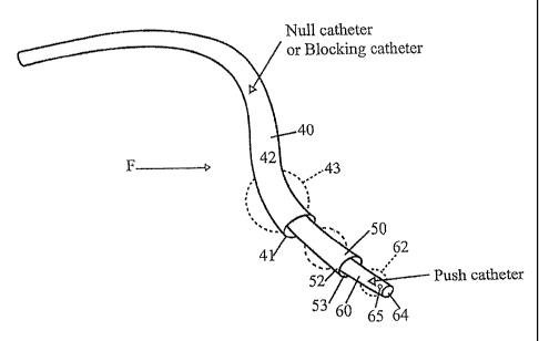
administered
by conventional intravenous delivery techniques.
1
CA 02592660 2007-06-28
WO 2006/073870 PCT/US2005/046607
[0004] Another problem has involved inadequate uptakes and nonoptimal
distribution
in tumors in vivo. As has been pointed out in Applicant's earlier U.S.
Patents: The tumor
blood flow is thus impaired, measuring only two to fifteen percent of that of
the
surrounding tissue, and this impaired circulation distinguishes the cancer
vasculature. The
probability of blood flow through the V--V shunts is far less than the
probability of blood
flow through the normal vasculature. Therefore, in any attempt to deliver
chemotherapy
to a tumor, the likelihood that the drug will spread to the remainder of the
body is far
greater than the likelihood that it will reach the tumor. There were problems
in making
certain that the tumor (rather than the entire body) received a significantly
high dose and
0 duration of exposure to the treatment agent. Another problem was in
determining and
controlling the routes of drug delivery within a target site, as well as that
of withdrawing
any excess drug.
[0005] Dynamic fluoroscopic maps enabled a physician to somewhat
visualize at a
macroscopic level delivery routes and a target site. However, the fluoroscopic
images
5 that captured macroscopic data were incapable of tracking the flow
dynamics at the
submicroscopic level of cellular activity.
[0006] Another problem has been isolation of the treatment agent to the
area of the
tumor in the patient. Avoiding systemic leakage of toxic drugs that cause
damage to
healthy tissue and organs has been a major problem in the delivery of
chemotherapy.
Obtaining precise delivery of genetic material to a target region has
continued to be a
desirable goal of gene therapy. Regardless of the agent being delivered,
localized,
precise, targeted therapy delivery to a specific site with negligible run-off
or leakage of
the agent to collateral sites has remained a concern.
[0007] There are certain agents which have proven effective in
chemotherapeutic
3 treatment of tumors, but which have potentially severe side effects. An
example is
doxorubicin, available under the trademark ADRIAMYCIN , which has been used as
an
anti-cancer drug for a number of years. That composition has been used to
treat many
forms of cancer including cancer of the breast and stomach, lymphoma and
multiple
myeloma. However, severe side effects have ensued. A common side effect if
dosage is
not controlled has been dilated cardiomyopathy. The use of this chemical to
treat tumors
2
CA 02592660 2007-06-28
WO 2006/073870 PCT/US2005/046607
has been limited, when systemically administered, due to its toxic side effect
on the
patient's heart.
SUMMARY OF THE INVENTION
[0008] Briefly, the present invention provides a new and improved computer-
implemented method of monitoring retrograde venous perfusion of a tumor in a
patient's
body According to the method of the present invention, the positioning is
monitored of a
withdrawal catheter within vasculature of a target vessel in the patient's
body near the
tumor, and of an infusion catheter within the vasculature of the target vessel
near the
tumor and beyond the withdrawal catheter. The positioning of a venous pressure
catheter
within the vasculature of the target vessel and intermediate the infusion
catheter and the
withdrawal catheter is also monitored. The location and positioning which are
monitored
allow observation of a closed loop flow path between the positioned infusion
catheter and
the positioned withdrawal catheter through the target vessel. Venous pressure
is
monitored in the closed loop flow path, and the circulation of fluids through
the closed
loop flow path is also monitored.
[0009] The process of the present invention allows control of the delivery
of therapy
via the retrograde perfusion modality. It provides for monitoring and
presentation of a
multitude of complex and continually changing variables during the tumor
treatment by
retrograde perfusion.
I [0010] The present invention also provides a computerized system for
monitoring the
retrograde perfusion of tumors. A processor of the computer system performs
the steps of
the computer implemented monitoring of the retrograde perfusion of the tumor.
The
present invention also provides a computer program product containing machine-
readable
code that causes the processor to implement the monitoring of the retrograde
perfusion.
; By virtue of the position of the catheters relative to one another and to
the target vessel,
the treating physician is provided with monitoring capablity to verify that
the perfusion
treatment is carefully controlled and monitored, and that the flow of fluids
in the
vasculature and tumor region is in accordance with fluid dynamic and flow
principles.
[0011] There is, however, no need to establish or define specific fluid
flow equations
of motion explicitly in order to verify that proper perfusion fluid flow paths
and relations
3
CA 02592660 2007-06-28
WO 2006/073870 PCT/US2005/046607
are established and maintained. The control or treatment unit when positioned
and
monitored with the present invention during its use and operation implicitly
computes the
solution to the equations of motion for the network, and performs the
perfusion treatment
according to the desired flow paths and relationships. This is done without
resorting to
i the explicit use of calculations, numbers, mathematical equations or
physical equations of
motion and such; proper positioning of the control unit during its use
performs those
kinds of computational tasks.
[0012] Recognizing that timely intervention and response is a critical
factor in the
management of disease processes, the present invention makes it possible to
synchronize
) the multiple disparate signals related to any one or more of a number of
factors of interest
during retrograde perfusion on a real-time basis. Data or images of interest
include:
(1) the catheter, i.e. infusion rate, withdrawal rate, fluid displacement,
pressure, concentration;
(2) the patient's history and present condition, i.e. prior surgeries and
treatments, current heart rate, blood pressure, respiration, temperature;
(3) 3-D high resolution imaging, i.e. spatial boundaries, borders, density;
and
(4) ongoing response to therapy at the cellular level.
[0013] The present invention is capable of putting these disparate signals in
a
) synchronized or zero-state of image retention in a manner that, so far as
is known, has not
been previously contemplated. In addition to providing a high degree of
control and
integration to the treatment process, the present invention offers a treating
physician with
up-to-the-minute support for planning, decision-making, and problem-solving.
[0014] All data including three-dimensional or 3-D models are data
archived in a
5 central repository so that data mining, predictive modeling, and
suggested action states
may be applied to various systems. Local systems can be networked to remote
systems so
that data available at a treatment center in one locality is simultaneously
available to
treatment centers in other localities.
4
CA 02592660 2011-03-29
[0015] Two examples or models help to explain by analogy the kinds of
differential
equations of motion that are implicitly solved by operation of the control
unit. One is a
water-flow model that cascades; the other is a moving crowd model. In the
water-flow
model, the size and shape of the catheters influence the motion of fluid
through the
catheters. Also, the motion of fluid in parallel and opposite directions, and
orientation
through the catheters and through the vascular beds obeys the physical laws
related to
pressure, flow rate, and volume. In the moving crowd model, the size and shape
of the
catheters influence the movement of particles through the catheters. Also, the
movement of
particles through the network conforms to the physical laws related to
pressure, flow rate,
and volume.
[0015a] In another aspect, the present invention resides in a computer-
implemented
method of monitoring retrograde venous perfusion of a tumor in a patient's
body, comprising
the steps of: monitoring the positioning of a withdrawal catheter within
vasculature of a
target vessel in the patient's body near the tumor and an infusion catheter
within the
vasculature of the target vessel extending beyond the withdrawal catheter and
near the
tumor; monitoring the positioning of a venous pressure catheter concentrically
disposed
between the infusion catheter and the withdrawal catheter forming a closed
loop flow path
between the positioned infusion catheter and the positioned withdrawal
catheter through the
target vessel; monitoring venous pressure in the closed loop flow paths;
andmonitoring the
circulation of a fluid through the closed loop flow path.
[0015b] In another aspect, the present invention resides in a data
processing system
for monitoring retrograde venous perfusion therapy of a tumor in a patient's
body as the
therapy is occurring, the data processing system comprising: a processor for
performing
the steps of: monitoring the positioning of a withdrawal catheter within the
vasculature
of a target vessel in the patient's body near the tumor and an infusion
catheter within the
vasculature of the target vessel extending beyond the withdrawal catheter and
near the
tumor; monitoring the positioning of a venous pressure catheter concentrically
disposed
between the infusion catheter and the withdrawal catheter, thereby forming a
closed loop
flow path between the positioned infusion catheter and the positioned
withdrawal catheter
through the target vessel; monitoring venous pressure in the closed loop flow
paths;
CA 02592660 2011-03-29
monitoring the circulation of a fluid through the closed loop flow path; and a
data output
display for providing the results of monitoring by the processor.
10015c1 In another aspect, the present invention resides in a computer
program
product stored in signal bearing media for causing a data processor to monitor
retrograde
venous perfusion on therapy of a tumor in a patient's body as the therapy is
occurring,
the computer program product containing instructions stored in machine-
readable code
and causing the processor to perform the following steps: monitoring the
positioning of a
withdrawal catheter within vasculature of a target vessel in the patient's
body near the tumor
and an infusion catheter within the vasculature of the target vessel extending
beyond the
withdrawal catheter and near the tumor; monitoring the positioning of a venous
pressure
catheter concentrically disposed between the infusion catheter and the
withdrawal
catheter, thereby forming a closed loop flow path between the positioned
infusion catheter
and the positioned withdrawal catheter through the target vessel; monitoring
venous pressure
in the closed loop flow paths; and monitoring the circulation of a fluid
through the closed
loop flow path.
[0015d] In another aspect, the present invention resides in a computer-
implemented
method of monitoring retrograde venous perfusion of a tumor in a patient's
body,
comprising the steps of: monitoring the positioning of a withdrawal catheter
within the
vasculature of a target vessel in the patient's body near the tumor, and
monitoring an
infusion catheter concentrically disposed within the withdrawal catheter and
near the tumor;
monitoring the positioning of a venous pressure catheter concentrically
disposed within the
withdrawal catheter; thereby forming one or more closed loop flow paths
between the
positioned infusion catheter, the positioned withdrawal catheter, and the
positioned
venous pressure monitoring catheter, through the target vessel; monitoring
venous pressure
in the closed loop flow paths; and monitoring the circulation of a fluid
through the closed
loop flow paths.
10015e1 In another aspect, the present invention resides in a A computer-
implemented
method of monitoring retrograde venous perfusion of a tumor in a patient's
body, comprising
the steps of: monitoring the positioning of a withdrawal catheter within
vasculature of a
target vessel in the patient's body near the tumor and an infusion catheter
within the
5a
CA 02592660 2011-03-29
vasculature of the target vessel extending beyond the withdrawal catheter and
near the
tumor; monitoring the positioning of a venous pressure catheter within the
vasculature of the
target vessel intermediate the infusion catheter and the withdrawal catheter
forming a closed
loop flow path between the positioned infusion catheter and the positioned
withdrawal
catheter through the target vessel; monitoring venous pressure in the closed
loop flow paths;
and monitoring the circulation of a fluid through the closed loop flow path.
[0015f] In a further aspect, the present invention resides in a data
processing system
for monitoring retrograde venous perfusion therapy of a tumor in a patient's
body as the
therapy is occurring, the data processing system comprising: a processor for
performing the
steps of: monitoring the positioning of a withdrawal catheter within
vasculature of a target
vessel in the patient's body near the tumor and an infusion catheter within
the vasculature of
the target vessel extending beyond the withdrawal catheter and near the tumor;
monitoring
the positioning of a venous pressure catheter within the vasculature of the
target vessel
intermediate the infusion catheter and the withdrawal catheter forming a
closed loop flow
path between the positioned infusion catheter and the positioned withdrawal
catheter
through the target vessel; monitoring venous pressure in the closed loop flow
paths;
monitoring the circulation of a fluid through the closed loop flow path; and a
data output
display for providing the results of monitoring by the processor.
[0015g] In yet another aspect, the present invention resides in a computer
program
product stored in signal bearing media for causing a data processor to monitor
retrograde
venous perfusion on therapy of a tumor in a patient's body as the therapy is
occurring, the
computer program product containing instructions stored in machine-readable
code and
causing the processor to perform the following steps: monitoring the
positioning of a
withdrawal catheter within vasculature of a target vessel in the patient's
body near the tumor
and an infusion catheter within the vasculature of the target vessel extending
beyond the
withdrawal catheter and near the tumor; monitoring the positioning of a venous
pressure
catheter within the vasculature of the target vessel intermediate the infusion
catheter and the
withdrawal catheter forming a closed loop flow path between the positioned
infusion
catheter and the positioned withdrawal catheter through the target vessel;
monitoring venous
5b
CA 02592660 2014-11-13
pressure in the closed loop flow paths; and monitoring the circulation of a
fluid through the
closed loop flow path.
[0015h] In yet another aspect, the present invention provides use of a
catheter
assembly and a computer for performing and monitoring retrograde venous
perfusion of a
tumor in a patient's body, the catheter assembly being positionable in an
inserted position
within vasculature of a target vessel in the patient's body near the tumor,
wherein the
catheter assembly comprises a withdrawal catheter, an infusion catheter and a
venous
pressure catheter, the venous pressure catheter being concentrically disposed
between the
withdrawal catheter and the infusion catheter forming a closed loop flow path
therebetween
in the inserted position through the target vessel, and wherein in the
inserted position the
infusion catheter is located to extend beyond the withdrawal catheter near the
tumor; and the
computer being for receiving a plurality of signals indicative of the catheter
assembly in the
inserted position, wherein the plurality of signals comprise: first, second
and third signals
indicative of monitored positions of the withdrawal catheter, the infusion
catheter, and the
venous pressure catheter, respectively; a fourth signal indicative of a
monitored venous
pressure in the closed loop flow path; and a fifth signal indicative of a
monitored circulation
of a fluid through the closed loop flow path.
[00151] In yet another aspect, the present invention provides use of a
catheter
assembly and a computer for performing and monitoring retrograde venous
perfusion of a
tumor in a patient's body, the catheter assembly being positionable in an
inserted position
within vasculature of a target vessel in the patient's body near the tumor,
wherein the
catheter assembly comprises a withdrawal catheter, an infusion catheter and a
venous
pressure catheter, the infusion catheter and the venous pressure catheter
being concentrically
disposed within the withdrawal catheter forming one or more closed loop flow
paths
between the withdrawal catheter, the infusion catheter and the venous pressure
catheter in
the inserted position through the target vessel; and the computer being for
receiving a
plurality of signals indicative of the catheter assembly in the inserted
position, wherein the
plurality of signals comprise: first, second and third signals indicative of
monitored positions
of the withdrawal catheter, the infusion catheter, and the venous pressure
catheter,
5c
CA 02592660 2014-11-13
respectively; a fourth signal indicative of a monitored venous pressure in the
closed loop
flow paths; and a
respectively; a fourth signal indicative of a monitored venous pressure in the
closed loop
flow paths; and a
fifth signal indicative of a monitored circulation of a fluid through the
closed loop flow
paths.
[0015j] In yet another aspect, the present invention provides use of a
catheter
assembly and a computer for performing and monitoring retrograde venous
perfusion of a
tumor in a patient's body, the catheter assembly being positionable in an
inserted position
within vasculature of a target vessel in the patient's body near the tumor,
wherein the
catheter assembly comprises a withdrawal catheter, an infusion catheter and a
venous
pressure catheter, the venous pressure catheter being disposed intermediate
the withdrawal
catheter and the infusion catheter to form a closed loop flow path
therebetween in the
inserted position through the target vessel, and wherein in the inserted
position the infusion
catheter is located to extend beyond the withdrawal catheter near the tumor;
and the
computer being for receiving a plurality of signals indicative of the catheter
assembly in the
inserted position, wherein the plurality of signals comprise: first, second
and third signal
indicative of monitored positions of the withdrawal catheter, the infusion
catheter, and the
venous pressure catheter, respectively; a fourth signal indicative of a
monitored venous
pressure in the closed loop flow path; and a fifth signal indicative of a
monitored circulation
of a fluid through the closed loop flow path.
[0015k] Further aspects of the invention will become apparent upon reading
the
following detailed description of the drawings, which illustrate the invention
and preferred
embodiments of the invention.
BRIEF DESCRIPTION OF THE DRAWINGS
[0016] A better understanding of the present invention can be obtained
when the
detailed description set forth below is reviewed in conjunction with the
accompanying
drawings, in which:
5d
CA 02592660 2014-11-13
[0017] Figure 1 is an illustration of a highly simplified model of the
circulatory
system in the liver of an animal.
[0018] Figures 2A and 2B are isometric views of catheter system portions
of the
present invention.
[0019] Figure 3 is a schematic diagram of a perfusion system according to
the
present invention.
[0020] Figure 4 is a schematic diagram of a treatment procedure with the
perfusion
system of Figure 3.
[0021] Figure 5 is a schematic drawing of a liver receiving treatment
during a
procedure with a perfusion system of the present invention.
[0022] Figure 6 is an illustration of a model like that of Figure 1 with
a catheter
according to Figure 2A.
[0023] Figure 7 is a photograph of an animal liver after a perfusion
treatment
procedure according to the present invention.
5e
CA 02592660 2007-06-28
WO 2006/073870 PCT/US2005/046607
[0024] Figure 8 is a display image of an animal liver during a perfusion
treatment
procedure according to the present invention.
[0025] Figures 9, 10, 11 12 and 13 are functional block diagrams of
computer
processing steps according to the present invention.
DETAILED DESCRIPTION OF THE PREFERRED EMBODIMENTS
[0026] In the drawings, a photographic model of the circulatory system of
blood flow
the liver of an animal, in this case a human, is shown in Fig. 1. The liver L
is located in
the body in communication through the common bile duct D with the gallbladder
G. As
indicated at 10, the hepatic artery connects to and transports blood into the
liver L for the
purpose of bile production, protein production, blood detoxification and other
liver
functions.
100271 In the treatment of tumors in other organs, a similar approach
applies. In the
case of a tumor of the kidney, for example, the renal artery carries blood
from the aorta to
the kidney while the renal vein carries blood from the kidney to the inferior
vena cava.
For the purpose of retrograde perfusion, access to a tumor of the kidney would
be via the
inferior vena cava to the renal vein.
[0028] Further, retrograde perfusion can also be performed via
percutaneous access to
any organ whereby the venous drainage of the target organ is accessed directly
via an
incision. In any given organ, the point of reference for the process of
retrograde perfusion
is the site of the venous drainage from the organ.
[0029] The other major blood flow paths in the liver in addition to the
hepatic artery
are also indicated in Figure 1, including the portal vein as indicated at 12
and the
inferior vena cava as indicated at 14. Blood enters the liver L from the heart
via the
hepatic artery 10 and from the stomach, intestines and other parts of the
digestive tract
; through the portal vein 12.
[0030] Incoming blood from the hepatic artery 10 and portal vein 12 merges
and
passes through the liver L to a series of hepatic veins (Figure 5), including
the left hepatic
vein 16, a middle hepatic vein 18 and a right hepatic vein 20. The hepatic
veins 16, 18
and 20 collect blood as it is processed in the liver L and empty into the
inferior vena cava
6
CA 02592660 2014-11-13
14. As can be seen in Figure 1, the hepatic artery 10 and the veins 12, 16, 18
and 20 are
only the major blood flow paths through the liver L. There are as indicated in
Figure 1 a
considerable number of other separate and distinct smaller or minor blood flow
paths or
veins branching off and in flow communication with the major flow paths.
Because of
the number of them, no reference indicators are assigned them in Figure 1.
[00311 Such branching structures are examples of fractal architecture found
commonly in a wide variety of physiological systems including the respiratory,
circulatory, and nervous systems. Examples of fractal anatomy can be seen in
anatomical
structures such as the hepatic arterial and venous trees shown in Figure 1.
10032] As opposed to classical geometric forms that are smooth and regular
having
integer dimensions such as one, two and three for line, surface, and volume,
fractals have
a fractional dimension between one and two and exhibit a pattern of repeating
smaller
scale sub-patterns that resemble the larger scale pattern, a property terms
self-similarity or
scale invariance. Such fractal scaling is seen in the lungs, the bronchial
tubes, capillaries,
intestinal lining, and bile ducts; and the heart comprises various fractal
networks
including the coronary arteries and veins, the fibers binding the valves to
the heart wall,
the cardiac muscles themselves, and the His-Purkinje system that transmits
electrical
impulses from atrium to ventricle.
[00331 Fractal structures exhibit another significant property, the
relationship between
perimeter and area. A physiologic advantage of self-similar fractal structures
is that they
serve a common physiological function that has been characterized in the
literature as
"rapid and efficient transport over a complex, spatially distributed system.
In the case of
the ventricular electrical conduction system, the quantity transported is the
electrical
stimulus regulating the timing of the cardiac contraction_ For the
vasculature, fractal
branchings provide a rich, redundant network for distribution of 02 and
nutrients and for
the collection of CO2 and other metabolic waste products. A variety of other
organ
systems contain fractal structures that serve functions related to information
distribution
(nervous system), nutrient absorption (bowel), as well as collection and
transport (biliary
duct system, renal calyces). "Nonlinear Dynamics, Fractals, and Chaos Theory:
Implications for Neuroautonomic Heart Rate Control in Health and Disease:, Ary
L.
Goldberger, in The Autonomic Nervous System, Bolis, C.L. and Licinio, J., eds.
Geneva:
World Health Organization, 1999, pages 135-152.
7
CA 02592660 2007-06-28
WO 2006/073870 PCT/US2005/046607
[0034] Further, the model liver L of Figure 1 although seemingly detailed
is instead
conceptual in that only a certain number of even the minor blood flow paths
are
represented, due to limits on the ability to form tangible representations of
a number of
the minor flow paths. The liver as in the case of other body organs or regions
has in
actuality a number of other smaller blood veins and flow paths, which are hard
to discern
and visualize. Further, the circulatory system embodied in the model of the
liver L is a
tangible, physical manifestation of the blood flow paths at a fixed moment.
[0035] Similar blood flow structure exists in other body organs as well.
Accordingly,
the liver as illustrated in Figure 1 is given by way of example. It should be
understood
[0 that the perfusion techniques of the present invention to be described
below are equally
applicable to other organs and portions of the body.
[0036] In the human or other animals, the flow of blood in flow paths
through an
organ such as the liver fluctuates in both pressure and flow rate in response
to heart rate
and blood pressure. As a result when an organ under investigation is viewed
through
body imaging systems as a display image by a treating physician, the organ
appears much
like a cloud or blurred image. Thus, in treating an organ, the display images
are less
articulated and defined in the body than the idealized, simplified flow path
models as
illustrated in the photograph of Figure 1.
[0037] As mentioned above, it is known that there are chemotherapeutic
agents of
demonstrated effectiveness in treatment of tumors. However, their use has been
significantly limited by the undesirable side effect of systemic toxicity on
other organs or
parts of the body. Although earlier retrograde perfusion efforts, as
exemplified in
Applicant's United States Patents mentioned above, have shown promise,
certainty of the
localization and isolation of the area of the patient's body receiving a
chemotherapeutic
agent is still a desirable goal. This holds true for chemotherapeutic agents
of any type,
but particularly those with undesirable systemic side effects, whether
toxicity or some
other undesirable effect.
[0038] The present invention provides a method and apparatus for
retrograde
perfusion of a patient with a therapeutic agent in a flow, controlled,
pressure regulated in
vivo closed loop in the vasculature of the patient. The apparatus of the
present invention
takes the form of a retrograde perfusion system P that includes a flow control
or
8
CA 02592660 2014-11-13
administration unit F (Figures 2A and 213) that is introduced into the body of
the patient.
The flow control unit F is in fluid communication with an external unit U
(Figures 3 and
4) with monitors and pumps with which treating physicians and their staff may
administer
the therapeutic agent, even one with substantial system toxicity, by
retrograde perfusion
in a closed loop, pressure regulated flow route in vivo. Typically, one or
more visual
monitors M are provides to display images formed for example by fluoroscopy or
by
computerized axial tomography or CAT scanner S. The monitors M allow the
treating
physician or physicians to gain visible confirmation of the formation,
establishment and
operation of the in vivo flow route.
0 [0039] The internal flow control unit F is a multicatheter system
introduced into the
vascular system of the patient at a suitable location, for example by femoral
or neck
cutdown, depending on the organ or portion of the patient's body to receive
the
therapeutic agent. The flow control unit F includes three catheters that may
be configured
to be concentrically mounted with each other (Figure 2A) or may have two of
the
5 catheters separately contained (Figure 213) within a third or larger
outer catheter.
[00401 In a flow control unit according to the present invention, a larger
catheter
40 to extract or pull fluid from the in vivo loop formed in the vasculature of
the patient
has a central venous pressure or cvp catheter 50 and an infusion or push
catheter 60
concentrically and telescopingly mounted therein. As will be set forth below,
each of
0 catheters 40, 50 and 60 is positioned with a proximal end within a vessel
in the patient's
vasculature and a distal end in flow communication with the external unit U of
the
perfusion system P.
[0041) The catheters of the flow control unit 30 are located near the tumor
to be
treated. In the context of the present invention, near the tumor is intended
to connote that
5 the tumor is located in vasculature between the infusion catheter 60 and
withdrawal
catheter 40. Further, near the tumor is intended according to the present
invention to
signify that the catheters of the flow control unit 30 are located in the
vasculature of the
patient with no unoecluded intervening vasculature present in the area between
the
infusion catheter 60 and withdrawal catheter 40.
0 [0042] The larger or pull catheter 40 is a size, such as a 10 to 12
French or Fr. sheath
42, with a compliant distal balloon 43 or other comparable mechanism for
occluding the
9
CA 02592660 2014-11-13
vessel of interest in the patient. The pull catheter 40 also has a large
enough internal
diameter to accommodate the push catheter 60 and the central venous pressure
catheter 50
concentrically and coaxially within it. Alternatively, the pull catheter 40
may, if desired,
be sufficiently large, such as 14 Fr. sheath, that its distal end 41 may be
used to occlude a
vein without balloon 43.
(0043] The length of the sheath 42 of pull catheter 40 may vary based on
the organ
site and the venous access, for example neck or femoral eutdown. A sheath
length of
approximately 34 ern typically permits the catheter 40 be routed via a jugular
cut-down
procedure to the target organ site. The sheath 42 preferably is suitably
flexible to permit
extensive maneuvering and routing in the vasculature. However, the sheath 42
should
also be structurally sturdy enough to avoid kinking or collapsing under
pressure. The
sheath 42 has a guide wire and/or introducer for proper placement. The guide
wire or
introducer is removed when the pull catheter 40 is established at the proper
in vivo,
closed loop position. An outflow port 46 (Figure 4) on the pull catheter 40 is
provided for
the purpose of withdrawing fluids. A distal end 47 of the pull catheter 40
routes the
outflow from pull catheter 40 to a withdrawal syringe 70 (Figure 3) of the
external unit U.
A proximal end 48 of the pull catheter 40 is connected via a T-port 72 to the
withdrawal
or pull syringe 70 for withdrawing fluids.
[0044] The push or infusion catheter 60 has similar properties of length,
flexibility
and structural strength to those of the pull or withdrawal catheter 40. The
push catheter
60 in the embodiment of Figure 2A has a sheath 61 with an outer diameter of
from about
3-7 Fr. fitted with a compliant balloon 62 for occluding a vessel. The sheath
61 is also
provided with a radio-opaque proximal tip 64 for visualizing the position of
the catheter
proximal end within a vessel. The push catheter 60 has an outer diameter that
enables it
to fit coaxially and telescopically within the central venous pressure
catheter 50 and the
pull catheter 40. An opening 65 at the distal tip 64 of the input catheter 60
serves the
purpose of infusing fluids. A proximal end of the input or infusion catheter
60 is
connected via a T-port 74 of the external unit U to a push syringe 76 for
infusing fluids
into the in vivo loop in the patient.
100451 The central venous pressure or cvp catheter 50 has similar
properties of length,
flexibility and structural strength to those of each of the push catheter 60
and the pull
CA 02592660 2014-11-13
catheter 40. In the embodiment shown in Figure 2A, the central venous pressure
catheter
50 has a sheath with an outer diameter intermediate that of the push catheter
60 and
the pull catheter 40. The central venous pressure catheter 50 is fitted at a
distal end 52
with a port or opening 53 and in fluid communication with a pressure
transducer 54. The
pressure transducer 54 may, if desired, be located with the external unit U in
fluid
communication through the port 53 with pressure and flow rate conditions in
the closed
loop formed in the patient's vasculature by the present invention between the
infusion
catheter 60 and the pull catheter 40. The pressure transducer 54 allows
monitoring of
central venous pressure in the closed loop to be certain that a stable central
venous
pressure is present between the push catheter 60 and the pull catheter 40. A
gauge or
meter 55 or other form of pressure readout indication or display, as indicated
schematically at 55, is present in the external unit U to indicate the central
venous
pressure sensed by transducer 54 to the monitoring/treating physician(s).
[0046] The pressure transducer 54 and indicator gauge or readout device 55
are
connected to the central venous pressure catheter 50 for monitoring and
tracking the
central venous pressure in the patient's vasculature in the organ to receive
perfusion
between the push catheter 60 and pull catheter 40. The pressure transducer 54
and
indicator gauge 55 thus provide the physician(s) with information about fluid
conditions
so that after formation of the closed loop at the treatment site, a steady
state or frame of
fluid pressure reference is obtained there. During the subsequent
perfusion/treatment
cycle, fluctuations or transient changes sensed through the transducer 54 and
central
venous pressure catheter 50 provide the physician with valuable information to
closely
control and monitor the infusion and extraction of fluid at the treatment
site.
[0047] By virtue of the position of the three catheters relative to one
another and to
the target vessel, a pressure differential is established in the catheter
network. One such
pressure differential relationship is that of a transient stability
established between the tip
of the push catheter and the central venous pressure catheter. Another is the
pressure
differential between the push catheter and the background noise of the venous
liver
circulation. The pressure differential thus established is in a forward
orientation and
direction from the tip of the infusion catheter to the venous circulation.
11
CA 02592660 2014-11-13
100481 In the opposite orientation and direction, a pressure differential
is established
between the pull catheter and the central venous pressure catheter. Another
pressure
differential is established between the venous circulation and the pull
catheter. The
perfusion treatment according to the present invention thus is in accordance
with fluid
dynamic and flow principles.
100491 The push syringe 76 of the external unit U as connected via the T-
port 74 to
the push catheter 60 measures and injects the desired amount of various fluids
during the
treatment cycle, whether saline, dye, or therapeutic drug to be infuser'.
[00501 The external unit U also includes the withdrawal syringe 70 that is
connected
via the T-port 72 to the pull catheter 40 for collecting the spent fluid used
during
treatment, whether saline, dye, or drug, once the fluid has been infused and
passed
through the closed loop treatment site. Each of the syringes 70 and 76 is
further
connected to its respective associated pump 71 and 75, such as a Harvard type
infusion
pump, for the purpose of infusing and withdrawing the saline, dye, or drug, as
the case
may be. The infusion by syringe 76 and withdrawal by syringe 70 is done by the
physician with the external unit U at the desired flow rate, and also to set
up the
differential pressure and related motions to physically impart characteristics
to the fluids
at the perfusion treatment site.
100511 According to the present invention, the external unit U includes a
computer
which obtains, organizes, stores and present data and images to a treating
physician or
physicians during the retrograde perfusion procedure. The data and images are
available
from the computer on a real time basis and include, for example, data relating
to the
operation and functioning of the internal flow control unit F. The data
include data from
or relating to operation of the multicatheter flow control unit 30 such as
infusion rate,
withdrawal rate, fluid displacement, pressure concentration and other fluid
flow and
pressure parameters and measurements.
10052) The operation of the syringes 70 and 76 and their respective
associated pumps
are preferably automated via the computer C and associated computer control
instructions, or software. As will be described the computer C and associated
software
instructions allow for the monitoring, organization, presentation and storage
to treating
physicians of multiple measurements, data and records relating to the patient
and the=
12
CA 02592660 2007-06-28
WO 2006/073870 PCT/US2005/046607
retrograde perfusion treatment on a real time basis of multiple disparate
measurements
and item of data as the retrograde perfusion procedure is in progress. The
computer C
and its associated software operate in the fluid monitoring phase according to
established
settings, taking into account various factors, such as:
(1) the volume of fluid (saline, dye, drug) to be infused;
(2) the rate of infusion of the fluid(s);
(3) the time duration of the infusion; and
(4) the ratio of withdrawal rate to infusion rate.
[0053] In addition, the computer C and software permit a database to be
formed and
maintained. The database so formed may be maintained and updated in the
computer C
and may also be networked and made available on a real time basis via data
communications links such as wire, optic, radio wave, satellite or other
communications
media to other computers and data storage systems and facilities. The database
contents
of the computer C and other computer systems in communication therewith also
preferably includes data relating to the patient's history and present
condition, current
heart rate, blood pressure, respiration and temperature obtained in any
suitable
conventional manner, as well as data records from prior treatments or
surgeries.
[0054] The computer C also is in communication with the visual monitor M
and the
imaging mechanisms, such as a scanner S, fluoroscopy and the like. The
computer C
receives data the content of the content of the image from such imaging
mechanisms and
includes such data as image data in the database. The image data is available
for 3-D
high resolution imaging to observe or define spatial boundaries or borders, or
densities of
portions of the body under treatment or investigation. The image data and the
physiological monitoring data also allow monitoring and observation of the
response to
therapy at the cellular level. The database allows data to be retained in
order to correlate
the location of various perfusion treatment sites, and established settings,
as well as the
factors mentioned above, along with the type and nature of images or fractals
obtained
therewith. Such a database allows, as will be set forth, a physician greater
flexibility in
treatment by retrograde perfusion.
13
CA 02592660 2014-11-13
[0055] The computer system of the computer C and its associated computer
executable instructions or software described herein is capable of organizing
disparate
sets of data in the form of signals or other information media from various
sources,
organizing the data, time-stamping the data, and presenting the data for use
by a physician
in the course of a treatment procedure.
[0056] The computer (Figs. 3 and 4) includes a processor or CPU which
operates
under the control of a series of computer-executable instructions. The
instructions may be
contained in memory of the computer, or on magnetic tape, conventional hard
disk drive,
electronic read-only memory, optical storage device, or other appropriate data
storage
device. Also, the instruction may be stored on a data storage device with a
computer
readable medium, such as a computer diskette, having a computer usable medium
stored
thereon. The CPU is connected by input/output interfaces to components of the
perfusion
system for data transfer purposes. The CPU receives data from the catheters 70
and 76
and other components of the external unit U, as well as the monitors M and
imaging
mechanisms described above. The CPU also includes a data display screen for
the
computer operator, as well. The CPU is also networked, as described above,
with other
computer systems for database compilation, transfer and storage purposes.
Generally, at
least one computer includes a file serve capability for database retention and
master
storage purposes. Also, if desired, the computer networked computer may
include a
mainframe computer of any conventional type of suitable processing capacity.
Other
digital processors, however, may be used, such as a laptop computer, or any
suitable
processing apparatus at any of the computer sites in the network.
[0057] A flow chart T (Figs. 9-13 herein) illustrates the structure of the
logic of the
present invention as embodied in computer program software. Those skilled in
the art
will appreciate that the flow charts illustrate the structures of computer
program code
elements that function according to this invention. Manifestly, the invention
is practiced
in its essential embodiment by a machine component that renders the program
code
elements in a form that instructs a digital processing apparatus (that is, a
computer) to
perform a sequence of function steps corresponding to those shown.
14
CA 02592660 2007-06-28
WO 2006/073870 PCT/US2005/046607
[0058] It is important to note that, while the present invention has been, and
will continue
to be, described in the context of a fully functional computer system, those
skilled in the
art will appreciate that the present invention is capable of being distributed
as a program
product in a variety of forms, and that the present invention applies equally
regardless of
the particular type of signal-bearing media utilized to actually carry out the
distribution.
Examples of signal-bearing media include: recordable-type media, such as
floppy disks,
hard disk drives, and CD ROMs, and transmission-type media such as digital and
analog
communication links.
[0059] With reference to Figs. 9-13, there is depicted a high-level logic
flowchart
illustrating a method according to the present invention. The method of the
present
invention performed in the computer C can be implemented utilizing the
computer
program steps of Figs. 9-13 stored in memory 82 and executable by system
processor 80
of computer C and the data resulting from the data collection steps performed
by the
components of the perfusion system F connected to the computer C, as described
above.
[0060] Several classes of data sets are to be controlled and synchronized
by the
computer system C of the present invention. There are
1) catheter control and monitoring data,
2) 3-D graphic modeling of data captured from fluoroscopy or other imaging
techniques,
3) patient history data,
4) physiological monitoring data, and
5) predictive statement.
[0061] The flow chart T illustrates computerized monitoring according to
the present
invention of a retrograde perfusion treatment procedure. The system and means
described
herein is capable of organizing disparate sets of data in the form of signals
or other means
from various sources, organizing the data, time-stamping the data, and
presenting the data
for use by a physician in the course of a treatment procedure.
CA 02592660 2007-06-28
WO 2006/073870 PCT/US2005/046607
[0062] The flow chart T (Figures 9-13) illustrates the flow and
interchange of
information from the computer C from start to end of a retrograde perfusion
treatment
procedure. The preliminary steps (Figure 9) include system initialization and
system
readiness. In the first stage also illustrated in Figure 9 patient data is
input and verified.
; The patient data sets include demographic patient data, patient history
data, and current
patient physiological monitoring data.
[0063] In the second stage of the flow chart T (Figure 10), the computer
system C
operates to aid in visually guiding the proper placement of each catheter, in
properly
inflating each balloon, and in initiating the saline infusion to achieve the
desired stability
) of the catheter system.
[0064] In the third state (Figure 11), the system C initiates the
catheter start-up
subroutine with the infusion of radioopaque dye to aid in monitoring the
catheter system
flow dynamics including the hydrostatic, hydrodynamic, hydrokinetic, and
hydrokinematic attributes.
[0065] In the fourth stage (Figure 12) delivery routes are confirmed as
data from the
fluoroscopic image is video captured and modeled in high-resolution 3-D
graphics. The
physician may query and examine data from both the local and the central
database to
obtain a predictive statement and to select a delivery route and treatment
process.
[0066] In the fifth and final stage (Figure 13) the physician has
verified the delivery
routes and begins the administration of a therapeutic agent. Each cycle of
delivery of
therapeutic agents during the treatment process is completed when the agent
has traversed
the route from input to withdrawal.
[0067] During the treatment process, as shown in Figure 13, the computer
C under
control of the operating instructions monitors the patient's condition,
including response
5 to therapy. Prior to shutting down at the completion of the treatment
process, predictive
and suggested action states for future treatment to the patient are presented
to the
physician, then the system shut-down process occurs.
[0068] To initiate a treatment procedure, the system is appropriately
initialized. An
initial step 200 (Figure 9) includes system initialization and system
readiness procedures
D of the conventional type. As indicated, if the system initialization step
is determined to
16
CA 02592660 2007-06-28
WO 2006/073870 PCT/US2005/046607
be not properly completed, a re-boot step 201 is performed. The process
continues until
satisfactory initialization occurs.
[0069] Next, following system initialization a step 202 existing patient
data is input
into the computer C from the database and verified. The patient data sets
include
demographic patient data, patient history data, and current patient
physiological
monitoring data. A query input is entered simultaneously as indicated a steps
204a and
204b at the local database and the remote central repository, respectively.
Data in each of
the local and remote databases is polled to determine proper patient
scheduling.
[0070] If a patient is not scheduled in either of the local or remote
databases, the
computer displays a "not ready" alert. If the patient is properly scheduled,
the computer
displays a "system ready" alert, as indicated at 206.
[0071] When the system is ready, a series of prompts request the user to
input the set
of patient identification data. A patient identifier code, number or other
indicator is
prompted to be entered at step 208. The computer system performs a search of
the
archived patient history data library to determine if the patient is a new or
established
patient. If the patient is a new patient, a prompt step 210 requests the user
as indicated in
step 211 to enter the set of patient history data and the data is archived in
the central
repository.
[0072] If the system finds the patient to be an established patient, the
system performs
a data mining function as indicated at step 212 to yield data regarding the
patient's prior
history and, as indicated at step 214, presents the results to the physician.
In addition,
given the patient's history, the computer system during step 214 makes
predictions as to
what treatment options might be best suited to the patient's current situation
and presents
its predictions to the physician.
[0073] After the patient demographic and historical data are entered,
analyzed and
archived, as under control of the steps illustrated in Figure 9, a
physiological monitoring
sequence illustrated beginning at step 218 in Figure 10 is next performed.
When patient
history data is archived during procedures in connection with the present
invention,
physiological data are obtained include, for example, heart rate, blood
pressure,
temperature, pulse, respiration, CO2 and the like, and are input as indicated
in step 220
17
CA 02592660 2007-06-28
WO 2006/073870
PCT/US2005/046607
into the database from various transducers and monitors. The different types
of such
physiological data are archived, continuously updated and presented on the
workstation
monitor, such as 86 (Figure 4) throughout the subsequent treatment procedure,
as
indicated in Figure 10. The computer system C thus continuously monitors all
physiological data and alerts the physician of any deviations from the normal
physiologic
parameters. If a transducer is detected as not being connected, prompts or
signals may be
sent out during a step 221 to inform the treating physician and staff that
reconnection
needs to be made for physiological monitoring by that transducer.
[0074] Steps 224, 226 and 228 in the flow chart T shown in Figure 10
subsequent to
step 220 cause the computer system C to receive a video input, permit initial
visualization
of the treatment area and form a high resolution output image. These steps aid
the
physician in visually guiding the subsequent proper placement of each
catheter, properly
inflating each balloon, and initiating a hydrostatic phase, and infusing a
saline infusion to
achieve the desired stability of the catheter system of the flow control unit
F.
[0075] The initial phase of catheter placement is that of assembly of the
flow control
unit F based on the planned perfusion treatment, the treatment site and other
factors.
Assembly can be regarded as a sequential assembly phase. The catheters 40, 50
and 60
are combined externally in sequence and placed sequentially coaxially relative
to one
another. In one possible configuration of the catheters shown in Figure 2A,
the pull or
proximal catheter 40 is the outermost catheter of the three. Coaxially
positioned within
the pull catheter 40 are the catheters 50 and 60, which are sequentially
placed based on
their respective sizes. In the embodiment of Figure 2A, the next catheter to
be positioned
coaxially within the pull catheter 40 is the central venous pressure or cvp
catheter 50.
Coaxially positioned within the central venous pressure catheter 50 is the
innermost
chamber and catheter, the push or distal catheter 60.
[0076] Assembled telescopically one inside the other in this manner, the
three
catheters 40, 50 and 60 form the internal flow control unit F. As noted above,
it may in
certain instances be desirable for the catheters 40, 50 and 60 to have an
alternate
configuration. For example, as shown in Figure 2B, an outer catheter 100 with
balloon
101 serves as the pull catheter, and catheters 110 and 120 with their
respective balloons
111 and 121 are separately and not co axially mounted with each other serve as
the
18
CA 02592660 2007-06-28
WO 2006/073870
PCT/US2005/046607
central venous pressure catheter and the infusion or push catheter,
respectively.
Appropriate connections to the respective syringes and pumps of the external
unit U are
made for these purposes.
[0077] Alternatively, the outer catheter 100 shown in Figure 2B may serve
as the
central venous pressure catheter and the catheter 110 serve as the pull
catheter, if desired.
Again, appropriate connections to the external unit U are made for this
purpose.
[0078] The control unit F with catheters of the various configurations
identified above
allows the physician to develop various strategies for how to organize
differential
pressures externally between the push syringe 76 and the pull syringe 70 for
moving fluid
outward through the perfusion system P to the closed loop to the treatment
site and
returning. The fluid movement is accomplished under control of the computer
system C
using the pressure-monitoring central venous pressure catheter 50 to
coordinate, monitor,
and visualize transient changes in central venous pressure sensed through
catheter 50
during the operation of the internal control unit F.
[0079] The assembly of the control unit F and the final determination of
its
configuration is adjustable with regard to the relative longitudinal placement
of the
catheters 40, 50 and 60 with respect to each other. Further, the configuration
and location
of the catheters 40, 50 and 60; the infusion flow rate and pressure; and the
extraction flow
rate and pressure may be monitored and adjusted "on the fly" with the computer
system C
under control of the treating physician while the retrograde perfusion is
under way. The
adjustments may be based on the variable requirements of the target vessel
(i.e. vessel
diameter, length) as well as on the objectives of the planned, controlled
treatment that is
to be performed to frame a search for a missing piece while trying to frame a
strategic
action and a strategic course of retrograde perfusion treatment, including
apriori goals of
a visual representation of mapping of a volumetric shape based upon an
emergent shape.
[0080] Prior to catheter placement and prior to beginning the hydrostatic
phase,
catheter attributes for the catheter of the flow control unit F (Figure 2A or
2B) being used.
are also input to the database of the computer system. Catheter
specifications, connectors,
connector sites, and balloon specifications for each catheter are input
manually into the
database and archived in the central repository. Catheter balloon
specifications are also
input to the database and archived. In addition, the input and withdrawal
syringes 76 and
19
CA 02592660 2007-06-28
WO 2006/073870 PCT/US2005/046607
70 are filled with a desired volume of saline solution and the automatic pumps
are set at
the desired flow rate. This information also is archived in the central
database.
[0081] Each catheter of the flow control unit F is fitted at both ends of
each lumen
with a transducer, and as needed, the transducers measure proprietary
attribute data at
each of the external and internal ends of each lumen. Data from the
transducers are
archived in the central repository and made available to the physician during
the
procedure. To achieve proper catheter placement, the physician manually guides
each of
the three catheters respectively to the target vessel, as indicated at step
230. As indicated
at step 232 and 234, catheter position is observed and re-adjustment of
catheter position is
made by the physician until satisfactory placement is achieved.
[0082] A visual representation (Figure 8) of the type shown on video
monitor M
illustrates the successful placement of the catheters 40, 50 and 60 in order
within a target
vessel, in this case an animal liver L. The pull catheter 40 is inserted first
in sequence into
the external jugular vein and routed with the help of guide wire 45 into the
desired
location of the venous vasculature of the liver selectively toward the target
area.
Subsequently in time the stable central venous pressure catheter 50 is
threaded coaxially
within the pull catheter 40 to its desired location distal to the tip of the
pull catheter 40.
Then, the push catheter 60 is threaded coaxially within the stable central
venous pressure
catheter 50 and is pushed forward via a selective route to a destination point
within the
target organ L. At the destination point, the catheters 40 and 60 are seated
at their
respective desired occluded positions in the vasculature. The sequential
assembly of the
flow control unit F is thus completed.
[0083] In the foregoing initial stage, the three catheters 40, 50 and 60
are put in
position in a selected venous site with no flow through the control unit F.
With the
catheters in place and without initiating flow, the measurement of the central
venous
pressure by transducer 54 gives a real-time initial model of the system fluid
dynamics of
blood at the treatment site.
[0084] As the manual placement process is visualized on fluoroscopy, the
image of
the catheters is captured and input to a computer graphics system of the
computer C
which renders the image on the workstation monitor 86 to aid the physician in
proper
placement. Having confirmed the proper catheter configuration and placement,
the
CA 02592660 2007-06-28
WO 2006/073870 PCT/US2005/046607
balloons are at step 234 inflated as needed to occlude the vessel, to insure a
tight seal, and
to prevent collateral leakage.
[0085] Then, to insure that a state of hydrostatic equilibrium exists
between each of
the push, pull, and central venous catheters and the organ, the external
central venous
pressure is measured and recorded in the database.
[0086] At this point the catheter configuration is considered fixed and
the position of
each catheter is input into the database as a topographical coordinate. Then
the time is set
to zero at the pump connected to each of the push and pull syringes and at
each end of the
external and internal catheter lumens.
[0087] As the saline solution delivery procedure begins during step 236,
the motion of
the pull syringe produces a vacuum that guides the saline from the tip of the
push catheter
toward the pull catheter. As the flow of saline travels from the tip of the
push catheter and
into the countervailing force of the hepatic flow, a resonant pattern results.
This resonance
is monitored and registered in the database.
[0088] Because the infusion of saline is not visible on fluoroscopy, the
computer
system C monitors the fluid cycle and registers the completion of distinct
fluid
trajectories. The completion of one fluid trajectory is registered as the
saline flows from
the tip of the push syringe to the tip of the pull syringe. Another and
simultaneous
trajectory is registered as the saline flows from the external tip of the push
catheter 60 to
the external tip of the pull catheter 50. A third and simultaneous trajectory
is registered as
the saline flows from the internal tip of the push catheter to the internal
tip of the pull
catheter. The different trajectories of the hydrodynamic phase are monitored
independently yet simultaneously by the system and the data is input into the
central
repository.
[0089] As the hydrostatic phase ends, the catheters are in position, the
fluid forces are
in equilibrium, and the spatio-temporal coordinates are registered; the data
relating
thereto is input into the central repository. The computer system C temporally
synchronizes all the data and the system is ready to begin stage 3, the
catheter system
start-up routine of step 240 (Figure 10) which marks the beginning of the
hydrodynamic
phase.
21
CA 02592660 2007-06-28
WO 2006/073870 PCT/US2005/046607
[0090] At the start of the hydrodynamic phase it is important to
distinguish the
various parallel, dynamic data sets that must be monitored, synchronized, and
integrated.
A first distinction is made between external parameters and internal
parameters. A second
distinction is made between push flow parameters, pull flow parameters, and
central
venous pressure parameters. A third distinction is made between input
trajectories and
withdrawal trajectories.
[0091] The first set of attribute data is the external parameters. One
set of external
parameters is the volume of saline to be infused, the infusion rate in ml/min,
the
orientation of flow, and the withdrawal rate in ml/min. These quantities are
input into the
database and, because infusion and withdrawal are to occur simultaneously, the
system
marks the start time at zero for each of the input and withdrawal syringes 76
and 70,
respectively.
[0092] A second set of attribute data is the internal parameters. The
internal
parameters are the infusion rate in ml/min through the push catheter, the flow
pressure,
the withdrawal rate in mi/mm through the withdrawal catheter, and the central
venous
pressure. The system marks the start time at zero for each of input and
withdrawal
catheters. The rate of infusion through the push catheter, the rate of
withdrawal through
the withdrawal catheter, and the central venous pressure are monitored
continuously as
signals and are input into the database for synchronization.
[0093] Having completed the hydrodynamic stage of saline infusion, the
physician
begins the hydrokinetic phase of radiopaque dye infusion through the
established route
created previously by the saline. Attribute data for this phase include the
volume of
radiopaque dye, its density, its concentration, the rate of infusion, and the
rate of
withdrawal. The time is set at zero, the data is input into the central
repository and the dye
infusion begins. The computer C system during step 240 initiates the catheter
start-up
subroutine with the infusion of radio opaque dye to aid in monitoring the
catheter system
flow dynamics including the hydrostatic, hydrodynamic, hydrokinetic, and
hydrokinematic attributes.
[0094] Next, (Figure 11) a radio-opaque dye delivery step 250 occurs,
during which a
radio-opaque dye is added into the saline solution already present in the
closed loop flow
path established as described above. The dye-containing solution is allowed to
flow into
22
CA 02592660 2007-06-28
WO 2006/073870
PCT/US2005/046607
the perfusion site so that a CAT image may be formed on monitor M of the
catheter
placement site. With the infusion of the radio-opaque dye and the resultant
image formed
on the monitor M, a visible, physically-imparted characteristic pattern
emerges in real
time of the region within the organ between the distal ends of the push or
infusion
catheter 60 and the pull catheter 50. The image is also formed at the same
time that the
treatment administering catheters are in vivo at the site where retrograde
perfusion of the
organ is indicated. The image so formed provides a visible indication on the
display
monitor M of the established flow path. Thus, during step 252 shown in Figure
11,
delivery routes are confirmed as data from the fluoroscopic image is video
captured and
modeled in high-resolution 3-D graphics during step 254 for display on the
monitors M.
The 3-D graphic data so obtained are stored in the central repository during
step 256.
[0095] As the radio-opaque dye fills the topographical region previously
demarcated
by the saline injection, the resonant pattern of flow known as a fractal
appears as a 2-D
image on the fluoroscopy screen. Attribute data from the 2-D fluoroscopy image
is video
captured, time stamped, and input into the computer graphics system of the
computer C.
The system correlates the flow data with 2-D fluoroscopic fractal image and
renders the
image as a high-resolution 3-D interactive model during step 254.
[0096] The high resolution model and corresponding flow parameters are
archived in
the central repository during step 256 and presented on the workstation
display 86 as
time-stamped flow data in 3-D, enabling the treating physician to interact
with and
manipulate the dynamic image.
[0097] At this point the computer system C as indicated at step 260 (Figure
12) offers
a query option to the physician. Given the system's extensive knowledge base
regarding
up-to-the-minute treatment modalities, pharmacokinetics, and the patient's
condition, the
computer system C has the capability of data mining to find the optimum
therapeutic
course to take for the current situation.
[0098] The physician may as indicated at step 262 query the local database
to ask
what drug would be the most effective, in the given situation, what dose rate
to use, for
what duration, etc. The computer system C software performs a data mining
function as
indicated at step 264 and predictive state generation function as indicated at
state 266 or
23
CA 02592660 2007-06-28
WO 2006/073870 PCT/US2005/046607
the contents of the local database and presents the treating physician a
suggested course
of action on the display 86.
[0099] Also at step 270, the physician may similarly query the central
repository for
the same information. The central repository again perform similar data mining
and
predictive state generation functions as indicated at steps 272 and 274 of the
contents of
the central database and presents the results to the treating physician.
[00100] Optionally, physicians as indicated at steps 276 and 278 may choose to
use
their own knowledge base and experience along with their own familiarity with
the
patient to determine the proper course of action and provide this information
as inputs to
the computer system C for inclusion in the knowledge base stored therein. The
ultimate
decision in regard to the course of action is in the hands of the physician.
[00101] Also, because the computer system C can track the flow dynamics at a
microscopic level beyond human perception, physicians can query the local
database,
query the central repository, or trust their own experience in determining the
need for
adjustments to the catheter system to validate system stability and to insure
that no
systemic leakage occurs prior to the infusion of therapy.
[00102] Once the required confirmatory data is registered in the central
repository, the
computer system C makes a comparative analysis with previous procedures and
presents
the physician with a predictive state or suggested action states.
[00103] With the continuous 3-D modeling and the continually updated flow
parameters available on the workstation monitor in real-time, the physician
prepares to
initiate the treatment procedure and as indicated at step 280 makes
appropriate entries
regarding attribute data into the database(s) of the treatment choice.
[00104] Attribute data for the treatment phase includes the name of the drug
or drugs,
and for each drug the volume, the flow rate, the concentration, the order of
infusion and
the threshold, intensity and duration of flow or time-on-target.
[00105] The present invention thus allows direct control and definition or
establishment of the retrograde perfusion flow path for delivery of therapy by
retrograde
perfusion to an organ site in the body. The image so formed also serves to
allow the
24
CA 02592660 2007-06-28
WO 2006/073870 PCT/US2005/046607
treating physician to formulate, predict and establish probable routes and
trajectories to be
taken thereafter by a desired therapeutic agent. As can be seen, a definite
and definable
flow path, and in effect an in vivo flow map of the perfusion site, is formed
and depicted.
The treating physician is not presented with a vague and undefined image of
the organ
and flow path of the therapeutic agent.
[001061 As a result and as indicated in the steps illustrated in Figure 13,
the physician
has verified the delivery routes during step 282 and thereafter at step 284
begins the
administration of a therapeutic agent. The chemotherapeutic agent is
introduced at the
established treatment site. The physician can with the in vivo loop so formed
develop
various strategies for the flow control unit F. The physician is given
alternatives by using
the flow control unit F as to how to organize differential pressures
externally between the
push syringe 76 moving fluid forward and the pull syringe 70 moving fluid
outward
through the in vivo loop formed at the tumor treatment site.
1001071 If desired, different chemotherapeutic agents, different dosages,
different
sequences and exposure times and various combinations of any one or more of
these
chemotherapeutic strategies may be implemented with the present invention
while the
flow control unit is at the treatment site. The pressure-monitoring central
venous pressure
catheter 50 is used to coordinate, monitor, and visualize transient changes in
the central
venous pressure at the in vivo treatment site during the operation of the flow
control unit
F. As noted, the closed loop in vivo flow path has been established and
verified before
the administration of the chemotherapeutic agent.
[001081 The chemotherapeutic agent may, in addition to doxorubicin previously
mentioned, be any of a number of treatment agents. Other treatment agents
which are
effective as anti-cancer treatment agents may, for example, include
cyclophosphamides
such as those known as Cytoxan , and others; methotrexate; and prednisone. The
present invention, with its closed loop flow path and mounting pressure within
such a
flow path is particularly adapted for administration of chemotherapeutic
agents having
possible side effects on other organs, even potentially severe side effects.
An example, as
mentioned above, is doxorubicin. The delivery process is completed when the
agent has
traversed the route from input to withdrawal.
CA 02592660 2007-06-28
WO 2006/073870 PCT/US2005/046607
[00109] When the push syringe has been filled and the time has been set at
zero, the
delivery of therapy begins at step 284, as indicated previously. As the drug
cycles
through the predetermined route, the therapy is monitored as indicated at step
286.
Physiological measurements are updated and cellular response is measured.
During
treatment, as indicated at step 288, modifications in the treatment may be
indicated.
These may result from observations of the patient's physiological data, the
location of the
catheters, images of the treatment in progress, comparisons of current data
with data
stored in one or more of the databases, or some combination of these, or other
factors. As
indicated at step 290, adjustments may be made as required and monitor
continued during
step 286.
[00110] The data obtained during step 286 is input into the database and
presented on
the workstation monitor 86 for the physician to use for right-time, on-line
decision-
making. When the desired end state is achieved, the infusion of drug is
halted, and saline
is washed through the catheter system. All end state parameters are registered
and input
into the central repository.
[00111] Throughout the treatment process, the computer system C is tracking
the
patient's physiological condition. In addition, computer system C registers
reaction to
therapy at the cellular level and monitors the ongoing response.
[00112] Prior to finalizing the procedure, given its knowledge base regarding
the
patient's history and present condition, the computer system C performs a
comparative
data analysis and as indicated at step 290 generates a predictive state and
suggested action
state as to what future treatment options might be best for the patient. This
information is
presented to the physician to aid in future decisions and courses of action
for the patient.
[00113] In the final stage during step 292, the catheters are withdrawn, all
data is time
stamped, updated and archived, and the system is shut down.
[00114] Figure 8 is, as noted above, a photographic image of such a flow path
established in this manner. The image so formed can be considered as an in
vivo
volumetric fractal map of the fluid trajectories induced in the organ under
treatment with
the present invention. The map or image so formed serves as a visible record,
much like
a fractal map, of each of the three-dimensional volume, two-dimensional area,
and
26
CA 02592660 2007-06-28
WO 2006/073870 PCT/US2005/046607
perimeter of the controlled dynamic flow routes taken by infusates from the
proximal end
of the catheters to the outer boundary of the organ for an during retrograde
perfusion.
Several advantages result from such volumetric mapping. Current techniques of
visualization provide no means to analyze self-similar fractal anatomical
structures from
the inside and extending to the outer boundary of the vascular venous tree.
Nor do
current techniques enable the correlation of the geometrical-visualizable
properties of a
physiological system with its dynamic physical properties. The image so formed
also
serves to allow the treating physician to formulate, predict and establish
probable routes
and trajectories to be taken thereafter by a desired therapeutic agent.
[00115] The system thus described can be said to have both fixed and variable
properties. The fixed properties refer to the fixed position of each of the
three catheters.
The variable properties refer both to the background noise dynamics of the
hepatic
circulation, i.e. the hepatic artery, the portal vein, and the hepatic veins,
and to the
variable hydrodynamics of the fluid trajectories and wave motions induced by
the actions
of the push, pull and central catheters. The fixed and the variable aspects of
the system
are coupled together, and inextricably interrelated.
[00116] There is, however, no need to establish or define specific fluid flow
equations
of motion explicitly in order to verify that proper perfusion fluid flow paths
and relations
are established. The control or treatment unit functions as an analog fluid
dynamic
computing unit that during its use and operation implicitly computes the
solution to the
equations of motion for the network, and performs the perfusion treatment
according to
the desired flow paths and relationships. This is done without resorting to
the explicit use
of calculations, numbers, mathematical equations or physical equations of
motion and
such; the control unit during its use performs those kinds of computational
tasks.
[00117] Two examples or models help to explain by analogy the kinds of
differential
equations of motion that are implicitly solved by operation of the control
unit. One is a
water-flow model that cascades; the other is a moving crowd model.
[00118] In the water-flow model, the size and shape of the catheters influence
the
motion of fluid through the catheters. Also, the motion of fluid in parallel
and opposite
directions, and orientation through the catheters and through the vascular
beds obeys the
physical laws related to pressure, flow rate, and volume. In the moving crowd
model, the
27
CA 02592660 2007-06-28
WO 2006/073870
PCT/US2005/046607
size and shape of the catheters influence the movement of particles through
the catheters.
Also, the movement of particles through the network conforms to the physical
laws
related to pressure, flow rate, and volume.
[00119] As shown in Figure 8 an iodinated contrast material has been injected
with the
control unit F into a peripheral branch of a hepatic vein of an adult
laboratory animal.
Figure 8 was obtained with the retrograde perfusion procedure described above
in an
equilibrium phase and with a net pressure of from about 8 to about 10 mm Hg.
It is to be
noted that opacification is obtained of the branches, with minimal parenchymal
stain.
Further, no opacification of the adjacent hepatic or portal veins is seen
present.
[00120] Figure 7 is a photograph of a portion of the same liver from which the
image
of Figure 8 was obtained. Figure 7 depicts the results from a wedged hepatic
venogram
with an equilibrium phase after injection with a yellow color dye. In the
equilibrium
phase, infusion and withdrawal parameters were monitored so that no
transsinusoidal
leakage has occurred. The sample depicted in Figure 7 confirms that no
significant
amount of any such leakage has occurred. No leakage of the fluids injected
into the
subject liver beyond the in vivo closed loop established with the present
invention is
perceptible in either of Figures 7 and 8. Because of this, compositions may be
administered according to the present invention, which have not often used in
the past,
due to adverse side effects, such as those described above for doxorubicin, or
due to
system toxicity.
[00121] The present invention augments the physician's senses by means of a
software
program capable of tracing the activity of the submicroscopic nonlinear fluid
dynamics
and a high-resolution, interactive 3-D imaging means to visualize and
manipulate the
dynamic images.
[00122] The effective local delivery of anticancer or other therapeutic agents
via the
retrograde perfusion delivery device and process is a very promising new
treatment
modality. Feasibility studies using the new delivery system have yielded
remarkable
efficiency in the local hepatic delivery of doxorubicin, a widely-used
anticancer agent
whose use in conventional delivery methods induces life-threatening damage to
the heart.
28
CA 02592660 2014-11-13
r001231 Tissue and blood analysis results from experimental data confirmed
that the
new system and method of drug delivery proved an astounding ability to deliver
therapy
to a local site with little or no systemic leakage and negligible effect on
the heart ¨
typically 0.07% concentration of doxorubicin in the target region, typically
0.03% in
heart tissue, and typically 99% collected in waste blood.
[00124] The disclosed system and means controls the delivery of therapy via
the
retrograde perfusion modality by providing the "right time" monitoring and
presentation
of a multitude of complex and continually changing variables and interactions
occurring
at the microscopic and sub-microscopic cellular level beyond human perception.
[00125] It should be noted and understood that there can be improvements and
modifications made of the present invention described in detail above without
departing
from the scope of the invention as set forth in the accompanying claims.
29