Note: Descriptions are shown in the official language in which they were submitted.
CA 02592991 2010-07-08
NANOPARTICLES FOR PROTEIN DRUG DELIVERY
FIELD OF THE INVENTION
[0002] The present invention is related to medical uses of nanoparticles
composed of
chitosan/poly-,y-glutamic acid with protein drugs as a drug delivery system
with enhanced paracellular
drug delivery capability.
BACKGROUND OF THE INVENTION
[0003] Production of pharmaceutically active peptides and proteins in large
quantities
has become feasible (Biomacromolecules 2004;5:1917-1925). The oral route is
considered the most
convenient way of drug administrations for patients. Nevertheless, the
intestinal epithelium is a major
barrier to the absorption of hydrophilic drugs such as peptides and proteins
(J. Control. Release
1996;39:131-138). This is because hydrophilic drugs cannot easily diffuse
across the cells through the
lipid-bilayer cell membranes. Attentions have been given to improving
paracellular transport of
hydrophilic drugs (J. Control. Release 1998;51:35-46). The transport of
hydrophilic molecules via the
paracellular pathway is severely restricted by the presence of tight junctions
that are located at the luminal
aspect of adjacent epithelial cells (Annu. Rev. Nutr. 1995; 15:35-55). These
tight junctions form a barrier
that limits the paracellular diffusion of hydrophilic molecules. The structure
and function of tight
junctions are described, inter alia, in Ann. Rev. Physiol. 1998;60:121-160 and
in Ballard TS et al., Annu.
Rev. Nutr. 1995;15:35-55. Tight junctions do not form a rigid barrier but play
an important role in the
diffusion through the intestinal epithelium from lumen to bloodstream and vice
versa.
[0004] Movement of solutes between cells, through the tight junctions that
bind cells
together into a layer as with the epithelial cells of the gastrointestinal
tract, is termed paracellular
transport. Paracellular transport is passive. Paracellular transport depends
on electrochemical gradients
generated by transcellular transport and on solvent drag through tight
junctions. Tight junctions form an
intercellular barrier that separates the apical and basolateral fluid
compartments of a cell layer. Movement
of a solute through a tight junction from apical to basolateral compartments
depends on the "tightness" of
the tight junction for that solute.
(0005] Polymeric nanoparticles have been widely investigated as carriers for
drug
delivery (Biomaterials 2002;23:3193-3201). Much attention has been given to
the nanoparticles made of
-1-
CA 02592991 2007-07-03
WO 2006/073950 PCT/US2005/047125
synthetic bioc[egradable polymers such ` as poly-s-caprolactone and
polylactide due to their good
biocompatibility (J. Drug Delivery 2000;7:215-232; Eur. J. Pharm. Biopharm.
1995;41:19-25). However,
these nanoparticles are not ideal carriers for hydrophilic drugs because of
their hydrophobic property.
Some aspects of the invention relate to a novel nanoparticle system, composed
of hydrophilic chitosan
and poly-y-glutamic acid hydrogels that is prepared by a simple ionic-gelation
method. This technique of
the nanoparticles are prepared under mild conditions without using harmful
solvents. It is known that
organic solvents may cause degradation of peptide or protein drugs that are
unstable and sensitive to their
environments (J. Control. Release 2001;73:279-291).
[0006] Following the oral drug delivery route, protein drugs are readily
degraded by the
low pH of gastric medium in the stomach. The absorption of protein drugs
following oral administration
has disadvantages of high molecular weight, hydrophilicity, and susceptibility
to enzymatic inactivation.
Protein drugs at the intestinal epithelium could not partition into the
hydrophobic membrane and thus can
only traverse the epithelial barrier via the paracellular pathway. However,
the tight junction forms a
barrier that limits the paracellular diffusion of hydrophilic molecules.
[0007] Chitosan (CS), a cationic polysaccharide, is generally derived from
chitin by
alkaline deacetylation (J. Control. Release 2004;96:285-300). It was reported
from literature that CS is
non-toxic and soft-tissue compatible (Biomacromolecules 2004;5:1917-1925;
Biomacromolecules
2004;5:828-833). Additionally, it is known that CS has a special feature of
adhering to the mucosal
surface and transiently opening the tight junctions between epithelial cells
(Pharm. Res. 1994;11:1358-
1361). Most commercially available CSs have a quite large molecular weight
(MW) and need to be
dissolved in an acetic acid solution at a pH value of approximately 4.0 or
lower that is sometimes
impractical. Given a low MW, the polycationic characteristic of CS can be used
together with a good
solubility at a pH value close to physiological ranges. Loading of peptide or
protein drugs at physiological
pH ranges would preserve their bioactivity. On this basis, a low-MW CS,
obtained by depolymerizing a
commercially available CS using cellulase, is disclosed herein to prepare
nanoparticles of the present
invention.
[0008] The y-PGA, an anionic peptide, is a natural compound produced as
capsular
substance or as slime by members of the genus Bacillus (Crit. Rev. Biotechnol.
2001;21:219-232). y-
PGA is unique in that it is composed of naturally occurring L-glutamic acid
linked together through
amide bonds. It was reported from literature that this naturally occurring y-
PGA is a water-soluble,
biodegradable, and non-toxic polymer. a-PGA is usually synthesized from poly(y-
benzyl-L-glutamate)
by removing the benzyl protecting group with the use of hydrogen bromide.
[0009] Thanou et al. reported chitosan and its derivatives as intestinal
absorption
enhancers (Adv Drug Deliv Rev 2001;50:S91-S101). Chitosan, when protonated at
an acidic pH, is able
to increase the paracellular permeability of peptide drugs across mucosal
epithelia. Co-administration of
- 2-
CA 02592991 2007-07-03
WO 2006/073950 PCT/US2005/047125
chitosan or trimethyl chitosan chloride*with peptide drugs were found to
substantially increase the
bioavailability of the peptide in animals compared with administrations
without the chitosan component.
SUMMARY OF THE INVENTION
[0010] It is one object of the present invention to provide a novel
nanoparticle system
and methods of preparation for paracellular transport drug delivery using a
simple and mild ionic-gelation
method upon addition of a poly-y-glutamic acid (y-PGA) solution into a low
molecular weight chitosan
(low-MW CS) solution. In one embodiment, the molecular weight of a low-MW CS
of the present
invention is about 80 kDa or less, preferably at about 40 kDa, adapted for
adequate solubility at a pH that
maintains the bioactivity of protein and peptide drugs. It is stipulated that
a chitosan particle with about
30-50 kDa molecular weight is kidney inert. The particle size and the zeta
potential value of the prepared
nanoparticles are controlled by their constituted compositions. The results
obtained by the TEM
(transmission electron microscopy) and AFM (atomic force microscopy)
examinations showed that the
morphology of the prepared nanoparticles was generally spherical in shape.
Evaluation of the prepared
nanoparticles in enhancing intestinal paracellular transport was investigated
in vitro in Caco-2 cell
monolayers. In some aspects of the present invention, it provides the
nanoparticles with CS dominated on
the surfaces to effectively reduce the transepithelial electrical resistance
(TEER) of Caco-2 cell
monolayers. The confocal laser scanning microscopy (CLSM) observations confirm
that the nanoparticles
with CS dominated on the surface are able to open the tight junctions between
Caco-2 cells and allows
transport of the nanoparticles via the paracellular pathways, which was
documented in an article by Sung
and associates (Biomacromolecules 2005;6:1104-1112).
[0011] Some aspects of the invention relate to a method of enhancing
intestinal or blood
brain paracellular transport configured for delivering at least one bioactive
agent in a patient comprising
administering nanoparticles composed of y-PGA and chitosan, wherein the step
of administering the
nanoparticles may be via oral administration or blood vessel injection. In one
embodiment, the chitosan
dominates on a surface of the nanoparticles. In another embodiment, a surface
of the nanoparticles is
characterized with a positive surface charge.
[0012] In a further embodiment, the chitosan of the nanoparticles is a low
molecular
weight chitosan, wherein the low molecular weight chitosan has a molecular
weight of about 80 kDa,
preferably having a molecular weight of less than about 50 kDa, and most
preferably at about 40 kDa or
less.
[0013] In a further embodiment, the nanoparticles have a mean particle size
between
about 50 and 400 nanometers, preferably between about 100 and 300 nanometers,
and most preferably
between about 100 and 200 manometers.
[0014] In some embodiments, the nanoparticles are loaded with a
therapeutically
- 3-
CA 02592991 2007-07-03
WO 2006/073950 PCT/US2005/047125
Ir", i%au if Il it -:ii I' ,i>
effective amount of at least one bioactive agent, wherein the bioactive agent
is selected from a group
consisting of proteins, peptides, nucleosides, nucleotides, antiviral agents,
antineoplastic agents,
antibiotics, and anti-inflammatory drugs. Further, the bioactive agent may be
selected from a group
consisting of calcitonin, cyclosporin, insulin, oxytocin, tyrosine,
enkephalin, tyrotropin releasing
hormone, follicle stimulating hormone, luteinizing hormone, vasopressin and
vasopressin analogs,
catalase, superoxide dismutase, interleukin-II, interferon, colony stimulating
factor, tumor necrosis factor
and melanocyte-stimulating hormone. In one preferred embodiment, the bioactive
agent is an Alzheimer
antagonist.
[0015] Some aspects of the invention relate to an oral dose of nanoparticles
that
effectively enhance intestinal or blood brain paracellular transport
comprising y-PGA and low molecular
weight chitosan, wherein the chitosan dominates on a surface of the
nanoparticles. Some aspects of the
invention relate to an oral dose of nanoparticles that effectively enhance
intestinal or blood brain
paracellular transport comprising a negative component, such as y-PGA or
heparin, in the core and low
molecular weight chitosan, wherein the chitosan dominates on a surface of the
nanoparticles with positive
charges. In a further embodiment, the nanoparticles comprise at least one
bioactive agent, such as insulin,
insulin analog, Alzheimer's disease antagonist, Parkison's disease antagonist,
or other protein/peptide.
The bioactive agent for treating Alzheimer's disease may include memantine
hydrochloride (Axura by
Merz Pharmaceuticals), donepezil hydrochloride (Aricept by Eisai Co. Ltd.),
rivastigmine tartrate
(Exelon by Novartis), galantamine hydrochloride (Reminyl by Johnson &
Johnson), and tacrine
hydrochloride (Cognex by Parke Davis). Examples of insulin or insulin analog
products include, but not
limited to, Humulin (by Eli Lilly), Humalog (by Eli Lilly) and Lantus (by
Aventis).
[0016] Some aspects of the invention relate to an oral dose of nanoparticles
that
effectively enhance intestinal or blood brain paracellular transport
comprising y-PGA and low molecular
weight chitosan, wherein the nanoparticles are crosslinked with a crosslinking
agent or with light, such as
ultraviolet irradiation.
[0017] Some aspects of the invention provide a dose of nanoparticles
characterized by
enhancing intestinal or brain blood paracellular transport, each nanoparticle
comprising a first component
of at least one bioactive agent, a second component that is negatively
charged, and a third component of
low molecular weight chitosan, wherein the third component dominates on a
surface of the nanoparticle.
In one embodiment, the second component is y-PGA, heparin or alginate. In
another embodiment, the
first component comprises insulin at a concentration range of 0.075 to 0.091
mg/ml, the second
component comprises y-PGA at a concentration range of 0.150 to 0.184 mg/ml,
and the third component
at a concentration range of 0.67 to 0.83 mg/ml in the nanoparticles
preparation solution.
[0018] Some aspects of the invention provide a dose of nanoparticles
characterized by
enhancing intestinal or brain blood paracellular transport, each nanoparticle
comprising a first component
-4-
CA 02592991 2007-07-03
WO 2006/073950 PCT/US2005/047125
of at le`asf orie`b16active.agent, a' seconcrtomponent that is negatively
charged, and a third component of
low molecular weight chitosan, wherein the third component dominates on a
surface of the nanoparticle,
wherein the at least one bioactive agent is an antagonist for Alzheimer's
disease or is for treating
Alzheimer's disease selected from the group consisting of memantine
hydrochloride, donepezil
hydrochloride, rivastigmine tartrate, galantamine hydrochloride, and tacrine
hydrochloride. In a further
embodiment, the at least one bioactive agent is insulin or insulin analog. In
still another embodiment, the
at least one bioactive agent is selected from the group consisting of
proteins, peptides, nucleosides,
nucleotides, antiviral agents, antineoplastic agents, antibiotics, and anti-
inflammatory drugs.
[0019] Some aspects of the invention provide a dose of nanoparticles
characterized by
enhancing intestinal paracellular transport or brain blood paracellular
transport, wherein the nanoparticles
are further encapsulated in a gelcap capsule, softgel, and the like.
[0020] Some aspects of the invention provide a dose of nanoparticles
characterized by
enhancing intestinal or brain blood paracellular transport, each nanoparticle
comprising a first component
of at least one bioactive agent, a second component that is negatively
charged, and a third component of
low molecular weight chitosan, wherein the third component dominates on a
surface of the nanoparticle,
wherein the third component is crosslinked. In one embodiment, the degree of
crosslinking is less than
50%. In another embodiment, the degree of crosslinking is ranged between 1%
and 20%.
[0021] Some aspects of the invention provide a dose of nanoparticles
characterized by
enhancing intestinal or brain blood paracellular transport, each nanoparticle
comprising a first component
of at least one bioactive agent, a second component that is negatively
charged, and a third component of
low molecular weight chitosan, wherein the third component dominates on a
surface of the nanoparticle,
wherein the third component is crosslinked with a crosslinking agent selected
from the group consisting
of genipin, its derivatives, analog, stereoisomers and mixtures thereof. In
one embodiment, the
crosslinking agent is selected from the group consisting of epoxy compounds,
dialdehyde starch,
glutaraldehyde, formaldehyde, dimethyl suberimidate, carbodiimides,
succinimidyls, diisocyanates, acyl
azide, reuterin, ultraviolet irradiation, dehydrothermal treatment,
tris(hydroxymethyl)phosphine,
ascorbate-copper, glucose-lysine and photo-oxidizers.
[0022] Some aspects of the invention provide a dose of nanoparticles
characterized by
enhancing intestinal or brain blood paracellular transport, wherein the low
molecule weight chitosan has a
molecular weight of 80 kDa or less. In one embodiment, the low molecule weight
chitosan is further
grafted with a polymer having a chemical formula as:
O = C " NH where R is >12
R
- 5-
CA 02592991 2007-07-03
WO 2006/073950 PCT/US2005/047125
[0023] Some aspects of the invention provide a method of enhancing intestinal
or brain
blood paracellular transport comprising administering a dose of nanoparticles,
wherein each nanoparticle
comprises a first component of at least one bioactive agent, a second
component that is negatively
charged, and a third component of low molecular weight chitosan, wherein the
third component
dominates on a surface of the nanoparticle. In one embodiment, the step of
administering the dose of
nanoparticles is via oral administration for enhancing intestinal paracellular
transport. In another
embodiment, the step of administering the dose of nanoparticles is via blood
vessel administration or
venous vessel injection for enhancing brain blood paracellular transport.
BRIEF DESCRIPTION OF THE DRAWINGS
[0024] Additional objects and features of the present invention will become
more
apparent and the disclosure itself will be best understood from the following
Detailed Description of the
Exemplary Embodiments, when read with reference to the accompanying drawings.
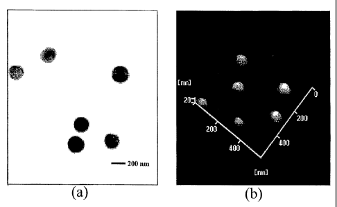
[0025] Figure 1 shows (a) a TEM micrograph of the prepared CS-y-PGA
nanoparticles
(0.10% y-PGA:0.20% CS) and (b) an AFM micrograph of the prepared CS-y-PGA
nanoparticles (0.01%
y-PGA:0.01% CS).
[0026] Figure 2 shows effects of the prepared CS-y-PGA nanoparticles on the
TEER
values of Caco-2 cell monolayers.
[0027] - Figure 3 shows the loading capacity and association efficiency of
insulin in
nanoparticles of chitosan and y-PGA.
[0028] Figure 4 shows the loading capacity and association efficiency of
insulin in
nanoparticles of chitosan as reference.
[0029] Figure 5 shows the stability of insulin-loaded nanoparticles.
[0030] Figure 6 show a representative in vitro study with insulin release
profile in a pH-
adjusted solution.
[0031] Figure 7 show the bioavailability of insulin of orally administered
insulin-loaded
nanoparticles in diabetic rats.
DETAILED DESCRIPTION OF THE EXEMPLARY EMBODIMENTS
[0032] The preferred embodiments of the present invention described below
relate
particularly to preparation of nanoparticles composed of chitosan/poly-y-
glutamic acid/insulin and their
permeability to enhance the intestinal or blood brain paracellular permeation
by opening the tight
junctions between epithelial cells. While the description sets forth various
embodiment specific details, it
will be appreciated that the description is illustrative only and should not
be construed in any way as
- 6-
CA 02592991 2007-07-03
WO 2006/073950 PCT/US2005/047125
limitin'g' the inwei t[oi '.'E EF& the Ere; " i6 s applications of the
invention, and modifications thereto,
which may occur to those who are skilled in the art, are also encompassed by
the general concepts
described below.
[0033] y-PGA is a naturally occurring anionic homo-polyamide that is made of L-
glutamic acid units connected by amide linkages between a-amino and y-
carboxylic acid groups (Crit.
Rev. Biotechnol. 2001;21:219-232). It is an exocellular polymer of certain
Bacillus species that is
produced within cells via the TCA cycle and is freely excreted into the
fermentation broth. Its exact
biological role is not fully known, although it is likely that 7-PGA is linked
to increasing the survival of
producing strains when exposed to environmental stresses. Because of its water-
solubility,
biodegradability, edibility, and non-toxicity toward humans and the
environment, several applications of
y-PGA in food, cosmetics, medicine, and water treatment have been investigated
in the past few years.
[0034] EXAMPLE NO. 1
[0035] Materials and methods of nanoparticles preparation
[0036] CS (MW -2.8 x 10⁵) with a degree of deacetylation of approximately 85%
was
acquired from Challenge Bioproducts Co. (Taichung, Taiwan). Acetic acid,
cellulase (1.92 units/mg),
fluorescein isothiocyanate (FITC), phosphate buffered saline (PBS), periodic
acid, sodium acetate,
formaldehyde, bismuth subnitrate, and Hanks' balanced salt solution (HBSS)
were purchased from Sigma
Chemical Co. (St. Louis, MO). Ethanol absolute anhydrous and potassium sodium
tartrate were obtained
from Merck (Darmstadt, Germany). Non-essential amino acid (NEAA) solution,
fetal bovine serum
(FBS), gentamicin and trypsin-EDTA were acquired from Gibco (Grand Island,
NY). Eagle's minimal
essential medium (MEM) was purchased from Bio West (Nuaille, France). All
other chemicals and
reagents used were of analytical grade.
[0037] EXAMPLE NO.2
[0038] Depolymerization of CS by enzymatic hydrolysis
[0039] Regular CS was treated with enzyme (cellulase) to produce low-MW CS
according to a method described by Qin et al. with some modifications (Food
Chein. 2004;84:107-115).
A solution of CS (20 g/1) was prepared by dissolving CS in 2% acetic acid.
Care was taken to ensure total
solubility of CS. Then, the CS solution was introduced into a vessel and
adjusted to the desired pH 5.0
with 2N aqueous NaOH. Subsequently, cellulase (0.1 g) was added into the CS
solution (100 ml) and
continuously stirred at 37 C for 12 hours. Afterward, the depolymerized CS was
precipitated with
aqueous NaOH at pH 7.0-7.2 and the precipitated CS was washed three times with
deionized water. The
resulting low-MW CS was lyophilized in a freeze dryer (Eyela Co. Ltd, Tokyo,
Japan).
[0040] The average molecular weight of the depolymerized CS was determined by
a gel
permeation chromatography (GPC) system equipped with a series of PL aquagel-OH
columns (one Guard
8 pm, 50 x 7.5 mm and two MIXED 8 m, 300 x 7.5 mm, PL Laboratories, UK) and a
refractive index
- 7-
CA 02592991 2007-07-03
WO 2006/073950 PCT/US2005/047125
(RI) dete6t6r`(RI20000-F,'SFD, Torrance, CA). Polysaccharide standards
(molecular weights range from
180 to 788,000, Polymer Laboratories, UK) were used to construct a calibration
curve. The mobile phase
contained 0.O 1M NaH2PO4 and 0.5MNaNO3 and was brought to a pH of 2Ø The
flow rate of mobile
phase was 1.0 ml/min, and the columns and the RI detector cell were maintained
at 30 C .
[0041] Factors limiting applications of most commercially available CSs are
their high
molecular weight and thus high viscosity and poor solubility at physiological
pH ranges. Low-MW CS
overcomes these limitations and hence finds much wider applications in
diversified fields. It was
suggested that low-MW CS be used as a parenteral drug carrier due to its lower
antigen effect (Eur. J.
Pharm. Biopharm. 2004;57:101-105). Low-MW CS was used as a non-viral gene
delivery system and
showed promising results (Int. J. Pharm. 1999;178:231-243). Several hydrolytic
enzymes such as
lysozyme, pectinase, cellulase, bromelain, hemicellulase, lipase, papain and
the like can be used to
depolymerize CS (Biochim. Biophys. Acta 1996;1291:5-15; Biochem. Eng. J.
2001;7:85-88; Carbohydr.
Res. 1992;237:325-332). GPC chromatograms of both standard-MW (also known as
regular-MW) and
low-MW CS are shown in an article by Sung and associates (Biomacromolecules
2005;6:1104-1112) . It
is known that cellulase catalyzes the cleavage of the glycosidic linkage in CS
(Food Chem. 2004;84:107-
115). The low-MW CS used in the study was obtained by precipitating the
depolymerized CS solution
with aqueous NaOH at pH 7.0-7.2. The obtained low-MW CS had a MW of about 50
kDa. In a preferred
embodiment, the low molecular weight chitosan has a molecular weight of about
40 kDa or less.
[0042] It was observed that the obtained low-MW CS can be readily dissolved in
an
aqueous solution at pH 6.0, while that before depolymerization needs to be
dissolved in an acetic acid
solution with a pH value about 4Ø Additionally, it was found that with the
low-MW CS, the prepared
nanoparticles had a significantly smaller size with a narrower distribution
than their counterparts prepared
with the high-MW (also known as standard-MW) CS (before depolymerization), due
to its lower
viscosity. As an example, upon adding a 0.10% y-PGA aqueous solution into a
0.20% high-MW CS
solution (viscosity 5.73 0.08 cp, measured by a viscometer), the mean
particle size of the prepared
nanoparticles was 878.3 28.4 nm with a polydispersity index of 1.0, whereas
adding a 0.10% 7-PGA
aqueous solution into the low-MW CS solution (viscosity 1.29 0.02 ep) formed
nanoparticles with a
mean particle size of 218.1 4.1 nm with a polydispersity index of 0.3 (n =
5).
[0043] EXAMPLE NO.3
[0044] Production and purification of y-PGA
[0045] y-PGA was produced by Bacillus lichenifornis (ATCC 9945, Bioresources
Collection and Research Center, Hsinchu, Taiwan) as per a method reported by
Yoon et al. with slight
modifications (Biotechnol. Lett. 2000;22:585-588). Highly mucoid colonies
(ATCC 9945a) were selected
from Bacillus licheniformis (ATCC 9945) cultured on the E medium (ingredients
comprising L-glutamic
acid, 20.0 g/l; citric acid, 12.0 g/1; glycerol, 80.0 g/1; NI-14C1, 7.0 g/l;
K2HPO4, 0.5 g/1; MgSO4.7H20, 0.5
- 8-
CA 02592991 2007-07-03
WO 2006/073950 PCT/US2005/047125
g/l; Fe'C13 `6J{ O, ~':04'`g7I;" aClz='2fI2 , `0 `I'S" g/l; MnSO4 H2O, 0.104
g/l, pH 6.5) agar plates at 37 C for
several times. Subsequently, young mucoid colonies were transferred into 10 ml
E medium and grown at
37 C in a shaking incubator at 250 rpm for 24 hours. Afterward, 500 l of
culture broth was mixed with
50 ml E medium and was transferred into a 2.5-liter jar-fermentor (KMJ-2B,
Mituwa Co., Osaka, Japan)
containing 950 ml of E medium. Cells were cultured at 37 C. The pH was
controlled at 6.5 by automatic
feeding of 25% (v/v) NH4OH and/or 2M HCl. The dissolved oxygen concentration
was initially
controlled at 40% of air saturation by supplying air and by controlling the
agitation speed up to 1000 rpm.
[0046] After 40 hours, cells were separated from the culture broth by
centrifugation for
20 minutes at 12,000 x g at 4C. The supernatant containing y-PGA was poured
into 4 volumes of
methanol and left overnight with gentle stirring. The resulting precipitate
containing crude y-PGA was
collected by centrifugation for 40 minutes at 12,000 x g at 4 C and then was
dissolved in deionized water
to remove insoluble impurities by centrifugation for 20 minutes at 24,000 x g
at 4C. The aqueous y-PGA
solution was desalted by dialysis (MWCO: 100,000, Spectrum Laboratories, Inc.,
Laguna Hills, CA)
against distilled water for 12 hours with water exchanges several times, and
finally was lyophilized to
obtain pure y-PGA.
[0047] EXAMPLE NO. 4
[0048] Preparation of the CS-y-PGA nanoparticles
[0049] Nanoparticles were obtained upon addition of y-PGA aqueous solution (pH
7.4, 2
ml), using a pipette (0.5-5 ml, PLASTIBRAND , BrandTech Scientific Inc.,
Germany), into a low-MW
CS aqueous solution (pH 6.0, 10 ml) at varying concentrations (0.01%, 0.05%,
0.10%, 0.15%, or 0.20%
by w/v) under magnetic stirring at room temperature. Nanoparticles were
collected by ultracentrifugation
at 38,000 rpm for 1 hour. Supernatants were discarded and nanoparticles were
resuspended in deionized
water for further studies. FT-IR was used to analyze peak variations of amino
groups of low-MW CS and
carboxylic acid salts of y-PGA in the CS-y-PGA nanoparticles.
[0050] As stated, nanoparticles were obtained instantaneously upon addition of
a y-PGA
aqueous solution (pH 7.4) into a low-MW CS aqueous solution (pH 6.0) under
magnetic stirring at room
temperature. The FT-IR spectra of the low-MW CS and the CSJy-PGA nanoparticles
are shown in an
article by Sung and associates (Biomacromolecules 2005;6:1104-1112). The
electrostatic interaction
between the two polyelectrolytes (y-PGA and CS) instantaneously induced the
formation of long
hydrophobic segments (or segments with a high density of neutral ion-pairs),
and thus resulted in highly
neutralized complexes that segregated into colloidal nanoparticles.
[0051] EXAMPLE NO. 5
[0052] Characterization of the CS-y-PGA nanoparticles
[0053] The morphological examination of the CS-7-PGA nanoparticles was
performed
- 9-
CA 02592991 2007-07-03
WO 2006/073950 PCT/US2005/047125
by TEM ftradsrhi9~id f &lbtf6n rAi6ro otpyy"and AFM (atomic force microscopy).
The TEM sample was
prepared by placing a drop of the nanoparticle solution onto a 400 mesh copper
grid coated with carbon.
About 2 minutes after deposition, the grid was tapped with a filter paper to
remove surface water and
positively stained by using an alkaline bismuth solution (Microbiol. Immunol.
1986;30:1207-1211). The
AFM sample was prepared by casting -a drop of the nanoparticle solution on a
slide glass and then dried in
vacuum. The size distribution and zeta potential of the prepared nanoparticles
were measured using a
Zetasizer (3000HS, Malvern Instruments Ltd., Worcestershire, UK).
[0054] The particle sizes and the zeta potential values of CS-y-PGA
nanoparticles,
prepared at varying concentrations of y-PGA and CS, were determined and the
results are shown in
Tables la and lb. It was found that the particle size and the zeta potential
value of the prepared
nanoparticles were mainly determined by the relative amount of the local
concentration of y-PGA in the
added solution to the surrounding concentration of CS in the sink solution. At
a fixed concentration of
CS, an increase in the y-PGA concentration allowed y-PGA molecules interacting
with more CS
molecules, and thus formed a lager size of nanoparticles (Table 1 a, p <
0.05).
[0055] When the amount of CS molecules exceeded that of local y-PGA molecules,
some of the excessive CS molecules were entangled onto the surfaces of CS-y-
PGA nanoparticles. Thus,
the resulting nanoparticles may display a structure of a neutral
polyelectrolyte-complex core surrounded
by a positively charged CS shell (Table lb) ensuring the colloidal
stabilization (Langmuir. 2004;20:7766-
7778). In contrast, as the amount of local y-PGA molecules sufficiently
exceeded that of surrounding CS
molecules, the formed nanoparticles had y-PGA exposed on the surfaces and thus
had a negative charge
of zeta potential. Therefore, the particle size and the zeta potential value
of the prepared CS-y-PGA
nanoparticles can-be controlled by their constituted compositions. The results
obtained by the TEM and
AFM examinations showed that the morphology of the prepared nanoparticles was
spherical in shape with
a smooth surface (Figure la and lb). Some aspects of the invention relate to
nanoparticles having a mean
particle size between about 50 and 400 nanometers, preferably between about
100 and 300 nanometers,
and most preferably between about, 100 and 200 nanometers. The morphology of
the nanoparticles shows
spherical in shape with a smooth surface at any pH between 2.5 and 6.6. In one
embodiment, the stability
of the nanoparticles of the present invention at a low pH around 2.5 or lower
enables the nanoparticles to
be intact when exposed to the acidic medium in the stomach.
[0056] EXAMPLE NO.6
[0057] Caco-2 cell cultures and TEER measurements
[0058] Caco-2 cells were seeded on the tissue-culture-treated polycarbonate
filters
(diameter 24.5 mm, growth area 4.7 cm2) in Costar Transwell 6 wells/plates
(Corning Costar Corp., NY)
at a seeding density of 3 x 10⁵ cells/insert. MEM (pH 7.4) supplemented with
20% FBS, 1% NEAA, and
40 g/ml antibiotic-gentamicin was used as the culture medium, and added to
both the donor and acceptor
-10-
CA 02592991 2007-07-03
WO 2006/073950 PCT/US2005/047125
compafitent.=" "1"fie"'WOUffif was' rrpfheer every 48 hours for the first 6
days and every 24 hours
thereafter. The cultures were kept in an atmosphere of 95% air and 5% CO2 at
37 C and were used for
the paracellular transport experiments 18-21 days after seeding (TEER values
in the range of 600-800
5lcm2).
[0059] TEER values of the Caco-2 cell monolayers were monitored with a
Millicell -
Electrical Resistance System (Millipore Corp., Bedford, MA) connected to a
pair of chopstick electrodes.
To initiate the transport experiments, the culture media in the donor and
acceptor compartments were
aspirated, and the cells were rinsed twice with pre-warmed transport media
(HBSS supplemented with
251nMglucose, pH 6.0). Following a 30-min equilibration with the transport
media at 37 C, the cells were
incubated for 2 hours with 2 ml transport media containing 0.5 ml test
nanoparticle solutions (0.2 mg/ml)
at 37 C . Subsequently, solutions of nanoparticles were carefully removed and
cells were washed three
times with HESS and replaced by fresh culture media. The TEER was measured for
another 20 hours to
study reversibility of the effect of test nanoparticles on Caco-2 cell
monolayers.
[0060] The intercellular tight junction is one of the major barriers to the
paracellular
transport of macromolecules (J. Control. Release 1996;39:131-138; J. Control.
Release 1998;51:35-46).
Trans-epithelial ion transport is contemplated to be a good indication of the
tightness of the junctions
between cells and was evaluated by measuring TEER of Caco-2 cell monolayers in
the study. It was
reported that the measurement of TEER can be used to predict the paracellular
transport of hydrophilic
molecules (Eur. J. Pharm. Biopharm. 2004;58:225-235). When the tight junctions
open, the TEER value
will be reduced due to the water and ion passage through the paracellular
route. Caco-2 cell monolayers
have been widely used as an in vitro model to evaluate the intestinal
paracellular permeability of
macromolecules.
[0061] Effects of the prepared CS-y-PGA nanoparticles on the TEER values of
Caco-2
cell monolayers are shown in Figure 2. As shown, the prepared nanoparticles
with a positive surface
charge (CS dominated on the surface, 0.01% y-PGA:0.05% CS, 0.10% y-PGA:0.2%
CS, and 0.20% y-
PGA:0.20% CS) were able to reduce the values of TEER of Caco-2 cell monolayers
significantly (p <
0.05). After a 2-hour incubation with these nanoparticles, the TEER values of
Caco-2 cell monolayers
were reduced to about 50% of their initial values as compared to the control
group (without addition of
nanoparticles in the transport media). This indicated that the nanoparticles
with CS dominated on the
surfaces could effectively open the tight junctions between Caco-2 cells,
resulting in a decrease in the
TEER values. It was reported that interaction of the positively charged amino
groups of CS with the
negatively charged sites on cell surfaces and tight junctions induces a
redistribution of F-actin and the
tight junction's protein ZO- 1, which accompanies the increased paracellular
permeability (Drug Deliv.
- 11-
CA 02592991 2007-07-03
WO 2006/073950 PCT/US2005/047125
Table la
Effects of concentrations of y-PGA and CS on the particle sizes of the
prepared
CS-y-PGA nanoparticles
Mean Particle Size (nm, n = 5)
CS
y0.01% a) 0.05% 0.10% 0.15% 0.20%
0.01% 79.0 3.0 103.1 4.6 96.7 1.9 103.6 1.9 140.5 2.0
0.05% 157.4 1.7 120.8 3.9 144.5 2.4 106.2 3.8 165.4 1.7
0.10% 202.2 3.1 232.6 1.2 161.0 1.8 143.7 2.7 218.1 4.1
0.15% 277.7 3.2 264.9 2.1 188.6 2.9 178.0 2.2 301.1 6.4
0.20% 284.1 2.1 402.2 4.0 A 225.5 3.1 365.5 5.1
a)
concentration of CS (by w/v)
b) concentration of 7-PGA (by w/v)
A precipitation of aggregates was observed
Table lb
Effects of concentrations of y-PGA and CS on the zeta potential values of the
prepared CS-y-PGA
nanoparticles.
Zeta Potential (mV, n = 5)
CS
y0.01% a) 0.05% 0.10% 0.15% 0.20%
0.01% 15.4 0.3 22.8 0.5 19.8 1.5 16.5 1.4 17.2 1.6
0.05% -32.7 0.7 23.7 1.7 27.6 0.7 20.3 0.8 19.2 0.6
0.10% -33.1 1.3 21.1 1.6 20.3 1.1 23.6 0.9 24.7 1.2
0.15% -33.2 2.1 -21.9 2.0 19.2 0.4 16.9 1.7 19.8 0.3
0.20% -34.5 0.5 -34.6 0.3 A 14.6 0.7 16.3 0.7
a) concentration of CS (by w/v)
b) concentration of y-PGA (by w/v)
A precipitation of aggregates was observed
-12-
CA 02592991 2007-07-03
WO 2006/073950 PCT/US2005/047125
Rev. 2001;50:S91-S101). It is suggested that an interaction between chitosan
and the tight junction
protein ZO-1, leads to its translocation to the cytoskeleton.
[0062] After removal of the incubated nanoparticles, a gradual increase in
TEER values
was noticed. This phenomenon indicated that the intercellular tight junctions
of Caco-2 cell monolayers
started to recover gradually; however, the TEER values did not recover to
their initial values (Figure 2).
In contrast, the TEER values of Caco-2 cell monolayers incubated with the
nanoparticles with a negative
surface charge (y-PGA dominated on the surface, 0.10% y-PGA:0.01% CS and 0.20%
y-PGA:0.01% CS,
Figure 2) showed no significant differences as compared to the control group
(p > 0.05). This indicated
that y-PGA does not have any effects on the opening of the intercellular tight
junctions.
[0063] EXAMPLE NO. 7
[0064] fCS-y-PGA nanoparticle preparation and CLSM visualization
[0065] Fluorescence (FITC)-labeled CS-y-PGA (fCS y-PGA) nanoparticles were
prepared for the confocal laser scanning microscopy (CLSM) study. The
nanoparticles of the present
invention display a structure of a neutral polyelectrolyte-complex core
surrounded by a positively charged
chitosan shell. Synthesis of the FITC-labeled low-MW CS (fCS) was based on the
reaction between the
isothiocyanate group of FITC and the primary amino groups of CS as reported in
the literature (Pharm.
Res. 2003;20:1812-1819). Briefly, 100 mg of FITC in 150 ml of dehydrated
methanol were added to 100
ml of 1% low-MW CS in O.1Macetic acid. After 3 hours of reaction in the dark
at ambient conditions,
fCS was precipitated by raising the pH to about 8-9 with 0.5M NaOH. To remove
the unconjugated
FITC, the precipitate was subjected to repeated cycles of washing and
centrifugation (40,000 x g for 10
min) until no fluorescence was detected in the supernatant. The fCS dissolved
in 80 ml of 0.1M acetic
acid was then dialyzed for 3 days in the dark against 5 liters of distilled
water, with water replaced on a
daily basis. The resultant fCS was lyophilized in a freeze dryer. The fCSy-PGA
nanoparticles were
prepared as per the procedure described in EXAMPLE No. 4.
[0066] Subsequently, the transport medium containing fCSy-PGA nanoparticles
(0.2
mg/ml) was introduced into the donor compartment of Caco-2 cells, which were
pre-cultured on the
transwell for 18-21 days. The experimental temperature was maintained at 37 C
by a temperature control
system (DH-35 Culture Dish Heater, Warner Instruments Inc., Hamden, CT). After
incubation for
specific time intervals, test samples were aspirated. The cells were then
washed twice with pre-warmed
PBS solution before they were fixed in 3.7% paraformaldehyde (Pharm. Res.
2003;20:1812-1819). Cells
were examined under an inversed CLSM (TCS SL, Leica, Germany). The
fluorescence images were
observed using an argon laser (excitation at 488 nm, emission collected at a
range of 510-540 nm).
[0067] CLSM was used to visualize the transport of the fluorescence-labeled CS-
y-PGA
(fCS-y-PGA) nanoparticles across the Caco-2 cell monolayers. This non-invasive
method allows for
- 13-
CA 02592991 2007-07-03
WO 2006/073950 PCT/US2005/047125
opticaf 6ctidnfiie-aitd'iMttki ng'bf thL `'t`rap port pathways across the
Caco-2 cell monolayers, without
disrupting their structures (J. Control. Release 1996;39:131-138).
[0068] After 60 minutes of incubation with the nanoparticles, the intensity of
fluorescence observed at intercellular spaces was stronger and appeared at a
deeper level than those
observed at 20 min after incubation. These observations confirmed with our
TEER results that the
nanoparticles with a positive surface charge (CS dominated on the surface)
were able to open the tight
junctions between Caco-2 cells and allowed transport of the nanoparticles by
passive diffusion via the
paracellular pathways. More detailed data can be found in an article by Sung
and associates
(Biomacromolecules 2005;6:1104-1112).
[0069] EXAMPLE NO. 8
[0070] In vivo study with Fluorescence-labeled nanoparticles
[0071] Fluorescence (FITC)-labeled CS-y-PGA (fCS-y-PGA) nanoparticles were
prepared for the confocal laser scanning microscopy (CLSM) study. After
feeding rats with fCSy-PGA
nanoparticles, the rats are sacrificed at a pre-determined time and the
intestine is isolated for CLSM
examination. The fluorescence images of the nanoparticles were clearly
observed by CLSM that
penetrates through the mouse intestine at appropriate time and at various
depths from the inner surface
toward the exterior surface of the intestine, including duodenum, jejunum, and
ileum, which is discussed
in EXAMPLE No. 12.
[0072] EXAMPLE NO. 9
[0073] Insulin loading capacity in nanoparticles
[0074] Fluorescence (FITC)-labeled y-PGA was added into chitosan solution to
prepare
fluorescence (FITC)-labeled, insulin-loaded CS-,y-PGA nanoparticles for in
vivo animal study with
confocal laser scanning microscopy (CLSM) assessment and bioactivity analysis.
The insulin-loaded
CS- y PGA nanoparticles are manufactured by using the ionic-gelation method
upon addition of insulin/y-
PGA solution into CS solution, followed by magnetic stirring in a container.
[0075] The nanoparticles with two insulin concentrations are prepared at a
chitosan to y-
PGA ratio of 0.75 mg/ml to 0.167 mg/ml. Their particle size and zeta potential
are shown in Table 2
below.
Table 2
Insulin Conc. Mean Particle Size Polydispersity Index Zeta Potential
(mg/ml) (n=5) (nm) (PI) (mV)
0* 145.6 1.9 0.14 0.01 +32.11 1.61
0.042 185.1 5.6 0.31 0.05 +29.91 1.02
0.083 198.4 6.2 0.30 0.09 +27.83 1.22
(*) control reference without insulin
- 14-
CA 02592991 2007-07-03
WO 2006/073950 PCT/US2005/047125
[0076] Further, their association efficiency of insulin and loading capacity
of insulin are
analyzed, calculated and shown in Figures 3 and 4, according to the following
formula:
(Total amount of insulin-Insulin in supernatant) x100%
Insulin Association Total amount of insulin
Efficiency (AE %)
_ (Total amount of insulin-Insulin in supernatant) x100%
Loading Capacity (LC) Weight of recovered particles
[0077] Figure 3 shows loading capacity and association efficiency of insulin
in
nanoparticles of chitosan and y-PGA, whereas Figure 4 shows loading capacity
and association efficiency
of insulin in nanoparticles of chitosan alone (in absence of y-PGA) as
reference. The data clearly
demonstrates that both the insulin loading capacity and insulin association
efficiency are statistically
higher for the nanoparticles with y-PGA in the core. The AE (4055%) and LC
(5.014.0%) of insulin for
CS-,r PGA nanoparticles was obtained by using ionic-gelation method upon
addition of insulin mixed
with y-PGA solution into CS solution, followed by magnetic stirring for
nanoparticle separation. Some
aspects of the invention relate to an oral dose of nanoparticles that
effectively enhance intestinal or blood
brain paracellular transport comprising a negative component (such as y-PGA,
heparin, or alginate) in the
core and low molecular weight chitosan, wherein the chitosan dominates on a
surface of the nanoparticles
with positive charges. Alginate is non-biodegradable; however, it is
stipulated that an alginate particle
with about 30-50 kDa molecular weight is kidney inert.
[0078] Calceti et al. reported an in vivo evaluation of an oral insulin-PEG
delivery
system (Eur J Pharma Sci 2004;22:315-323). Insulin-PEG was formulated into
mucoadhesive tablets
constituted by the thiolated polymer poly(acrylic acid)-cysteine. The
therapeutic agent was sustained
released from these tablets within 5 hours. In vivo, by oral administration to
diabetic mice, the glucose
levels were found to decrease significantly over the time. Further, Krauland
et al. reported another oral
insulin delivery study of thiolated chitosan-insulin tablets on non-diabetic
rats (J. Control. Release 2004,
95:547-555). The delivery tablets utilized 2-Iminothiolane covalently linked
to chitosan to form chitosan-
TBA (chitosan-4-thiobutylamidine) conjugate. After oral administration of
chitosan-TBA-insulin tablets
to non-diabetic conscious rats, the blood glucose level decreased
significantly for 24 hours; supporting the
observation of sustained insulin release of the presently disclosed
nanoparticles herein through intestinal
absorption. In a further report by Morcol et al. (Int. J. Pharm. 2004;277:91-
97), an oral delivery system
comprising calcium phosphate-PEG-insulin-casein particles displays a prolonged
hypoglycemic effect
after oral administration to diabetic rats.
[0079] Pan et al. disclosed chitosan nanoparticles improving the intestinal
absorption of
-15-
CA 02592991 2007-07-03
WO 2006/073950 PCT/US2005/047125
insulini''int'vi./b'(Iri't'`'J"inh~r i`a`2d(1 ;249:"1'39='147) with insulin-
chitosan nanoparticles at a particle size of
250-400 nm, a polydispersity index smaller than 0.1, positively charged and
stable. After administering
the insulin-chitosan nanoparticles, it was found that the hypoglycemic was
prolonged with enhanced
pharmacological bioavailability. Their data confirmed our observation as shown
in Figures 3 and 4;
however, the insulin loading capacity and insulin association efficiency of
the present invention are
substantially higher for the chitosan-insulin nanoparticles with 'Y-PGA in the
core.
[0080] EXAMPLE NO. 10
[0081] Insulin nanoparticle stability
[0082] Figure 5 shows the stability of insulin-loaded nanoparticles of, the
present
invention with an exemplary composition of CS 0.75mg/ml, y-PGA 0.167mg/ml, and
insulin 0.083
mg/ml. The prepared insulin-loaded nanoparticles suspended in deionized water
are stable during storage
up to 40 days. First (as shown in Figure 5), the insulin content in the
nanoparticle storage solution
maintains at about a constant level of 9.5%. The nanoparticle stability is
further evidenced by the
substantially constant particle size at about 200 nm and substantially
constant zeta potential of about +28
mV over the period of about 40 days. It is contemplated that the insulin-
containing nanoparticles of the
present invention would further maintain their biostability when formulated in
a softgel or gelcap capsule
configuration that further isolates the nanoparticles from environmental
effects, such as sunlight, heat, air
conditions, and the like. In one embodiment, the surface of the gelcap capsule
may further treated with
glycerin or hydrophilicity to allow easy swallowing. Some aspects of the
invention provide a gelcap pill
or capsule containing a dosage of insulin nanoparticles effective amount of
the insulin to treat or manage
the diabetic patients, wherein the stability of the insulin-containing
nanoparticles is at least 40 days,
preferably more than 6 months, and most preferably more than a couple of
years. By "effective amount of
the insulin", it is meant that a sufficient amount of insulin will be present
in the dose to provide for a
desired therapeutic, prophylatic, or other biological effect when the
compositions are administered to a
host in the single dosage forms.
[0083] Thus, for convenient and effective oral administration,
pharmaceutically effective
amounts of the nanoparticles of this invention can be tabletted with one or
more excipient, encased in
capsules such as gel capsules, and suspended in a liquid solution and the
like. The nanoparticles can be
suspended in a deionized solution or the like for parenteral administration.
The nanoparticles may be
formed into a packed mass for ingestion by conventional techniques. For
instance, the nanoparticles may
be encapsulated as a "hard-filled capsule" or a "soft-elastic capsule" using
known encapsulating
procedures and materials. The encapsulating material should be highly soluble
in gastric fluid so that the
particles are rapidly dispersed in the stomach after the capsule is ingested.
Each unit dose, whether
capsule or tablet, will preferably contain nanoparticles of a suitable size
and quantity that provides
pharmaceutically effective amounts of the nanoparticles. One example is a size
0 gelatin capsule.
- 16-
CA 02592991 2007-07-03
WO 2006/073950 PCT/US2005/047125
"[00841' "t MANTLE NO: l 1"
[0085] In vitro Insulin release study
[0086] Figure 6 show a representative protein drug (for example, insulin)
release profile
in a pH-adjusted solution for pH-sensitivity study with an exemplary
composition of CS 0.75mg/ml, y-
PGA 0.167mg/ml, and insulin 0.083 mg/ml in nanoparticles. In one embodiment,
the exemplary
composition may include each component at a concentration range of 10% as
follows: CS 0.75mg/ml (a
concentration range of 0.67 to 0.83 mg/ml), ,y-PGA 0.167mg/ml (a concentration
range of 0.150 to 0.184
mg/ml), and insulin 0.083 mg/ml (a concentration range of 0.075 to 0.091
mg/ml). First, solution of the
insulin-loaded nanoparticles was adjusted to pH 2.5 to simulate the gastric
environment in a DISTEK-
2230A container at 37 C and 100 rpm. Samples (n=5) were taken at a pre-
determined particular time
interval and the particle-free solution was obtained by centrifuging at 22,000
rpm for 30 minutes to
analyze the free or released insulin in solution by HPLC. Until the free
insulin content in the sample
solution approaches about constant of 26% (shown in Figure 6), the pH was
adjusted to 6.6 to simulate
the entrance portion of the intestine. The net released insulin during this
particular time interval is about
(from 26% to 33%) 7%. In other words, the nanoparticles are quite stable
(evidenced by minimal
measurable insulin in solution) for both the pH 2.5 and pH 6.6 regions.
[0087] To further simulate the exit portion of the intestine, the insulin-
containing
nanoparticle solution is adjusted to pH 7.4. The remaining insulin (about 67%)
is released from the
nanoparticles. As discussed above, the insulin in nanoparticles would be more
effective to penetrate the
intestine wall in paracellular transport mode than the free insulin because of
the nanoparticles of the
present invention with chitosan at the outer surface (preferential mucosal
adhesion on the intestinal wall)
and positive charge (enhancing paracellular tight junction transport).
[0088] Some aspects of the invention provide a dose of nanoparticles to a
patient
characterized by enhancing intestinal paracellular transport or brain blood
paracellular transport, each
nanoparticle comprising a first component of at least one bioactive agent, a
second component that is
negatively charged, and a third component of low molecular weight chitosan,
wherein the first and second
components occupy a center core and the third component dominates on a surface
of the nanoparticle. In
one embodiment, the second component is y-PGA, wherein a weight ratio of the
chitosan to y-PGA is
0.75 to 0.167 or higher. A preparation solution with excess chitosan to y-PGA
would yield nanoparticles
with stable positive charge at the surface of the nanoparticles. The surface
charge (zeta potential) of the
nanoparticles of the present invention is between about 15 and 40 mV,
preferably between about 25 to 40
mV.
[0089] By way of illustration, the dose of nanoparticles for treating diabetes
comprises a
first component of insulin, a second component of y-PGA, and a third component
of low molecular
weight chitosan, wherein a weight ratio of the three components (insulin to y-
PGA to CS) is about
-17-
CA 02592991 2007-07-03
WO 2006/073950 PCT/US2005/047125
0.083'6.1 7:0.75. 'As shown in" Tigur'e_47," the insulin content (i.e.,
insulin loading capacity) of a
conventional chitosan-insulin composite is at about 7.1 0.6 w/w% at a CS to
insulin ratio of 0.75:0.083 or
an insulin loading capacity at about 0.7 0.1 w/w% at a CS to insulin ratio of
0.75:0.043 in the preparation
solution. In some embodiments of the present invention, the insulin loading
capacity is least 8 w/w% of
the nanoparticles, preferably at least 14 wlw% of the nanoparticles.
[0090] EXAMPLE NO. 12
[0091] In vivo study with Insulin-loaded fluorescence-labeled nanoparticles
[0092] In the in vivo study, rats were injected with streptozotocin (STZ
75mg/kg
intraperitoneal) in 0.01M citrate buffer (pH 4.3) to induce diabetes rats. The
blood from the rat's tail was
analyzed with a commercially available glucometer for blood glucose. The blood
glucose level on Wistar
male rats at no fasting (n=5) is measured at 107.2 8.1 mg/dL for normal rats
while the blood glucose
level is at 469.7 34.2 mg/dL for diabetic rats. In the animal study, diabetic
rats were fasting for 12 hours
and subjected to four different conditions: (a) oral deionized water (DI)
administration; (b) oral insulin
administration at 3 OU/kg; (c) - oral insulin-loaded nanoparticles
administration at 3 OU/kg; and (d)
subcutaneous (SC) insulin injection at 5U/kg as positive control. The blood
glucose concentration from
rat's tail was measured over the time in the study.
[0093] Figure 7 shows glucose change (hypoglycemic index) versus time of the
in vivo
animal study (n=5). The glucose change as a percentage of base lines for both
oral DI administration and
oral insulin administration over a time interval of 8 hours appears relatively
constant within the
experimental measurement error range. It is illustrative that substantially
all insulin from the oral
administration route has been decomposed in rat stomach. As anticipated, the
glucose decrease for the SC
insulin injection route appears in rat blood in the very early time interval
and starts to taper off after 3
hours in this exemplary study. The most important observation of the study
comes from the oral
administration route with insulin-loaded nanoparticles. The blood glucose
begins to decrease from the
base line at about 2 hours after administration and sustains at a lower
glucose level at more than 8 hours
into study. It suggests that the current insulin-loaded nanoparticles modulate
the glucose level in animals
in a sustained or prolonged effective mode.
[0094] Some aspects of the invention relate to a novel nanoparticle system
that is
composed of a low-MW CS and -(-PGA with CS dominated on the surfaces being
configured to
effectively open the tight junctions. The surface of the nanoparticles is
characterized with a positive
surface charge. In one embodiment, the nanoparticles of the invention enables
effective intestinal delivery
for bioactive agent, including peptide, polypeptide, protein drugs, other
large hydrophilic molecules, and
the like. Such polypeptide drugs can be any natural or synthetic polypeptide
that may be orally
administered to a human patient. Exemplary drugs include, but are not limited
to, insulin; growth factors,
such as epidermal growth factor (EGF), insulin-like growth factor (IGF),
transforming growth factor
-18-
CA 02592991 2007-07-03
WO 2006/073950 PCT/US2005/047125
Irl, n f`ve" growAh" a`ctor`(NOV), "p"1906-derived growth factor (PDGF), bone
morphogenic protein
(TGF),
(BMP), fibroblast growth factor and the like; somatostatin; somatotropin;
somatropin; somatrem;
calcitonin; parathyroid hormone; colony stimulating factors (CSF); clotting
factors; tumor necrosis
factors: interferons; interleukins; gastrointestinal peptides, such as
vasoactive intestinal peptide (VIP),
cholecytokinin (CCK), gastrin, secretin, and the like; erythropoietins; growth
hormone and GRF;
vasopressins; octreotide; pancreatic enzymes; dismutases such as superoxide
dismutase; thyrotropin
releasing hormone (TRH); thyroid stimulating hormone; luteinizing hormone;
LHRH; GHRH; tissue
plasminogen activators; macrophage activator; chorionic gonadotropin; heparin;
atrial natriuretic peptide;
hemoglobin; retroviral vectors; relaxin; cyclosporin; oxytocin; vaccines;
monoclonal antibodies; and the
like; and analogs and derivatives of these compounds. The bioactive agent of
the present invention may
be selected from group consisting of oxytocin, vasopressin,
adrenocorticotrophic hormone, prolactin,
luliberin or luteinising hormone releasing hormone, growth hormone, growth
hormone releasing factor,
somatostatin, glucagon, interferon, gastrin, tetragastrin, pentagastrin,
urogastroine, secretin, calcitonin,
enkephalins, endorphins, angiotensins, renin, bradykinin, bacitracins,
polymixins, colistins, tyrocidin,
gramicidines, and synthetic analogues, modifications and pharmacologically
active fragments thereof,
monoclonal antibodies and soluble vaccines. In one embodiment, the bioactive
agent comprises stem
cells.
[0095] In another embodiment, the nanoparticles of the invention increase the
absorption
of bioactive agents across the blood brain barrier and/or the gastrointestinal
barrier. In still another
embodiment, the nanoparticles with chitosan at an outer layer and surface
positive charge serve as an
enhancer in enhancing paracellular drug (bioactive agent) transport of an
administered bioactive agent
when the bioactive agent and nanoparticles are orally administrated in a two-
component system, or orally
administered substantially simultaneously.
[0096] Some aspects of the invention relate to a method of enhancing
intestinal or blood
brain paracellular transport of bioactive agents configured and adapted for
delivering at least one
bioactive agent in a patient comprising administering nanoparticles composed
of y-PGA and chitosan,
wherein the nanoparticles are loaded with a therapeutically effective amount
or dose of the at least one
bioactive agent. The nanoparticle of the present invention is an effective
intestinal delivery system for
peptide and protein drugs and other large hydrophilic molecules. In a further
embodiment, the bioactive
agent is selected from the group consisting of proteins, peptides,
nucleosides, nucleotides, antiviral
agents, antineoplastic agents, antibiotics, and anti-inflammatory drugs. In a
further embodiment, the
bioactive agent is selected from the group consisting of calcitonin,
cyclosporin, insulin, oxytocin,
tyrosine, enkephalin, tyrotropin releasing hormone (TRH), follicle stimulating
hormone (FSH),
luteinizing hormone (LH), vasopressin and vasopressin analogs, catalase,
superoxide dismutase,
interleukin-II (IL2), interferon, colony stimulating factor (CSF), tumor
necrosis factor (TNF) and
- 19-
CA 02592991 2010-07-08
melanocyte-stimulating hormone. In a further embodiment, the bioactive agent
is an Alzheimer
antagonist.
[0097] In a co-pending application, U.S. patent publication 2005/19404, it is
disclosed that a biomaterial with free amino groups of lysine, hydroxylysine,
or
arginine residues within biologic tissues is crosslinkable with genipin, a
crosslinker (Biomaterials
1999;20:1759-72). It is also disclosed that the crosslinkable biomaterial may
be crosslinked with a
crosslinking agent or with light, such as ultraviolet irradiation, wherein the
crosslinkable biomaterial may
be selected from the group consisting of collagen, gelatin, elastin, chitosan,
NOCC (N, 0, carboxylmethyl
chitosan), fibrin glue, biological sealant, and the like. Further, it is
disclosed that a crosslinking agent may
be selected from the group consisting of genipin, its derivatives, analog (for
example, aglycon geniposidic
acid), stereoisomers and mixtures thereof. In one embodiment, the crosslinking
agent may further be
selected from the group consisting of epoxy compounds, dialdehyde starch,
glutaraldehyde,
formaldehyde, dimethyl suberimidate, carbodiimides, succinimidyls,
diisocyanates, acyl azide, reuterin,
ultraviolet irradiation, dehydrothermal treatment,
tris(hydroxymethyl)phosphine, ascorbate-copper,
glucose-lysine and photo-oxidizers, and the like. In one embodiment, the
nanoparticles comprised of
crosslinkable biomaterial is crosslinked, for example up to about 50% degree
or more of crosslinking,
preferably about 1 to about 20% degree of crosslinking of the crosslinkable
components of the
biomaterial, enabling sustained biodegradation of the biomaterial and/or
sustained drug release.
[0098] By modifying the chitosan structure to alter its charge
characteristics, such as
grafting the chitosan with methyl, alkyl (for example, ethyl, propyl, butyl,
isobutyl, etc.), polyethylene
glycol (PEG), or heparin, the surface charge density (zeta potential) of the
CS- y PGA nanoparticles may
become more pH resistant or hydrophilic. In one embodiment, the chitosan is
grafted with polyacrylic
acid or a polymer with a chemical formula:
0=C -1 NH where Ris>_12
R
[0099] By way of illustration, trimethyl chitosan chloride might be used in
formulating
the CS- T PGA nanoparticles for maintaining its spherical biostability at a pH
lower than pH 2.5,
preferably at a pH as low as 1Ø Some aspects of the invention provide a drug-
loaded chitosan-containing
biological material crosslinked with genipin or other crosslinking agent as a
biocompatible drug carrier
for enhancing biostability at a pH lower than pH 2.5, preferably within at a
pH as low as 1Ø
[0100] Although the present invention has been described with reference to
specific
details of certain embodiments thereof, it is not intended that such details
should be regarded as
limitations upon the scope of the invention except as and to the extent that
they are included in the
accompanying claims. Many modifications and variations are possible in light
of the above disclosure.
- 20-