Note: Descriptions are shown in the official language in which they were submitted.
CA 02593074 2007-06-29
WO 2006/071597 PCT/US2005/045857
Title: Preloaded IOL Injector and Method
Background of the Invention
The present invention relates to ophthalmic surgical devices and methods. More
particularly, the present invention relates to a device and method for
inserting an
intraocular lens (IOL) into an eye wherein the IOL may be conveniently
preloaded in and
packaged together with the injector device.
IOLs are artificial lenses used to replace the natural crystalline lens of the
eye
when the natural lens has cataracts or is otherwise diseased. IOLs are also
sometimes
implanted into an eye to correct refractive errors of the eye in which case
the natural lens
may remain in the eye together with the implanted IOL. The IOL may be placed
in either
the posterior chamber or anterior chamber of the eye. IOLs come in a variety
of
configurations and materials. Some common IOL styles include the so-called
open-
looped haptics which include the three-piece type having an optic and two
haptics
attached to and extending from the optic; the one-piece type wherein the optic
and
haptics are integrally formed (e.g., by machining the optic and haptics
together from a
single block of material); and also the closed looped haptic IOLs. Yet a
further style of
IOL is called the plate haptic type wherein the haptics are configured as a
flat plate
extending from opposite sides of the optic. The IOL may be made from a variety
of
materials or combination of materials such as PMMA, silicone, hydrogels and
silicone
hydrogels, etc.
Various instruments and methods for implanting the IOL in the eye are known.
In
one method, the surgeon simply uses surgical forceps having opposing blades
which are
CA 02593074 2007-06-29
WO 2006/071597 PCT/US2005/045857
used to grasp the IOL and insert it through the incision into the eye. While
this method is
still practiced today, more and more surgeons are using more sophisticated IOL
inserter
devices which offer advantages such as affording the surgeon more control when
inserting the IOL into the eye. IOL inserter devices have recently been
developed with
reduced diameter insertion tips which allow for a much smaller incision to be
made in
the cornea than is possible using forceps alone. Smaller incision sizes (e.g.,
less than
about 3mm) are preferred over larger incisions (e.g., about 3.2 to 5+mm) since
smaller
incisions have been attributed to reduced post-surgical healing time and
complications
such as induced astigmatism.
Since IOLs are very small and delicate articles of manufacture, great care
must be
taken in their handling. In order for the IOL to fit through the smaller
incisions, they
need to be folded and/or compressed prior to entering the eye wherein they
will assume
their original unfolded/uncompressed shape. The IOL inserter device must
therefore be
designed in such a way as to permit the easy passage of the IOL through the
device and
into the eye, yet at the same time not damage the delicate IOL in any way.
Should the
IOL be damaged during delivery into the eye, the surgeon will most likely need
to
extract the damaged IOL from the eye and replace it with a new IOL, a highly
undesirable surgical outcome.
Thus, as explained above, the IOL inserter device must be designed to permit
easy passage of the IOL therethrough. It is equally important that the IOL be
expelled
from the tip of the IOL inserter device and into the eye in a predictable
orientation and
manner. Should the IOL be expelled from the tip too quickly or in the wrong
orientation,
the surgeon must further manipulate the IOL in the eye which could result in
trauma to
the surrounding tissues of the eye. It is therefore highly desirable to have
an inserter
2
CA 02593074 2007-06-29
WO 2006/071597 PCT/US2005/045857
device which allows for precise loading of the IOL into the inserter device
and which
will pass and expel the IOL from the inserter device tip and into the eye in a
controlled,
predictable and repeatable manner.
To ensure controlled expression of the IOL through the tip of the IOL inserter
device, the IOL must first be loaded into the IOL inserter device. The loading
of the IOL
into the inserter device is therefore a precise and very important step in the
process.
Incorrect loading of an IOL into the inserter device is oftentimes cited as
the reason for a
failed IOL delivery sequence. Many IOL injector devices on the market today
require the
IOL to be loaded into the injector at the time of surgery by the attending
nurse and/or
surgeon. Due to the delicate nature of the IOL, there is a risk that the nurse
and/or
surgeon will inadvertently damage the IOL and/or incorrectly load the IOL into
the
injector device resulting in a failed implantation. Direct handling and/or
loading of the
IOL into the injector by the nurse and/or surgeon is therefore undesirable.
There remains a need for an IOL inserter and method which removes the need for
direct handling of the IOL by the nurse and/or surgeon and which generally
simplifies
operation of the IOL injector device and IOL delivery process.
Summary of the Invention
In a broad aspect of the invention, an injector device is provided having a
proximal tubular body section having a longitudinal passageway extending
between
opposite, open ends thereof, a shuttle which holds the IOL in an initially
unstressed state,
and a nozzle section which are packaged and then assembled together at the
time of
surgery. A plunger component is inserted into the proximal open end of the
tubular body
and telescopes within the longitudinal passageway thereof. The IOL is
preloaded in an
3
CA 02593074 2007-06-29
WO 2006/071597 PCT/US2005/045857
unstressed condition in the shuttle component of the device (i.e., in a
condition where at
least the IOL optic is not compressed or folded). The shuttle and IOL are
packaged either
dry or submersed in a container of sterile solution which maintains the IOL in
a hydrated
state during shipping and storage, a necessary requirement for IOLs made of
certain
materials such as acrylic.
In a particularly advantageous embodiment of the invention, the shuttle
component is releasably attached to a cover of the storage container whereby
the cover
may be manually grasped and used to plug the shuttle component (with IOL
already
contained therein) into the proximal body section of the injector device. At
the time of
surgery, the nurse or surgeon simply opens the container containing the
shuttle and IOL
and removes them from the container by grasping the container top to which the
shuttle
is attached. While still grasping the cover, the shuttle is then plugged into
the device
body at which time the cover may be detached from the shuttle. A quick release
mechanism may be employed to allow for the quick and easy detachment of the
cover
from the shuttle once the shuttle has been plugged into the device body.
The nozzle section includes a distal tip through which the IOL is ultimately
expelled from the injector device and is attached to the device body to
complete the
assembly. The body, shuttle and nozzle each include a longitudinal passageway
which
preferably lie along a common longitudinal axis when the body, shuttle and
nozzle
sections are assembled together. A plunger is provided which telescopes within
the
device body and may be advanced therein to enter the shuttle component and
engage and
push the IOL through the shuttle and nozzle, the IOL ultimately being expelled
from the
injector device at the nozzle tip. The injector device includes means for
compressing or
otherwise urging the IOL into a smaller cross-section for delivery through the
injector. In
4
CA 02593074 2007-06-29
WO 2006/071597 PCT/US2005/045857
a preferred embodiment of the invention, the shuttle and nozzle passageways
are
configured with a narrowing taper towards the distal tip. The plunger is
advanced at the
proximal end of the injector device causing the distal tip of the plunger to
engage the
IOL optic. As the plunger is advanced further, the IOL is pushed through the
narrowing
passageway, thereby compressing the IOL into a smaller cross-section and
finally exiting
at the nozzle tip and expressed into the eye in the intended manner.
In an alternate embodiment, the shuttle is first attached to the nozzle
section and
the nozzle/shuttle unit is then attached to the device body.
In yet a further alternate embodiment, the nozzle section and shuttle are
connected together and placed in the storage container. In this alternate
embodiment, the
nozzle and shuttle are releasably connected to the cover and handled as a unit
whereby
the cover may be manually grasped to plug the nozzle and shuttle unit into the
device
body.
Brief Description of the Drawings
Figure 1 is a perspective view of the fully assembled injector device showing
the
an IOL expressed from the distal tip thereof,
Figure 2a is a perspective view of the shuttle component and IOL packaged in a
vial with an outer cover thereof being removed;
Figure 2b is the view of Figure 2a showing the inside cover and shuttle
component being removed from the vial;
Figure 3a is a side elevational view showing a user plugging the shuttle
component into the proximal body section of the device;
CA 02593074 2007-06-29
WO 2006/071597 PCT/US2005/045857
Figure 3b is the view of Fig. 3a showing the shuttle component fully inserted
into
the proximal body section and the nozzle section being attached to the
proximal body
section;
Figure 4a is a side elevational view of an alternate embodiment showing the
shuttle being connected to the nozzle section;
Figure 4b is a perspective view showing the nozzle section and shuttle
combination being connected to the proximal body section;
Figure 4c is a perspective view showing an alternate embodiment where the
nozzle section and shuttle are attached together and releasably attached to
the vial cover;
Figure 5 is an enlarged, perspective view of the plunger component of the
device;
Figure 6 is an enlarged, perspective view of the proximal body section of the
device;
Figure 7a is an enlarged, perspective view of the shuttle component of the
device;
Figure 7b is the view of Figure 7a showing the shuttle component in the open
position and an IOL placed therein; and
Figure 8 is a perspective view of the nozzle section of the device.
Detailed Description
The invention comprises a preloaded injector device for injecting an IOL into
an
eye. The term "preloaded" as used herein means that a packaged component of
the
injector device includes an IOL positioned therein. Direct handling and
loading of an
IOL into the injector device is therefore not necessary.
The basic components of injector device 10 include a proximal body section 12,
a
plunger 20, a distal nozzle section 14, and a shuttle component 16 which are
assembled
6
CA 02593074 2007-06-29
WO 2006/071597 PCT/US2005/045857
at the time of surgery to ready the device 10 for delivery of an IOL 30
therethrough. An
IOL 30 is preloaded into the shuttle component 16 of the device which is
packaged in
either a dry state or in a hydrated state in a vial 11 (Figs. 2a,b) containing
solution to
maintain the IOL in a hydrated state during shipping and storage. Whether the
IOL is
packaged and stored in the dry or wet state depends on the type of material
from which
the IOL is made. Examples of IOL materials which may be packaged in the dry
state
include silicone while IOL materials which require wet storage include
acrylic.
The different embodiments of the invention will now be briefly described
followed by a more detailed description of the individual components thereof.
In a first embodiment seen in Figs. 2 and 3, at the time of surgery, the vial
11 is
opened and the shuttle 16 having an IOL 30 preloaded therein is removed from
the vial.
The vial may contain a quantity of storage solution (not shown) if required to
maintain
the IOL 30 in a hydrated state during storage. Once removed from vial 11,
shuttle 16 is
plugged into the proximal body section 12 followed by attachment of the distal
nozzle
section 14 whereupon the device is ready for injecting the IOL 30 into a
patient's eye. In
an advantageous embodiment, the shuttle is releasably attached to a cover of
the vial
which may be manually grasped and used to plug the shuttle into the device
body at
which point the cover is removed from the shuttle.
In a second embodiment shown in Figs. 4a,b, at the time of surgery, the vial
11 is
opened and the shuttle 16 having IOL 30 preloaded therein is removed from the
vial. The
shuttle is then plugged into the nozzle section 14 followed by attachment of
the nozzle
section and shuttle to the proximal body section 12 whereupon the device is
ready for
injecting an IOL 30 into a patient's eye. In an advantageous embodiment, the
shuttle is
releasably attached to a cover of the vial which may be manually grasped and
used to
7
CA 02593074 2008-06-19
plug the shuttle into the nozzle section at which point the cover is removed
from the
shuttle.
In a third embodiment seen in Fig. 4c, the nozzle section 14 is attached to
the
shuttle 16 having the IOL preloaded therein and the nozzle/shuttle/IOL
combination is
releasably connected to the vial cover and sealed in the vial 11. At the time
of surgery,,
the vial 11 is opened and the nozzle/shuttle/IOL combination is removed
therefrom and
then attached to the proximal body section 12 whereupon the cover is released
from the
nozzle section. The device is then ready for injecting IOL 30 into a patient's
eye.
The proximal body section 12 includes a longitudinal passageway 12a extending
between the open proximal and distal ends 12b, 12c thereof, respectively. The
passageway 12a may assume any desired cross-sectional shape such as a rounded
rectangular shape as shown.
The distal nozzle section 14 includes a longitudinal passageway 14a extending
between the open proximal end 14b and open distal tip 14c thereof (Fig. 3b).
The
passageway 14a tapers inwardly toward distal nozzle tip 14c so that the IOL is
gradually
compressed to a very small cross-section (e.g., sub 3mm) as it exits the
device at tip 15c.
Once delivered into the patient's eye, the IOL returns to its original shape
due to the
elastic memory of the material from which it is made.
Shuttle 16 also includes a longitudinal passageway 16a extending between the
open proximal end 16b and open distal end 16c thereof. When shuttle 16 is
positioned in
distal section 14, it is preferred, though not necessary, that the
longitudinal passageways
16a, 14a of each are aligned along the same axis X-X. When the proximal body
section
12 is attached to the distal nozzle section 14, the longitudinal passageway
12a is aligned
along the common axis X-X of the nozzle and shuttle passageways 14a, 16a (Fig.
1).
8
CA 02593074 2007-06-29
WO 2006/071597 PCT/US2005/045857
Referring again to proximal body section 12, a finger flange 17 may be formed
at
the proximal end 12b thereof for ease in operating the injector device in the
manner of a
syringe. Finger flange is preferably configured with a straight edge 17a as
shown (Fig. 1)
for resting device 10 on a flat surface.
A plunger 20 having proximal and distal lengths 20a, 20b, respectively, a
distal
plunger tip 22, and a thumb press 24 telescopes within the proximal body
section 12.
When the proximal body section 12, shuttle 16 and nozzle 14 are attached
together, the
plunger 20 extends sequentially through proximal body section passageway 12a
and the
shuttle passageway 16a so as to engage and push the IOL 30 through passageway
16a
and out distal tip 15c. The IOL delivery sequence will be explained in more
detail below.
It is understood that the overall configuration of the injector body 12 may
vary
from that shown and described herein. It is furthermore understood that the
components
of the injector device may be made of any suitable material (e.g.,
polypropylene) and
may be wholly or partly opaque, transparent or translucent to better visualize
the IOL
within the injector device and the IOL delivery sequence. In a preferred
embodiment of
the injector device, the components thereof which require wet storage in vial
11 are
steam sterilized, requiring that the components are made from a material which
can
withstand the heat generated during steam sterilization. Examples of such
materials
include, but are not limited to, polypropylene, polycarbonate, polysulfone,
ALTEM (by
Dupont), and PFA.
As stated above, shuttle 16 is used for holding an IOL 30 in the preloaded
position. As seen best in Figures 7a and 7b, shuttle 16 includes an IOL
loading area 16d
wherein the IOL 30 is positioned in an unstressed state. Loading area 16d is
in open
communication with longitudinal passageway 16a and is configured to position
the IOL
9
CA 02593074 2007-06-29
WO 2006/071597 PCT/US2005/045857
30 along axis X-X in an unstressed state and may include one or more optic
support
elements 16e,f each having a radius or other feature for aligning the IOL
optic 31 along
passageway 16a (and hence also axis X-X) about the periphery 31a thereof
Alternatively
or in addition to the optic support elements, one or more haptic support
elements 16g -j
are provided on shuttle 16, each of which include a radius or other feature
for aligning
one or more haptics 30b-e which attach to and extend from the optic 31. In
this regard, it
is understood that the IOL configuration 30 shown and described herein is for
discussion
purposes only, and that the present invention is not to be limited thereby.
The invention
may be easily adapted to IOLs of any configuration and type (e.g., IOLs with
plate, open
or closed loop haptics, anterior chamber IOLs, posterior chamber IOLs,
accommodating
IOLs (including single and double lens types), etc.). The overall
configuration of the IOL
shuttle 16 and IOL loading area 16a may thus likewise vary so as to be
cooperatively
configured with and align the particular IOL style being used with the device.
For ease of
description, the shuttle embodiment will be described with reference to IOL
30. In all
embodiments, the shuttle 16 holds at least the IOL optic 31 in the unstressed
state. It is
furthermore preferable that shuttle 16 hold the IOL haptics at the correct
vault angle (i.e.,
the angle from which they normally extend from the IOL optic periphery). It is
even
furthermore preferable that, in the case of an IOL having open looped haptics,
the haptic
support elements maintain the looped haptics at the correct angle of curvature
by
constraining the haptics along the outer curved edges thereof. This ensures
that the haptic
curvature, which is designed and set at manufacture of the haptics, does not
increase or
bend out of specification during storage of the IOL and shuttle.
At manufacture, the IOL 30 is placed in the shuttle 16. Positioning the IOL 30
in
the shuttle 16 may be done by a worker using a pair of forceps, for example,
although
CA 02593074 2007-06-29
WO 2006/071597 PCT/US2005/045857
other methods may be used as desired, including automated or semi-automated
means in
an assembly line. To facilitate loading of the IOL in the shuttle, the IOL
loading area 16a
may be formed with two wall sections 16k and 16L which are pivotally connected
(e.g.,
via a living hinge 16m) to enable opening and closing of the IOL loading area
16d. Wall
sections 16k and 16L are spread open in a coplanar relationship in the open
position of
the shuttle loading area 16d. In this open position, IOL loading area 16d is
easily
accessible and an IOL 30 may be simply placed upon one of the two sections,
preferably
upon section 16k. This may be done by aligning the IOL optic 31 with the IOL
supporting elements 16g,j and aligning the haptics 30b-e with the haptic
support
elements 16d, 16e, respectively.
Once the IOL 30 is properly positioned in the shuttle IOL loading area 16a,
the
two sections 16g, 16h are pivoted together (in the direction of arrow "a" in
Fig. 7b) to the
closed position which encases IOL 30 between the now facing wall sections 16k,
16L
(Fig. 7a). With the IOL 30 thus positioned in the shuttle 16, the shuttle 16
is closed and is
then deposited into a dry container (for IOL material that may be dry
packaged) or a vial
11 with a quantity of storage solution (for IOL material that requires wet
storage). For
wet packaging, to ensure storage solution reaches the IOL 30, the shuttle may
include
one or more through-holes 14p, 16p which permit fluid communication with the
IOL 30.
The container or vial 11 is sealed and sterilized using known methods.
To assist in attaching the shuttle to the distal section in the correct
manner, a
longitudinal groove 14h (Fig. 6d) may be formed on an inner wall surface of
distal
section 14 which aligns with a longitudinal flange 16h formed on an outer wall
surface
of shuttle 16 (Fig. 5b). As such, the shuttle 16 may be slidingly received
within distal
section 14 with groove 14h and flange 16h providing a "key" to prevent
incorrect
11
CA 02593074 2007-06-29
WO 2006/071597 PCT/US2005/045857
coupling between the shuttle and distal section. Furthermore, the shuttle 16
and distal
section 14 may be fixed in the assembled condition through suitable mechanical
locking
features. For example, the shuttle 16 may be provided with a detent 16n and
the distal
section provided with a slot 14n which engage upon full advancement of the
shuttle
within the distal section. It will thus be realized that the shuttle 16 is
then fixed to the
distal section 12. It is further noted that the shuttle 16 may be provided
with a proximal
flange 16q at proximal end 16b to assist in maintaining proper alignment
between the
proximal section passageway 12a and the shuttle 16. Flange 16q may or may not
touch
the inner wall surface defining proximal section passageway 12a.
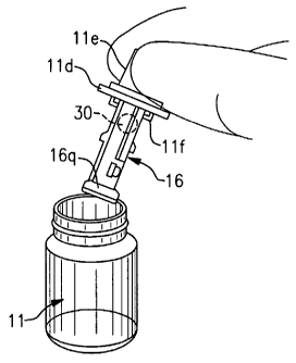
Turning attention to Figs. 2a,b, at the time of surgery, the container or vial
11 is
removed from any outer packaging in a sterile field and the outer vial cover
11 c is
removed to open vial 11 and access shuttle 16. As stated above, shuttle 16 may
be
releasably attached to the cover 11 c, or an inner cap 11 d located beneath
cover 11 c,
whereby the user may manually grasp inner cap 11 d by a cover flange 11 a and
lift the
shuttle 16 from vial 11 without having to directly handle the shuttle 16.
In the first embodiment described above, while still grasping cap 11 d, the
user
then proceeds to plug the shuttle 16 into the device body 12 as seen in Fig.
3a. Shuttle 16
slides within the longitudinal opening 12a at the distal end 12c of proximal
body section
12. Once fully received therein, the user releases cap 11 d from shuttle 16.
In this regard,
it is noted that any appropriate mechanism may be employed to releasably
connect
shuttle 16 to cap 11 d. In the embodiment shown in the figures, one or more
latches 11 If
extend from cap 11 d to releasable engage shuttle 16. The latches 11 f may be
operable
between an engaged position and a release position with respect to the shuttle
16, for
example, by spring loading the latches biased in the engage position. To move
the
12
CA 02593074 2007-06-29
WO 2006/071597 PCT/US2005/045857
latches to the release position, the user would squeeze the cap to spread the
latches away
from each other and thereby release the shuttle. Other shuttle engage and
release
mechanisms are of course possible and within the scope of the invention.
Once the shuttle 16 is received in the body section 12, nozzle section 14 is
attached thereto as seen in Fig. 3b. The nozzle section 14 may include one or
more
detents 14d for aligning and engaging one or more respective holes 12d formed
in body
section 12 to interconnect these components. Other connection means are of
course
possible and within the scope of the invention. Once connected, the device 10
is ready
for injecting IOL 30 into an eye.
Referring now to Figs. 4a,b and to the second, alternate embodiment described
above, while still grasping cap l ld, the user removes the shuttle 16 from
vial 11 as
described with reference to the first embodiment. The user then proceeds to
plug the
shuttle 16 into the nozzle section 14 as seen in Fig. 4a. Shuttle 16 slides
within the
longitudinal opening 14a at the proximal end 14b of nozzle section 14. Once
fully
received therein, the user proceeds to connect the nozzle section 12 and
shuttle 16 as a
unit to the proximal body section 14 as seen in fig. 4b. The user may then
release cover
l I d from nozzle section 14.
In the third embodiment seen in Fig. 4c, the nozzle section 14 and shuttle 16
(with IOL 30 preloaded therein) are connected together and releasably attached
to vial
cover 11 d for storage in vial 11. The user removes the nozzle section and
shuttle together
from the vial and attaches the nozzle section 14 to the proximal body section
12 as seen
in Fig. 4b whereupon the vial cover 11 d may be removed.
Upon further pressing of proximal section 12 against nozzle section 14 results
in
the two sections attaching together. Various mechanical connection features
may be
13
CA 02593074 2007-06-29
WO 2006/071597 PCT/US2005/045857
employed to permit the quick and easy attachment of the proximal body section
12 to the
nozzle section 14 by simply pressing the two sections together as described
above. Such
features may include cooperating detents and recesses or a friction fit
between the two
sections, for example. In the embodiment shown in the Figures, a pair of
detents 14d,e
(Figs. 6a-d) are provided on the outer wall surface of distal section 14 which
align with
and engage a pair of through-holes 12d,e formed on proximal section 12
adjacent open
distal end 12c thereof (Figs. 3a,b). When the proximal section 12 is pressed
against the
distal section 14, the detents 14d,e engage the through-holes 12d,e,
respectively, and the
sections become attached together. A radial flange 14f may be provided on
distal section
14 to act as a stop against further advancement of the proximal section 12 on
the distal
section 14, i.e., to prevent advancement beyond the point of detent
engagement. The
assembly of the injector device is now complete and the surgeon may proceed to
inject
the IOL 30 into a patient's eye by inserting tip 14c into an incision formed
in the eye and
pressing plunger 20 to advance the IOL 30 through and out the nozzle tip 14c
(see Fig. 2;
the eye not shown for sake of clarity).
Referring to Figures 1, 3a,b and 5, it is seen that the plunger 20 includes
distal
and proximal plunger shaft lengths 20a, 20b, respectively, having a plunger
tip 22 at the
distal end thereof and a thumb press 24 at the proximal end thereof for
manually
operating the injector device. The plunger tip 22 is configured for engaging
the IOL optic
31 at the periphery 31a thereof as the plunger 20 is advanced toward the
distal tip 14c of
distal section 14. It is very important that the plunger tip 22 not damage the
IOL optic 31.
The plunger tip 22 is thus designed to prevent damage to the IOL optic 31. In
the
preferred embodiment, the tip is bifurcated into first and second tip portions
22a and 22b,
whereby the IOL optic periphery 31 a becomes engaged between tip portions 22a,
22b as
14
CA 02593074 2007-06-29
WO 2006/071597 PCT/US2005/045857
seen in Figure 2B. It is understood that other plunger tip designs may be used
with the
present invention as desired. It is furthermore preferred that the plunger
shaft is
rotationally fixed within passageway 12a to prevent unexpected rotation of the
shaft (and
thus the tip 22) therein. For example, the plunger shaft may be rotationally
fixed by
forming the proximal shaft length 20b and passageway 12a non-circular in cross-
section
as shown.
In a particularly advantageous embodiment, the proximal length 20b of the
plunger shaft is provided with one or more elongated flanges 20a' which align
with a like
number of slots 12a' formed between radially extending fins 21 a-d formed on
the inner
wall surfaces of proximal section 12 adjacent proximal end 12b thereof (Fig.
3c). The
purpose of flanges 20a' and slots 12a' is to provide tactile resistance
therebetween and
thereby allowing the surgeon more precise control and feel when advancing the
plunger.
The fins 21 a-d may be made flexible yet resilient to provide the amount of
tactile
resistance desired. It is understood that other ways of providing tactile
resistance
between the plunger and injector body are within the scope of this invention.
This
provides the surgeon with continuous tactile feedback allowing the surgeon to
advance
the plunger (and thus the IOL) through the injector device in a very concise
and
controlled manner. Additionally, the flanges 20a' and slots 12a' help provide
proper
centering of the plunger shaft 20 and tip 22 relative to axis X-X along which
the
passageways of the components lie as explained above. Upon full advancement of
the
plunger, it is desirable to have the plunger automatically retract to some
degree upon
release of finger pressure against plunger finger press 24. In this regard, a
spring 20c
may be provided on a finger 20d on shaft length 20a. As the plunger is
advanced, the
CA 02593074 2007-06-29
WO 2006/071597 PCT/US2005/045857
spring 20c will interact with the one or more of the fins 21 a-d as the
plunger 20 is
advanced therethrough.
When it is time to use the injector device, the surgeon selects a package or
vial 11
having the appropriate IOL style and power preloaded in the shuttle stored in
the vial as
described above. The outer packaging is removed in a sterile field of the
surgical suite.
The proximal section having the plunger coupled thereto is also removed from
its
associated packaging in the sterile filed. If desired, all the injector
components including
the vial may be placed in a single outer packaging to present all the injector
device
components together. The nurse or surgeon proceeds to remove the shuttle and
IOL from
the vial in the manner described above. While holding the vial cover, the
shuttle and IOL
are connected to the nozzle section which is then attached to the proximal
body section
as described above. Once the device 10 is fully assembled as shown in Fig. 1,
the
surgeon inserts the distal tip 14c into an incision cut into the eye and
begins advancing
the plunger 20. As the plunger 20 is advanced, the plunger tip 22 engages the
optic
periphery 31 a and pushes IOL 30 forwardly. Upon continued advancement of the
plunger 20, the IOL 30 is pushed through the shuttle passageway 16a and is
expressed
from distal tip 14c and into the eye. As stated above, the spring 20c provides
increasing
bias in the reverse direction as the plunger reaches the fully advanced
position. This
occurs as spring 20c is compressed against one or more of the fins 21 a-d.
This assists the
surgeon in maintaining precise control over plunger (and hence IOL)
advancement and
allows automatic retraction of the plunger upon relieving the pushing pressure
being
exerted against the plunger thumb press 24. This is useful for easily
executing a second
stroke of the plunger in order to engage and manipulate the trailing haptic
into place in
the eye. This feature, together with the bifurcated plunger tip 22, allows a
more precise
16
CA 02593074 2007-06-29
WO 2006/071597 PCT/US2005/045857
control and manipulation of the IOL with the plunger tip in-situ than would be
possible
with an injector device not having these features.
As discussed above, the device may be used for IOLs of any type and style. The
configuration of the various component parts may likewise vary to accommodate
the
particular IOL style being employed with the device. It may thus be realized
that the
present invention provides an injector device method and apparatus that may be
provided
in a variety of embodiments.
17